
4-Year Follow-Up after Transatrial Transcatheter Tricuspid Valve Replacement with the LuX-Valve
Abstract
:1. Introduction
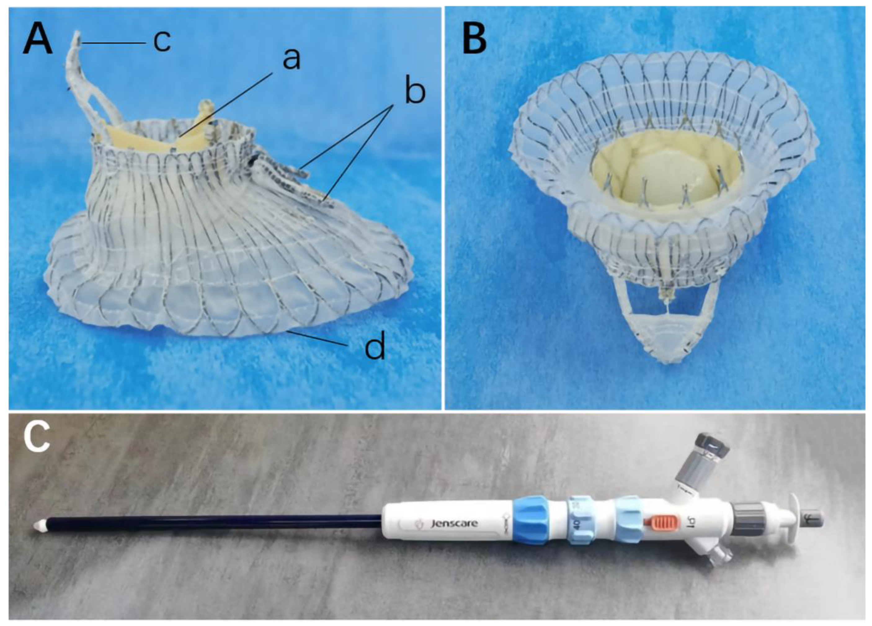
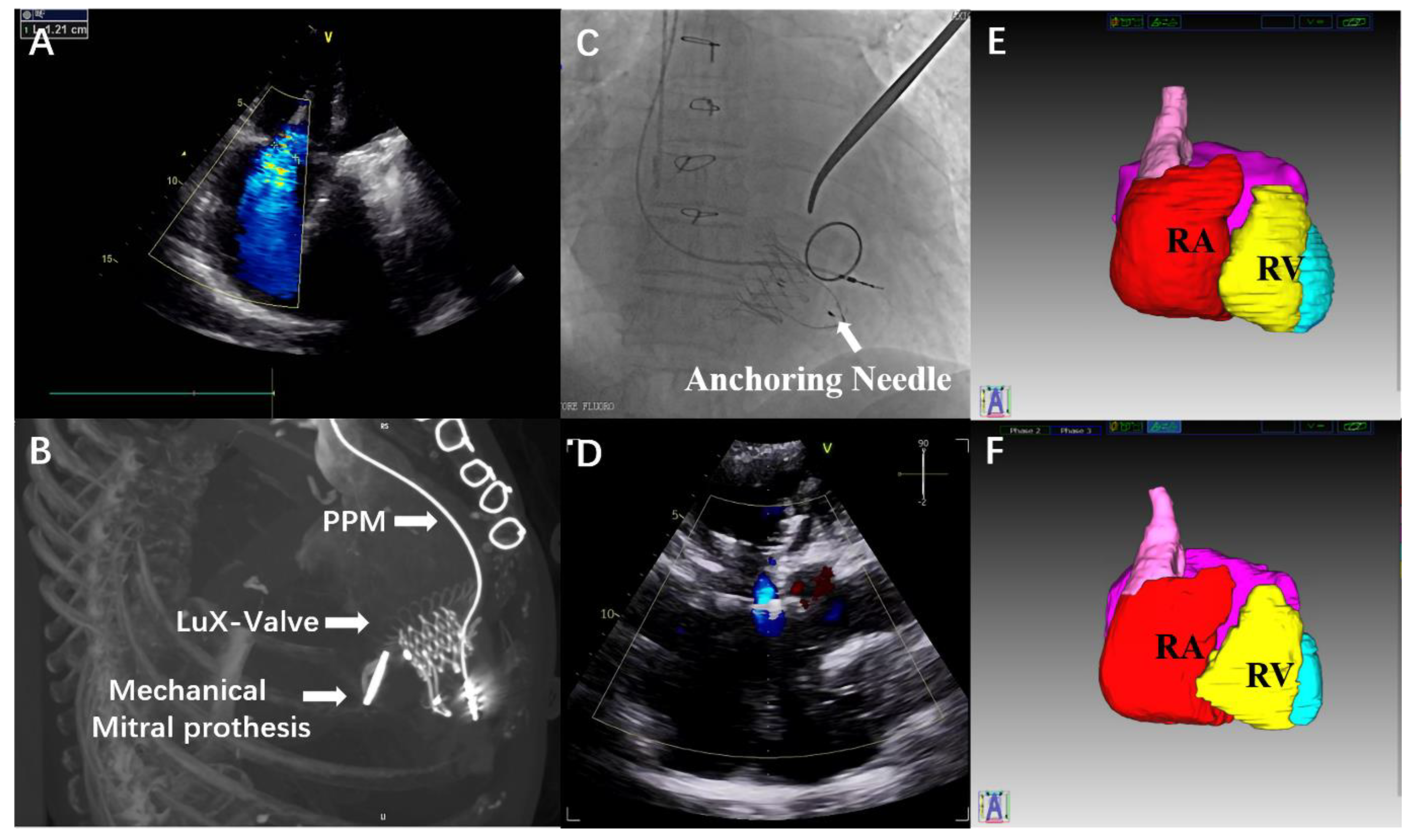
2. Case Report
3. Discussion
4. Conclusions
Supplementary Materials
Author Contributions
Funding
Institutional Review Board Statement
Informed Consent Statement
Data Availability Statement
Conflicts of Interest
References
- Nath, J.; Foster, E.; Heidenreich, P.A. Impact of tricuspid regurgitation on long-term survival. J. Am. Coll. Cardiol. 2004, 43, 405–409. [Google Scholar] [CrossRef] [PubMed] [Green Version]
- Voelkel, N.F.; Quaife, R.A.; Leinwand, L.A.; Barst, R.J.; McGoon, M.D.; Meldrum, D.R.; Dupuis, J.; Long, C.S.; Rubin, L.J.; Smart, F.W.; et al. Right ventricular function and failure: Report of a National Heart, Lung, and Blood Institute working group on cellular and molecular mechanisms of right heart failure. Circulation 2006, 114, 1883–1891. [Google Scholar] [CrossRef] [PubMed] [Green Version]
- Nishimura, R.A.; Otto, C.M.; Bonow, R.O.; Carabello, B.A.; Erwin, J.P., 3rd; Fleisher, L.A.; Jneid, H.; Mack, M.J.; McLeod, C.J.; O’Gara, P.T.; et al. 2017 AHA/ACC Focused Update of the 2014 AHA/ACC guideline for the management of patients with valvular heart disease: A report of the American College of Cardiology/American Heart Association Task Force on clinical practice guidelines. Circulation 2017, 135, e1159–e1195. [Google Scholar] [CrossRef] [Green Version]
- Hausleiter, J.; Braun, D.; Orban, M.; Latib, A.; Lurz, P.; Boekstegers, P.; von Bardeleben, R.S.; Kowalski, M.; Hahn, R.T.; Maisano, F.; et al. Patient selection, echocardiographic screening and treatment strategies for interventional tricuspid repair using the edge-to-edge repair technique. EuroIntervention 2018, 14, 645–653. [Google Scholar] [CrossRef] [PubMed]
- Rodés-Cabau, J.; Hahn, R.T.; Latib, A.; Laule, M.; Lauten, A.; Maisano, F.; Schofer, J.; Campelo-Parada, F.; Puri, R.; Vahanian, A. Transcatheter therapies for treating tricuspid regurgitation. J. Am. Coll. Cardiol. 2016, 67, 1829–1845. [Google Scholar] [CrossRef] [PubMed]
- Alqahtani, F.; Berzingi, C.O.; Aljohani, S.; Hijazi, M.; Al-Hallak, A.; Alkhouli, M. Contemporary trends in the use and outcomes of surgical treatment of tricuspid regurgitation. J. Am. Heart Assoc. 2017, 6, e007597. [Google Scholar] [CrossRef] [PubMed] [Green Version]
- Zack, C.J.; Fender, E.A.; Chandrashekar, P.; Reddy, Y.N.V.; Bennett, C.E.; Stulak, J.M.; Miller, V.M.; Nishimura, R.A. National trends and outcomes in isolated tricuspid valve surgery. J. Am. Coll. Cardiol. 2017, 70, 2953–2960. [Google Scholar] [CrossRef] [PubMed]
- Mesnier, J.; Alperi, A.; Panagides, V.; Bédard, E.; Salaun, E.; Philippon, F.; Rodés-Cabau, J. Transcatheter tricuspid valve interventions: Current devices and associated evidence. Prog. Cardiovasc. Dis. 2021, 69, 89–100. [Google Scholar] [CrossRef] [PubMed]
- Lu, F.L.; Ma, Y.; An, Z.; Cai, C.L.; Li, B.L.; Song, Z.G.; Han, L.; Wang, J.; Qiao, F.; Xu, Z.Y. First-in-man experience of transcatheter tricuspid valve replacement with LuX-Valve in high-risk tricuspid regurgitation patients. JACC Cardiovasc. Interv. 2020, 13, 1614–1616. [Google Scholar] [CrossRef] [PubMed]
- Lu, F.L.; An, Z.; Ma, Y.; Song, Z.G.; Cai, C.L.; Li, B.L.; Zhou, G.W.; Han, L.; Wang, J.; Bai, Y.F.; et al. Transcatheter tricuspid valve replacement in patients with severe tricuspid regurgitation. Heart 2021, 107, 1664–1670. [Google Scholar] [CrossRef] [PubMed]
- Hahn, R.T.; Kodali, S.; Fam, N.; Bapat, V.; Bartus, K.; Rodés-Cabau, J.; Dagenais, F.; Estevez-Loureiro, R.; Forteza, A.; Kapadia, S.; et al. Early multinational experience of transcatheter tricuspid valve replacement for treating severe tricuspid regurgitation. JACC Cardiovasc. Interv. 2020, 13, 2482–2493. [Google Scholar] [CrossRef] [PubMed]
- Webb, J.G.; Chuang, A.M.; Meier, D.; von Bardeleben, R.S.; Kodali, S.K.; Smith, R.L.; Hausleiter, J.; Ong, G.; Boone, R.; Ruf, T.; et al. Transcatheter tricuspid valve replacement with the EVOQUE System: 1-year outcomes of a multicenter, first-in-human experience. JACC Cardiovasc. Interv. 2022, 15, 481–491. [Google Scholar] [CrossRef]


Publisher’s Note: MDPI stays neutral with regard to jurisdictional claims in published maps and institutional affiliations. |
© 2022 by the authors. Licensee MDPI, Basel, Switzerland. This article is an open access article distributed under the terms and conditions of the Creative Commons Attribution (CC BY) license (https://creativecommons.org/licenses/by/4.0/).
Share and Cite
Ning, X.; Cao, J.; Wang, W.; Zhou, G.; Yang, F.; Xu, Z.; Han, L.; Qiao, F.; Lu, F. 4-Year Follow-Up after Transatrial Transcatheter Tricuspid Valve Replacement with the LuX-Valve. J. Cardiovasc. Dev. Dis. 2022, 9, 435. https://doi.org/10.3390/jcdd9120435
Ning X, Cao J, Wang W, Zhou G, Yang F, Xu Z, Han L, Qiao F, Lu F. 4-Year Follow-Up after Transatrial Transcatheter Tricuspid Valve Replacement with the LuX-Valve. Journal of Cardiovascular Development and Disease. 2022; 9(12):435. https://doi.org/10.3390/jcdd9120435
Chicago/Turabian StyleNing, Xiaoping, Jingyi Cao, Wei Wang, Guangwei Zhou, Fan Yang, Zhiyun Xu, Lin Han, Fan Qiao, and Fanglin Lu. 2022. "4-Year Follow-Up after Transatrial Transcatheter Tricuspid Valve Replacement with the LuX-Valve" Journal of Cardiovascular Development and Disease 9, no. 12: 435. https://doi.org/10.3390/jcdd9120435