Article

Colonisation Patterns of Nosema ceranae in the Azores Archipelago

by
Ana Rita Lopes
1,
Raquel Martín-Hernández
2,3,
Mariano Higes
2,
Sara Kafafi Segura
4,
Dora Henriques
1 and
Maria Alice Pinto
1,*
1
Centro de Investigação de Montanha, Instituto Politécnico de Bragança, Campus de Santa Apolónia, 5300-253 Bragança, Portugal
2
Laboratorio de Patología Apícola, IRIAF—Instituto Regional de Investigación y Desarrollo Agroalimentario y Forestal, Centro de Investigación Apícola y Agroambiental (CIAPA), Consejería de Agricultura de la Junta de Comunidades de Castilla-La Mancha, Camino de San Martín, 19180 Marchamalo, Spain
3
Instituto de Recursos Humanos para la Ciencia y la Tecnología (INCRECYT-FSE/EC-ESF), Fundación Parque Científico y Tecnológico de Castilla—La Mancha, 02006 Albacete, Spain
4
Zoología y Antropología Física, Facultad de Ciencias Biológicas, Universidad Complutense de Madrid, 28014 Madrid, Spain
*
Author to whom correspondence should be addressed.
Vet. Sci. 2022, 9(7), 320; https://doi.org/10.3390/vetsci9070320
Submission received: 31 May 2022 / Revised: 21 June 2022 / Accepted: 23 June 2022 / Published: 25 June 2022
(This article belongs to the Special Issue Challenges and Advances in Bee Health and Diseases)

Abstract

:

Simple Summary

Nosema ceranae is an emergent honey bee pathogen that has now invaded most of the world. However, geographically-isolated places that are free of this pathogen may still exist, and the Azores may be one of them. Here, we used molecular tools to see whether N. ceranae has entered the Azores and how far it has colonised the archipelago. In 2014/2015 we sampled 474 colonies from eight islands, and in 2020 we re-sampled 91 colonies from four islands. Our results showed that N. ceranae was present on all islands but Santa Maria and Flores. In the 2014/2015 sampling, Pico, the island of Varroa destructor entry in the Azores, showed the greatest prevalence. Resampling in 2020 revealed that N. ceranae built-up on Terceira and São Jorge. Our findings suggest that N. ceranae colonised the archipelago recently, and it spread across the other islands. Santa Maria is also free of V. destructor, making it one of the remaining areas in the world where bees are naive to both stressors. This study will help the veterinary authority establish biosecurity rules for the movement of bees and hive products among islands to maintain the N. ceranae-free status of Santa Maria and Flores.

Abstract

Nosema ceranae is a highly prevalent pathogen of Apis mellifera, which is distributed worldwide. However, there may still exist isolated areas that remain free of N. ceranae. Herein, we used molecular tools to survey the Azores to detect N. ceranae and unravel its colonisation patterns. To that end, we sampled 474 colonies from eight islands in 2014/2015 and 91 from four islands in 2020. The findings revealed that N. ceranae was not only present but also the dominant species in the Azores. In 2014/2015, N. apis was rare and N. ceranae prevalence varied between 2.7% in São Jorge and 50.7% in Pico. In 2020, N. ceranae prevalence increased significantly (p < 0.001) in Terceira and São Jorge also showing higher infection levels. The spatiotemporal patterns suggest that N. ceranae colonised the archipelago recently, and it rapidly spread across other islands, where at least two independent introductions might have occurred. Flores and Santa Maria have escaped the N. ceranae invasion, and it is remarkable that Santa Maria is also free of Varroa destructor, which makes it one of the last places in Europe where the honey bee remains naive to these two major biotic stressors.

1. Introduction

Western honey bees (Apis mellifera Linnaeus; Hymenoptera: Apidae) play a crucial role in the maintenance of ecosystems’ biodiversity through their pollination services. Yet, in recent decades, honey bee populations have declined worldwide [1,2], compromising not only food security but also current and future income for farmers and beekeepers [3]. Various factors, such as climate change, pesticide exposure, and malnutrition, together with predators (e.g., Vespa velutina Lepeletier; Hymenoptera: Vespidae), and pathogens (e.g., Varroa destructor Anderson and Trueman (Mesostigmata: Varroidae), viruses, bacteria, and fungi) have been identified as interacting contributors to honey bee colony losses [1,2,4,5]. Within fungi, the microsporidia of the former genus Nosema play an important role in honey bee health [6,7]. A recent phylogenetic study reclassified the genus Nosema as Vairimorpha [8]. However, here we will keep the name Nosema, for the sake of consistency with the existing literature.
Nosema spp. (Microsporidia: Nosematidae) are obligate intracellular parasites of the ventricular host cells, which are infectious to honey bees and other Hymenoptera [9,10]. In honey bees, the following three species have been identified: Nosema apis Zander [11], Nosema ceranae Fries et al. [12] and, most recently, Nosema neumanni Chemurot et al. [13]. Until the early 2000s, nosemosis in A. mellifera was thought to be caused only by N. apis, while N. ceranae was thought to be species-specific to Apis cerana Fabricius. However, this view changed when Higes et al. [14] and Huang et al. [15] detected N. ceranae for the first time in A. mellifera, in Spain and Taiwan, respectively. Since the host shift episode, N. ceranae has become the most prevalent microsporidia in A. mellifera populations across the globe [16,17,18,19], whereas N. neumanni has only been described in Uganda. Of the three species, N. ceranae is the one that raises more concern and has been pointed out as an important culprit of colony demise, at least in countries with warmer climates [6,20,21,22,23,24]. N. ceranae can harm honey bees in many different ways, namely, the following: (i) by decreasing lifespan and nursing ability [25,26], (ii) by inducing precocious foraging [26,27,28,29], (iii) by affecting olfactory learning, memory [30], and flight [31]. All these effects can cause decreases in colony size, honey production, and brood-rearing capacity, eventually leading to the collapse of the entire colony [6,32,33].
The islands offer an interesting stage for studying the colonisation dynamics of invasive pathogens such as N. ceranae, due to the geographic isolation of the host populations. Nonetheless, there are only a few reports on N. ceranae’s prevalence on islands, and these include the Canadian Prince Edward Island and Newfoundland [34], Caribbean Sea islands such as Cuba [35] and Dominica [36], and Pacific Ocean islands such as New Zealand and Norfolk [37,38]. In Europe, surveys of N. ceranae have been reported for Ireland, Great Britain [39,40], Isle of Man [41], Tuscan archipelago [42], and the Canary Islands [43]. The presence of N. ceranae was confirmed on all islands with a prevalence ranging from 7% (Isle of Man) to 100% (Norfolk), whereas N. apis was detected only in Dominica, New Zealand, Prince Edward Island, Newfoundland, Ireland, and Great Britain, with a prevalence ranging from 21.9% (Prince Edward Island and Newfoundland) to 82.3% (Dominica).
The Azores is a Portuguese archipelago in the Macaronesia region, located over 1400 km west of Lisbon. The archipelago is composed of nine islands, all harbouring honey bee colonies that are predominantly near the coastline. The first introduction of honey bees dates back to the XVI century, when Portuguese settlers brought in colonies to the largest island of São Miguel [44], probably from northern Portugal [45]. The last introduction occurred in 2015 when colonies from the island of Terceira were taken to the smallest island of Corvo [46]. There are now nearly 8000 managed hives in the Azores [47], with the islands of São Miguel, Terceira, and Pico harbouring the largest number of apiaries (76%) and the most active beekeeping [48].
Due to geographical isolation, the Azores were free of one of the most important honey bee stressors, the invasive ectoparasitic mite V. destructor, until 2000 [45]. The mite hitchhiked in the queen’s parcels illegally imported to Pico in 2000 and to the island of Flores in 2001 [45]. Eight years after the first introduction, the mite was spotted on the island of Faial, which is only 8.3 km apart from Pico. Contrary to V. destructor, which introduction history is well-known, whether N. ceranae reached and spread throughout the archipelago was uncertain.
Since 2008, Nosema spp. has been annually surveyed in the Azores by the regional veterinary authority. According to the annual reports, the only islands that are negative to the microsporidia are the islands of Santa Maria and Flores, together with the most recently colonised island of Corvo, while the island of São Jorge became positive in 2018. [48]. However, since spore detection was based on morphological methods under light microscopy, the question remains as to which Nosema species underlies the detection. This is because the spores of N. apis and N. ceranae are very similar, and there is no data on field symptoms that could help discriminate between the two species. It was supposed that nosemosis was caused by N. apis, the species associated with A. mellifera before the recent worldwide spread of N. ceranae [16,18,19,49,50], as the geographical isolation and the early biosecurity measures, imposed by the veterinary authority after arrival of the V. destructor to Pico (Dr. Paula Vieira, Direção Regional da Agricultura e Desenvolvimento Rural dos Açores, pers. comm.), could be able to deter the new honey bee pathogen from entering the Azores. However, if N. ceranae succeeded in reaching the Azores, its transmission among honey bee populations that are isolated on each island would be difficult, allowing unique conditions for studying an emergent pathogen colonising a pristine territory. While V. destructor and N. ceranae are serious pests for a colony [32,51,52], when they are simultaneously present, it is possible that their impact is further aggravated by a summative or synergistic interaction. Whether this interaction exists is unclear, although some studies have found a positive correlation between the two parasites [53,54,55,56,57]. In the event that N. ceranae is detected, then the Azores provide a unique stage for addressing this issue because there are islands where the mite is present and islands that are mite free. Hereby, the objectives of this study were to (i) determine whether N. ceranae is present in the Azores; (ii) infer its prevalence and levels of infection throughout the archipelago; (iii) search for any association between N. ceranae and V. destructor; finally, (iii) explore possible colonisation scenarios of this emergent pathogen.

2. Materials and Methods

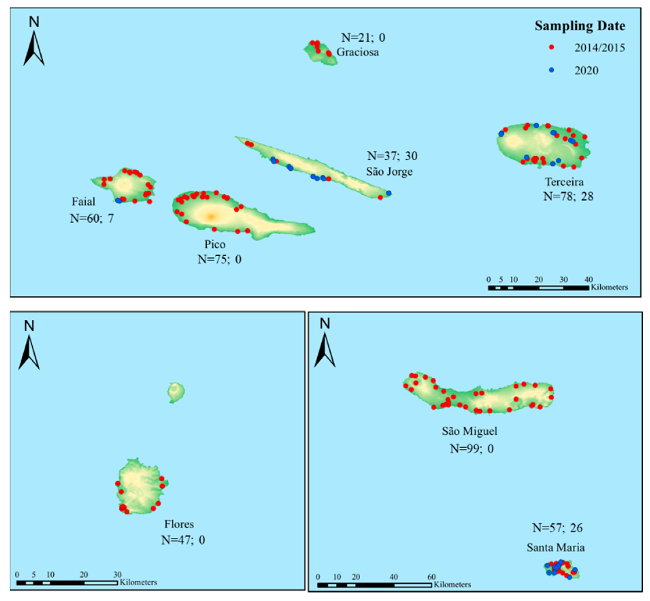
2.1. Survey and Sampling Collection

The number of colonies to be sampled was calculated in relation to the number of apiaries registered in 2013 with an expected prevalence of Nosema species of 15%, a precision rate of 10% and a confidence level of 95%. The number of samples was subsequently distributed in proportion to the number of apiaries on each island in which colonies were selected randomly (Table S1). In this cross-sectional study, 474 colonies (representing a total of 156 apiaries) were sampled from eight Azorean islands in July and August of 2014 and 2015 (Figure 1 and Table 1). While most apiaries were represented by three colonies, there were a few apiaries that were represented by either two or four colonies (Table S2). Over 150 workers were collected from the outside frame of brood nest of the 474 colonies to appropriate card boxes and then shipped alive to Centro de Investigação de Montanha (CIMO; latitude 41°47′53.19″ N, longitude 6°45′56.89″ W).
Given the rise of Nosema spp. on São Jorge in 2018, as detected by the veterinary authority, in 2020 we decided to re-sample this island and the following three additional islands for comparison purposes: Santa Maria (island negative to Nosema spp., according to the morphological reports), Faial (island positive to V. destructor) and Terceira (island negative to V. destructor but positive to Nosema spp., according to the morphological reports). A total of 91 colonies, representing 34 apiaries, were sampled between July and August of 2020 using the same protocol as in 2014/2015 (Figure 1 and Table 1). The 565 samples were stored at −80 °C until molecular analyses.

2.2. Nosema spp. Extraction and Detection for the Prevalence Study

Total DNA was extracted from a pool of 120 workers for each of the 474 samples (collected in 2014/2015), following the protocol described in Martín-Hernández et al. [58] with minor modifications. Briefly, 120 workers were added to a double bag strainer (BA6040, Seward, Worthing, UK) with 18 mL of 50% AL Buffer (Qiagen®, Hilden, Germany) and crushed in Stomacher 80 (Stomacher 80-Microbiomaster®, Seward, Worthing, UK) for two minutes at low speed, followed by adding 9 mL of 50% AL buffer and one more cycle of homogenization (60 s). Subsequently, those macerates were centrifuged at 3000 rpm for 10 min, and the pellet was resuspended in 3 mL of MilliQ water. To improve DNA yield, the mechanical lysis of the spores was applied by adding 400 µL of resuspended pellet to a 96-well plate (Qiagen®, Hilden, DE) containing glass beads (2 mm diameter, Sigma), which was shaken in a TissueLyser machine (Qiagen®, Hilden, DE) for three minutes. A negative control with no honey bee sample was set for every 20 samples and processed in parallel. Finally, 150 µL of macerate was used for DNA extraction following the BS96 DNA Tissue extraction in a BioSprint workstation (Qiagen®, Hilden, DE). The DNA extracts were frozen at −20 °C until the next analysis.
N. ceranae, N. apis and an internal control of A. mellifera DNA (COI gene) were simultaneously screened by using a multiplex-PCR developed by Martín-Hernández et al. [58]. PCR primers are shown in Table S3 whereas the thermal profile and reactions set up are detailed in Martín-Hernández et al. [58]. Nontemplate (NTC) and positive controls were included in each run. PCR products were analysed by capillary electrophoresis in the QIAxcel advanced apparatus using a QIAxcel DNA High Resolution Kit (Qiagen®, Hilden, DE).

2.3. N. ceranae Extraction and Load Determination

Since the previous DNA extracts of the 2014/2015 sampling period were accidentally lost, DNA was isolated de novo from a pool of 30 remaining workers and from all the samples collected in 2020, for real-time qPCR purposes. Fifteen per cent of the colonies were excluded for further analysis due to the insufficient number of workers to pool, reducing the sample size of the 2014/2015 collection to 403 (Table S2). These samples were used to determine the N. ceranae load and to compare the prevalence between both periods (2014/2015 and 2020). Briefly, the 30 workers were placed in a double bag strainer (BA6040, Seward, Worthing, UK) and added 6 mL of cool DEPC water (E476, VWR, Pennsylvania, US). Subsequently, the homogenization step was carried out using the MixWell Lab Blender (Alliance Bio Expertise®, Guipry-Messac, FR) for the following two cycles: one of 60 s followed by another 30 s, with 30 s pause between each cycle. DNA was extracted from the tissue homogenates by using the standard protocol for animal tissue from NucleoSpin Tissue commercial kit (Macherey-NagelTM, Düren, Germany), with minor modifications. To that end, 50 µL of the homogenates and 180 µL of T1 buffer were added to 2 mL tubes containing two zirconia beads (3 mm, Specanalítica Lda., Carcavelos, PT) to proceed with the mechanical tissue disruption using Precellys (Bertin Instruments, Montigny-le-Bretonneux, FR) with the following protocol: 6200 rpm; 5 s; 3 times. Subsequently, the NucleoSpin Tissue protocol extraction was followed without any modification. The yield and DNA quality were verified through spectrophotometry (Lvis chip, SpectroStar Nano, BMG Labtech, Ortenberg, Germany) and DNA extracts were normalized to 10 ng/µL.
Our approach to quantifying N. ceranae load per colony combined the primers described by Martín-Hernández et al. [58] and the SYBR® Green chemistry (primers shown in Table S3). Real-time qPCR assays were performed in the QuantStudio 5 apparatus (Applied Biosystems®, Masschusetts, USA). qPCR reactions were carried out in 10 µL total volume (two replicates per sample), containing 10 ng of DNA, 5 µL of 2× iTaq Universal SYBR® Green Supermix (Biorad®, California, USA), 300 nM of each primer (Table S3) and 2 µL of DEPC water. The thermal conditions were set according to the SYBR® Green manufacturer’s instructions. Positive controls used to establish the standard curves were made by concentrating known positive samples for N. ceranae through PCR. PCR products were quantified using the LVis Chip (SpectroStar Nano, BMG Labtech, Ortenberg, Germany) apparatus. Subsequently, PCR products were used to prepare ten-fold serial dilutions with concentrations (7 points in duplicate) ranging from 1.75 × 10⁻² to 1.75 × 10⁻⁸ ng/µL (Figure S1). Melting curve analysis was conducted to detect primer dimers and/or unspecific amplicons. All samples amplifying before the last point of the standard curve, with an amplification plot showing an exponential increase and a melting profile matching the melting temperature of the positive controls, were classified as N. ceranae positive. In addition, the RPL8 gene of A. mellifera was analysed in those samples to check whether DNA extraction was successful ([59]; see primers in Table S3). N. ceranae loads were used to establish the following three infection classes: low for loads < 10−8 ng/µL, medium for loads between 10−5 ng/µL and 10−8 ng/µL and, high for loads ≥ 10−5 ng/µL.

2.4. Statistical Analysis

To evaluate N. ceranae prevalence differences between sampling periods and its association with V. destructor, the Chi-square test (χ2) was applied. Whenever the sample size of one of the variables was smaller than 5 counts, one of the Chi-square test assumptions was violated, and the Fisher’s exact test was applied. The strength of the association between N. ceranae prevalence and V. destructor (presence/absence at the island level) was assessed with Cramer’s V measurement (φc).
Since the qPCR data did not meet the assumptions of parametric tests, differences in N. ceranae loads among islands were assessed using the Kruskal–Wallis or Mann–Whitney tests. All statistical analyses and graphical representations were carried out in R studio software (version 4.0.2) [60] and the level of significance was set at 95% (α = 0.05).

3. Results

3.1. Quality Control

All samples analysed with the multiplex PCR method amplified the internal control of A. mellifera DNA (COI gene), confirming DNA integrity. Furthermore, none of the DNA extraction controls or non-template controls (NTCs) were amplified, suggesting no cross-contamination during sample processing and analysis. Regarding real-time qPCR, all samples validated DNA integrity by amplifying the A. mellifera RPL8 gene, and all NTCs showed no amplification, indicating that there was no cross-contamination throughout the study. The amplification efficiency of N. ceranae was 97.4% (Figure S1). Altogether, these quality control results indicate that the data analysed below is of good quality.

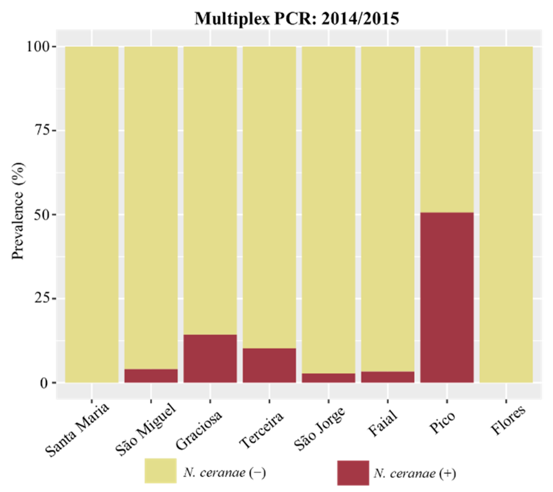
3.2. Detection and Prevalence of Nosema spp. across the Azores

The prevalence of Nosema infection in the Azores was inferred from the multiplex-PCR output for the samples from the 2014/2015 period, as allowed by the sampling design. A total of 56 (11.8%), 9 (1.9%), and 6 (1.3%), out of 474 colonies, tested positive for N. ceranae, N. apis, and co-infection, respectively, using the multiplex PCR approach (Table 2 and Figure 2). N. ceranae showed the highest prevalence on Pico (50.7%), followed by Graciosa and Terceira, with 14.3% and 10.3%, respectively. On São Miguel, Faial and São Jorge, N. ceranae prevalence was ≤4.0% and Santa Maria and Flores did not have any positive colony. N. apis was detected in five islands (Flores, Graciosa, São Jorge, São Miguel, and Terceira), in general, with a lower prevalence than N. ceranae, ranging from 1.3 to 5.1% (Table 2). The co-infection was even rarer, with one positive colony in São Jorge (2.7%), one positive colony in Terceira (1.3%), and four positive colonies in São Miguel (4.0%).
To assess whether the infection changed between 2014/2015 and 2020, samples were processed for both periods using the same qPCR conditions. The overall prevalence for the first sampling period showed that 34 (8.4%) of the 403 screened colonies were positive for N. ceranae (Table 2 and Figure 3). N. ceranae could be detected only on Pico (43.7%), Graciosa (5.6%), Terceira (1.4%), and São Miguel (1.1%). On Faial, the two colonies that tested positive for N. ceranae by the multiplex PCR approach were negative by qPCR. The single positive colony of São Jorge was not re-analysed because there were not enough workers for pooling. In the 2020 sampling, the highest number of N. ceranae positive colonies was recorded on Terceira (57.1%), closely followed by São Jorge (50%). No infection of N. ceranae was identified on Faial and Santa Maria. Notably, the months of July and August of 2020 were considerably dryer than those of 2014 and 2015 (Table 1), which may have influenced the build-up of the infection observed on these two islands.
While the overall prevalence patterns are consistent between the qualitative PCR (multiplex) and the quantitative approach (qPCR), a reanalysis of the 2014/2015 dataset by qPCR, using a lower number of pooled workers (30 instead of 120), led to the detection of a lower number of positives, indicating that false negatives may arise from small pool sizes. This is expected when the proportion of infected individuals in the colony is low, requiring a higher pool size for detecting infected colonies [61].
Statistical analysis of N. ceranae prevalence data, obtained from qPCR, revealed a highly significant difference (p < 0.001; Chi-square test) between sampling periods (Figure 3), which is explained by the dramatic rise in positive colonies observed in 2020 for Terceira and São Jorge (p = 0.001 for both islands; Fisher’s exact test).

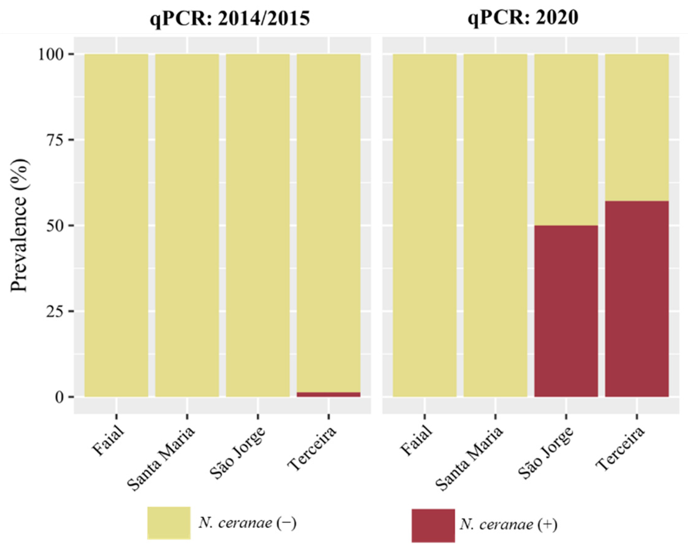
3.3. N. ceranae Loads across the Azores

N. ceranae loads were statistically different (p < 0.0001; Mann–Whitney test) between the 2014/2015 and 2020 sampling periods, with the latter showing the highest median loads (Figure 4A). In the 2014/2015 sampling period, the level of N. ceranae infection in positive colonies ranged from 5.44 × 10⁻¹⁰ ± 5.49 × 10⁻¹¹ to 7.34 × 10⁻⁵ ± 1.69 × 10⁻⁶ ng/µL (Figure 4A). Despite this wide range, the levels of N. ceranae infection across islands were not significantly different from each other (p = 0.175; Kruskal–Wallis test), since most of the positive colonies in this sampling period originated from Pico. The other three positive islands (Graciosa, Terceira, and São Miguel), as detected by the qPCR approach (Table 2), only had one positive colony each and the infection level was generally low (<1.04 × 10⁻⁸ ng/µL). In the 2020 sampling period, the two islands (São Jorge and Terceira) with N. ceranae positive colonies had a broad range of infection levels (from 5.74 × 10⁻¹⁰ ± 6.72 × 10⁻¹¹ to 1.35 × 10⁻⁴ ± 1.52 × 10⁻⁵ ng/µL; Figure 4B). São Jorge recorded the highest median (2.40 × 10⁻⁴ ± 9.10 × 10⁻⁶ ng/µL) of N. ceranae loads, being statistically different from Terceira (p = 0.001; Mann–Whitney test).
When categorising the infection level of N. ceranae according to the qPCR loads in the 2014/2015 sampling period, most colonies exhibited a medium infection (23; 75%), one colony exhibited a severe infection (3.1%), and seven colonies exhibited a low infection (21.9%). Notably, the majority of the medium-infected colonies originated from Pico, and they were located on the north-western part of the island (Figure 4C). In the 2020 sampling period, 14 (45.2%) positive colonies were categorised as having a high infection, 15 (48.4%) a medium infection, and 2 (6.4%) a low infection. The most highly infected colonies originated from São Jorge (Figure 4B).

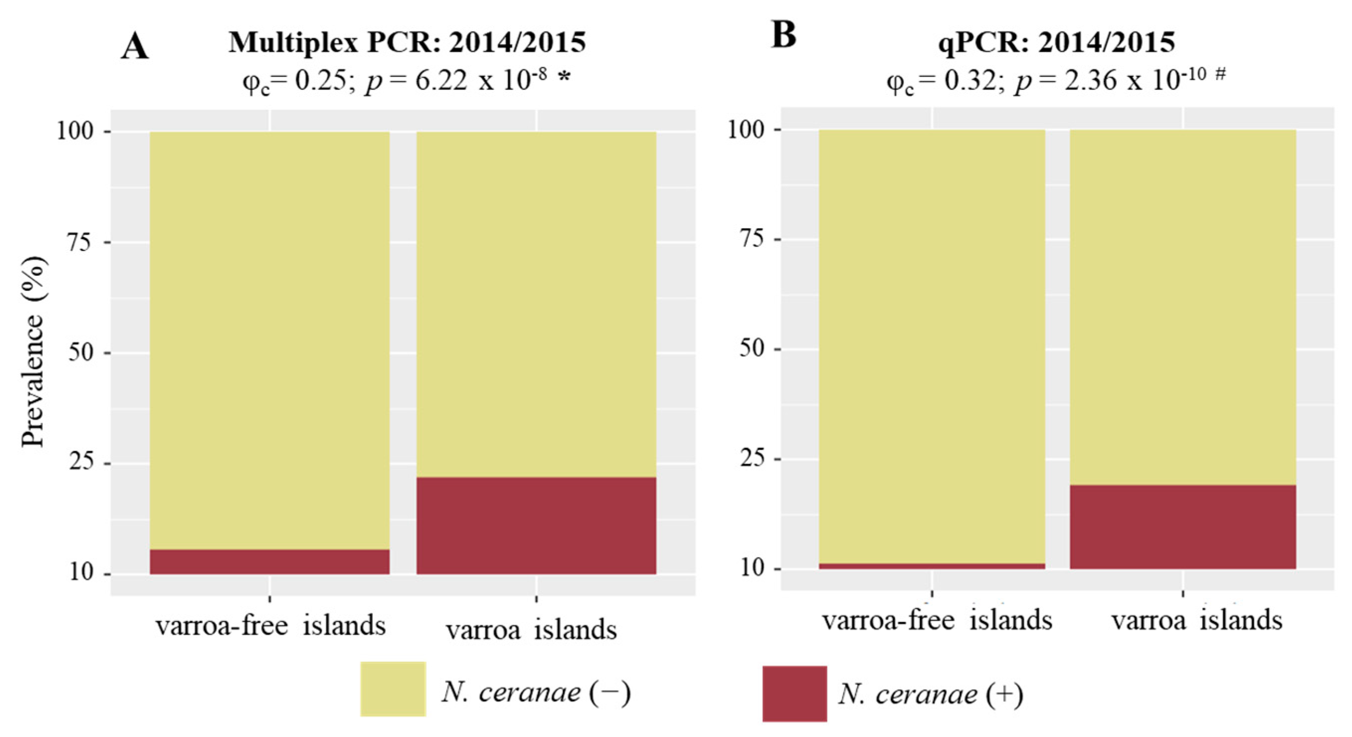
3.4. N. ceranae and V. destructor Associations

The association between N. ceranae prevalence and V. destructor was tested using the multiplex PCR and the qPCR datasets separately. A statistically significant positive association was found in the 2014/2015 sampling period (φc = 0.25; p < 0.001, Chi-square test) (Figure 5A). This finding was corroborated by the qPCR qualitative data (φc = 0.32; p < 0.001, Fisher’s exact test), despite the lower number of tested samples (Figure 5B). In contrast, the prevalence of N. apisc = 0.08; p = 0.163, Fisher’s exact test) or co-infection (φc = 0.09; p = 0.087, Fisher’s exact test) did not reveal any relationship with V. destructor presence. Since only one (Faial) of the sampled islands from 2020 has V. destructor, the association analysis could not be performed.

4. Discussion

This study documents for the first time the presence of N. ceranae in the Azores, further extending its distributional range in the world (reviewed by Klee et al. [16] and Grupe and Quandt [49]) and specifically in Macaronesia, where it had only been reported for the Canaries [43]. Nosema infection has occurred in the Azores at least since 2008, according to the first spore identification by microscopical analysis produced by the local veterinary authority [46]. Whether the early cases were due to N. apis or N. ceranae is unknown. Moreover, unknown is the year and exact location of the first arrival of N. ceranae to the archipelago, contrasting with the known putative N. ceranae-free status of the following two of its islands: Santa Maria and Flores. Remarkably, despite the widespread distribution of N. ceranae in the Azores, samples from these two islands were all negative in both the 2014/2015 and 2020 sampling periods. These molecular results, together with the morphological results reported by the Azorean veterinary authority for several years (from 2008 to 2021; Direção Regional da Agricultura e Desenvolvimento Rural dos Açores [46,48]), strongly suggest that Santa Maria and Flores are still free of this harmful pathogen.
In the 2014/2015 sampling, N. ceranae prevalence was below 10.3% for six of the eight surveyed islands, with Santa Maria and Flores exhibiting values of 0%. However, in the 2020 sampling, while N. ceranae went undetected on Santa Maria and Faial, the epidemiological situation aggravated considerably in São Jorge and Terceira, as suggested by over 50% of samples testing positive in qPCR on both islands. Due to the geographically limited sampling effort in 2020, these results should be interpreted with caution, particularly on Faial where only two apiaries were examined. Nonetheless, the increasing trend observed on São Jorge and Terceira, which is supported by the morphological data [48], cannot be ignored. These findings call for a thorough molecular survey across the entire archipelago so that a more rigorous appraisal of the epidemiological status of the Azorean honey bee populations, and a further confirmation of the N. ceranae-free status of Flores and Santa Maria, can be carried out. This is particularly important for Santa Maria as this island is also free of V. destructor and harbours the purest Iberian honey bee (A. m. iberiensis) population in the Azores [45].
Since the first discovery in colonies sampled in 2005 in Spain [14] and in Taiwan [15], N. ceranae has been detected with high prevalence rates in all continents where A. mellifera is present, indicating a very high invasive potential [16,49,62]. In the European continent, N. ceranae reached 95–98% prevalence in Hungary [63], 84% in the Balkan countries [20], 77.2% in the northern part of Bulgaria [64], 59.7% in Belgium [65], over 63.2% in Italy [23,66], or 58.5% in Spain [67]. In mainland Portugal, the possible origin of the spores introduced in the Azores, N. ceranae prevalence was over 60% in a survey conducted in 2012 (report published by Federação Nacional dos Apicultores de Portugal [68]). High prevalence rates have also been reported for islands. For instance, N. ceranae reached 97.1% prevalence in Cuba [35], 63% in the Dominica Islands [36], or 75% in the Macaronesia archipelago of the Canaries [43].
All those regions were in stark contrast with the epidemiological situation of the Azores, where two islands seemed to remain free of N. ceranae and five islands exhibited prevalence rates lower than 14.3% in the 2014/2015 sampling. Only Pico, the island where V. destructor was first introduced, registered a large proportion of infected colonies, regardless of the molecular approach used (43.7–50.7%). This result suggests that, as for V. destructor, Pico might have acted as the first entry point for N. ceranae in the Azores. If this was the case, it is possible that N. ceranae spores hitchhiked along with V. destructor in the queens parcels illegally imported in 2000, as this pathogen has been shown to be infecting A. mellifera since at least 1995 in the USA [17] and 1998 in Europe [69]. Alternatively, Pico did not act as the entry point in the Azores and N. ceranae could have been introduced later to this island through contaminated hive products [70,71,72], originating from the varroa-free islands, mainland or elsewhere, and V. destructor aided infection development by lowering the immune defences of the honey bees [73,74,75]. If the first hypothesis is true, then the stringent restrictions on the circulation of honey bees and hive products from the varroa-invaded islands onto the varroa-free islands, imposed early by the veterinary authority (Dr. Paula Vieira, pers. comm.), imply that at least one additional independent introduction of N. ceranae occurred on São Miguel, São Jorge, Terceira, or Graciosa. On Flores, the other illegal queen import was seemingly free of N. ceranae, as suggested by the negative results obtained from either molecular or morphological methods.
The introduction of N. ceranae on the varroa-free islands likely occurred via commercial hive products, as the importation of queens or packaged honey bees has been restricted for over 22 years. Honey, pollen, royal jelly, and wax foundation can all contain viable spores and thus operate as transmission vehicles of N. ceranae through trading [70,71,72]. Among these, the wax foundation is the more probable original source of introduction in the varroa-free islands because there is a high demand for this hive product in the Azores and sterilisation of wax imports has only been compulsory since 2010 (Dr. Paula Vieira, pers. comm.). Regardless of the places and means of N. ceranae’s entrance, the spatial and temporal prevalence patterns suggest that N. ceranae arrived recently in the Azores, and it is invading the archipelago at a fast pace. Since its entry, it is behaving as an emerging pathogen, as evidenced by the rising prevalence observed in some islands.
In addition to prevalence, N. ceranae infection loads were determined for all positive samples identified by real-time qPCR. In 2014/2015, the three single positive samples, originating from varroa-free islands, had low (Graciosa and São Miguel) and medium (Terceira) infection intensities. On Pico, there were samples with low as well as high infection intensities, but the great majority (75%) exhibited medium intensity, and these were mostly located in the northwest. This part of the island has a higher concentration of apiaries, which might have facilitated the transmission of N. ceranae as the spores are considered bioaerosols that can be carried in the air and deposited in natural environments, including flowers [76]. Other ways to spread the infection could be through sharing food or water sources, or the effects of drifting or robbing infected colonies. In the 2020 sampling, the two islands (Terceira and São Jorge) that showed high prevalence also showed high loads, particularly São Jorge. Strikingly, in the previous sampling, only one sample was identified as positive on this island, and now, in addition to the high prevalence (50.0%), most samples displayed infection levels classified as high. The infection loads, together with the temporal prevalence patterns, suggest a recent establishment of N. ceranae on these two varroa-free islands, and now the pathogen is rapidly spreading, showing the typical behaviour of an emergent pathogen, with an increase in the prevalence and in the level of infection. Therefore, despite the positive association between V. destructor and N. ceranae prevalence detected in an earlier stage of the invasion (2014/2015), and also reported in other studies [53,54,55,56,57], these findings suggest that the mite is not a mandatory condition for a successful invasion of the pathogen.
Once N. ceranae is introduced into a pristine territory, several beekeeper-independent factors, such as pesticide exposure, quality of pollen forage, and climate, can influence the establishment of the pathogen and the build-up of the infection [77,78,79,80,81,82]. Yet, new introductions into geographically isolated places such as the Azores are dependent on humans for transportation of propagules. In the case of N. ceranae, long-distance transmission is facilitated by the trade of infected honey bees or contaminated hive products [70,71]. There are only two islands, Flores and Santa Maria (and perhaps Corvo), that remain free of N. ceranae (according to the veterinary report for 2021 [48]), and this status can only be perpetuated if biosecurity restrictions are capable of continuing to prohibit any importation attempt of honey bees and decontamination of imported hive products used in beekeeping (e.g., wax) is assured.

5. Conclusions

This is the first molecular survey of Nosema spp. carried out in the Azores and consequently the first detection of N. ceranae in this Macaronesia region. Nosema spp. was identified on some of the islands in the first morphological report released by the local veterinary authority in 2008. Whether these early cases were due to N. apis or N. ceranae is unknown. However, there is a chance that N. ceranae was already present in 2008, at least on Pico, as suggested by the high prevalence rate and infection loads found on this island in the 2014/2015 sampling. The spatial and temporal patterns are compatible with a recent colonisation hypothesis, after N. ceranae has been introduced in the Azores, likely multiple times. Flores and Santa Maria have so far seemed to avoid N. ceranae invasion, a situation that can be sustained if beekeepers comply with biosecurity regulations. While Flores has V. destructor, Santa Maria is free of this parasite. Therefore, Santa Maria is one of the last places in Europe, and perhaps in the world, where honey bees remain naive to two of the major honey bee biotic stressors, making this island unique for beekeeping activity.

Supplementary Materials

The following supporting information can be downloaded at: https://www.mdpi.com/article/10.3390/vetsci9070320/s1; Figure S1: Real-time qPCR standard curves for N. ceranae; Table S1: Distribution of registered apiaries and colonies per island in 2013; Table S2: Metadata of sampled apiaries; Table S3: Primers used for molecular detection of N. ceranae; N. apis and internal controls for Apis mellifera.

Author Contributions

Conceptualization; M.A.P., R.M.-H. and M.H.; methodology; M.A.P., R.M.-H. and M.H.; formal analysis; A.R.L. and D.H.; investigation; S.K.S. and A.R.L.; resources; M.A.P.; data curation; A.R.L.; writing—original draft preparation; A.R.L. and M.A.P.; writing—review and editing; A.R.L., M.A.P., R.M.-H. and D.H.; supervision; M.A.P. and R.M.-H.; project administration; M.A.P.; funding acquisition; M.A.P. All authors have read and agreed to the published version of the manuscript.

Funding

This research was funded by Portuguese funds through FCT (Fundação para a Ciência e a Tecnologia) in the framework of the project BeeHappy grant number POCI-01-0145-FEDER-029871. ARL was supported by a PhD scholarship (SFRH/BD/143627/2019) from the FCT. FCT provided financial support by national funds (FCT/MCTES) to CIMO (UIDB/00690/2020).

Institutional Review Board Statement

Not applicable.

Informed Consent Statement

Not applicable.

Data Availability Statement

The data analyzed for the study are available from the corresponding author on reasonable request.

Acknowledgments

We are grateful to all veterinarians and technicians of the “Direção Regional da Agricultura e Desenvolvimento Rural dos Açores”, who collected the samples across the Azores, namely: Frank Aguiar, Nuno Salvador, Janyne Sousa, Ivan castro, Célia Mesquita, Ana Jorge, José Dias, Paulo Rico, Pedro Leal, Vagner Paulos, Luis Xavier, Luís Silva, Martins Silva, Carlos Gouveia, Ana Carina Coimbra, João Ramos, João Arruda, Edgardo Melo, João Luís and Moniz da Ponte. Special thanks to Paula Vieira and Frank Aguiar for coordinating the sampling and to Stephen Smith for kindly proofreading the English.

Conflicts of Interest

The authors declare no conflict of interest.

References

  1. Goulson, D.; Nicholls, E.; Botías, C.; Rotheray, E.L. Bee declines driven by combined stress from parasites, pesticides, and lack of flowers. Science 2015, 347, 1255957. [Google Scholar] [CrossRef]
  2. Potts, S.G.; Biesmeijer, J.C.; Kremen, C.; Neumann, P.; Schweiger, O.; Kunin, W.E. Global pollinator declines: Trends, impacts and drivers. Trends Ecol. Evol. 2010, 25, 345–353. [Google Scholar] [CrossRef]
  3. Gallai, N.; Salles, J.M.; Settele, J.; Vaissiere, B.E. Economic valuation of the vulnerability of world agriculture confronted with pollinator decline. Ecol. Econ. 2009, 68, 810–821. [Google Scholar] [CrossRef]
  4. Hristov, P.; Shumkova, R.; Palova, N.; Neov, B. Honey bee colony losses: Why are honey bees disappearing? Sociobiology 2021, 68, e5851. [Google Scholar] [CrossRef]
  5. Evans, J.D.; Schwarz, R.S. Bees brought to their knees: Microbes affecting honey bee health. Trends Microbiol. 2011, 19, 614–620. [Google Scholar] [CrossRef] [PubMed]
  6. Higes, M.; Martín-Hernández, R.; Botías, C.; Bailón, E.G.; González-Porto, A.V.; Barrios, L.; del Nozal, M.J.; Bernal, J.L.; Jiménez, J.J.; Palencia, P.G.; et al. How natural infection by Nosema ceranae causes honeybee colony collapse. Environ. Microbiol. 2008, 10, 2659–2669. [Google Scholar] [CrossRef] [PubMed]
  7. Emsen, B.; De la Mora, A.; Lacey, B.; Eccles, L.; Kelly, P.G.; Medina-Flores, C.A.; Petukhova, T.; Morfin, N.; Guzman-Novoa, E. Seasonality of Nosema ceranae infections and their relationship with honey bee populations, food stores, and survivorship in a North American region. Vet. Sci. 2020, 7, 131. [Google Scholar] [CrossRef]
  8. Tokarev, Y.S.; Huang, W.F.; Solter, L.F.; Malysh, J.M.; Becnel, J.J.; Vossbrinck, C.R. A formal redefinition of the genera Nosema and Vairimorpha (Microsporidia: Nosematidae) and reassignment of species based on molecular phylogenetics. J. Invertebr. Pathol. 2020, 169, 7. [Google Scholar] [CrossRef]
  9. Plischuk, S.; Martín-Hernández, R.; Prieto, L.; Lucía, M.; Botías, C.; Meana, A.; Abrahamovich, A.H.; Lange, C.; Higes, M. South American native bumblebees (Hymenoptera: Apidae) infected by Nosema ceranae (Microsporidia), an emerging pathogen of honeybees (Apis mellifera). Env. Microbiol. Rep. 2009, 1, 131–135. [Google Scholar] [CrossRef]
  10. Bravi, M.E.; Alvarez, L.J.; Lucia, M.; Pecoraro, M.R.I.; García, M.L.G.; Reynaldi, F.J. Wild bumble bees (Hymenoptera: Apidae: Bombini) as a potential reservoir for bee pathogens in northeastern Argentina. J. Apic. Res. 2019, 58, 710–713. [Google Scholar] [CrossRef]
  11. Fries, I. Nosema apis—A parasite in the honey bee colony. Bee World 1993, 74, 5–19. [Google Scholar] [CrossRef]
  12. Fries, I.; Feng, F.; daSilva, A.; Slemenda, S.B.; Pieniazek, N.J. Nosema ceranae n sp (Microspora, Nosematidae), morphological and molecular characterization of a microsporidian parasite of the Asian honey bee Apis cerana (Hymenoptera, Apidae). Eur. J. Protistol. 1996, 32, 356–365. [Google Scholar] [CrossRef]
  13. Chemurot, M.; De Smet, L.; Brunain, M.; De Rycke, R.; de Graaf, D.C. Nosema neumanni n. sp (Microsporidia, Nosematidae), a new microsporidian parasite of honeybees, Apis mellifera in Uganda. Eur. J. Protistol. 2017, 61, 13–19. [Google Scholar] [CrossRef]
  14. Higes, M.; Martín-Hernández, R.; Meana, A. Nosema ceranae, a new microsporidian parasite in honeybees in Europe. J. Invertebr. Pathol. 2006, 92, 93–95. [Google Scholar] [CrossRef] [PubMed]
  15. Huang, W.F.; Jiang, J.H.; Chen, Y.W.; Wang, C.H. A Nosema ceranae isolate from the honeybee Apis mellifera. Apidologie 2007, 38, 30–37. [Google Scholar] [CrossRef]
  16. Klee, J.; Besana, A.M.; Genersch, E.; Gisder, S.; Nanetti, A.; Tam, D.Q.; Chinh, T.X.; Puerta, F.; Ruz, J.M.; Kryger, P.; et al. Widespread dispersal of the microsporidian Nosema ceranae, an emergent pathogen of the western honey bee, Apis mellifera. J. Invertebr. Pathol. 2007, 96, 1–10. [Google Scholar] [CrossRef] [PubMed]
  17. Chen, Y.; Evans, J.D.; Smith, I.B.; Pettis, J.S. Nosema ceranae is a long-present and wide-spread microsporidian infection of the European honey bee (Apis mellifera) in the United States. J. Invertebr. Pathol. 2008, 97, 186–188. [Google Scholar] [CrossRef]
  18. Giersch, T.; Berg, T.; Galea, F.; Hornitzky, M. Nosema ceranae infects honey bees (Apis mellifera) and contaminates honey in Australia. Apidologie 2009, 40, 117–123. [Google Scholar] [CrossRef] [Green Version]
  19. Williams, G.R.; Shafer, A.B.A.; Rogers, R.E.L.; Shutler, D.; Stewart, D.T. First detection of Nosema ceranae, a microsporidian parasite of European honey bees (Apis mellifera), in Canada and central USA. J. Invertebr. Pathol. 2008, 97, 189–192. [Google Scholar] [CrossRef]
  20. Stevanovic, J.; Stanimirovic, Z.; Genersch, E.; Kovacevic, S.R.; Ljubenkovic, J.; Radakovic, M.; Aleksic, N. Dominance of Nosema ceranae in honey bees in the Balkan countries in the absence of symptoms of colony collapse disorder. Apidologie 2011, 42, 49–58. [Google Scholar] [CrossRef] [Green Version]
  21. Fries, I. Nosema ceranae in European honey bees (Apis mellifera). J. Invertebr. Pathol. 2010, 103, S73–S79. [Google Scholar] [CrossRef] [PubMed]
  22. Bacandritsos, N.; Granato, A.; Budge, G.; Papanastasiou, I.; Roinioti, E.; Caldon, M.; Falcaro, C.; Gallina, A.; Mutinelli, F. Sudden deaths and colony population decline in Greek honey bee colonies. J. Invertebr. Pathol. 2010, 105, 335–340. [Google Scholar] [CrossRef] [PubMed]
  23. Papini, R.; Mancianti, F.; Canovai, R.; Cosci, F.; Rocchigiani, G.; Benelli, G.; Canale, A. Prevalence of the microsporidian Nosema ceranae in honeybee (Apis mellifera) apiaries in Central Italy. Saudi J. Biol. Sci. 2017, 24, 979–982. [Google Scholar] [CrossRef]
  24. Soroker, V.; Hetzroni, A.; Yakobson, B.; David, D.; David, A.; Voet, H.; Slabezki, Y.; Efrat, H.; Levski, S.; Kamer, Y.; et al. Evaluation of colony losses in Israel in relation to the incidence of pathogens and pests. Apidologie 2011, 42, 192–199. [Google Scholar] [CrossRef] [Green Version]
  25. Higes, M.; García-Palencia, P.; Martín-Hernández, R.; Meana, A. Experimental infection of Apis mellifera honeybees with Nosema ceranae (Microsporidia). J. Invertebr. Pathol. 2007, 94, 211–217. [Google Scholar] [CrossRef]
  26. Goblirsch, M.; Huang, Z.Y.; Spivak, M. Physiological and behavioral changes in honey bees (Apis mellifera) induced by Nosema ceranae infection. PLoS ONE 2013, 8, 8. [Google Scholar] [CrossRef]
  27. Mayack, C.; Naug, D. Energetic stress in the honeybee Apis mellifera from Nosema ceranae infection. J. Invertebr. Pathol. 2009, 100, 185–188. [Google Scholar] [CrossRef]
  28. Lecocq, A.; Jensen, A.B.; Kryger, P.; Nieh, J.C. Parasite infection accelerates age polyethism in young honey bees. Sci Rep. 2016, 6, 11. [Google Scholar] [CrossRef]
  29. Li, Z.; He, J.; Yu, T.; Chen, Y.; Huang, W.-F.; Huang, J.; Zhao, Y.; Nie, H.; Su, S. Transcriptional and physiological responses of hypopharyngeal glands in honeybees (Apis mellifera L.) infected by Nosema ceranae. Apidologie 2019, 50, 51–62. [Google Scholar] [CrossRef] [Green Version]
  30. Gage, S.L.; Kramer, C.; Calle, S.; Carroll, M.; Heien, M.; DeGrandi-Hoffman, G. Nosema ceranae parasitism impacts olfactory learning and memory and neurochemistry in honey bees (Apis mellifera). J. Exp. Biol. 2018, 221, jeb161489. [Google Scholar] [CrossRef] [Green Version]
  31. Dussaubat, C.; Maisonnasse, A.; Crauser, D.; Beslay, D.; Costagliola, G.; Soubeyrand, S.; Kretzchmar, A.; Le Conte, Y. Flight behavior and pheromone changes associated to Nosema ceranae infection of honey bee workers (Apis mellifera) in field conditions. J. Invertebr. Pathol. 2013, 113, 42–51. [Google Scholar] [CrossRef] [PubMed]
  32. Higes, M.; Martín-Hernández, R.; Garrido-Bailon, E.; González-Porto, A.V.; García-Palencia, P.; Meana, A.; del Nozal, M.J.; Mayo, R.; Bernal, J.L. Honeybee colony collapse due to Nosema ceranae in professional apiaries. Environ. Microbiol. Rep. 2009, 1, 110–113. [Google Scholar] [CrossRef]
  33. Botías, C.; Martín-Hernández, R.; Barrios, L.; Meana, A.; Higes, M. Nosema spp. infection and its negative effects on honey bees (Apis mellifera iberiensis) at the colony level. Vet. Res. 2013, 44, 14. [Google Scholar] [CrossRef] [Green Version]
  34. Burgher-MacLellan, K.L.; Williams, G.R.; Shutler, D.; MacKenzie, K.; Rogers, R.E.L. Optimization of duplex real-time PCR with melting-curve analysis for detecting the microsporidian parasites Nosema apis and Nosema ceranae in Apis mellifera. Can. Entomol. 2010, 142, 271–283. [Google Scholar] [CrossRef]
  35. Luis, A.R.; García, C.A.Y.; Invernizzi, C.; Branchiccela, B.; Piñeiro, A.M.P.; Morfi, A.P.; Zunino, P.; Antúnez, K. Nosema ceranae and RNA viruses in honey bee populations of Cuba. J. Apic. Res. 2020, 59, 468–471. [Google Scholar] [CrossRef]
  36. Rangel, J.; Gonzalez, A.; Stoner, M.; Hatter, A.; Traver, B.E. Genetic diversity and prevalence of Varroa destructor, Nosema apis, and N. ceranae in managed honey bee (Apis mellifera) colonies in the Caribbean island of Dominica, West Indies. J. Apic. Res. 2018, 57, 541–550. [Google Scholar] [CrossRef]
  37. Hall, R.J.; Pragert, H.; Phiri, B.J.; Fan, Q.H.; Li, X.; Parnell, A.; Stanislawek, W.L.; McDonald, C.M.; Ha, H.J.; McDonald, W.; et al. Apicultural practice and disease prevalence in Apis mellifera, New Zealand: A longitudinal study. J. Apic. Res. 2021, 60, 644–658. [Google Scholar] [CrossRef]
  38. Malfroy, S.F.; Roberts, J.M.K.; Perrone, S.; Maynard, G.; Chapman, N. A pest and disease survey of the isolated Norfolk Island honey bee (Apis mellifera) population. J. Apic. Res. 2016, 55, 202–211. [Google Scholar] [CrossRef]
  39. Budge, G.E.; Pietravalle, S.; Brown, M.; Laurenson, L.; Jones, B.; Tomkies, V.; Delaplane, K.S. Pathogens as predictors of honey bee colony strength in England and Wales. PLoS ONE 2015, 10, e0133228. [Google Scholar] [CrossRef]
  40. Bollan, K.A.; Hothersall, J.D.; Moffat, C.; Durkacz, J.; Saranzewa, N.; Wright, G.A.; Raine, N.E.; Highet, F.; Connolly, C.N. The microsporidian parasites Nosema ceranae and Nosema apis are widespread in honeybee (Apis mellifera) colonies across Scotland. Parasitol. Res. 2013, 112, 751–759. [Google Scholar] [CrossRef] [Green Version]
  41. Fürst, M.A.; McMahon, D.P.; Osborne, J.L.; Paxton, R.J.; Brown, M.J.F. Disease associations between honeybees and bumblebees as a threat to wild pollinators. Nature 2014, 506, 364–366. [Google Scholar] [CrossRef] [PubMed]
  42. Cilia, G.; Sagona, S.; Giusti, M.; dos Santos, P.E.J.; Nanetti, A.; Felicioli, A. Nosema ceranae infection in honeybee samples from Tuscanian Archipelago (Central Italy) investigated by two qPCR methods. Saudi J. Biol. Sci. 2019, 26, 1553–1556. [Google Scholar] [CrossRef] [PubMed]
  43. Muñoz, I.; Cepero, A.; Pinto, M.A.; Martín-Hernández, R.; Higes, M.; De la Rúa, P. Presence of Nosema ceranae associated with honeybee queen introductions. Infect. Genet. Evol. 2014, 23, 161–168. [Google Scholar] [CrossRef]
  44. Canto, E. Arquivo dos Açores—Volume 1; University of Toronto: Ponta Delgada, Portugal, 1878; Volume 1, pp. 232–238. [Google Scholar]
  45. Ferreira, H.; Henriques, D.; Neves, C.J.; Machado, C.A.S.; Azeved, J.C.; Francoy, T.M.; Pinto, M.A. Historical and contemporaneous human-mediated processes left a strong genetic signature on honey bee populations from the Macaronesian archipelago of the Azores. Apidologie 2020, 51, 316–328. [Google Scholar] [CrossRef] [Green Version]
  46. Direção Regional da Agricultura (DRA). Anexo I—PSA 2016- Situação Epidemiológica (2008-2015); Secretaria Regional da Agricultura e Ambiente: Angra do Heroísmo, Portugal, 2016.
  47. Direção Regional da Agricultura (DRA). Programa Sanitário Apícola—Região Autónoma dos Açores; Secretaria Regional da Agricultura e Ambiente: Angra do Heroísmo, Portugal, 2022.
  48. Direção Regional da Agricultura (DRA). Anexo I—PSA 2021—Situação Epidemiológica (2016–2021); Secretaria Regional da Agricultura e Ambiente: Angra Heroísmo, Portugal, 2021.
  49. Grupe, A.C.; Quandt, C.A. A growing pandemic: A review of Nosema parasites in globally distributed domesticated and native bees. PLoS Pathog. 2020, 16, e1008580. [Google Scholar] [CrossRef]
  50. Higes, M.; Martín-Hernández, R.; Meana, A. Nosema ceranae in Europe: An emergent type C nosemosis. Apidologie 2010, 41, 375–392. [Google Scholar] [CrossRef] [Green Version]
  51. Meana, A.; Llorens-Picher, M.; Euba, A.; Bernal, J.L.; Bernal, J.; García-Chao, M.; Dagnac, T.; Castro-Hermida, J.A.; González-Porto, A.V.; Higes, M.; et al. Risk factors associated with honey bee colony loss in apiaries in Galicia, NW Spain. Span. J. Agric. Res. 2017, 15, e0501. [Google Scholar] [CrossRef] [Green Version]
  52. Le Conte, Y.; Ellis, M.; Ritter, W. Varroa mites and honey bee health: Can Varroa explain part of the colony losses? Apidologie 2010, 41, 353–363. [Google Scholar] [CrossRef] [Green Version]
  53. Giacobino, A.; Pacini, A.; Molineri, A.; Rodríguez, G.; Crisanti, P.; Bulacio Cagnolo, N.; Merke, J.; Orellano, E.; Bertozzi, E.; Pietronave, H.; et al. Potential associations between the mite Varroa destructor and other stressors in honeybee colonies (Apis mellifera L.) in temperate and subtropical climate from Argentina. Prev. Vet. Med. 2018, 159, 143–152. [Google Scholar] [CrossRef]
  54. Little, C.M.; Shutler, D.; Williams, G.R. Associations among Nosema spp. fungi, Varroa destructor mites, and chemical treatments in honey bees, Apis mellifera. J. Apic. Res. 2016, 54, 378–385. [Google Scholar] [CrossRef]
  55. Michalczyk, M.; Sokol, R.; Bancerz-Kisiel, A. Coexistence between selected pathogens in honey bee workers. J. Apic. Res. 2021, 61, 346–351. [Google Scholar] [CrossRef]
  56. Pacini, A.; Giacobino, A.; Molineri, A.; Bulacio Cagnolo, N.; Aignasse, A.; Zago, L.; Mira, A.; Izaguirre, M.; Schnittger, L.; Merke, J.; et al. Risk factors associated with the abundance of Nosema spp. in apiaries located in temperate and subtropical conditions after honey harvest. J. Apic. Res. 2016, 55, 342–350. [Google Scholar] [CrossRef]
  57. Mariani, F.; Maggi, M.; Porrini, M.; Fuselli, S.; Caraballo, G.; Brasesco, C.; Barrios, C.; Principal, J.; Martin, E. Parasitic interactions between Nosema spp. and Varroa destructor in Apis mellifera colonies. Zootec. Trop. 2012, 30, 081–090. [Google Scholar]
  58. Martín-Hernández, R.; Botías, C.; Bailón, E.G.; Martínez-Salvador, A.; Prieto, L.; Meana, A.; Higes, M. Microsporidia infecting Apis mellifera: Coexistence or competition. Is Nosema ceranae replacing Nosema apis? Environ. Microbiol. 2012, 14, 2127–2138. [Google Scholar] [CrossRef]
  59. Evans, J.D. Beepath: An ordered quantitative-PCR array for exploring honey bee immunity and disease. J. Invertebr. Pathol. 2006, 93, 135–139. [Google Scholar] [CrossRef] [PubMed]
  60. R Core Team. R: A Language and Environment for Statistical Computing; R Foundation for Statistical Computing: Vienna, Austria, 2020; Available online: https://www.R-project.org/ (accessed on 1 April 2022).
  61. Botías, C.; Martín-Hernández, R.; Meana, A.; Higes, M. Critical aspects of the Nosema spp. diagnostic sampling in honey bee (Apis mellifera L.) colonies. Parasitol. Res. 2012, 110, 2557–2561. [Google Scholar] [CrossRef]
  62. Martín-Hernández, R.; Bartolomé, C.; Chejanovsky, N.; Le Conte, Y.; Dalmon, A.; Dussaubat, C.; García-Palencia, P.; Meana, A.; Pinto, M.A.; Soroker, V.; et al. Nosema ceranae in Apis mellifera: A 12 years postdetection perspective. Environ. Microbiol. 2018, 20, 1302–1329. [Google Scholar] [CrossRef] [Green Version]
  63. Csaki, T.; Heltai, M.; Markolt, F.; Kovacs, B.; Bekesi, L.; Ladanyi, M.; Pentek-Zakar, E.; Meana, A.; Botías, C.; Martín-Hernández, R.; et al. Permanent prevalence of Nosema ceranae in honey bees (Apis mellifera) in Hungary. Acta Vet. Hung. 2015, 63, 358–369. [Google Scholar] [CrossRef] [Green Version]
  64. Shumkova, R.; Georgieva, A.; Radoslavov, G.; Sirakova, D.; Dzhebir, G.; Neov, B.; Bouga, M.; Hristov, P. The first report of the prevalence of Nosema ceranae in Bulgaria. PeerJ 2018, 6, 11. [Google Scholar] [CrossRef] [Green Version]
  65. Matthijs, S.; de Waele, V.; Vandenberge, V.; Verhoeven, B.; Evers, J.; Brunain, M.; Saegerman, C.; de Winter, P.J.J.; Roels, S.; de Graaf, D.C.; et al. Nationwide screening for bee viruses and parasites in belgian honey bees. Viruses 2020, 12, 890. [Google Scholar] [CrossRef]
  66. Porrini, C.; Mutinelli, F.; Bortolotti, L.; Granato, A.; Laurenson, L.; Roberts, K.; Gallina, A.; Silvester, N.; Medrzycki, P.; Renzi, T.; et al. The status of honey bee health in italy: Results from the nationwide bee monitoring network. PLoS ONE 2016, 11, e0155411. [Google Scholar] [CrossRef] [PubMed]
  67. Botías, C.; Martín-Hernández, R.; Garrido-Bailon, E.; Gonzalez-Porto, A.; Martinez-Salvador, A.; De La Rúa, P.; Meana, A.; Higes, M. The growing prevalence of Nosema ceranae in honey bees in Spain, an emerging problem for the last decade. Res. Vet. Sci. 2012, 93, 150–155. [Google Scholar] [CrossRef] [PubMed]
  68. FNAP. Investigação no PAN 2011–2013. Available online: http://fnap.pt/projectos/projectos-de-investigacao-pan-2011-2013/ (accessed on 13 May 2022).
  69. Paxton, R.J.; Klee, J.; Korpela, S.; Fries, I. Nosema ceranae has infected Apis mellifera in Europe since at least 1998 and may be more virulent than Nosema apis. Apidologie 2007, 38, 558–565. [Google Scholar] [CrossRef]
  70. MacInnis, C.I.; Keddie, B.A.; Pernal, S.F. Nosema ceranae (Microspora: Nosematidae): A sweet surprise? investigating the viability and infectivity of N. ceranae spores maintained in honey and on beeswax. J. Econ. Entomol. 2020, 113, 2069–2078. [Google Scholar] [CrossRef]
  71. Teixeira, É.W.; Guimarães-Cestaro, L.; Alves, M.L.T.M.F.; Martins, M.F.; Luz, C.F.P.d.; Serrão, J.E. Spores of Paenibacillus larvae, Ascosphaera apis, Nosema ceranae and Nosema apis in bee products supervised by the Brazilian Federal Inspection Service. Rev. Bras. Entomol. 2018, 62, 188–194. [Google Scholar] [CrossRef]
  72. Marín-García, P.J.; Peyre, Y.; Ahuir-Baraja, A.E.; Garijo, M.M.; Llobat, L. The role of Nosema ceranae (Microsporidia: Nosematidae) in honey bee colony losses and current insights on treatment. Vet. Sci. 2022, 9, 12. [Google Scholar] [CrossRef] [PubMed]
  73. Gregory, P.G.; Evans, J.D.; Rinderer, T.; De Guzman, L. Conditional immune-gene suppression of honeybees parasitized by Varroa mites. J. Insect Sci. 2005, 5, 1–5. [Google Scholar] [CrossRef]
  74. Yang, X.; Cox-Foster, D.L. Impact of an ectoparasite on the immunity and pathology of an invertebrate: Evidence for host immunosuppression and viral amplification. Proc. Natl. Acad. Sci. USA 2005, 102, 7470–7475. [Google Scholar] [CrossRef] [Green Version]
  75. Reyes-Quintana, M.; Espinosa-Montaño, L.G.; Prieto-Merlos, D.; Koleoglu, G.; Petukhova, T.; Correa-Benítez, A.; Guzman-Novoa, E. Impact of Varroa destructor and deformed wing virus on emergence, cellular immunity, wing integrity and survivorship of Africanized honey bees in Mexico. J. Invertebr. Pathol. 2019, 164, 43–48. [Google Scholar] [CrossRef]
  76. Graystock, P.; Goulson, D.; Hughes, W.O.H. Parasites in bloom: Flowers aid dispersal and transmission of pollinator parasites within and between bee species. Proc. R. Soc. B-Biol. Sci. 2015, 282, 7. [Google Scholar] [CrossRef] [Green Version]
  77. Pettis, J.S.; vanEngelsdorp, D.; Johnson, J.; Dively, G. Pesticide exposure in honey bees results in increased levels of the gut pathogen Nosema. Naturwissenschaften 2012, 99, 153–158. [Google Scholar] [CrossRef] [PubMed] [Green Version]
  78. Castelli, L.; Branchiccela, B.; Garrido, M.; Invernizzi, C.; Porrini, M.; Romero, H.; Santos, E.; Zunino, P.; Antunez, K. Impact of nutritional stress on honeybee gut microbiota, immunity, and Nosema ceranae infection. Microb. Ecol. 2020, 80, 908–919. [Google Scholar] [CrossRef] [PubMed]
  79. Invernizzi, C.; Santos, E.; García, E.; Daners, G.; Di Landro, R.; Saadoun, A.; Cabrera, C. Sanitary and nutritional characterization of honeybee colonies in Eucalyptus grandis plantations. Arch. De Zootec. 2011, 60, 1303–1314. [Google Scholar] [CrossRef] [Green Version]
  80. Gisder, S.; Hedtke, K.; Mockel, N.; Frielitz, M.C.; Linde, A.; Genersch, E. Five-year cohort study of Nosema spp. in Germany: Does climate shape virulence and assertiveness of Nosema ceranae? Appl. Environ. Microbiol. 2010, 76, 3032–3038. [Google Scholar] [CrossRef] [PubMed] [Green Version]
  81. Chen, Y.W.; Chung, W.P.; Wang, C.H.; Softer, L.F.; Huang, W.F. Nosema ceranae infection intensity highly correlates with temperature. J. Invertebr. Pathol. 2012, 111, 264–267. [Google Scholar] [CrossRef]
  82. Martín-Hernández, R.; Meana, A.; García-Palencia, P.; Marín, P.; Botías, C.; Garrido-Bailón, E.; Barrios, L.; Higes, M. Effect of temperature on the biotic potential of honeybee microsporidia. Appl. Environ. Microbiol. 2009, 75, 2554–2557. [Google Scholar] [CrossRef] [Green Version]
Figure 1. Geographic distribution of the apiaries sampled in 2014/2015 (red dots) and 2020 (blue dots) across the Azores. N is the number of colonies sampled in 2014/2015 (left) and 2020 (right).
Vetsci 09 00320 g001
Figure 2. N. ceranae prevalence result of multiplex approach in 2014/2015.
Vetsci 09 00320 g002
Figure 3. N. ceranae prevalence result of qPCR in 2014/2015 compared with 2020.
Vetsci 09 00320 g003
Figure 4. N. ceranae infection loads by sampling period and island. (A) Boxplot showing the distribution of the positive samples in 2014/2015 (green dots) and 2020 (orange dots); (B) Boxplot comparing the N. ceranae loads between the island sampled in 2014/2015 (green dots) and 2020 (orange dots); (C) Geographic distribution of the samples with infection levels classified as high (red dots); medium (yellow dots) and low infection (green dots). Negative samples are denoted by a cross mark.
Vetsci 09 00320 g004
Figure 5. N. ceranae prevalence for the islands with and without V. destructor for the 2014/2015 sampling period. (A) multiplex PCR prevalence between islands with and without V. destructor; (B) real-time qPCR prevalence between islands with and without V. destructor. The strength of the association between the two parasites was evaluated by Cramer’s V (<0.1–0.3: weak; 0.3–0.5: moderate; >0.5: strong) and the significance of the association by the Chi-square Test (*) or Fisher’s exact test (#).
Vetsci 09 00320 g005
Table 1. Sample sizes and climate data for the 2014/2015 and 2020 sampling periods. Islands with V. destructor are represented in bold.
Island2014/20152020
ColoniesApiariesTemperature (°C) *Rainfall (mm) *ColoniesApiariesTemperature (°C) **Rainfall (mm) **
Santa Maria571922.4/23.154.7/30.9261221.4/23.415.0/36.7
São Miguel993321.6/22.549.7/58.3--20.8/22.87.1/19.5
Graciosa21722.0/22.564.3/91.6--******
Terceira782622.5/23.252.8/35.8281021.6/23.19.1/33.8
São Jorge371321.5/21.767.1/45.3301021.2/22.818.0/33.8
Faial602022.1/22.8117.5/63.37221.6/22.816.5/87.0
Pico752522.6/23.3127.6/43.1--22.5/23.411.8/86.5
Flores471322.3/22.8116.6/165.2--22.3/23.012.2/131.3
Total474156 9134
* Monthly average July (2014/2015)/August (2014/2015); ** Monthly average July/August 2020; *** Unavailable information; Data obtained from Instituto Português do Mar e da Atmosfera.
Table 2. Prevalence of N. ceranae; N. apis; co-infection by sampling year and molecular method. The number of positive samples and sample sizes are shown within parenthesis for each island. Islands with V. destructor are represented in bold.
IslandN. ceranaeN. apisCo-Infection
2014–201520202014–20152014–2015
Multiplex PCRqPCRqPCRMultiplex PCRMultiplex PCR
Santa Mariand (0/57)nd (0/45)nd (0/26)nd (0/57)nd (0/57)
São Miguel4.0% (4/99)1.1% (1/91)-5.1% (5/99)4.0% (4/99)
Graciosa14.3% (3/21)5.6% (1/18)-4.8% (1/21)nd (0/21)
Terceira10.3% (8/78)1.4% (1/74)57.1% (16/28)1.3% (1/78)1.3% (1/78)
São Jorge2.7% (1/37)nd (0/13)50.0% (15/30)2.7% (1/37)2.7% (1/37)
Faial3.3% (2/60)nd (0/54)nd (0/7)nd (0/60)nd (0/60)
Pico50.7% (38/75)43.7% (31/71)-nd (0/75)nd (0/75)
Floresnd (0/47)nd (0/37)-2.1% (1/47)nd (0/47)
nd = not detected.
Publisher’s Note: MDPI stays neutral with regard to jurisdictional claims in published maps and institutional affiliations.

