
Digital Lesions in Dogs: A Statistical Breed Analysis of 2912 Cases

Abstract
:1. Introduction
2. Materials and Methods
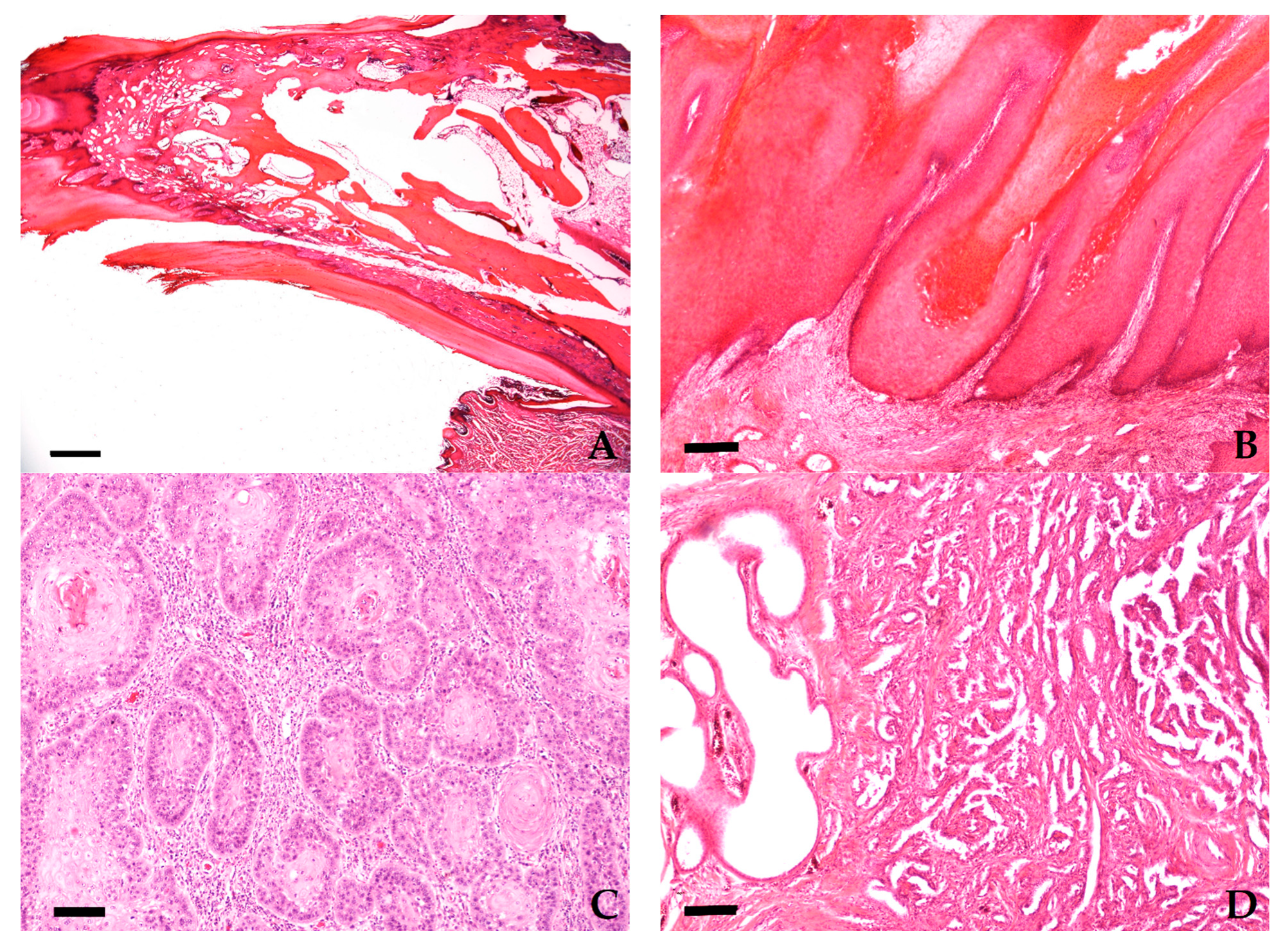
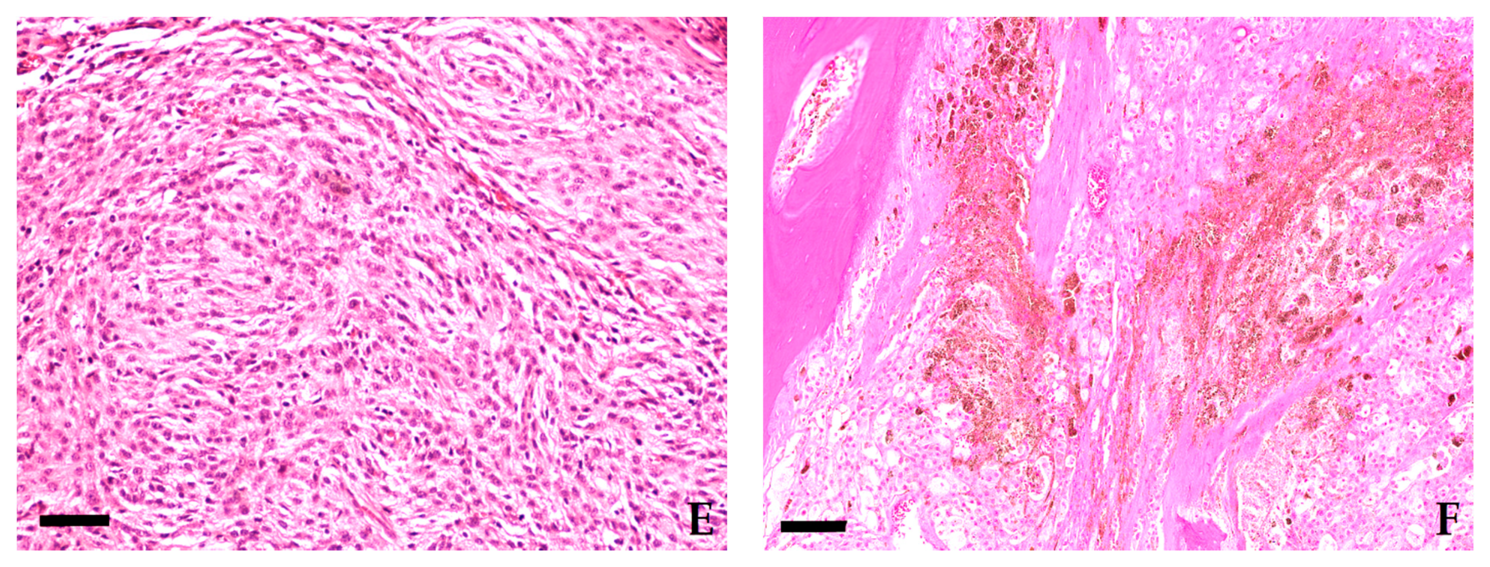
2.1. Histopathology
2.2. Statistics
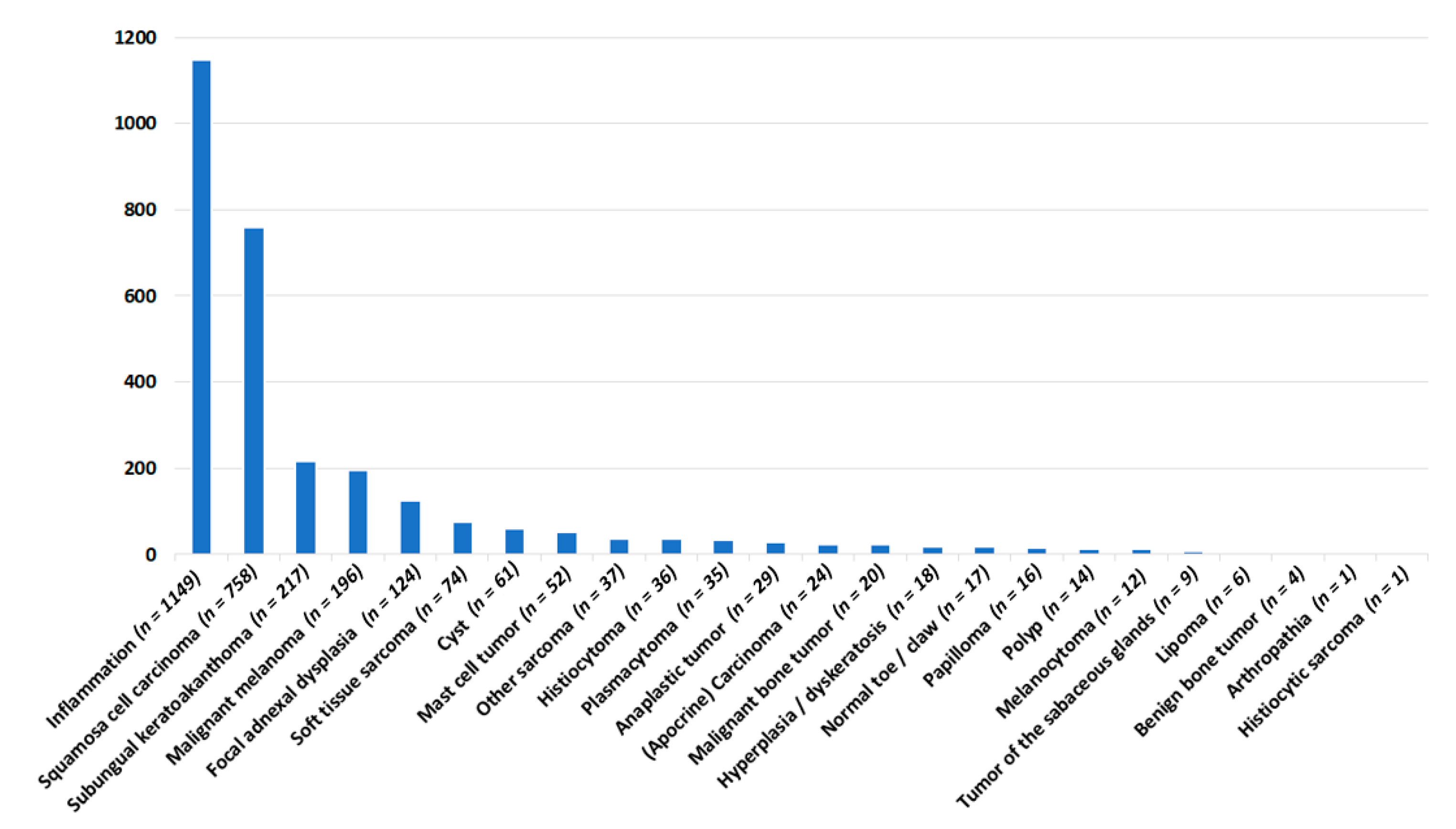
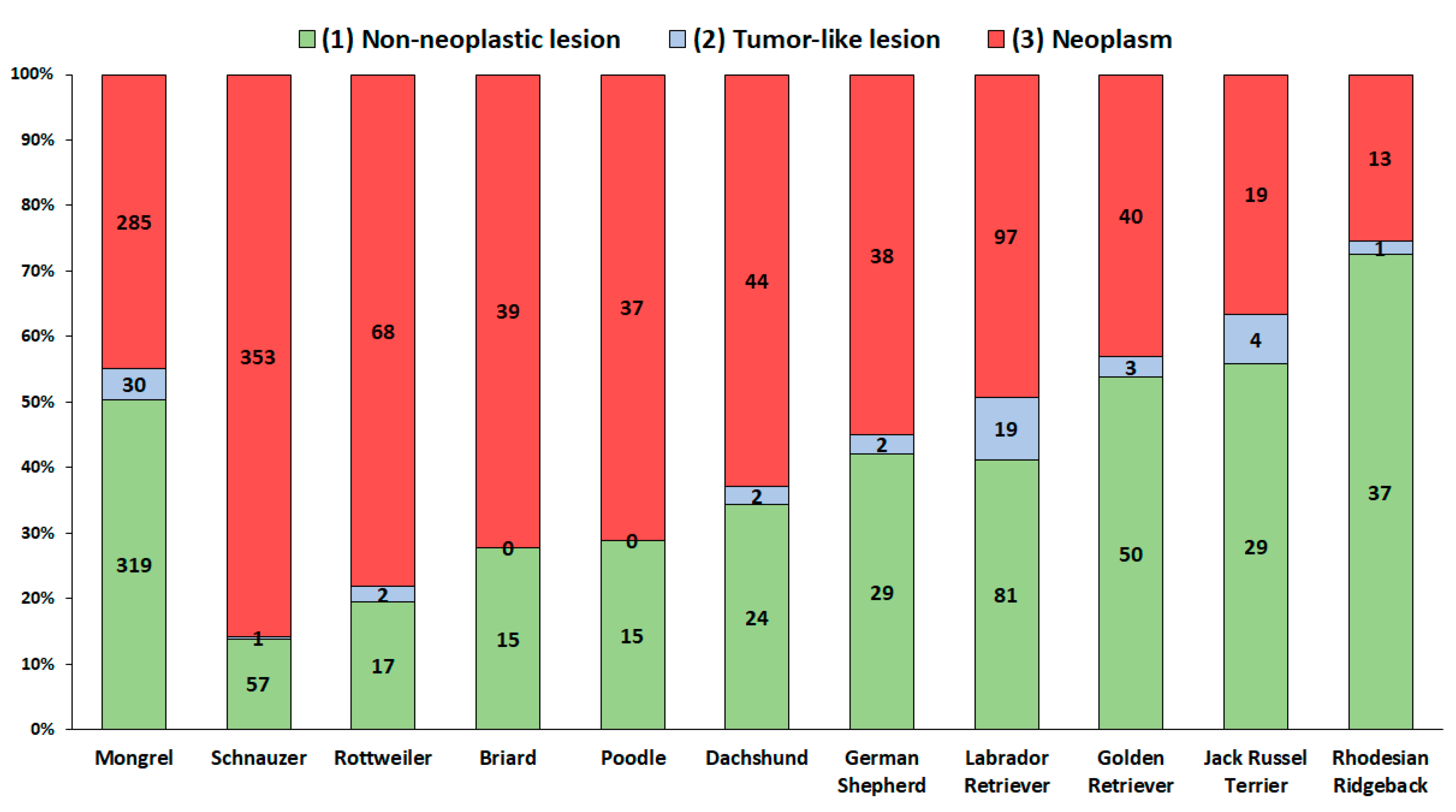
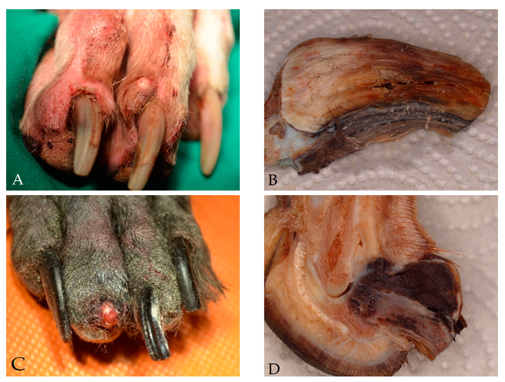
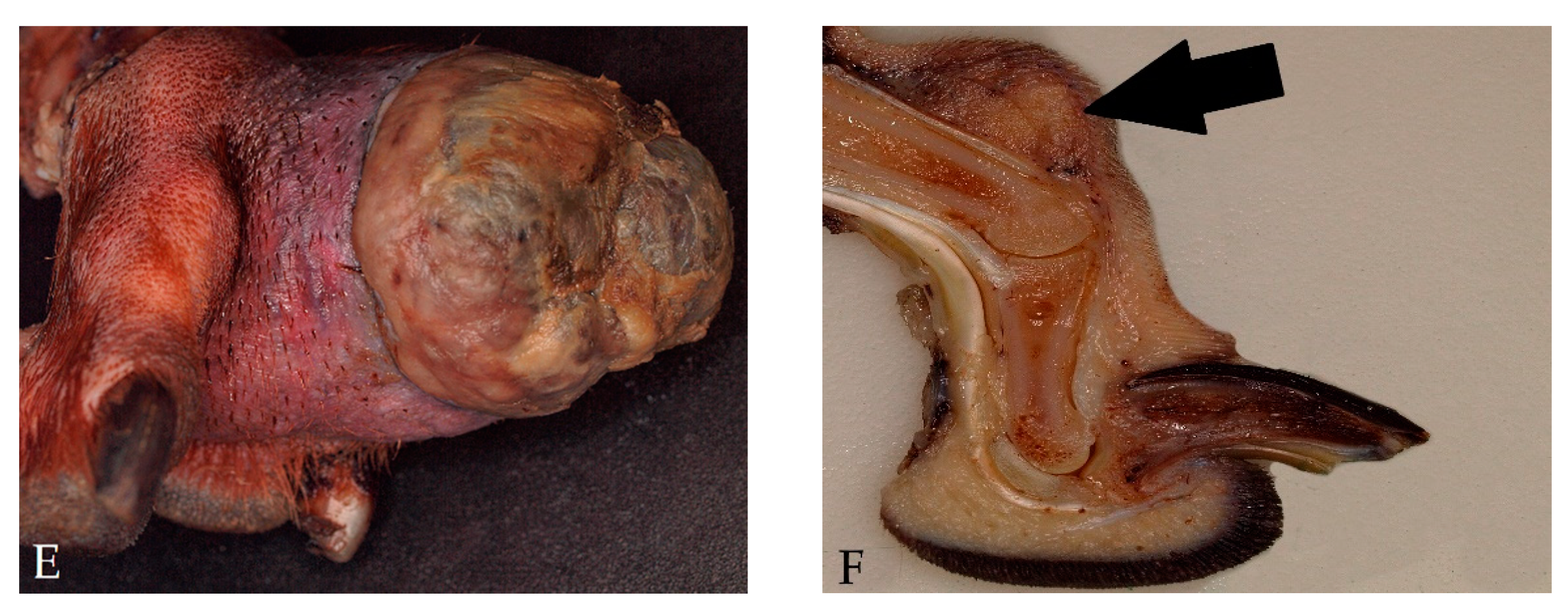
3. Results
4. Discussion
5. Conclusions
Author Contributions
Funding
Institutional Review Board Statement
Informed Consent Statement
Data Availability Statement
Conflicts of Interest
Abbreviations
References
- Wobeser, B.K.; Kidney, B.A.; Powers, B.E.; Withrow, S.J.; Mayer, M.N.; Spinato, M.T.; Allen, A.L. Diagnoses and clinical outcomes associated with surgically amputated canine digits submitted to multiple veterinary diagnostic laboratories. Vet. Pathol. 2007, 44, 355–361. [Google Scholar] [CrossRef]
- Henry, C.J.; Brewer, W.G.; Whitley, E.M.; Tyler, J.W.; Ogilvie, G.K.; Norris, A.; Fox, L.E.; Morrison, W.B.; Hammer, A.; Vail, D.M.; et al. Canine Digital Tumors: A Veterinary Cooperative Oncology Group Retrospective Study of 64 Dogs. J. Vet. Int. Med. 2005, 19, 720. [Google Scholar] [CrossRef]
- Marino, D.J.; Matthiesen, D.T.; Stefanacci, J.D.; Moroff, S.D. Evaluation of dogs with digit masses: 117 cases (1981–1991). J. Am. Vet. Med. Assoc. 1995, 207, 726–728. [Google Scholar] [PubMed]
- Gruber-Beckmann, B.; Aupperle, H.; Staudacher, M. Histopathologische Diagnosen nach Amputation umfangsvermehrter Zehen bei Hund und Katze. Kleintier Konkret 2016, 19, 37–39. [Google Scholar] [CrossRef]
- Belluco, S.; Brisebard, E.; Watrelot, D.; Pillet, E.; Marchal, T.; Ponce, F. Digital squamous cell carcinoma in dogs: Epidemiological, histological, and immunohistochemical study. Vet. Pathol. 2013, 50, 1078–1082. [Google Scholar] [CrossRef] [Green Version]
- Grüntzig, K.; Graf, R.; Boo, G.; Guscetti, F.; Hässig, M.; Axhausen, K.W.; Fabrikant, S.; Welle, M.; Meier, D.; Folkers, G.; et al. Swiss Canine Cancer Registry 1955–2008: Occurrence of the Most Common Tumour Diagnoses and Influence of Age, Breed, Body Size, Sex and Neutering Status on Tumour Development. J. Comp. Pathol. 2016, 155, 156–170. [Google Scholar] [CrossRef] [PubMed] [Green Version]
- O’Brien, M.G.; Berg, J.; Engler, S.J. Treatment by digital amputation of subungual squamous cell carcinoma in dogs: 21 cases (1987–1988). J. Am. Vet. Med. Assoc. 1992, 201, 759–761. [Google Scholar]
- Madewell, B.R.; Pool, R.R.; Theilen, G.H.; Brewer, W.G. Multiple subungual squamous cell carcinomas in five dogs. J. Am. Vet. Med. Assoc. 1982, 180, 731–734. [Google Scholar] [PubMed]
- Nishiya, A.T.; Massoco, C.O.; Felizzola, C.R.; Perlmann, E.; Batschinski, K.; Tedardi, M.V.; Garcia, J.S.; Mendonça, P.P.; Teixeira, T.F.; Zaidan Dagli, M.L. Comparative Aspects of Canine Melanoma. Vet. Sci. 2016, 3, 7. [Google Scholar] [CrossRef]
- Smith, S.H.; Goldschmidt, M.H.; McManus, P.M. A comparative review of melanocytic neoplasms. Vet. Pathol. 2002, 39, 651–678. [Google Scholar] [CrossRef] [PubMed]
- Gillard, M.; Cadieu, E.; de Brito, C.; Abadie, J.; Vergier, B.; Devauchelle, P.; Degorce, F.; Dréano, S.; Primot, A.; Dorso, L.; et al. Naturally occurring melanomas in dogs as models for non-UV pathways of human melanomas. Pigment Cell Melanoma Res. 2014, 27, 90–102. [Google Scholar] [CrossRef]
- Kamstock, D.A.; Ehrhart, E.J.; Getzy, D.M.; Bacon, N.J.; Rassnick, K.M.; Moroff, S.D.; Liu, S.M.; Straw, R.C.; McKnight, C.A.; Amorim, R.L.; et al. Recommended guidelines for submission, trimming, margin evaluation, and reporting of tumor biopsy specimens in veterinary surgical pathology. Vet. Pathol. 2011, 48, 19–31. [Google Scholar] [CrossRef] [Green Version]
- Goldschmidt, M.H.; Dunstan, R.W.; Stannard, A.A.; von Tscharner, C.; Walder, E.J.; Yager, J.A. Epithelial and Melanocytic Tumors of the Skin of Domestic Animals; Armed Forces Institute of Pathology, American Registry of Pathology, World Health Organization Collaborating Center for Comparative Oncology: Washington, DC, USA, 1998. [Google Scholar]
- Roccabianca, P.; Schulman, F.Y.; Avallone, G.; Kiupel, M.; Foster, R.A.; Scruggs, J.L.; Dittmer, K. Surgical Pathology of Tumors of Domestic Animals: Volume 3: Tumors of Soft Tissue; Davis Thompson Foundation: Gurnee, IL, USA, 2020; ISBN 9781733749121. [Google Scholar]
- Goldschmidt, M.H.; Kiupel, M.; Klopfleisch, R.; Munday, J.S.; Sruggs, J.L. Surgical Pathology of Tumors of Domestic Animals Volume 1: Epithelial Tumors of the Skin; Davis Thompson Foundation: Gurnee, IL, USA, 2018; ISBN 9781733749107. [Google Scholar]
- Heinze, G.; Ploner, M.; Jiricka, L. Logistf: Firth’s Bias-Reduced Logistic Regression, R Package Version 1.24; 2020. Available online: https://cran.r-project.org/web/packages/logistf/index.html (accessed on 17 July 2021).
- Lüdecke, D. _sjPlot: Data Visualization for Statistics in Social Science_. R. R Package Version 2.8.7. 2021. Available online: https://CRAN.R-project.org/package=sjPlot> (accessed on 9 February 2021).
- Benjamini, Y.; Hochberg, Y. Controlling The False Discovery Rate—A Practical And Powerful Approach to Multiple Testing. J. R. Stat. Soc. 1995, 57, 289–300. [Google Scholar] [CrossRef]
- Lapsley, J.; Selmic, L.E. Common Neoplastic Diseases Affecting the Forelimb. Vet. Clin. N. Am. Small Anim. Pract. 2021, 51, 343–356. [Google Scholar] [CrossRef]
- Horisberger, U.; Stärk, K.D.; Rüfenacht, J.; Pillonel, C.; Steiger, A. Demographie der Hundepopulation in der Schweiz. Schweiz. Arch. Tierheilkd. 2004, 146, 223–232. [Google Scholar] [CrossRef] [Green Version]
- Pospischil, A.; Hässig, M.; Vogel, R.; Salvini, M.M.; Fabrikant, S.; Axhausen, K.; Schenker, S.N.; Erni, D.; Guscetti, F. Hundepopulation und Hunderassen in der Schweiz von 1955 bis 2008. Schweiz. Arch. Tierheilkd. 2013, 155, 219–228. [Google Scholar] [CrossRef] [PubMed] [Green Version]
- Asher, L.; Buckland, E.L.; Phylactopoulos, C.I.; Whiting, M.C.; Abeyesinghe, S.M.; Wathes, C.M. Estimation of the number and demographics of companion dogs in the UK. BMC Vet. Res. 2011, 7, 74. [Google Scholar] [CrossRef] [PubMed] [Green Version]
- Metz, G. Die Geschichte des Pinscher-Schnauzer-Klub 1895 e.V. (PSK). Pinscher und Schnauzer-Klub feiert 125. Jubiläum 2020, 1, 11–14. [Google Scholar]
- Wobeser, B.K.; Kidney, B.A.; Powers, B.E.; Withrow, S.J.; Mayer, M.N.; Spinato, M.T.; Allen, A.L. Agreement among surgical pathologists evaluating routine histologic sections of digits amputated from cats and dogs. J. Vet. Diagn. Investig. 2007, 19, 439–443. [Google Scholar] [CrossRef] [PubMed] [Green Version]
- Karyadi, D.M.; Karlins, E.; Decker, B.; vonHoldt, B.M.; Carpintero-Ramirez, G.; Parker, H.G.; Wayne, R.K.; Ostrander, E.A. A copy number variant at the KITLG locus likely confers risk for canine squamous cell carcinoma of the digit. PLoS Genet. 2013, 9, e1003409. [Google Scholar] [CrossRef] [PubMed] [Green Version]
- Cerezo-Echevarria, A.; Grassinger, J.M.; Beitzinger, C.; Klopfleisch, R.; Aupperle-Lellbach, H. Evaluating the Histologic Grade of Digital Squamous Cell Carcinomas in Dogs with Dark and Light Haircoat-A Comparative Study of the Invasive Front and Tumor Cell Budding Systems. Vet. Sci. 2020, 8, 3. [Google Scholar] [CrossRef] [PubMed]
- Karlsson, E.K.; Baranowska, I.; Wade, C.M.; Salmon Hillbertz, N.H.C.; Zody, M.C.; Anderson, N.; Biagi, T.M.; Patterson, N.; Pielberg, G.R.; Kulbokas, E.J.; et al. Efficient mapping of mendelian traits in dogs through genome-wide association. Nat. Genet. 2007, 39, 1321–1328. [Google Scholar] [CrossRef]
- Federation Cynologique Internationale. FCI-Standard N° 345: Jack Russell Terrier; Geschäftstelle der FCI; Schlütersche: Hannover, Germany, 2012. [Google Scholar]
- Tompkins, S.; Fosgate, G.T.; Williams, J.; Clift, S. Breed and anatomical predisposition for canine cutaneous neoplasia in South Africa during 2013. Vet. Rec. 2020, 186, 218. [Google Scholar] [CrossRef]
- Tang, N.; Maloney, M.E.; Clark, A.H.; Jellinek, N.J. A Retrospective Study of Nail Squamous Cell Carcinoma at 2 Institutions. Dermatol. Surg. 2016, 42 (Suppl. 1), S8–S17. [Google Scholar] [CrossRef]
- Yan, W.; Wistuba, I.I.; Emmert-Buck, M.R.; Erickson, H.S. Squamous Cell Carcinoma—Similarities and Differences among Anatomical Sites. Am. J. Cancer Res. 2011, 1, 275–300. [Google Scholar] [CrossRef]
- vonHoldt, B.M.; Pollinger, J.P.; Lohmueller, K.E.; Han, E.; Parker, H.G.; Quignon, P.; Degenhardt, J.D.; Boyko, A.R.; Earl, D.A.; Auton, A.; et al. Genome-wide SNP and haplotype analyses reveal a rich history underlying dog domestication. Nature 2010, 464, 898–902. [Google Scholar] [CrossRef] [Green Version]
- Hendricks, W.P.D.; Zismann, V.; Sivaprakasam, K.; Legendre, C.; Poorman, K.; Tembe, W.; Perdigones, N.; Kiefer, J.; Liang, W.; DeLuca, V.; et al. Somatic inactivating PTPRJ mutations and dysregulated pathways identified in canine malignant melanoma by integrated comparative genomic analysis. PLoS Genet. 2018, 14, e1007589. [Google Scholar] [CrossRef] [Green Version]
- Wong, K.; van der Weyden, L.; Schott, C.R.; Foote, A.; Constantino-Casas, F.; Smith, S.; Dobson, J.M.; Murchison, E.P.; Wu, H.; Yeh, I.; et al. Cross-species genomic landscape comparison of human mucosal melanoma with canine oral and equine melanoma. Nat. Commun. 2019, 10, 353. [Google Scholar] [CrossRef] [PubMed]
- Mochizuki, H.; Kennedy, K.; Shapiro, S.G.; Breen, M. BRAF Mutations in Canine Cancers. PLoS ONE 2015, 10, e0129534. [Google Scholar] [CrossRef] [Green Version]
- Graf, R.; Pospischil, A.; Guscetti, F.; Meier, D.; Welle, M.; Dettwiler, M. Cutaneous Tumors in Swiss Dogs: Retrospective Data From the Swiss Canine Cancer Registry, 2008–2013. Vet. Pathol. 2018, 55, 809–820. [Google Scholar] [CrossRef]
- Pierini, A.; Lubas, G.; Gori, E.; Binanti, D.; Millanta, F.; Marchetti, V. Epidemiology of Breed-Related Mast Cell Tumour Occurrence and Prognostic Significance of Clinical Features in a Defined Population of Dogs in West-Central Italy. Vet. Sci. 2019, 6, 53. [Google Scholar] [CrossRef] [Green Version]
- Kok, M.K.; Chambers, J.K.; Tsuboi, M.; Nishimura, R.; Tsujimoto, H.; Uchida, K.; Nakayama, H. Retrospective study of canine cutaneous tumors in Japan, 2008–2017. J. Vet. Med. Sci. 2019, 81, 1133–1143. [Google Scholar] [CrossRef] [Green Version]
- Patnaik, A.K.; Ehler, W.J.; MacEwen, E.G. Canine cutaneous mast cell tumor: Morphologic grading and survival time in 83 dogs. Vet. Pathol. 1984, 21, 469–474. [Google Scholar] [CrossRef]
- Brønden, L.B.; Eriksen, T.; Kristensen, A.T. Mast cell tumours and other skin neoplasia in Danish dogs—Data from the Danish Veterinary Cancer Registry. Acta Vet. Scand. 2010, 52, 6. [Google Scholar] [CrossRef] [PubMed] [Green Version]
- Kiupel, M.; Webster, J.D.; Bailey, K.L.; Best, S.; DeLay, J.; Detrisac, C.J.; Fitzgerald, S.D.; Gamble, D.; Ginn, P.E.; Goldschmidt, M.H.; et al. Proposal of a 2-tier histologic grading system for canine cutaneous mast cell tumors to more accurately predict biological behavior. Vet. Pathol. 2011, 48, 147–155. [Google Scholar] [CrossRef] [PubMed]
- Bray, J.P. Soft tissue sarcoma in the dog—Part 1: A current review. J. Small Anim. Pract. 2016, 57, 510–519. [Google Scholar] [CrossRef] [PubMed] [Green Version]
- Simpson, S.; Dunning, M.D.; de Brot, S.; Grau-Roma, L.; Mongan, N.P.; Rutland, C.S. Comparative review of human and canine osteosarcoma: Morphology, epidemiology, prognosis, treatment and genetics. Acta Vet. Scand. 2017, 59, 71. [Google Scholar] [CrossRef] [PubMed]
- Egenvall, A.; Nødtvedt, A.; von Euler, H. Bone tumors in a population of 400,000 insured Swedish dogs up to 10 y of age: Incidence and survival. Can. J. Vet. Res. 2007, 71, 292–299. [Google Scholar]
- Goldfinch, N.; Argyle, D.J. Feline lung-digit syndrome: Unusual metastatic patterns of primary lung tumours in cats. J. Feline Med. Surg. 2012, 14, 202–208. [Google Scholar] [CrossRef]
- Moore, A.S.; Middleton, D.J. Pulmonary adenocarcinoma in three cats with nonrespiratory signs only. J. Small Anim. Pract. 1982, 23, 501–509. [Google Scholar] [CrossRef]
- Nødtvedt, A.; Berke, O.; Bonnett, B.N.; Brønden, L. Current status of canine cancer registration—Report from an international workshop. Vet. Comp. Oncol. 2012, 10, 95–101. [Google Scholar] [CrossRef] [PubMed]
- Villaescusa, A.; García-Sancho, M.; Delgado, A.M.; Tesouro, M.Á.; Rodríguez-Franco, F.; Sainz, A. Immunophenotypic evaluation of working Labrador Retrievers and German Shepherd dogs living in the same environment. Vet. J. 2012, 193, 602–605. [Google Scholar] [CrossRef]
- Prouteau, A.; André, C. Canine Melanomas as Models for Human Melanomas: Clinical, Histological, and Genetic Comparison. Genes 2019, 10, 501. [Google Scholar] [CrossRef] [PubMed] [Green Version]
- Wycislo, K.L.; Fan, T.M. The immunotherapy of canine osteosarcoma: A historical and systematic review. J. Vet. Intern. Med. 2015, 29, 759–769. [Google Scholar] [CrossRef] [PubMed]
- Hernandez, B.; Adissu, H.A.; Wei, B.-R.; Michael, H.T.; Merlino, G.; Simpson, R.M. Naturally Occurring Canine Melanoma as a Predictive Comparative Oncology Model for Human Mucosal and Other Triple Wild-Type Melanomas. Int. J. Mol. Sci. 2018, 19, 394. [Google Scholar] [CrossRef] [PubMed] [Green Version]
- Weich, K.; Affolter, V.; York, D.; Rebhun, R.; Grahn, R.; Kallenberg, A.; Bannasch, D. Pigment Intensity in Dogs is Associated with a Copy Number Variant Upstream of KITLG. Genes 2020, 11, 75. [Google Scholar] [CrossRef] [Green Version]


| Diagnosis | Breeds | Median Age (Range) in Years | Sex |
|---|---|---|---|
| Inflammation (n = 1149) | 291 Mongrels, 76 Labrador Retrievers, 51 Schnauzers, 47 Golden Retrievers, 38 German Shepherds, 37 Rhodesian Ridgebacks, 28 JRT, 22 Belgian Shepherds, 22 Boxers, 21 Dachshunds, 20 French Bulldogs, 20 Spaniels, 18 Great Danes, 17 Dobermanns, 16 Rottweilers, 15 BMDs, 15 WHWTs, 14 Bull Terriers, 14 Pugs, 14 Poodles, 13 Briards, 12 Beagles, 12 Setters, 11 Collies, 11 Irish Wolfhounds, 10 YTs, 8 Australian Shepherds, 8 Tibet Terriers, 7 Old German Shepherds, 7 ASTs, 7 English Bulldogs, 7 Fox Terriers, 7 Greyhounds, 7 Shih Tzus, 6 Bearded Collies, 6 Border Collies, 6 German Wire-haired Pointers, 6 Miniature Schnauzers, 5 Dalmatians, 5 Huskies, 5 Maltese, 5 White Swiss Shepherds, 4 Chihuahuas, 4 German Short-haired Pointers, 4 FCRs, 4 Spanish Greyhounds, 4 Hovawarts, 4 Olde English Bulldogs, 4 Parson Russell Terriers, 4 Russian Black Terriers, 4 Shetland Sheepdogs (Sheltie), 4 Weimaraners, 4 Whippets, 3 Airedale Terriers, 3 Akitas, 3 American Bulldogs, 3 Beaucerons, 3 Cairn Terriers, 3 Chow Chows, 3 Dogos Argentinos, 3 Irish Terriers, 3 Leonbergers, 3 Magyar Viszlas, 3 Podencos, 3 SBTs, 102 dogs from 58 other breeds | 7 (0.1–17) 121 U | 221 F, 206 FS, 415 M, 221 MC, 86 U |
| Cyst (n = 61) | 19 Mongrels, 3 Chihuahuas, 3 Great Danes, 3 Pugs, 33 dogs from 28 other breeds | 9 (2–14) 9 U | 13 F, 18 FS, 15 M, 9 MC, 6 U |
| Hyperplasia/dyskeratosis (n = 18) | 6 Mongrels, 3 Labrador Retrievers, 9 dogs from 9 other breeds | 6 (1–14) 1 U | 2 F, 3 FS, 8 M, 4 MC, 1 U |
| Normal toe/claw (n = 17) | 4 Schnauzers, 3 Mongrels, 2 Golden Retrievers, 8 dogs from 8 other breeds | 8 (2–11) 2 U | 5 FS, 6 M, 3 MC, 3 U |
| Arthropathia (n = 1) | 1 Dobermann | 1 U | 1 F |
| Diagnosis | Breeds | Median Age (Range) in Years | Sex |
|---|---|---|---|
| Focal adnexal dysplasia (n = 124) | 25 Mongrels, 19 Labrador Retrievers, 10 Airedale Terriers, 8 ASTs, 4 Old German Herding Dogs, 4 JRTs, 4 Greyhounds, 3 Alaska Malamutes, 3 American Bulldogs, 3 Boxers, 3 Golden Retrievers, 3 Huskies, 3 Magyar Vizslas, 32 dogs from 26 other breeds | 9 (1–16) 10 U | 15 F, 25 FS, 43 M, 32 MC, 9 U |
| Polyp (n = 14) | 5 Mongrel, 9 dogs from 9 other breeds | 9 (2–14) 9 U | 2 F, 1 FS, 3 M, 6 MC, 2 U |
| Diagnosis | Breeds | Median Age (Range) in Years | Sex |
|---|---|---|---|
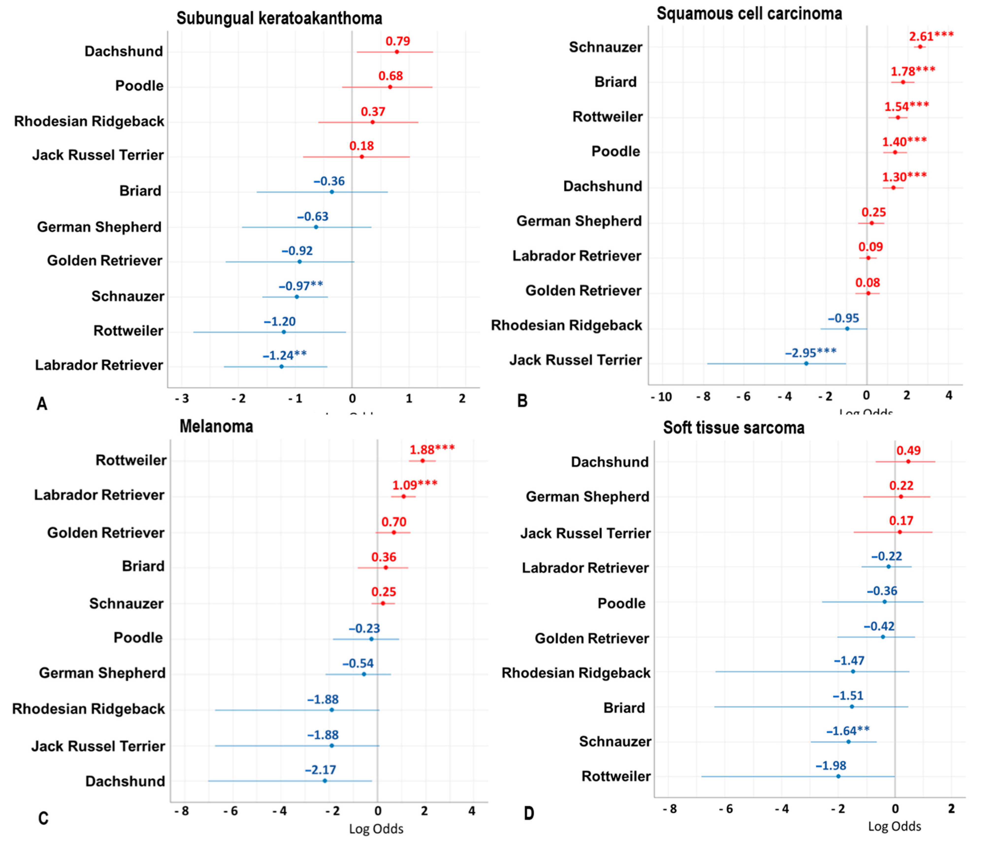
| Subungual keratoacanthoma (n = 217) | 55 Mongrels, 14 Schnauzers, 12 Dachshunds, 8 Poodles, 6 BMDs, 6 FCRs, 6 Rhodesian Ridgebacks, 6 YTs, 5 JRTs, 5 Labrador Retrievers, 5 Spaniels, 4 Beagles, 4 Hovawarts, 4 Setters, 3 Beaucerons, 3 Bolonki Zwetnas, 3 Border Collies, 3 Briards, 3 German Shepherds, 3 Dobermanns, 3 English Pointers, 3 Golden Retrievers, 3 Greyhounds, 3 Shetland Sheepdogs (Sheltie), 3 Shih Tzus, 44 dogs from 38 other breeds | 10 (1–16) 21 U | 42 F, 48 FS, 68 M, 49 MC, 10 U |
| Histiocytoma (n = 36) | 12 Mongrels, 7 French Bulldogs, 4 JRTs, 2 Boxers, 2 Magyar Vizslas, 9 dogs from 9 other breeds | 5 (1–12) 4 U | 7 F, 5 FS, 11 M, 11 MC, 2 U |
| Plasma cell tumor (n = 35) | 6 Mongrels, 4 JRTs, 3 French Bulldogs, 3 Labrador Retrievers, 3 Schnauzers, 3 WHWTs, 2 YTs, 11 dogs from 11 other breeds | 10 (3–16) 3 U | 6 F, 6 FS, 14 M, 7 MC, 2 U |
| Papilloma (n = 16) | 6 Mongrels, 2 Pinschers, 2 JRTs, 2 Huskies, 4 dogs from 4 other breeds | 8 (2–14) 3 U | 3 F, 5 FS, 4 M, 2 MC, 2 U |
| Melanocytoma (n = 12) | 3 Labrador Retrievers, 2 Briards, 7 dogs from 7 other breeds | 10 (5–12) 4 U | 1 F, 4 FS, 3 M, 4 MC |
| Tumor of the sebaceous glands (n = 9) | 3 Mongrels, 2 Labrador Retrievers, 4 dogs from 4 other breeds | 11 (11–14) | 4 FS, 3 M, 2 MC |
| Lipoma (n = 6) | 4 Mongrels, 2 dogs from 2 other breeds | 10 (4–12) 1 U | 2 FS, 2 MC, 2 U |
| Benign bone tumor (n = 4) | 1 French Bulldog, 1 Great Dane, 1 Australian Shepherd, 1 English Bulldog | 6 (3–9) | 1 FS, 1 M, 1 MC, 1 U |
| Diagnosis | Breeds | Median Age (Range) in Years | Sex |
|---|---|---|---|
| Squamous cell carcinoma (n = 758) | 298 Schnauzers, 98 Mongrels, 40 Rottweilers, 33 Labrador Retrievers, 28 Briards, 28 Dachshunds, 27 FCRs, 22 Poodles, 20 Setters, 15 Golden Retrievers, 13 German Shepherds, 11 Miniature Schnauzers, 8 BMD, 8 Hovawarts, 7 Russian Black Terriers, 7 Spaniels, 6 Airedale Terriers, 6 German Short-haired Pointers, 4 Australian Shepherds, 3 Beaucerons, 3 Boxers, 3 Kerry Blue Terriers, 3 Leonbergers, 3 Rhodesian Ridgebacks, 3 WHWTs, 3 YTs, 58 dogs from 42 other breeds | 10 (3–16) 72 U | 155 F, 165 FS, 268 M, 111 MC, 59 U |
| Malignant melanoma (n = 196) | 39 Mongrels, 31 Labrador Retrievers, 31 Schnauzers, 25 Rottweilers, 9 Golden Retrievers, 6 Spaniels, 4 Briards, 4 Dobermanns, 3 Russian Black Terriers, 3 Scottish Terriers, 41 dogs from 31 other breeds | 10 (2–16) 20 U | 47 F, 30 FS, 75 M, 29 MC, 15 U |
| Soft tissue sarcoma (n = 76) | 21 Mongrels, 5 Labrador Retrievers, 4 Dachshunds, 3 German Shepherds, 43 dogs from 33 other breeds | 11 (5–17) 4 U | 16 F, 19 FS, 29 M, 8 MC, 4 U |
| Mast cell tumor (n = 52) | 8 Boxers, 6 Mongrels, 6 Labrador Retrievers, 5 Golden Retrievers, 4 BMDs, 4 French Bulldogs, 19 dogs from 19 other breeds | 8 (2–16) 8 U | 13 F, 8 FS, 18 M, 9 MC, 4 U |
| Other sarcoma (n = 37) | 10 Mongrels, 3 Labrador Retrievers, 3 German Shepherds, 3 Australian Shepherds, 2 Beagles, 16 dogs from 16 other breeds | 10 (5–16) 1 U | 10 F, 12 FS, 8 M, 5 MC, 2 U |
| Anaplastic tumor (n = 29) | 9 Mongrels, 4 Golden Retrievers, 3 Labrador Retrievers, 3 Schnauzers, 10 dogs from 10 other breeds | 10 (2–17) 6 U | 9 F, 5 FS, 10 M, 4 MC, 1 U |
| (Apocrine) Carcinoma (n = 24) | 9 Mongrels, 15 dogs from 15 other breeds | 11 (8–14) 4 U | 3 F, 6 FS, 7 M, 7 MC, 1 U |
| Malignant bone tumor (n = 20) | 6 Mongrels, 2 German Shepherds, 2 Labrador Retrievers, 2 Italian Cani Corsi, 8 dogs from 8 other breeds | 9 (3–13) 4 U | 6 F, 4 FS, 5 M, 4 MC, 1 U |
| Histiocytic sarcoma (n = 1) | 1 Mongrel | 9 | 1 MC |
Publisher’s Note: MDPI stays neutral with regard to jurisdictional claims in published maps and institutional affiliations. |
© 2021 by the authors. Licensee MDPI, Basel, Switzerland. This article is an open access article distributed under the terms and conditions of the Creative Commons Attribution (CC BY) license (https://creativecommons.org/licenses/by/4.0/).
Share and Cite
Grassinger, J.M.; Floren, A.; Müller, T.; Cerezo-Echevarria, A.; Beitzinger, C.; Conrad, D.; Törner, K.; Staudacher, M.; Aupperle-Lellbach, H. Digital Lesions in Dogs: A Statistical Breed Analysis of 2912 Cases. Vet. Sci. 2021, 8, 136. https://doi.org/10.3390/vetsci8070136
Grassinger JM, Floren A, Müller T, Cerezo-Echevarria A, Beitzinger C, Conrad D, Törner K, Staudacher M, Aupperle-Lellbach H. Digital Lesions in Dogs: A Statistical Breed Analysis of 2912 Cases. Veterinary Sciences. 2021; 8(7):136. https://doi.org/10.3390/vetsci8070136
Chicago/Turabian StyleGrassinger, Julia Maria, Andreas Floren, Tobias Müller, Argiñe Cerezo-Echevarria, Christoph Beitzinger, David Conrad, Katrin Törner, Marlies Staudacher, and Heike Aupperle-Lellbach. 2021. "Digital Lesions in Dogs: A Statistical Breed Analysis of 2912 Cases" Veterinary Sciences 8, no. 7: 136. https://doi.org/10.3390/vetsci8070136