Paleopathological Changes in Animal Bones from Croatian Archaeological Sites from Prehistory to New Modern Period

Abstract
:Simple Summary
Abstract
1. Introduction
2. Materials and Methods
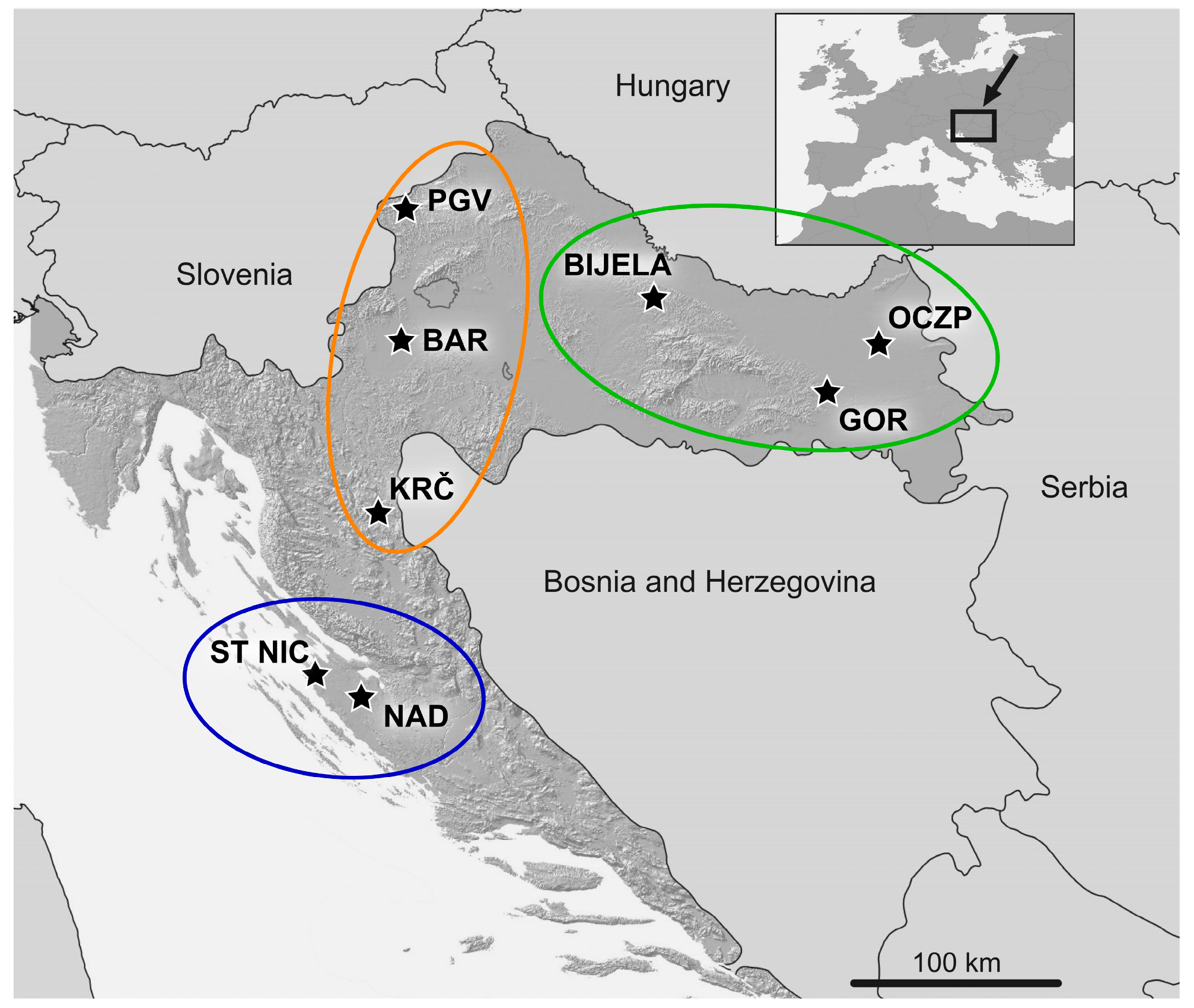
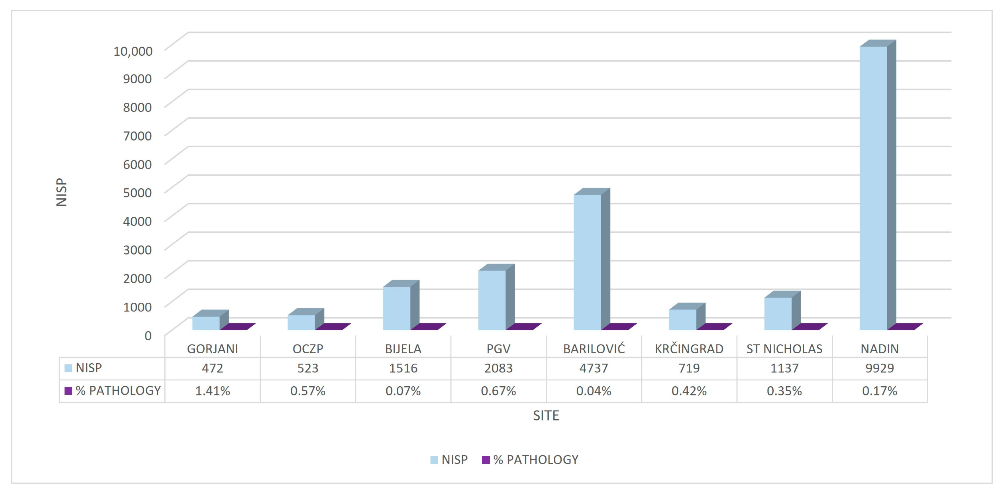
2.1. Archaeological Sites
2.1.1. Eastern Continental Croatia
2.1.2. Western Continental Croatia
2.1.3. Northern Dalmatia
2.2. Archaeozoological Analysis and Diagnostic Imaging
3. Results
3.1. Skull
3.2. Ribs and Vertebrae
3.3. Thoracic Limb Bones
3.4. Pelvic Limb Bones
3.5. Bones of the Metapodium and Acropodium
4. Discussion
4.1. Working/Keeping Etiology
4.2. Traumatic Changes
4.3. Oral Cavity Group
5. Conclusions
Author Contributions
Funding
Institutional Review Board Statement
Informed Consent Statement
Data Availability Statement
Acknowledgments
Conflicts of Interest
References
- Thomas, R. NonHuman Paleopathology. In The Global History of Paleopathology; Oxford University Press: Oxford, UK, 2012; pp. 654–666. [Google Scholar]
- Bartosiewicz, L. Pathological Lesions on Prehistoric Animal Remains from Southwest Asia. In Archaeozoology of the Near East V; Taylor & Francis: Abingdon, UK, 2002; pp. 320–336. [Google Scholar]
- O’Connor, T. The Archaeology of Animal Bones, 1st ed.; Sutton Publishing Ltd.: Stroud, UK, 2000. [Google Scholar]
- Bendrey, R.; Taylor, G.M.; Bouwman, A.S.; Cassidy, J.P. Suspected Bacterial Disease in Two Archaeological Horse Skeletons from Southern England: Palaeopathological and Biomolecular Studies. J. Archaeol. Sci. 2008, 35, 1581–1590. [Google Scholar] [CrossRef]
- Janeczek, M.; Chroszcz, A.; Miklikova, Z.; Fabis, M. The Pathological Changes in the Hind Limb of a Horse from the Roman Period. Vet. Med. 2010, 55, 331–335. [Google Scholar] [CrossRef]
- Bendrey, R. Care in the Community? Interpretations of a Fractured Goat Bone from Neolithic Jarmo, Iraq. Int. J. Paleopathol. 2014, 7, 33–37. [Google Scholar] [CrossRef]
- Bartosiewicz, L. Taphonomy and Palaeopathology in Archaeozoology. Geobios 2008, 41, 69–77. [Google Scholar] [CrossRef]
- Bulatović, J.; Marković, N.; Krstić, N.; Bulatović, A.; Mitrović, M.; Marinković, D. Animal Diseases in the Central Balkan Eneolithic (ca. 4500–2500 BC)—A Diachronic Perspective on the Site of Bubanj, South-Eastern Serbia. Int. J. Osteoarchaeol. 2022, 32, 303–316. [Google Scholar] [CrossRef]
- Siegel, J. Animal Palaeopathology: Possibilities and Problems. J. Archaeol. Sci. 1976, 3, 349–384. [Google Scholar] [CrossRef]
- Upex, B.; Dobney, K. More Than Just Mad Cows: Exploring Human-Animal Relationships through Animal Paleopathology. In A Companion to Paleopathology; Wiley-Blackwell: Oxford, UK, 2012; pp. 191–213. [Google Scholar] [CrossRef]
- Bartosiewicz, L.; Demeure, R.; Mottet, I.; Van Neer, W.; Lentacker, A. Magnetic resonance imaging in the study of spavin in recent and subfossil cattle. Anthropozoologica 1997, 25, 57–60. [Google Scholar]
- Bendrey, R. New Methods for the Identification of Evidence for Bitting on Horse Remains from Archaeological Sites. J. Archaeol. Sci. 2007, 34, 1036–1050. [Google Scholar] [CrossRef]
- Pluskowski, A.; Seetah, K.; Maltby, M. Potential Osteoarchaeological Evidence for Riding and the Military Use of Horses at Malbork Castle, Poland. Int. J. Osteoarchaeol. 2010, 20, 335–343. [Google Scholar] [CrossRef]
- Onar, V.; Pazvant, G.; Chrószcz, A. Byzantine Horse Skeletons of Theodosius Harbour: 1.Paleopathology. Rev. Med. Vet. 2012, 163, 139–146. [Google Scholar]
- Marković, N.; Stevanović, O.; Nešić, V.; Marinković, D.; Krstić, N.; Nedeljković, D.; Radmanović, D.; Janeczek, M. Paleopathological Study of Cattle and Horse Bone Remains of the Ancient Roman City of Sirmium (Pannonia/Serbia). Rev. Med. Vet. 2014, 165, 77–88. [Google Scholar]
- Makowiecki, D.; Janeczek, M.; Pasicka, E.; Rozwadowska, A.; Ciaputa, R.; Kocińska, M.K. Pathologies of a Horse Skeleton from the Early Medieval Stronghold in Gdańsk (Poland). Int. J. Osteoarchaeol. 2022, 32, 866–877. [Google Scholar] [CrossRef]
- Babo, V. Pferdebestattungen Auf Dem Frühmittelalterlichen Gräberfeld Drantumer Mühle (Gemeinde Emstek, Kreis Cloppenburg, Niedersachsen); Tierärztliche Hochschule Hannover: Hannover, Germany, 2004. [Google Scholar]
- Trbojević Vukičević, T.; Rapan Papeša, A.; Alić, I.; Ekert Kabalin, A.; Ostović, M.; Kužir, S. Contribution to Understanding Avar Burials with Equids in Croatia: Detailed Archaeozoological Analysis. Rev. Med. Vet. (Toulouse) 2017, 168, 73–80. [Google Scholar]
- Telldahl, Y. Skeletal Changes in Lower Limb Bones in Domestic Cattle from Eketorp Ringfort on the Öland Island in Sweden. Int. J. Paleopathol. 2012, 2, 208–216. [Google Scholar] [CrossRef]
- Holmes, M.; Thomas, R.; Hamerow, H. Periodontal Disease in Sheep and Cattle: Understanding Dental Health in Past Animal Populations. Int. J. Paleopathol. 2021, 33, 43–54. [Google Scholar] [CrossRef]
- Lin, M.; Luan, F.; Fang, H.; Xu, H.; Zhao, H.; Barker, G. Pathological Evidence Reveals Cattle Traction in North China by the Early Second Millennium BC. Holocene 2018, 28, 1205–1215. [Google Scholar] [CrossRef]
- Maldre, L. Pathological Bones amongst the Archaeozoological Material from Estonian Towns. Vet. Ir Zootech. 2008, 64, 51–57. [Google Scholar]
- Harcourt, R. Animal Bones from Thistleton; Ancient Monuments Laboratory: London, UK, 1971. [Google Scholar]
- Stingl, S.; Hirschler Marić, I.; Sekulić, P. Metal finds from the area of the Yahya-bey tower, present day chapel of the magi, in Gorjani. Vjesnik Arheološkog Muzeja Zagrebu. 2022, 55, 301–333. [Google Scholar] [CrossRef]
- Krmpotić, M.; Trbojević Vukičević, T.; Essert, S. Bronze and Iron Age settlements at the site of Osijek–Ciglana and Zeleno polje. Pril. Inst. Arheol. Zagreb. 2022, 39, 81–127. [Google Scholar] [CrossRef]
- Janeš, A.; Sekulić, P. Rudina and Bijela: Benedictine monasteries of Late Mediaeval Slavonia. Starohrv. Prosvj. 2014, 14, 185–204. [Google Scholar]
- Tkalčec, T. Burg Vrbovec u Klenovcu Humskome, 1st ed.; Horjan, G., Tomičić, Ž., Eds.; Muzeji Hrvatskog zagorja, Institut za arheologiju: Zagreb, Croatia, 2010; Volume 1. [Google Scholar]
- Tkalčec, T. Castle Vrbovec in Klenovec Humski—Continuation of Archaeological and Conservation Works on the Keep in 2016. In Annales Instituti Archaeologici; Botić, K., Krznar, S., Ožanić Roguljić, I., Konestra, A., Kudelić, A., Tonc, A., Ugarković, M., Eds.; Institute of Archaeology: Zagreb, Croatia, 2017; pp. 92–96. [Google Scholar]
- Azinović Bebek, A.; Krmpotić, M. Stari Grad Barilović—10 Godina Arheoloških Istraživanja, 1st ed.; Azinović Bebek, A., Krmpotić, M., Eds.; Hrvatski restauratorski zavod: Zagreb, Croatia, 2014. [Google Scholar]
- Kekez, H.; Pleše, T.; Sekulić, P. Krčingrad at Plitvice and the Babonić—Contextualizing the Date of Construction. Povij. Pril. 2018, 54, 65–99. [Google Scholar] [CrossRef]
- Čače, S. Broj Liburnijskih Općina i Vjerodostojnost Plinija. Razdio Povij. Znan. 1993, 32, 1–36. [Google Scholar]
- Zaro, G.; Gusar, K.; Čelhar, M. On the Edge of Empires: Exploring an Ottoman Legacy on the Venetian Frontier. J. Field Archaeol. 2020, 45, 188–208. [Google Scholar] [CrossRef]
- Bekić, L.; Ferenčić, N. Saint Nicholas in Zadar: Archaeological Excavations at Zadar’s St Nicholas Monastery Complex 2014–2016; Međunarodni Centar za Podvodnu Arheologiju: Zadar, Croatia, 2017; p. 1. [Google Scholar]
- Schmid, E. Atlas of Animal Bones for Prehistorians, Archaeologists and Quaternary Geologists; Elsevier Publishing Company: Amsterdam, The Netherlands, 1972; Volume 1. [Google Scholar]
- Prummel, W.; Frisch, H.J. A Guide for the Distinction of Species, Sex and Body Side in Bones of Sheep and Goat. J. Archaeol. Sci. 1986, 13, 567–577. [Google Scholar] [CrossRef]
- Hillson, S.; Brothwell, D.; Cunliffe, B.; Fleming, S.; Powler, P. (Eds.) Teeth; Press Syndicate of the University of Cambridge: Cambridge, UK, 1996. [Google Scholar]
- König, H.E.; Liebich, H.-G. Veterinary Anatomy of Domestic Animals, 7th ed.; Georg Thieme Verlag KG: Stuttgart, Germany, 2020. [Google Scholar] [CrossRef]
- Rackham, J.D. Animal Bones; University of California Press: Berkeley, CA, USA, 1994. [Google Scholar]
- Lyman, R.L. Vertebrate Taphonomy; Cambridge University Press: Cambridge, UK, 1994. [Google Scholar] [CrossRef]
- Baker, J.; Brothwell, D. Animal Diseases in Archaeology; Academic Press Inc.: London, UK, 1980. [Google Scholar]
- Thrall. Veterinary Diagnostic Radiology, 7th ed.; Elsevier Inc.: Amsterdam, The Netherlands, 2018. [Google Scholar]
- von den Driesch, A. A Guide to the Measurement of Animal Bones from Archaeological Sites; Peabody Museum of Archaeology & Ethnology: Cambridge, MA, USA, 1976. [Google Scholar]
- Stevanović, O.; Janeczek, M.; Chrószcz, A.; Marković, N. Joint Diseases in Animal Paleopathology: Veterinary Approach. Maced. Vet. Rev. 2015, 38, 5–12. [Google Scholar] [CrossRef]
- Thomas, R. Diachronic Trends in Lower Limb Pathologies in Later Medieval and Post-Medieval Cattle from Britain. In Limping Together through the Ages. Joint Afflictions and Bone Infections; Grupe, G., McGlynn, G., Peters, J., Eds.; Verlag Marie Leidorf: Rahden, Germany, 2008; pp. 187–201. [Google Scholar]
- Craig, L.E.; Dittmer, K.E.; Thompson, K.G. Bones and Joints. In Jubb, Kennedy & Palmer’s Pathology of Domestic Animals; Elsevier: Amsterdam, The Netherlands, 2015; Volume 1, pp. 16–163. [Google Scholar]
- Connor, T. On the Differential Diagnosis of Arthropathy in Bovids. Doc. Archaeobiol. 2008, 6, 165–186. [Google Scholar]
- Zachary, J.F. Pathologic Basis of Veterinary Disease, 7th ed.; Elsevier: St. Louis, MO, USA, 2022. [Google Scholar]
- Bartosiewicz, L. Shuffling Nags, Lame Ducks: The Archaeology of Animal Disease; Oxbow Books Ltd.: Oakville, ON, Canada, 2013. [Google Scholar]
- De Cupere, B.; Lentacker, A.; Van Neer, W.; Waelkens, M.; Verslype, L. Osteological Evidence for the Draught Exploitation of Cattle: First Applications of a New Methodology. Int. J. Osteoarchaeol. 2000, 10, 254–267. [Google Scholar] [CrossRef]
- Janeczek, M.; Chrószcz, A.; Onar, V.; Henklewski, R.; Piekalski, J.; Duma, P.; Czerski, A.; Całkosiński, I. Anatomical and Biomechanical Aspects of the Horse Spine: The Interpretation of Vertebral Fusion in a Medieval Horse from Wroclaw (Poland). Int. J. Osteoarchaeol. 2014, 24, 623–633. [Google Scholar] [CrossRef]
- Telldahl, Y. Can Palaeopathology Be Used as Evidence for Draught Animals? In Diet and Health in Past Animal Populations; Oxbow Books: Oxford, UK, 2002; pp. 63–67. [Google Scholar]
- Latham, K.J.; Losey, R.J. Spondylosis Deformans as an Indicator of Transport Activities in Archaeological Dogs: A Systematic Evaluation of Current Methods for Assessing Archaeological Specimens. PLoS ONE 2019, 14, e0214575. [Google Scholar] [CrossRef]
- Groot, M. Understanding Past Human-Animal Relationships through the Analysis of Fractures: A Case Study from a Roman Site in The Netherlands. In Current Research in Animal Palaeopathology Proceedings of the Second ICAZ Animal Palaeopathology Working Group Conference; BAR Publishing: Oxford, UK, 2008; Volume 1, pp. 40–50. [Google Scholar]
- Teegen, W.R.; Wussow, J. Maltreatment of Animals in the Late 19th and Early 20th Century AD? Evidence from the Julius-Kuehn Collection; University of Halle-Wittenberg (Germany): Halle, Germany, 2000. [Google Scholar]
- Teegen, W.R. Diet and Health in Past Animal Populations: Current Research and Future Directions. In Proceedings of the 9th ICAZ Conference, Durham 2002; Davies, J., Fabiš, M., Mainland, I., Richards, M., Thomas, R., Eds.; Oxbow Books Ltd.: Oxford, UK, 2005. [Google Scholar]


| Sample No. | Site | Period | Bone Element | Side | Species | AGE |
|---|---|---|---|---|---|---|
| P1 | PGV | High Middle Ages | MTC | left | cattle | >2–2.5 y |
| P2 | PGV | High Middle Ages | talus | right | pig | |
| P3 | PGV | High Middle Ages | MTC IV | right | pig | <2 y |
| P4 | PGV | High Middle Ages | femur, dist | right | pig | <3.5 y |
| P5 | PGV | Late Middle Ages | metapodium, dist | cattle | <2–2.5 y | |
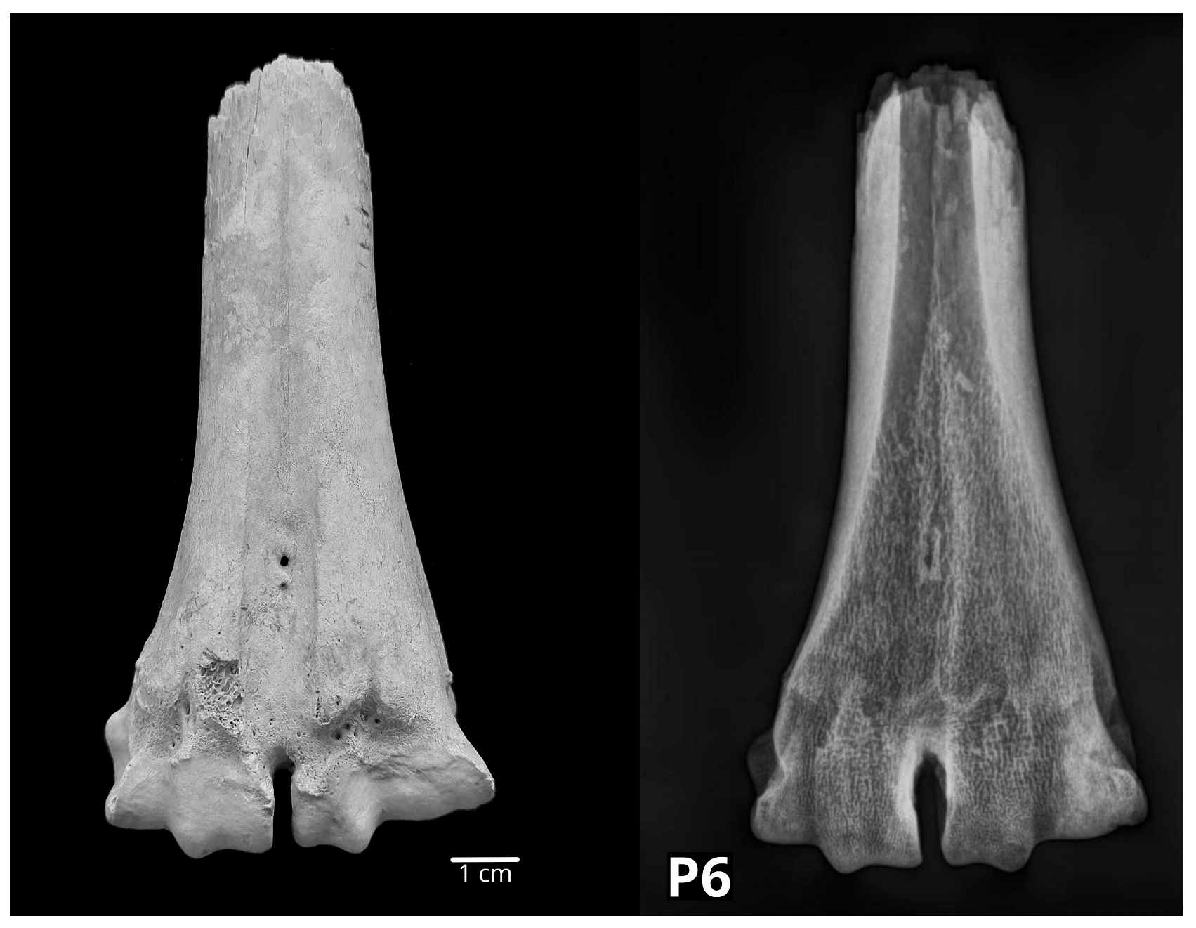
| P6 | PGV | High Middle Ages | MTT, dist | left | cattle | <2–2.5 y |
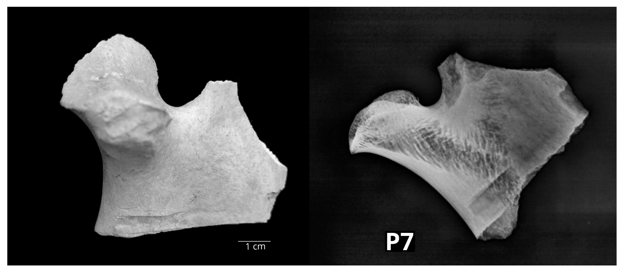
| P7 | PGV | Early Modern Period | mandibula, caput | left | cattle | |
| P8 | PGV | Late Middle Ages | humerus, dist | right | small R | >11–13 m |
| P9 | PGV | High Middle Ages | costa | small R | ||
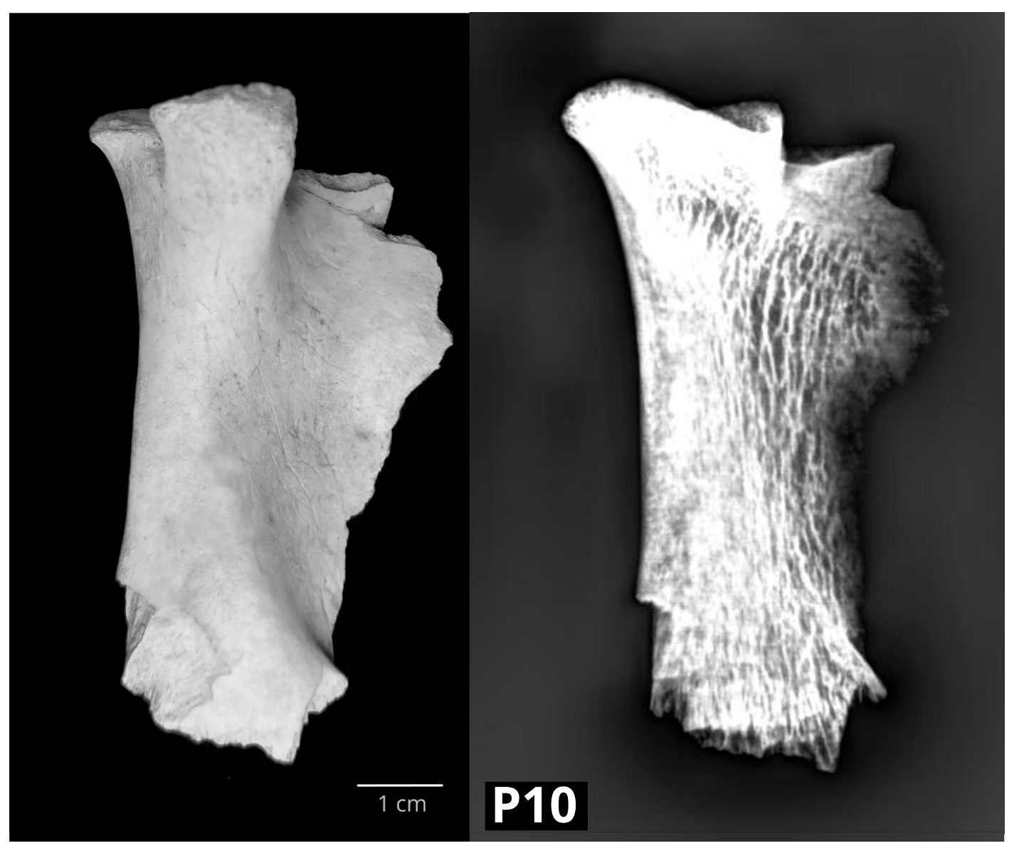
| P10 | PGV | High Middle Ages | mandibula | left | cattle | |
| P11 | PGV | High Middle Ages | vertebra lumbalis | pig | <4–7 y | |
| P12 | PGV | Early Modern Period | calcaneus | left | cattle | >3–3.5 y |
| P13 | PGV | Early Modern Period | calcaneus | left | cattle | >3–3.5 y |
| P14 | PGV | Early Modern Period | acetabulum | left | cattle | >7–10 m |
| P15 | KRČ | High/Late Middle Ages | mandibula, rostral | right | small R | |
| P16 | KRČ | High/Late Middle Ages | humerus, dist | right | pig | >1–1.5 y |
| P17 | KRČ | High/Late Middle Ages | tibia, dist | left | pig | >2 y |
| P18 | BIJELA | Early Modern Period | mandibula, rostral | left | goat | |
| P19 | BAR | Late Middle Ages | MTC, prox | right | cattle | |
| P20 | BAR | High/Late Middle Ages | ulna | right | small R | >2.5–3 y |
| P21 | OCZP | Late phase of the Middle Bronze Age | mandibula | right | pig | >1.5–2 y |
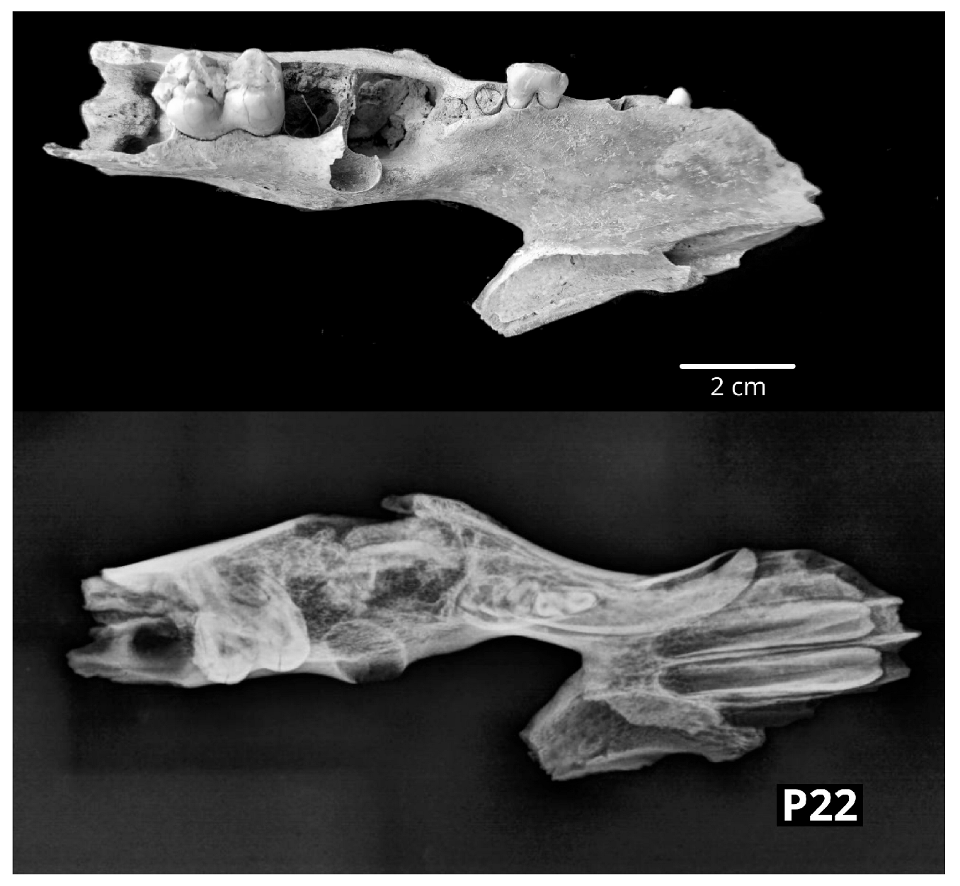
| P22 | OCZP | Early phase of the Middle Bronze Age | mandibula | left | pig | |
| P23 | OCZP | Early phase of the Middle Bronze Age | vertebrae lumbales (4) | carnivore | ||
| P24 | GOR | Early Modern Period | radius, dist | left | horse | >3.5 y |
| P25 | GOR | Early Modern Period | MTC, dist | right | cattle | >2–2.5 y |
| P26 | GOR | Early Modern Period | phalanx proximalis | cattle | >1.5 y | |
| P27 | GOR | Early Modern Period | phalanx media | cattle | >1.5 y | |
| P28 | GOR | Early Modern Period | MTC, dist | right | cattle | >2–2.5 y |
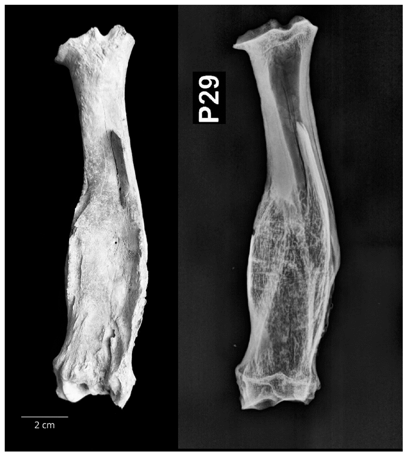
| P29 | GOR | Early Modern Period | radius | right | small R | >3–4 y |
| P30 | ST NIC | Iron age | phalanx proximalis | cattle | >1.5 y | |
| P31 | ST NIC | Iron age | phalanx proximalis | cattle | >1.5 y | |
| P32 | ST NIC | Early Modern Period | phalanx proximalis | cattle | >1.5 y | |
| P33 | ST NIC | Early Modern Period | MTC, dist | right | cattle | >2–2.5 y |
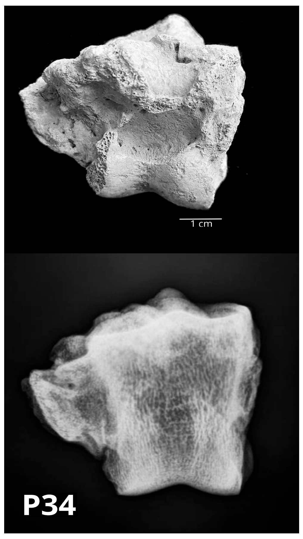
| P34 | NADIN | Antiquity | phalanx media | cattle | >1.5 y | |
| P35 | NADIN | Antiquity | os tarsi centroquartale | right | cattle | |
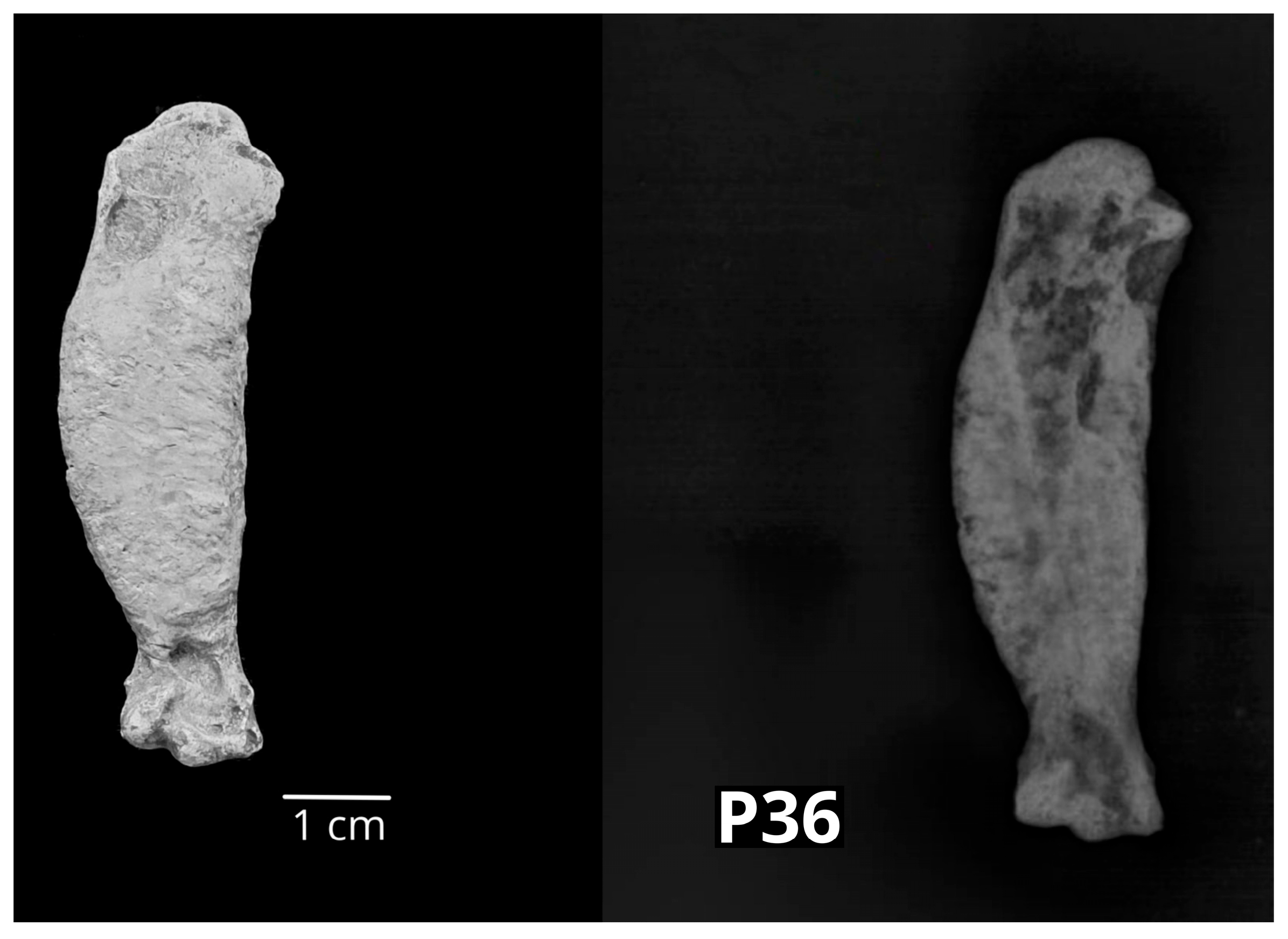
| P36 | NADIN | Late Middle Ages/Early Modern Period | humerus | right | chicken | |
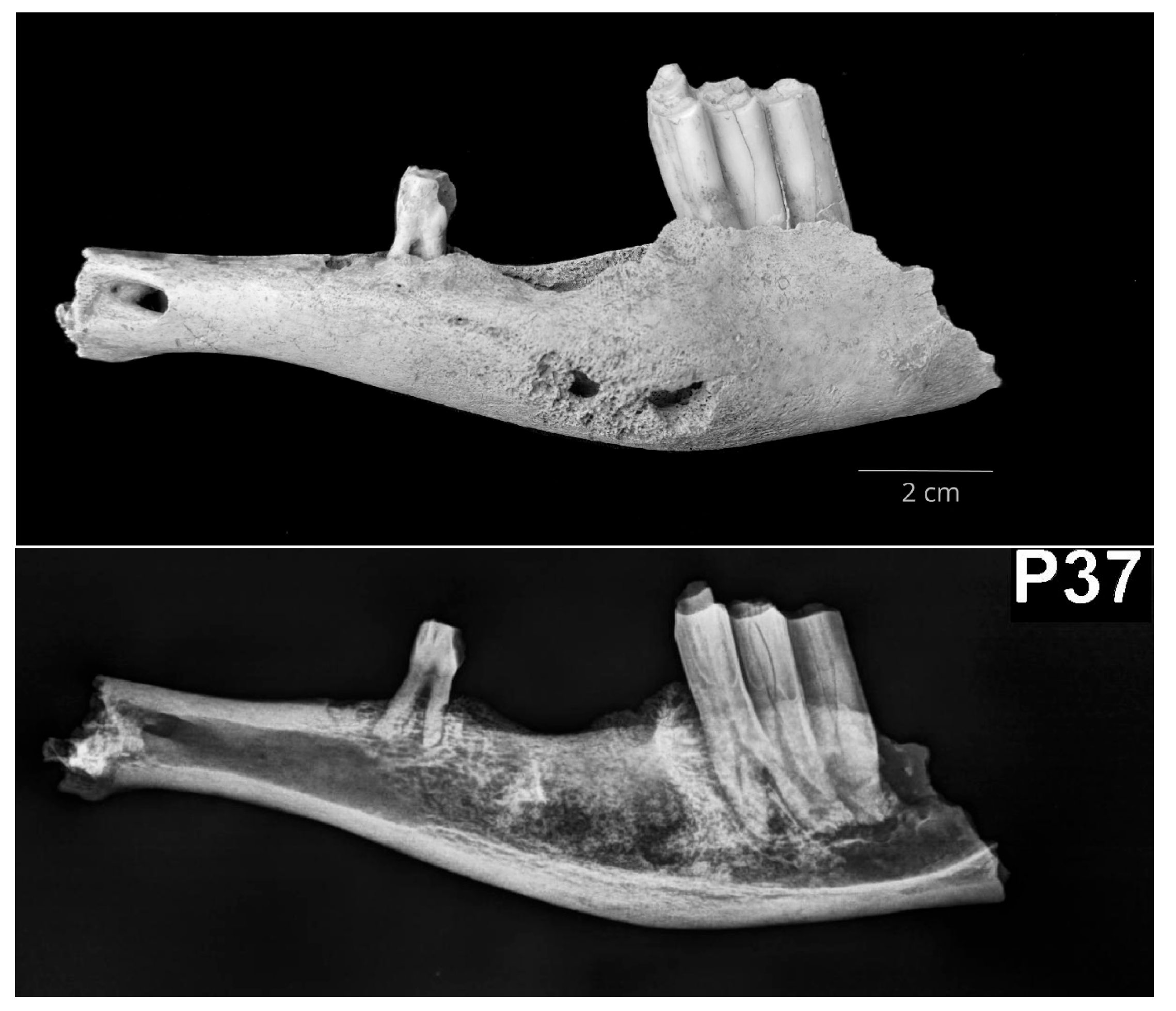
| P37 | NADIN | Late Middle Ages/Early Modern Period | mandibula | left | small R | around 2.5–3 y |
| P38 | NADIN | Antiquity | phalanx proximalis | cattle | >1.5 y | |
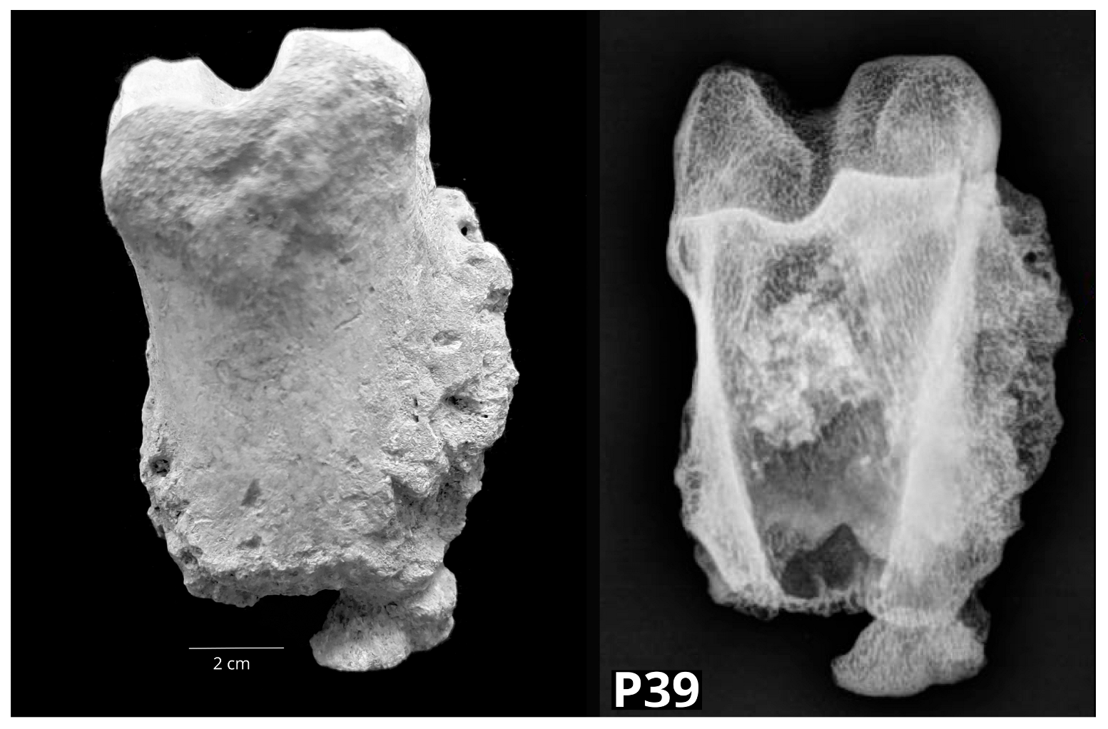
| P39 | NADIN | Antiquity | phalanx proximalis | cattle | >1.5 y | |
| P40 | NADIN | Antiquity | phalanx proximalis | cattle | >1.5 y | |
| P41 | NADIN | Antiquity and Late Antiquity | phalanx proximalis | cattle | >1.5 y | |
| P42 | NADIN | Antiquity and Late Antiquity | phalanx proximalis | cattle | >1.5 y | |
| P43 | NADIN | Antiquity and Late Antiquity | MTC, prox | right | cattle | |
| P44 | NADIN | Early Iron Age | MTT, prox | left | small R | |
| P45 | NADIN | Early Iron Age | MTC, prox | right | sheep | |
| P46 | NADIN | Early Iron Age | MTC, prox | left | goat | |
| P47 | NADIN | Early Iron Age | axis | small R | >4–5 y | |
| P48 | NADIN | Early Iron Age | os carpi radiale | cattle | ||
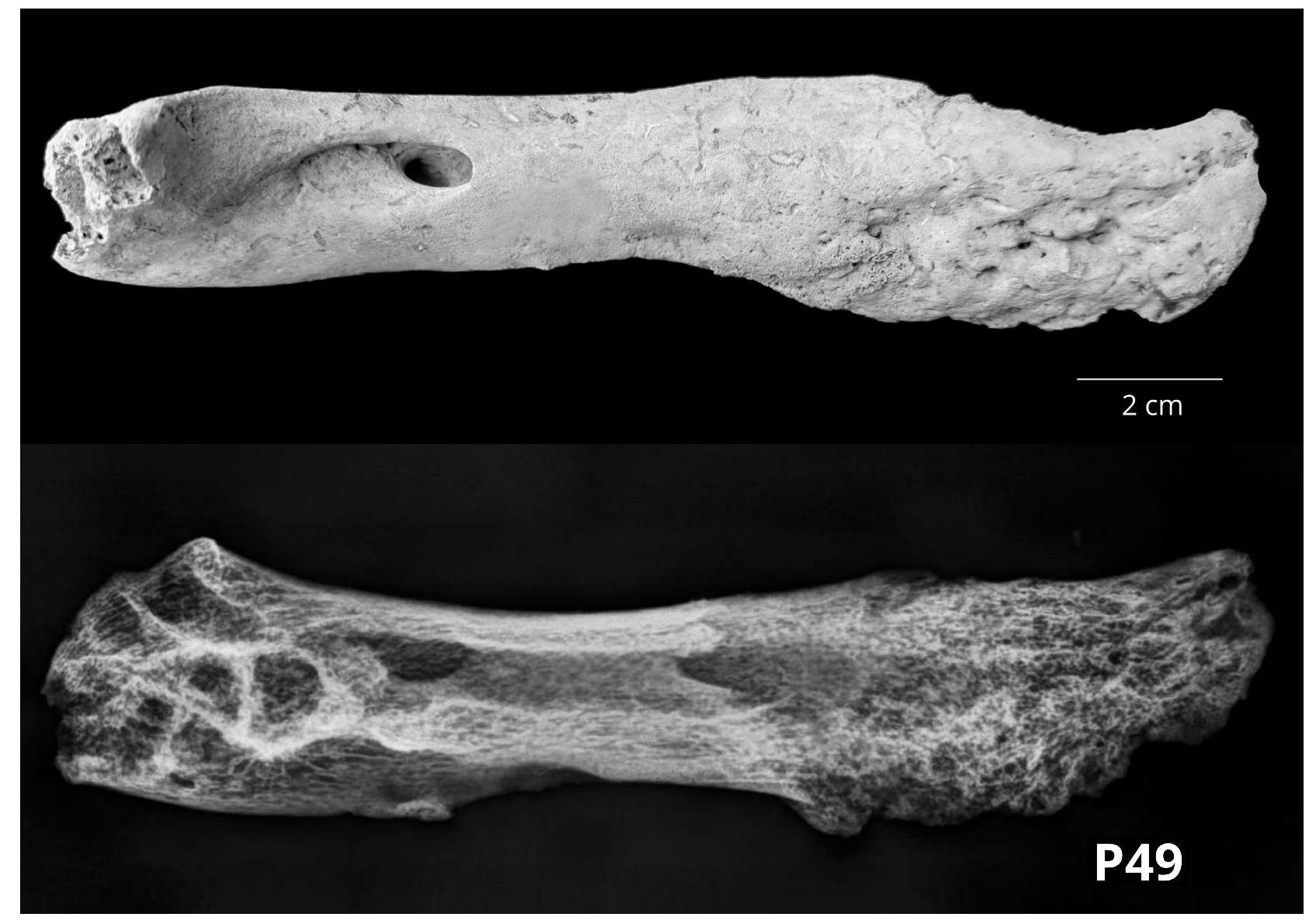
| P49 | NADIN | Early Iron Age | mandibula, rostral | left | cattle | |
| P50 | NADIN | Iron Age | radius, prox | right | sheep | >3–3.5 y |
| Site | Cattle | Small Ruminants | Pig | Horse | Carnivore | Chicken | Total |
|---|---|---|---|---|---|---|---|
| GORJANI | 4 | 1 | 1 | 6 | |||
| OCZP | 2 | 1 | 3 | ||||
| BIJELA | 1 | 1 | |||||
| PGV | 8 | 2 | 4 | 14 | |||
| BARILOVIĆ | 1 | 1 | 2 | ||||
| KRČINGRAD | 1 | 2 | 3 | ||||
| ST NICHOLAS | 4 | 4 | |||||
| NADIN | 10 | 6 | 1 | 17 | |||
| Total | 27 | 12 | 8 | 1 | 1 | 1 | 50 |
| % of total | 54% | 24% | 16% | 2% | 2% | 2% | 100% |
| Aethiolog. Classif. * | ORAL CAVITY | Working/Keeping | TRAUMA | WITHOUT PATHOL. CHANGES | ||||
|---|---|---|---|---|---|---|---|---|
| Radiol. Classif. | MET | INFL/ NEOPL | DEG | MET | INFL/NEOPL | Degenerative | TRAUMA | |
| CATTLE | mandible (P7) | mandible (P49) | mandible (P10) | centroquartal tarsal (P35) | metacarpal (P1) | metacarpal (P43) | calcaneus (P12) | |
| ph. prox (P39) | metapodium (P5) | calcaneus (P13) | ||||||
| ph. prox (P41) | metatarsal (P6) | |||||||
| radial carpal (P48) | acetabulum (P14) | |||||||
| metacarpal (P19) | ||||||||
| metacarpal (P25) | ||||||||
| ph. prox. (P26) | ||||||||
| ph. media (P27) | ||||||||
| metacarpal (P28) | ||||||||
| ph. prox (P30) | ||||||||
| ph. prox (P31) | ||||||||
| ph. prox (P32) | ||||||||
| metacarpal (P33) | ||||||||
| ph. media (P34) | ||||||||
| ph. prox (P38) | ||||||||
| ph. prox (P40) | ||||||||
| ph. prox (P42) | ||||||||
| Total for cattle | 1 | 1 | 1 | 0 | 4 | 17 | 1 | 2 |
| SMALL RUM. | mandible (P18) | ulna (P20) | humerus (P8) | rib (P9) | ||||
| mandible (P37) | metacarpal (P45) | mandible (P15) | ||||||
| metacarpal (P46) | radius (P29) | |||||||
| radius (P50) | metatarsal (P44) | |||||||
| axis (P47) | ||||||||
| Total for small rum. | 0 | 2 | 0 | 1 | 0 | 5 | 4 | 0 |
| PIG | mandible (P21) ** | talus (P2) | metacarpal IV (P3) | mandible (P21) ** | ||||
| lumbar vertebrae (P11) | femur (P4) | |||||||
| humerus (P16) | tibia (P17) | |||||||
| mandible (P22) | ||||||||
| Total for pigs | 0 | (+1) | 0 | 0 | 0 | 3 | 4 | 1 |
| HORSE | radius (P24) | |||||||
| Total for horse | 0 | 0 | 0 | 0 | 0 | 1 | 0 | 0 |
| DOG | lumbar vertebrae (P23) | |||||||
| Total for dog | 0 | 0 | 1 | |||||
| CHICKEN | humerus (P36) | |||||||
| Total for chicken | 0 | 0 | 0 | 0 | 0 | 0 | 1 | 0 |
| Total | 1 | 3 (+1) | 1 | 1 | 4 | 27 | 10 | 3 (−1) |
| Total by group | 5 (+1) | 32 | 10 | 3 (−1) | ||||
Disclaimer/Publisher’s Note: The statements, opinions and data contained in all publications are solely those of the individual author(s) and contributor(s) and not of MDPI and/or the editor(s). MDPI and/or the editor(s) disclaim responsibility for any injury to people or property resulting from any ideas, methods, instructions or products referred to in the content. |
© 2023 by the authors. Licensee MDPI, Basel, Switzerland. This article is an open access article distributed under the terms and conditions of the Creative Commons Attribution (CC BY) license (https://creativecommons.org/licenses/by/4.0/).
Share and Cite
Trbojević Vukičević, T.; Korpes, K.; Đuras, M.; Vrbanac, Z.; Javor, A.; Kolenc, M. Paleopathological Changes in Animal Bones from Croatian Archaeological Sites from Prehistory to New Modern Period. Vet. Sci. 2023, 10, 361. https://doi.org/10.3390/vetsci10050361
Trbojević Vukičević T, Korpes K, Đuras M, Vrbanac Z, Javor A, Kolenc M. Paleopathological Changes in Animal Bones from Croatian Archaeological Sites from Prehistory to New Modern Period. Veterinary Sciences. 2023; 10(5):361. https://doi.org/10.3390/vetsci10050361
Chicago/Turabian StyleTrbojević Vukičević, Tajana, Kim Korpes, Martina Đuras, Zoran Vrbanac, Ana Javor, and Magdalena Kolenc. 2023. "Paleopathological Changes in Animal Bones from Croatian Archaeological Sites from Prehistory to New Modern Period" Veterinary Sciences 10, no. 5: 361. https://doi.org/10.3390/vetsci10050361