Article

Individual Genomic Loci and mRNA Levels of Immune Biomarkers Associated with Pneumonia Susceptibility in Baladi Goats

1
Department of Animal Husbandry and Animal Wealth Development, Faculty of Veterinary Medicine, Mansoura University, Mansoura 35516, Egypt
2
Department of Biology, College of Science, University of Jeddah, Jeddah 21589, Saudi Arabia
3
Department of Animal Histology and Anatomy, School of Veterinary Medicine, Badr University in Cairo (BUC), Cairo 11829, Egypt
4
Department of Anatomy and Embryology, Faculty of Veterinary Medicine, University of Sadat City, Sadat City 32897, Egypt
5
Department of Biology and Plant Protection, Faculty of Agricultural Sciences, University of Life Sciences King Michael I, 300645 Timisoara, Romania
6
Department of Animal Husbandry and Animal Wealth Development, Faculty of Veterinary Medicine, Damanhour University, Damanhour 22511, Egypt
*
Authors to whom correspondence should be addressed.
Vet. Sci. 2023, 10(3), 185; https://doi.org/10.3390/vetsci10030185
Submission received: 31 January 2023 / Revised: 20 February 2023 / Accepted: 24 February 2023 / Published: 1 March 2023
(This article belongs to the Special Issue Role of Immune Biomarkers in Animal Diseases)

Abstract

:

Simple Summary

Pneumonia is a commonly encountered problem in small ruminant medicine practice that negatively affects goat breeding. Some of the limitations of the conventional approaches used by animal breeders to promote animal health may be overcome with the use of molecular genetic tools. Using PCR-DNA sequencing in pneumonia-affected and -resistant Baladi goats, SNPs associated with pneumonia resistance/susceptibility were found in the SLC11A1, CD-14, CCL2, TLR1, TLR7, TLR8, TLR9, β defensin, SP110, SPP1, BP1, A2M, ADORA3, CARD15, IRF3, and SCART1 genes. These indicators’ mRNA levels were different in both the healthy and affected goats. Therefore, to improve the effectiveness of animal selection for innate resistance, finding potential genes linked to pneumonia risk may be crucial.

Abstract

The effectiveness of breeding for inherent disease resistance in animals could be considerably increased by identifying the genes and mutations that cause diversity in disease resistance. One hundred and twenty adult female Baladi goats (sixty pneumonic and sixty apparently healthy) were used in this study. DNA and RNA were extracted from blood samples collected from the jugular vein of each goat. SLC11A1, CD-14, CCL2, TLR1, TLR7, TLR8, TLR9, β defensin, SP110, SPP1, BP1, A2M, ADORA3, CARD15, IRF3, and SCART1 SNPs that have been previously found to be associated with pneumonia resistance/susceptibility were identified via PCR-DNA sequencing. The pneumonic and healthy goats differed significantly, according to a Chi-square analysis of the discovered SNPs. The mRNA levels of the studied immune markers were noticeably greater in the pneumonic goats than in the healthy ones. The findings could support the significance of the use of immune gene expression profiles and nucleotide variations as biomarkers for the susceptibility/resistance to pneumonia and provide a practical management technique for Baladi goats. These results also suggest a potential strategy for lowering pneumonia in goats by employing genetic markers linked to an animal’s ability to fend off infection in selective breeding.

1. Introduction

The domestic goat (Capra hircus), one of the original livestock species, is an important component of animal management [1]. The ability to adapt is one of the core qualities of goats; for example, the majority of dairy goat breeds in use today are ancestors of European kinds [2]. There are 4.3 million goats in Egypt. Around the world, goats are raised extensively, especially in poor countries, where they are a valuable resource for the production of goods and a variety of household items [3,4,5,6,7].
Pneumonia is a frequent respiratory disease in the majority of animals worldwide [8]. Acute lung infection, known as pneumonia, restricts oxygen intake due to fluid-filled alveoli, which is a crucial feature of the disease’s pathogenesis [9]. Pneumonia can be caused by a variety of factors, although the most frequent ones are bacterial or viral infections [8]. Traditionally, farm management practices and the creation of vaccines have always been the key priorities for pneumonia prevention. The use of pharmaceutical interventions in the treatment of pneumonia is significantly influenced by the source of the infection; for example, antibiotics are used to treat bacterially generated pneumonia [10]. Even in the industrialized world, where access to routine clinical treatment and antibiotics is typically unrestricted, annual mortality rates from pneumonia are nevertheless significant [11,12]. This calls for a deeper comprehension of the mechanisms behind pneumonia susceptibility and pathogenesis, which might be used to find new cures and guide drug repositioning [13].
Lung infections typically develop as a result of an interplay between pathogenic microorganisms (bacteria, mycoplasmas, viruses, and fungi), host defense, and distressing environmental conditions, such as traffic, crowding, abrupt climate changes, poor ventilation, and malnutrition [14]. The peak seasons for small ruminant pneumonia in Egypt are often autumn and the beginning of winter, spring, and the start of summer [15]. This is a result of the reserve or sudden temperature fluctuations in the weather from warm to cold. Additionally, it has been discovered that there are more dust particles delivering infections deep into the respiratory system in the spring and autumn [16].
A comprehensive strategy for disease control may be found in genetic selection for the improvement of animal health [17]. Due to the complex interactions between pathogens, the environment, and variables specific to animals that favor their development, pneumonia, and other economically significant illnesses of farm animals pose a challenge to their effective management or eradication [18]. Changes in the management, treatment, vaccination, pathogen control, movement control, slaughter, isolation, and quarantine of diseased animals are just a few of the initial methods of control that are no longer effective due to increased pathogen resistance to chemotherapeutic and prophylactic drugs, antibiotic residues in the environment, and animal products, to name a few [17]. The key to the low-cost and effective control of chronic diseases is to make use of host genetic resistance because it does not suffer from these restrictions in the broadest sense [19]. Unfortunately, in order to attain effective control of these disorders, several gene loci must be identified and defined [18]. In order to find disease-resistance markers, quantitative trait loci (QTLs) are mapped and numerous potential gene variations are evaluated for associations with disease resistance [20]. A great deal of attention has been paid to the genetics of innate and acquired immunity related to resistance, and there is now evidence linking changes in some of these genes with resistance to diseases [21].
The diagnosis and management of complex disorders under multigenic control are more challenging than those under unigenic control [22]. Pathogenic and environmental variables also exacerbate the interplay of several host genes [23]. However, this does not rule out the possibility of genetic control; rather, it necessitates a greater understanding of the interrelated factors [24]. Finding host variables that affect the onset and clinical course of pneumonia in small ruminants has been the subject of extensive research [25,26].
The objective of this study was to determine how potential immune markers (SLC11A1, CD-14, CCL2, TLR1, TLR7, TLR8, TLR9, β defensin, SP110, SPP1, BP1, A2M, ADORA3, CARD15, IRF3, and SCART1) have an impact on the prevalence of pneumonia resistance/susceptibility in Baladi goats using PCR-DNA sequencing and quantitative real-time PCR.

2. Materials and Methods

2.1. Ethics Statement

The methods used to collect samples and to care for the animals in this experiment were permitted by the Institutional Committee for the Care and Use of Animals (IACUC) of the Veterinarian Medical Faculty, Damanhour University, Egypt (code DMU/VetMed-2023/001).

2.2. Animals and Research Subjects

A total of 120 adult female Baladi goats (60 pneumonic and 60 seemingly healthy), aged 4 years old, weighing 48 ± 3.5 kg, and being raised on a private farm in Damanhour province, Egypt, were employed in this study. The does were housed in shaded, semi-open pens and nourished with 650 g of a concentrate feed mixture (CFM) and 650 g of alfalfa hay per head each day, with unlimited access to water. The CFM was composed of wheat bran (220 kg), soya bean (210 kg), corn (520 kg), sodium chloride (4 kg), calcium carbonate (8 kg), Premix (1.25 kg), Netro-Nill (0.5 kg), and Fylax (0.5 kg). When available, the natural pasture (green vegetation, such as berseem and darawa) was used as feed.
A thorough clinical examination was performed on the goats under investigation in accordance with previously described standard protocols [27,28], and the results were simultaneously recorded. The jugular veins of each doe were punctured to obtain five milliliters of blood. Blood samples were collected from healthy and pneumonic does during active pneumonia. To obtain whole blood for the purpose of extracting DNA and RNA, the samples were placed in vacuum tubes with anticoagulants (sodium fluoride or EDTA).

2.3. DNA Isolation and Amplification

Following the established protocol, the extraction of DNA from the blood samples was carried out using a commercial kit, a QI-Aamp DNA Mini Kit (Qiagen, Hilden, Germany) (DNA purification from blood or body fluids). The only samples judged to be appropriate for analyses had DNA levels between 5 and 40 ng/μL and A260/A280 ratios of 1.7 to 1.9, as determined utilizing the DNA quantification and extraction software from Nanodrop (NanoDrop Technologies, Wilmington, DE, USA).
PCR was used to amplify the coding regions of the immune genes (SLC11A1, CD-14, CCL2, TLR1, TLR7, TLR8, TLR9, β defensin, SP110, SPP1, BP1, A2M, ADORA3, CARD15, IRF3, and SCART1). The Capra hircus sequence available in PubMed was used to design the primer sequences. Table S1 lists the primers that were employed during the amplification.
The mixture for the polymerase chain amplification was passed through a thermal cycler with a concluding capacity of 150 μL. The following ingredients were used in each reaction volume: 75 μL of master mixture (Jena Bioscience, Jena, Germany), 6 μL of DNA, 1.5 μL of each complementary primer, and 66 μL of d.d. water. The reaction mixture spent 4 min at a 95 °C starting denaturation temperature. One-minute denaturation cycles at 95 °C, annealing for one minute at the temperatures shown in Table S1, extending for one minute at 72 °C, and then extending for a further ten minutes at 72 °C were all included in the 35-cycle procedure. The samples were stored at 4 °C. Following the discovery of representative PCR findings using agarose gel electrophoresis, fragment patterns under UV light were visualized using a gel documentation system.

2.4. Sequencing DNA and Detecting Polymorphism

Jena Bioscience # pp-201s/Munich, Hamburg, Germany, provided kits for PCR purification to remove non-specific bands, primer dimmers, and other impurities before DNA sequencing, resulting in the desired PCR product of the expected size [29]. For PCR quantification, using a Nanodrop (UV-Vis spectrophotometer Q5000/Waltham, MA, USA), good quality and good concentrations were obtained [30]. The PCR results bearing the objective PCR product were forward- and reverse-sequenced to search for SNPs in the healthy and pneumonic goats. An ABI 3730XL DNA sequencer was used to sequence these products utilizing the enzymatic chain terminator approach discovered by Sanger et al. [31] (Applied Biosystems, Waltham, MA, USA).
The DNA sequencing data were analyzed using the software programs Chromas 1.45 and BLAST 2.0 [32]. SNPs were found as differences between the immune gene products generated using the PCR and the reference GenBank-sourced gene sequences. The MEGA4 tool uncovered variations in the amino acid sequences of the investigated genes on the basis of a sequence alignment among the does [33].

2.5. RNA Isolation and Immune Gene Quantitation

The blood samples from the does under investigation were used to obtain their total RNA via the Trizol reagent, in line with the directions provided by the manufacturer (RNeasy Mini Ki, Catalogue No. 74104). We measured and verified the quantity of the isolated RNA using a NanoDrop® ND-1000 spectrophotometer. The manufacturer’s procedure was used to synthesize the cDNA for each sample (Thermo Fisher, Waltham, MA, USA, Catalog No, EP0441). The expression patterns of the genes SLC11A1, CD-14, CCL2, TLR1, TLR7, TLR8, TLR9, β defensin, SP110, SPP1, BP1, A2M, ADORA3, CARD15, IRF3, and SCART1 were assessed using quantifiable RT-PCR and the SYBR Green PCR Master Mix (2x SensiFastTM SYBR, Bio-line, CAT No: Bio-98002). With the use of the SYBR Green PCR Master Mix, a comparative amount of mRNA was quantified (Quantitect SYBR green PCR kit, Toronto, ON, Canada, Catalog No, 204141). The Capra hircus sequence available in PubMed was used to design the primer sequences (Table S2). The constitutive control for normalization was the ß. actin gene. The reaction mixture contained the following components: 25 µL of total RNA, 0.5 µL of each complementary primer, 8.25 µL of RNase-free water, 0.25 µL of reverse transcriptase, 12.5 µL of Quantitect SYBR green PCR master solution, and 4 µL of Trans Amp buffer. The finished reaction blend underwent a subsequent program in a thermal cycler: reverse transcription at 50 °C for 30 min; preliminary denaturation at 94 °C for 8 min, 40 cycles at 94 °C for 15 s, and the annealing temperatures listed in Table S2; and elongation for 30 s at 72 °C. In order to confirm the specificity of the PCR product, a melting curve analysis was performed after the amplification stage. The 2−ΔΔCt method was utilized to assess how the expression of each gene varied in the tested sample in comparison to that of the ß. actin gene [34,35].

2.6. Statistical Analysis

H0: The individual genomic loci and gene expressions of SLC11A1, CD-14, CCL2, TLR1, TLR7, TLR8, TLR9, β defensin, SP110, SPP1, BP1, A2M, ADORA3, CARD15, IRF3, and SCART1 do not account for the resistance/susceptibility to pneumonia in Baladi goats.
HA: The individual genomic loci and gene expressions of SLC11A1, CD-14, CCL2, TLR1, TLR7, TLR8, TLR9, β defensin, SP110, SPP1, BP1, A2M, ADORA3, CARD15, IRF3, and SCART1 account for the resistance/susceptibility to pneumonia in Baladi goats.
The considerable variations in the genes’ SNPs that were found between the investigated does were determined using a chi-square analysis. Utilizing the statistical software application Graphpad, a statistical analysis was carried out for this aim (p < 0.05). It was determined whether the difference between the pneumonic and healthy Baladi does was statistically significant using the t-test and version 17 of the Statistical Package for Social Science (SPSS) computer program (SPSS Inc., Chicago, IL, USA). To present the findings, mean and standard error (Mean ± SE) were employed. p < 0.05 was used to determine whether the differences were significant.
The relevance of several variables was evaluated using a discriminant analysis model, and the gene expression profile of the examined immune genes was used as an independent variable to categorize the pneumonic and healthy does as a dependent variable. The examined genes’ transcript levels were used to differentiate between the pneumonic and healthy does. The interaction exerted by two components (gene kind and pneumonia incidence) and its influence on transcript levels were examined using a two-way ANOVA and the univariate general linear model (GLM).

3. Results

3.1. Clinical Findings

Based on health, the examined does were split into two equal-sized groups (60 each). Clinically healthy individuals made up the first group (i.e., those with a normal appetite, a normal body temperature, normal respiratory and pulse rates, bright eyes, and no lacrimal or nasal discharge), which was named the healthy group. Pneumonia was present in the second group (with mucopurulent nasal discharge, hyperthermia, abdominal respiration, weakness, an off appetite, crackles, exercise intolerance, gasping, and wheezing observed via auscultation).

3.2. Polymorphisms of Immune Markers

First, the genomic DNA from the investigated does was extracted to amplify immune genes (Figure S1). Purified and good concentration DNA was indicated by Nanodrop (Figure S2). Utilizing the PCR-DNA sequence findings of the SLC11A1 (523-bp), CD-14 (538-bp), CCL2 (534-bp), TLR1 (471-bp), TLR7 (398-bp), TLR8 (799-bp), TLR9 (460-bp), β defensin (253-bp), SP110 (537-bp), SPP1 (943-bp), BP1 (645-bp), A2M (325-bp), ADORA3 (521-bp), CARD15 (394-bp), IRF3 (468-bp), and SCART1 (475-bp) genes, the DNA base sequence SNPs associated with pneumonia were found to vary between the healthy and affected Baladi does. There were no common SNPs between healthy and pneumonic does. The nucleotide sequence variations between the immune genes examined in the does in this study and the reference GenBank-sourced gene sequences were applied to approve all the discovered SNPs (Figures S3–S18). Based on the chi-square analysis of the discovered SNPs, the healthy and pneumonia-affected does showed substantially different frequencies of the studied genes (p ˂ 0.05) (Table 1). The immunological indicators under examination all contained the changes described in Table 1 in the exonic region, resulting in coding mutations between the pneumonic does and the healthy does.

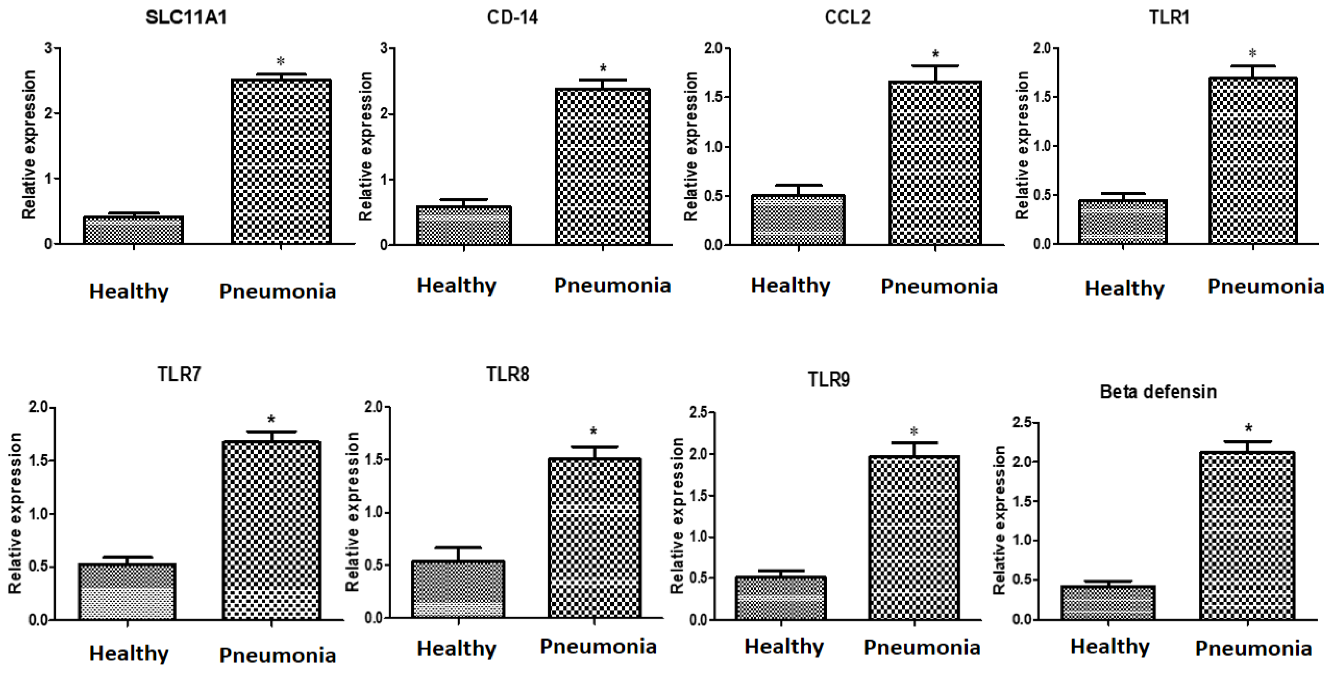
3.3. Immune Markers’ Patterns of Gene Expressions

The evaluated indicators’ gene expression profiles are shown in Figure 1. The expression levels of SLC11A1, CD-14, CCL2, TLR1, TLR7, TLR8, TLR9, β defensin, SP110, SPP1, BP1, A2M, ADORA3, CARD15, IRF3, and SCART1 were considerably greater in the diarrheic Baladi goats than in the healthy ones.
The mRNA levels of the investigated indicators were strongly influenced by the type of gene and pneumonia resistance/susceptibility. For every gene analyzed in the pneumonic does, IRF3 exhibited the greatest level of mRNA (2.51 ± 0.11); ADORA3 had the lowest level (1.62 ± 0.16). The gene CD14 exhibited the highest possible quantity of mRNA amongst all the genes inspected in the healthy does (0.59 ± 0.08), whereas ADORA3 had the lowest (0.38 ± 0.07).

4. Discussion

4.1. Individual Genomic Loci of Immune Markers Associated with Pneumonia Susceptibility

It is obvious that we still do not fully understand how genetics and epigenetics affect pneumonia in farm animals [36]. Using traditional breeding techniques of selecting disease features with high heritability has been beneficial in reducing the prevalence of various infectious diseases in livestock [37]. The majority of disease features, however, have relatively low heritability, making it difficult to achieve genetic control through breeding [37]. There is optimism that using molecular data will truly help to significantly reduce the number of animal diseases with the advancement of molecular genetic technology and our knowledge of farm animal genomes [38]. The availability of genome sequences and the high-resolution molecular mapping of specific livestock species has allowed for new insights into the genetic causes of infectious diseases [39]. Infection control has been made possible via the identification of the genes and causal SNPs responsible for particular disease traits, with the aid of the location of significant QTLs for those traits [40].
Finding immunological genomic regions that have a quantitative impact on a trait is critical for understanding the genetic architecture of a feature linked to pneumonia susceptibility. In this study, SLC11A1, CD-14, CCL2, TLR1, TLR7, TLR8, TLR9, β defensin, SP110, SPP1, BP1, A2M, ADORA3, CARD15, IRF3, and SCART1 were molecularly characterized in pneumonia-free and pneumonia-affected Baladi goats. The results demonstrate differences in the SNPs between the two groups. The SNP distribution among the investigated does was significant (p ˂ 0.05) according to the chi-square test. It is imperative to emphasize that the polymorphisms discovered and available herein provide additional data about the examined genes when contrasted with those in the associated GenBank-sourced sequences.
Immune-function-related gene mutations have been connected to the susceptibility of animals to pathogenic microorganisms [41,42]. We think that this is the first investigation to find nucleotide sequence variants in immune system genes (SLC11A1, CD-14, CCL2, TLR1, TLR7, TLR8, TLR9, β defensin, SP110, SPP1, BP1, A2M, ADORA3, CARD15, IRF3, and SCART1) as potential candidates for pneumonia resistance/susceptibility in Baladi goats. However, the candidate gene method was utilized to evaluate ruminant susceptibility to pneumonia. For example, in sheep, a link between MHC-DRB1 genotypes for mycoplasma ovipneumonia resistance or susceptibility has been documented [25,43]. Additionally, it has been established that progressive pneumonia virus susceptibility is affected by the effects of ovine TMEM154 gene polymorphisms after normal exposure [26]. In humans, TLRs gene polymorphisms have been reported to be associated with pneumonia [44].

4.2. Transcript Levels of Immune Markers Associated with Pneumonia Susceptibility

As a heritable endophenotype, transcript abundance is influenced by chromosomal polymorphisms, according to the theory of hereditary genomics [45]. The latter approach supports the notion that integrating information on the mRNA levels of investigated genes and chromosomal variations may improve our comprehension of the genetics underlying the development of disease [46]. Quantitative trait loci (QTLs) for expression are polymorphisms connected to mRNA quantification [47]. The present work postulated that an individual’s hereditary phenotypic variation in the response of mRNA levels to pneumonia incidence may have an impression on the disease course. Real-time PCR was conducted to quantify the expression levels of immunological markers (SLC11A1, CD-14, CCL2, TLR1, TLR7, TLR8, TLR9, β defensin, SP110, SPP1, BP1, A2M, ADORA3, CARD15, IRF3, and SCART1) in Baladi goats with and without pneumonia resistance. Our research revealed that the resistant Baladi goats expressed lower levels of immune genes than the pneumonic goats. As a means of remedying the shortcomings of past research, gene expression and genetic SNP markers were used to assess gene polymorphisms. Therefore, the processes that were studied to control the immunological indicators in both the healthy and pneumonic does are well recognized. To the best of our knowledge, this work is the first to thoroughly analyze the gene expressions of the immunological markers associated with the risk of goat pneumonia. However, gene expression profiling revealed that Toll-like receptors (TLRs) and complement genes were associated with infectious pneumonia in sheep [48].
SLC11A1, a trans-membrane protein, is one of the best-known probable potential genes for innate immunity against a number of intracellular infections [49]. An essential component of innate immunity is the cluster of differentiation 14 (CD14). One of the most crucial molecules that binds and neutralizes bacterial endotoxins is the anti-bacterial peptide CD14 [50]. Lipopolysaccharide (LPS) and peptidoglycan, two of the most prevalent components in the bacterial cell wall, both function as receptors for CD14 [51]. Leucocytes are thought to be transported to the mammary glands by chemokines and their receptors, and they may significantly affect how the host immune system responds to both acute and chronic intramammary infections [52]. CCL2 works as a potent chemokine for monocytes and neutrophils by ligating with the CCR2 receptors expressed on their surface [53].
Pattern-recognition receptors (PAMPs) are distinguished from other molecules by their leucine-rich repeat (LRR) domains, which are essential structural components and are reported to make up the majority of the ectodomains of TLR molecules [54]. In a previous study, when non-synonymous SNP prevalence was evaluated among TLR coding sequences from different animals, it was discovered that the sequences encoding LRR domains are more abundant in non-synonymous SNPs [55]. A molecular system’s ability to detect extracellular infections may be considerably altered by non-synonymous SNPs in LRR domains [56]. Leucocytes and epithelial cells have been thought to be the only sources of β-defensins, which have been thought to be the first line of defense against pathogens [57]. In a previous study, this viewpoint was corroborated by the way in which they responded to various bacterial, viral, and fungal diseases [58]. Goats have two beta-defensins, which have been named goat β-defensin (GBD)-1 and GBD-2. PreproGBD-1 and PreproGBD-2 genes are 96.8% and 88.2% identical in terms of nucleotide sequence and amino acid sequence, respectively [59]. Alpha- and beta-defensin genes in mammals have one intron and two exons [60].
The SP110 nuclear body protein (SP110) gene is implicated in inducing innate immune responses to combat many intracellular infections [61]. Mycobacterium tuberculosis appears to be under the control of SP110 in host macrophages [62]. According to reports, Mycobacterium avium subspecies paratuberculosis infection is associated with bovine SP110 nuclear body protein (SP110) gene polymorphisms [63]. Macrophages and T cells that have been activated generate the cytokine known as secreted phosphoprotein 1 (SPP1), also known as Osteopontin [64]. SPP1 has been identified to be a key player in the T-cell activation process. It boosts T-helper 1 (Th1) cell-mediated immunity via the release of Th1 cytokines and, when T cells are enhanced, attracts macrophages to the infection site [65]. The bactericidal/permeability-increasing endogenous cationic protein (BPI) neutralizes Gram-negative bacteria, endotoxins, and lipopolysaccharides [66]. Inhibiting angiogenesis and the production of inflammatory mediators, boosting complement activation and opsonization for enhanced phagocytosis, and protecting against infection by fungus and protozoan pathogens are biological functions of BPI [66]. As a result, BPI is crucial for the host’s natural defense.
The alpha-macroglobulin (aM) family of proteins, which includes C3, C4, and C5, also includes alpha-2-macroglobulin (A2M) [67]. The innate immune system’s A2M component has been conserved throughout evolution [68]. Additionally, it promotes the growth of macrophages and T cells [69]. The host defense mechanism uses A2M as a non-specific protease inhibitor to inactivate both endogenous and foreign proteases, including metalloproteases and serine, threonine, cysteine, aspartic, and cyclooxygenases [70]. The adenosine A3 receptor, also known as ADORA3, is an adenosine receptor. G-protein-coupled adenosine A3 receptors bind to Gi/Gq and participate in a number of intracellular signaling pathways and physiological processes [71]. They participate in the prevention of neutrophil degranulation in neutrophil-mediated tissue injury, and they exert a sustained cardio-protective role during myocardial ischemia [72].
The CARD15 protein, a pattern-recognition receptor (PRR) with leucine-rich repeats (LRR), a NACHT domain, and two caspase recruitment domains (CARDs), is crucial for both innate and acquired immunity [73]. The interferon regulatory transcription factor (IRF) family includes IRF3 [74]. IRF3 has a profound impact on the innate immune system’s response to microbial infection [75]. A protein that is only present in a specific subset of delta gamma T cells and serves to identify important pathogens is produced by the scavenger receptor family member expressed on T cells (SCART1) gene [76]. A genome-wide association analysis revealed that the SCART1 gene is related to the hoof health trait in Holstein dairy cattle [77].
The development of inflammatory markers driven by lipopolysaccharides or endotoxins may be the cause of the pneumonia-affected does’ noticeably altered immune and antioxidant expression patterns [78]. In macrophages stimulated by LPSs, inflammatory gene expressions triple, mRNA levels rise by about 100 fold, and the release of the protein itself may increase by as much as 10,000 fold [79]. Furthermore, compared to healthy tissues, damaged tissues produce higher free radical responses [80,81]. The susceptible genotypes promote the inflammatory response via the high production of inflammatory markers, whereas the resistant genotypes significantly upregulate the expressions of IL-2 and IL-10 to limit the inflammatory response [82]. Additionally, pneumonia-affected does create a variety of cellular immune components that attach to cell surface receptors and mediate and regulate immune activity and the inflammatory response [83]. According to several studies, there are complex relationships between various cell immunological components. These proteins, for instance, have been found to interact with one another and have an impact on the development of different immune globulins, complements, and acute phase reactive proteins, creating a complex network structure [84,85]. As a result, we anticipate that infectious pneumonia was the primary cause of the majority of the pneumonia cases in our study. Furthermore, the outcomes of our real-time PCR provide a believable indication that animals with pneumonia produce a strong inflammatory response.

5. Conclusions

By using PCR-DNA sequencing to analyze immunological markers (SLC11A1, CD-14, CCL2, TLR1, TLR7, TLR8, TLR9, β defensin, SP110, SPP1, BP1, A2M, ADORA3, CARD15, IRF3, and SCART1), it was possible to identify an association between pneumonia resistance/susceptibility and nucleotide sequence differences in Baladi goats. Additionally, these immune markers’ mRNA levels varied between the healthy and pneumonic does. With the help of these many functional variants, researchers can better understand how infections work, which could result in the development of novel pharmacological and therapeutic approaches to prevent the spread of diseases. The different transcripts of the immune makers in the pneumonic and healthy does may act as monitors and benchmarks to evaluate health.

Supplementary Materials

The following supporting information can be downloaded at: https://www.mdpi.com/article/10.3390/vetsci10030185/s1. Figure S1: Representative gel electrophoresis for extracted DNA samples from the investigated does, title Figure S2: Nanodrop curve showing concentration of representative DNA extracted from the investigated does, Figure S3: Comparison of healthy and pneumonia-affected Baladi goats using GenBank-sourced reference sequence gb|KX151137.1| and the SLC11A1 gene (523-bp), demonstrating DNA sequence alignment, Figure S4: Comparison of healthy and pneumonia-affected Baladi goats using GenBank-sourced reference sequence gb|NM_001361647.1| and the CD14 gene (538-bp), demonstrating DNA sequence alignment, Figure S5: Comparison of healthy and pneumonia-affected Baladi goats using GenBank-sourced reference sequence gb|XM_004012471.4| and the CCL2 gene (534-bp), demonstrating DNA sequence alignment, Figure S6: Comparison of healthy and pneumonia-affected Baladi goats using GenBank-sourced reference sequence gb|KJ210567.1| and the TLR1 gene (471-bp), demonstrating DNA sequence alignment, Figure S7: Comparison of healthy and pneumonia-affected Baladi goats using GenBank-sourced reference sequence gb|GU289401.1| and the TLR7 gene (398-bp), demonstrating DNA sequence alignment, Figure S8: Comparison of healthy and pneumonia-affected Baladi goats using the GenBank-sourced reference sequence gb|GU936189.1| and the TLR8 gene (799-bp), demonstrating DNA sequence alignment, Figure S9: Comparison of healthy and pneumonia-affected Baladi goats using GenBank-sourced reference sequence gb|EU747825.1| and the TLR9 gene (460-bp), demonstrating DNA sequence alignment, Figure S10: Comparison of healthy and pneumonia-affected Baladi goats using GenBank-sourced reference sequence gb|DQ532360.1| and the β defensin gene (253-bp), demonstrating DNA sequence alignment, Figure S11: Comparison of healthy and pneumonia-affected Baladi goats using GenBank-sourced reference sequence gb|XM_013973239.2|and the SP110 gene (537-bp), demonstrating DNA sequence alignment, Figure S12: Comparison of healthy and pneumonia-affected Baladi goats using GenBank-sourced reference sequence gb|NM_001285667.1| and the SPP1 gene (943-bp), demonstrating DNA sequence alignment, Figure S13: Comparison of healthy and pneumonia-affected Baladi goats using the GenBank-sourced reference sequence gb|KC894598.1| and the BP1 gene (645-bp), demonstrating DNA sequence alignment, Figure S14: Comparison of healthy and pneumonia-affected Baladi goats using GenBank-sourced reference sequence gb|XM_018048527.1| and the A2M gene (325-bp), demonstrating DNA sequence alignment, Figure S15: Comparison of healthy and pneumonia-affected Baladi goats using GenBank-sourced reference sequence gb|XM_013962543.2| and the ADORA3 gene (521-bp), demonstrating DNA sequence alignment, Figure S16: Comparison of healthy and pneumonia-affected Baladi goats using GenBank-sourced reference sequence gb|KJ001638.1| and the CARD15 gene (394-bp), demonstrating DNA sequence alignment, Figure S17: Comparison of healthy and pneumonia-affected Baladi goats using GenBank-sourced reference sequence gb|JQ308793.1|and the IRF3 gene (468-bp), demonstrating DNA sequence alignment, Figure S18: Comparison of healthy and pneumonia-affected Baladi goats using GenBank-sourced reference sequence gb|XR_001917340.1| and the SCART1 gene (475-bp), demonstrating DNA sequence alignment, Table S1: Immune gene oligonucleotide primer sequence used in PCR-DNA sequencing, Table S2: Sequence of the oligonucleotide primers used in real-time PCR for immunological genes under investigation.

Author Contributions

A.A. created the experiment, carried out the PCR, and wrote the manuscript. B.E. assisted in the writing of the manuscript and in the collection of blood samples. DNA sequencing was carried out by M.A.-S., who also helped with the manuscript’s writing. M.A. and L.F. made contributions to the planning of the text and data analyses. All authors have read and agreed to the published version of the manuscript.

Funding

This paper is published from the project 6PFE of the University of Life Sciences “King Mihai I” from Timisoara and the Research Institute for Biosecurity and Bioengineering from Timisoara.

Institutional Review Board Statement

The sample collection and animal care procedures used in this investigation were approved by the Institutional Animal Care and Use Committee (IACUC), Faculty of Veterinary Medicine, Damanhour University, Egypt (code DMU/VetMed-2023/001).

Informed Consent Statement

Every participant gave their informed consent to participate in the study.

Data Availability Statement

Upon reasonable request, the corresponding author will provide the data backing up the study’s conclusions.

Acknowledgments

The authors acknowledge the staff at Animal Husbandry and Animal Wealth Development at Damanhour University’s Faculty of Veterinary Medicine for their support.

Conflicts of Interest

The authors declare no conflict of interest.

References

  1. Skapetas, B.; Bampidis, V. Goat production in the world: Present situation and trends. Livest. Res. Rural. Dev. 2016, 28, 200. [Google Scholar]
  2. Gall, C. Goat Breeds Around the World; CTA; Margraf/FAO: Weikersheim, Germany, 1996; p. 186. [Google Scholar]
  3. Zeder, M.A.; Hesse, B. The initial domestication of goats (Capra hircus) in the Zagros Mountains 10,000 years ago. Science 2000, 287, 2254–2257. [Google Scholar] [CrossRef] [PubMed]
  4. MacHugh, D.E.; Bradley, D.G. Livestock genetic origins: Goats buck the trend. Proc. Natl. Acad. Sci. USA 2001, 98, 5382–5384. [Google Scholar] [CrossRef] [PubMed] [Green Version]
  5. Qureshi, M. Review of modern strategies to enhance livestock genetic performance: From molecular markers to next-generation sequencing technologies in goats. J. Food Agric. Environ. 2014, 12, 752–761. [Google Scholar]
  6. Haftu, A.B.B.; Gebrehiwot, T. Study on prevalence of gastrointestinal nematodes and coccidian parasites affecting cattle in West Arsi zone, Ormia Regional State, Ethiopia. J. Biol. Agric. Healthc. 2014, 4, 32–38. [Google Scholar]
  7. Ahmed, J.; Ararsa, D.; Dareje, R.; Dinaol, B.; Roba, J. Gastrointestinal nematode parasites of small ruminants and anthelmintics efficacy test in sheep of Haramaya District, Eastern Ethiopia. Anim. Vet. Sci. 2017, 5, 39. [Google Scholar] [CrossRef] [Green Version]
  8. Crespo, H.; Bertolotti, L.; Proffiti, M.; Cascio, P.; Cerruti, F.; Acutis, P.L.; de Andrés, D.; Reina, R.; Rosati, S. Low proviral small ruminant lentivirus load as biomarker of natural restriction in goats. Vet. Microbiol. 2016, 192, 152–162. [Google Scholar] [CrossRef]
  9. Ayling, R.D.; Nicholas, R.A.J. Mycoplasma Respiratory Infections. In Diseases of Sheep, 4th ed.; Aitken, I.D., Ed.; Blackwell Publishing: Oxford, UK, 2007; pp. 231–235. [Google Scholar]
  10. Bottinelli, M.; Schnee, C.; Lepri, E.; Stefanetti, V.; Filippini, G.; Gobbi, M.; Martina Sebastianelli, M.; Antenucci, P.; Rampacci, E.; Coletti, M.; et al. Investigation on mycoplasma populations in pneumonic dairy lamb lungs using a DNA microarray assay. Small Rumin. Res. 2017, 147, 96–100. [Google Scholar] [CrossRef]
  11. Medina, I.; Carbonell, J.; Pulido, L.; Madeira, S.; Goetz, S.; Conesa, A.; Tárraga, J.; Pascual-Montano, A.; Nogales-Cadenas, R.; Santoyo-Lopez, J.; et al. Babelomics: An integrative platform for the analysis of transcriptomics, proteomics and genomic data with advanced functional profiling. Nucleic Acids Res. 2010, 38, W210–W213. [Google Scholar] [CrossRef]
  12. Legastelois, I.; Cottin, V.; Mornex, J.F.; Cordier, G. Alveolar macrophages from sheep naturally infected by visna-maedi virus contribute to IL-8 production in the lung. Vet. Immunol. Immunopathol. 1997, 59, 131–139. [Google Scholar] [CrossRef]
  13. Song, Q.Y.; Zhang, Y.J.; Liu, Y.Q.; Li, T.Q. Antimicrobial activities of eighteen antimicrobials against Mycoplasma ovipneumoniae and Mycoplasma mycoides isolates in vitro. Pro. Vet. Med. 2011, 32, 14–18. [Google Scholar]
  14. Abendaño, N.; Tyukalova, L.; Barandika, J.F.; Balseiro, A.; Sevilla, I.A.; Garrido, J.M.; Juste, R.A.; Alonso-Hearn, M. Mycobacterium avium subsp. paratuberculosis isolates induce in vitro granuloma formation and show successful survival phenotype, common anti-inflammatory and antiapoptotic responses within ovine macrophages regardless of genotype or host of origin. PLoS ONE 2014, 9, e104238. [Google Scholar] [CrossRef] [PubMed] [Green Version]
  15. Rizk, M.A.; El-Sayed, S.A.E.-S.; Salman, D.; Marghani, B.H.; Gadalla, H.E.; Sayed-Ahmed, M.Z. Immunomodulatory Effect of Vitamin C on Proinflammatory Cytokines Production in Ossimi Lambs (Ovis aries) with Pneumonic Pasteurellosis. Animals 2021, 11, 3374. [Google Scholar] [CrossRef] [PubMed]
  16. Fernández, S.; Galapero, J.; Rey, J.; Pérez, C.J.; Ramos, A. Cytokines Study in Lungs Infected with Pasteurellaceae and Mycoplasma spp. from Fattening Lambs. J. Med. Microbiol. Immunol. Res. 2018, 2, 1. [Google Scholar]
  17. Stear, M.J.; Bishop, S.C.; Mallard, B.A.; Raadsma, H.W. The sustainability, feasibility and desirability of breeding livestock for disease resistance. Res. Vet. Sci. 2001, 71, 1–7. [Google Scholar] [CrossRef] [PubMed]
  18. Bishop, S.C.; Morris, C.A. Genetics of disease resistance in sheep and goats. Small Rumin. Res. 2007, 70, 48–59. [Google Scholar] [CrossRef]
  19. Berry, D.P.; Bermingham, M.L.; Good, M.; More, S.J. Genetics of animal health and disease in cattle. Ir. Vet. J. 2011, 64, 5. [Google Scholar] [CrossRef] [PubMed] [Green Version]
  20. White, S.N.; Mousel, M.R.; Reynolds, J.O.; Lewis, G.S.; Herrmann-Hoesing, L.M. Common promoter deletion is associated with 3.9-fold differential transcription of ovine CCR5 and reduced proviral level of ovine progressive pneumonia virus. Anim. Genet. 2009, 40, 583–589. [Google Scholar] [CrossRef]
  21. Woodall, C.J.; Maclaren, L.J.; Watt, N.J. Differential levels of mRNAs for cytokines, the interleukin-2 receptor and class II DR/DQ genes in ovine interstitial pneumonia induced by Maedi Visna virus infection. Vet. Pathol. 1997, 34, 204–211. [Google Scholar] [CrossRef] [Green Version]
  22. Ibeagha-Awemu, E.M.; Kgwatalala, P.; Ibeagha, A.E.; Zhao, X. A critical analysis of disease-associated DNA polymorphisms in the genes of cattle, goat, sheep, and pig. Mamm. Genome 2008, 19, 226–245. [Google Scholar] [CrossRef] [Green Version]
  23. Cortes, O.; Cañon, J.; Gama, L.T. Applications of Microsatellites and Single Nucleotide Polymorphisms for the Genetic Characterization of Cattle and Small Ruminants: An Overview. Ruminants 2022, 2, 456–470. [Google Scholar] [CrossRef]
  24. Marwaha, S.; Knowles, J.W.; Ashley, E.A. A guide for the diagnosis of rare and undiagnosed disease: Beyond the exome. Genome Med. 2022, 14, 23. [Google Scholar] [CrossRef] [PubMed]
  25. Wang, K.; Liu, X.; Li, Q.; Wan, K.; Gao, R.; Han, G.; Li, C.; Xu, M.; Jia, B.; Shen, X. MHC-DRB1 exon 2 polymorphism and its association with mycoplasma ovipneumonia resistance or susceptibility genotypes in sheep. J. Genet. 2020, 99, 22. [Google Scholar] [CrossRef] [PubMed]
  26. Leymaster, K.A.; Chitko-McKown, C.G.; Clawson, M.L.; Harhay, G.P.; Heaton, M.P. Effects of TMEM154 haplotypes 1 and 3 on susceptibility to ovine progressive pneumonia virus following natural exposure in sheep. J. Anim Sci. 2013, 91, 5114–5121. [Google Scholar] [CrossRef]
  27. Pugh, D.G.; Baird, A.N. Sheep & Goat Medicine-E-Book; Elsevier/Saunders: Maryland Heights, Mo, USA, 2012. [Google Scholar]
  28. Radostits, O.M.; Gay, C.C.; Hinchcliff, K.W.; Constable, P.D. Veterinary Medicine: A Textbook of the Diseases of Cattle, Horses, Sheep, Pigs and Goats, 10th ed.; Elsevier/Saunders: London, UK, 2006; pp. 966–994. Available online: https://www.scirp.org/(S(351jmbntvnsjt1aadkposzje))/reference/ReferencesPapers.aspx?ReferenceID=1679700 (accessed on 2 January 2023).
  29. Boom, R.; Sol, C.J.; Salimans, M.M.; Jansen, C.L.; Wertheim-van Dillen, P.M.; Noordaa, J.V.D. Rapid and simple method for purification of nucleic acids. J. Clin. Microbiol. 1990, 28, 495–503. [Google Scholar] [CrossRef] [Green Version]
  30. Boesenberg-Smith, K.A.; Pessarakli, M.M.; Wolk, D.M. Assessment of DNA Yield and Purity: An Overlooked Detail of PCR Troubleshooting. Clin. Microbiol. Newsl. 2012, 34, 1–6. [Google Scholar] [CrossRef]
  31. Sanger, F.; Nicklen, S.; Coulson, A.R. DNA sequencing with chain-terminating inhibitors. Proc. Natl. Acad. Sci. USA 1977, 74, 5463–5467. [Google Scholar] [CrossRef] [Green Version]
  32. Altschul, S.F.; Gish, W.; Miller, W.; Myers, E.W.; Lipman, D.J. Basic local alignment search tool. J. Mol. Biol. 1990, 215, 403–410. [Google Scholar] [CrossRef]
  33. Tamura, K.; Dudley, J.; Nei, M.; Kumar, S. MEGA4: Molecular Evolutionary Genetics Analysis (MEGA) Software Version 4.0. Mol. Biol. Evol. 2007, 24, 1596–1599. [Google Scholar] [CrossRef]
  34. Livak, K.J.; Schmittgen, T.D. Analysis of Relative Gene Expression Data Using Real-Time Quantitative PCR and the 2−∆∆CT. Methods. 2001, 25, 402–408. [Google Scholar] [CrossRef]
  35. Pfaffl, M.W. A new mathematical model for relative quantification in real-time RT–PCR. Nucleic Acids Res. 2001, 29, e45. [Google Scholar] [CrossRef] [PubMed]
  36. Triantaphyllopoulos, K.A.; Ikonomopoulos, I.; Bannister, A.J. Epigenetics and inheritance of phenotype variation in livestock. Epigenetics Chromatin. 2016, 9, 31. [Google Scholar] [CrossRef] [PubMed] [Green Version]
  37. Bishop, S.C.; Woolliams, J.A. Genomics and disease resistance studies in livestock. Livest. Sci. 2014, 166, 190–198. [Google Scholar] [CrossRef] [PubMed] [Green Version]
  38. Zonaed Siddiki, A.M.A.M.; Miah, G.; Islam, M.S.; Kumkum, M.; Rumi, M.H.; Baten, A.; Hossain, M.A. Goat Genomic Resources: The search for genes associated with its economic traits. Int. J. Genom. 2020, 18, 5940205. [Google Scholar] [CrossRef]
  39. Kirkpatrick, B.W.; Cooke, M.E.; Frie, M.; Sporer, K.R.B.; Lett, B.; Wells, S.J.; Coussens, P.M. Genome-wide association analysis for susceptibility to infection by Mycobacterium avium ssp. paratuberculosis in US Holsteins. J. Dairy Sci. 2022, 105, 4301–4313. [Google Scholar] [CrossRef]
  40. Cartegni, L.; Chew, S.L.; Krainer, A.R. Listening to silence and understanding nonsense: Exonic mutations that affect splicing. Nat. Rev. Genet. 2002, 3, 285–298. [Google Scholar] [CrossRef]
  41. Michel, O.; LeVan, T.D.; Stern, D.; Dentener, M.; Thorn, J.; Gnat, D.; Beijer, M.L.; Cochaux, P.; Holt, P.G.; Martinez, F.D.; et al. Systemic responsiveness to lipopolysaccharide and polymorphisms in the toll-like receptor 4 gene in human beings. J. Allergy Clin. Immunol. 2003, 112, 923–929. [Google Scholar] [CrossRef]
  42. Smirnova, I.; Poltorak, A.; Chan, E.K.; McBride, C.; Beutler, B. Phylogenetic variation and polymorphism at the toll-like receptor 4 locus (TLR-4). Genome Biol. 2000, 1, RESEARCH002. [Google Scholar] [CrossRef] [Green Version]
  43. Yaman, Y.; Bay, V.; Aymaz, R.; Keleş, M.; Öner, Y.; Teferedegn, E.Y.; Ün, C. A novel 2 bp deletion variant in Ovine-DRB1 gene is associated with increased Visna/maedi susceptibility in Turkish sheep. Sci. Rep. 2021, 11, 14435. [Google Scholar] [CrossRef]
  44. Salamaikina, S.; Karnaushkina, M.; Korchagin, V.; Litvinova, M.; Mironov, K.; Akimkin, V. TLRs Gene Polymorphisms Associated with Pneumonia before and during COVID-19 Pandemic. Diagnostics 2023, 13, 121. [Google Scholar] [CrossRef]
  45. Jansen, R.C.; Nap, J.P. Genetical genomics: The added value from segregation. Trends Genet. 2001, 17, 388–391. [Google Scholar] [CrossRef] [PubMed] [Green Version]
  46. Fairfax, B.P.; Knight, J.C. Genetics of gene expression in immunity to infection. Curr. Opin. Immunol. 2014, 30, 63–71. [Google Scholar] [CrossRef] [PubMed] [Green Version]
  47. Cloney, R. Complex traits: Integrating gene variation and expression to understand complex traits. Nat. Rev. Genet. 2016, 17, 194. [Google Scholar] [CrossRef] [PubMed]
  48. Abendaño, N.; Esparza-Baquer, A.; Bernales, I.; Reina, R.; de Andrés, D.; Jugo, B.M. Gene Expression Profiling Reveals New Pathways and Genes Associated with Visna/Maedi Viral Disease. Animals 2021, 11, 1785. [Google Scholar] [CrossRef] [PubMed]
  49. Kumar, N.; Ganguly, N.; Singh, R.; Deb, S.M.; Kumar, S.; Sharma, A.; Mitra, A. DNA polymorphism in SLC11A1 gene and its association with brucellosis resistance in Indian zebu (Bos indicus) and crossbred (Bos indicus×Bos taurus) Cattle. Asian-Aust. J. Anim. Sci. 2011, 24, 898–904. [Google Scholar] [CrossRef]
  50. Wright, S.D.; Ramos, R.A.; Tobias, P.S.; Ulevitch, R.J.; Mathison, J.C. CD14, a receptor for complexes of lipopolysaccharide (LPS) and LPS binding protein. Science 1990, 249, 1431–1433. [Google Scholar] [CrossRef] [PubMed]
  51. Hailman, E.; Lichenstein, H.S.; Wurfel, M.M.; Miller, D.S.; Johnson, D.A.; Kelley, M.; Busse, L.A.; Zukowski, M.M.; Wright, S.D. Lipopolysaccharide (LPS)-binding protein accelerates the binding of LPS to CD14. J. Exp. Med. 1994, 179, 269–277. [Google Scholar] [CrossRef] [Green Version]
  52. Nishimura, T. Expression of potential lymphocyte trafficking mediator molecules in the mammary gland. Vet. Res. 2003, 34, 3–10. [Google Scholar] [CrossRef] [Green Version]
  53. Proudfoot, A.I. Chemokine receptors: Multifaceted therapeutic targets. Nat. Rev. Immunol. 2002, 2, 106–115. [Google Scholar] [CrossRef]
  54. Botos, I.; Segal, D.M.; Davies, D.R. The structural biology of Toll-like receptors. Structure 2011, 13, 447–459. [Google Scholar] [CrossRef] [Green Version]
  55. Fujita, M.; Into, T.; Yasuda, M.; Okusawa, T.; Hamahira, S.; Kuroki, Y.; Eto, A.; Nisizawa, T.; Morita, M.; Shibata, K. Involvement of leucine residues at positions 107, 112, and 115 in a leucine-rich repeat motif of human Toll-like receptor 2 in the recognition of diacylated lipoproteins and lipopeptides and Staphylococcus aureus Peptidoglycans. J. Immunol. 2003, 171, 3675–3683. [Google Scholar] [CrossRef] [PubMed] [Green Version]
  56. White, S.N.; Taylor, K.H.; Abbey, C.A.; Gill, C.A.; Womack, J.E. Haplotype variation in bovine Toll-like receptor 4 and computational prediction of a positively selected ligand-binding domain. Proc. Natl. Acad. Sci. USA 2003, 100, 10364–10369. [Google Scholar] [CrossRef] [PubMed] [Green Version]
  57. Caverly, J.M.; Diamond, G.; Gallup, J.M.; Brogden, K.A.; Dixon, R.A.; Ackermann, M.R. Coordinated Expression of Tracheal Antimicrobial Peptide and Inflammatory-Response Elements in the Lungs of Neonatal Calves with Acute Bacterial Pneumonia. Infect. Immun. 2003, 71, 2950–2955. [Google Scholar] [CrossRef] [PubMed] [Green Version]
  58. Hall, T.J.; McQuillan, C.; Finlay, E.K.; O’Farrelly, C.; Fair, S.; Meade, K.G. Comparative genomic identification and validation of β-defensin genes in the Ovis aries genome. BMC Genom. 2017, 18, 278. [Google Scholar] [CrossRef] [PubMed] [Green Version]
  59. Zhao, C.; Nguyen, T.; Liu, L.; Shamova, O.; Brogden, K.; Lehrer, R.I. Differential expression of caprine β-defensins in digestive and respiratory tissues. Infect Immun. 1999, 67, 6221–6224. [Google Scholar] [CrossRef] [PubMed] [Green Version]
  60. Huttner, K.M.; Lambeth, M.R.; Burkin, H.R.; Burkin, D.J.; Broad, T.E. Localization and genomic organization of sheep antimicrobial peptide genes. Gene 1998, 206, 85–91. [Google Scholar] [CrossRef] [PubMed]
  61. Wu, H.; Wang, Y.; Zhang, Y.; Yang, M.; Lv, J.; Liu, J.; Zhang, Y. TALE nickase-mediated SP110 knockin endows cattle with increased resistance to tuberculosis. Proc. Natl. Acad. Sci. USA 2015, 112, E1530–E1539. [Google Scholar] [CrossRef] [PubMed] [Green Version]
  62. Bloch, D.B.; Nakajima, A.; Gulick, T.; Chiche, J.D.; Orth, D.; de La Monte, S.M.; Bloch, K.D. Sp110 localizes to the PML-Sp100 nuclear body and may function as a nuclear hormone receptor transcriptional coactivator. Mol Cell Biol. 2000, 20, 6138–6146. [Google Scholar] [CrossRef] [Green Version]
  63. Ruiz-Larrañaga, O.; Garrido, J.M.; Iriondo, M.; Manzano, C.; Molina, E.; Montes, I.; Vazquez, P.; Koets, A.P.; Rutten, V.P.; Juste, R.A.; et al. SP110 as a novel susceptibility gene for Mycobacterium avium subspecies paratuberculosis infection in cattle. J. Dairy Sci. 2010, 93, 5950–5958. [Google Scholar] [CrossRef] [Green Version]
  64. Chabas, D.; Baranzini, S.E.; Mitchell, D.; Bernard, C.C.; Rittling, S.R.; Denhardt, D.T.; Sobel, R.A.; Lock, C.; Karpuj, M.; Pedotti, R.; et al. The influence of the proinflammatory cytokine, osteopontin, on autoimmune demyelinating disease. Science 2001, 294, 1731–1735. [Google Scholar] [CrossRef]
  65. Denhardt, D.T.; Noda, M.; O’Regan, A.W.; Pavlin, D.; Berman, J.S. Osteopontin as a means to cope with environmental insults: Regulation of inflammation, tissue remodeling, and cell survival. J. Clin. Investig. 2001, 107, 1055–1061. [Google Scholar] [CrossRef] [PubMed] [Green Version]
  66. Balakrishnan, A.; Marathe, S.A.; Joglekar, M.; Chakravortty, D. Bactericidal/permeability increasing protein: A multifaceted protein with functions beyond LPS neutralization. Innate Immun. 2013, 19, 339–347. [Google Scholar] [CrossRef] [PubMed] [Green Version]
  67. Levashina, E.A.; Moita, L.F.; Blandin, S.; Vriend, G.; Lagueux, M.; Kafatos, F.C. Conserved role of a complement-like protein in phagocytosis revealed by dsRNA knockout in cultured cells of the mosquito, Anopheles gambiae. Cell 2001, 104, 709–718. [Google Scholar] [CrossRef]
  68. Banks, R.E.; Evans, S.W.; Van Leuven, F.; Alexander, D.; McMahon, M.J.; Whicher, J.T. Measurement of the ‘fast’ or complexed form of alpha 2-macroglobulin in biological fluids using a sandwich enzyme immunoassay. J. Immunol. Methods 1990, 126, 13–20. [Google Scholar] [CrossRef] [PubMed]
  69. Bonacci, G.R.; Caceres, L.C.; Sanchez, M.C.; Chiabrando, G.A. Activated alpha(2)-macroglobulin induces cell proliferation and mitogen-activated protein kinase activation by LRP-1 in the J774 macrophage-derived cell line. Arch. Biochem. Biophys. 2007, 460, 100–106. [Google Scholar] [CrossRef]
  70. Borth, W. Alpha 2-macroglobulin, a multifunctional binding protein with targeting characteristics. FASEB J. 1992, 6, 3345–3353. [Google Scholar] [CrossRef]
  71. Sommerschild, H.T.; Kirkebøen, K.A. Adenosine and cardioprotection during ischaemia and reperfusion—An overview. Acta Anaesthesiol. Scand. 2000, 44, 1038–1055. [Google Scholar] [CrossRef]
  72. He, H.R.; Li, Y.J.; He, G.H.; Qiang, H.; Zhai, Y.J.; Ma, M.; Wang, Y.J.; Wang, Y.; Zheng, X.W.; Dong, Y.L.; et al. The Polymorphism in ADORA3 Decreases Transcriptional Activity and Influences the Chronic Heart Failure Risk in the Chinese. Biomed. Res. Int. 2018, 31, 4969385. [Google Scholar] [CrossRef]
  73. Wang, Y.; Wang, S.; Liu, T.; Tu, W.; Li, W.; Dong, G.; Xu, C.; Qin, B.; Liu, K.; Yang, J.; et al. CARD15 Gene Polymorphisms Are Associated with Tuberculosis Susceptibility in Chinese Holstein Cows. PLoS ONE 2015, 10, e0135085. [Google Scholar] [CrossRef]
  74. Li, C.; Ma, L.; Chen, X. Interferon regulatory factor 3-CL, an isoform of IRF3, antagonizes activity of IRF3. Cell. Mol. Immunol. 2011, 8, 67–74. [Google Scholar] [CrossRef] [Green Version]
  75. Collins, S.E.; Noyce, R.S.; Mossman, K.L. Innate cellular response to virus particle entry requires IRF3 but not virus replication. J. Virol. 2004, 78, 1706–1717. [Google Scholar] [CrossRef] [PubMed] [Green Version]
  76. Baldwin, C.L.; Hsu, H.; Chen, C.; Palmer, M.; McGill, J.; Waters, W.R.; Telfer, J.C. The role of bovine γδ T cells and their WC1 co-receptor in response to bacterial pathogens and promoting vaccine efficacy: A model for cattle and humans. Vet. Immunol. Immunopathol. 2014, 159, 144–155. [Google Scholar] [CrossRef] [PubMed]
  77. Butty, A.M.; Chud, T.C.S.; Cardoso, D.F.; Lopes, L.S.F.; Miglior, F.; Schenkel, F.S.; Cánovas, A.; Häfliger, I.M.; Drögemüller, C.; Stothard, P.; et al. Genome-wide association study between copy number variants and hoof health traits in Holstein dairy cattle. J. Dairy Sci. 2021, 104, 8050–8061. [Google Scholar] [CrossRef] [PubMed]
  78. Pham, T.X.; Park, Y.K.; Bae, M.; Lee, J.Y. The Potential Role of an Endotoxin Tolerance-Like Mechanism for the Anti-Inflammatory Effect of Spirulina platensis Organic Extract in Macrophages. J. Med. Food. 2017, 20, 201–210. [Google Scholar] [CrossRef] [PubMed]
  79. Bannerman, D.D. Pathogen-dependent induction of cytokines and other soluble inflammatory mediators during intramammary infection of dairy cows. J. Anim Sci. 2009, 87, 10–25. [Google Scholar] [CrossRef] [PubMed] [Green Version]
  80. Aslan, O.; Goksu, A.G.; Apaydin, N. The evaluation of oxidative stress in lambs with Pestivirus infection. J. Hellenic Vet. Med. Soc. 2017, 68, 299–306. [Google Scholar] [CrossRef]
  81. Wei, W.; Feng, W.; Xin, G.; Niu, T.; Yan, X. Enhanced effect of κ-carrageenan on TNBS-induced inflammation in mice. Int. Immunopharmacol. 2016, 39, 218–228. [Google Scholar] [CrossRef]
  82. Blattman, A.W.; Hulme, D.J.; Kinghom, B.P.; Woolaston, R.R.; Gray, G.D.; Beh, K.J. A search for associations between major histocompatibility complex restriction fragment length polymorphism bands and resistances to Haemonchus contours infection in sheep. Anim. Genet. 1993, 24, 277–282. [Google Scholar] [CrossRef]
  83. Zhang, W.; Li, J.; McManus, D.P. Concepts in immunology and diagnosis of hydatid disease. Clin. Microbiol. Rev. 2003, 16, 18–36. [Google Scholar] [CrossRef] [Green Version]
  84. Torcal, M.; Lozano, L.; Larrad, J.C. Immune response and in vivo production of cytokines in patients with liver hydatidosis. Clin. Exp. Immunol. 1996, 106, 317–322. [Google Scholar] [CrossRef]
  85. Moroz, A.; Czopowicz, M.; Sobczak-Filipiak, M.; Dolka, I.; Rzewuska, M.; Kizerwetter-Swida, M.; Chrobak-Chmiel, D.; Mickiewicz, M.; Witkowski, L.; Szalu´s-Jordanow, O.; et al. The Prevalence of Histopathological Features of Pneumonia in Goats with Symptomatic Caprine Arthritis-Encephalitis. Pathogens 2022, 11, 629. [Google Scholar] [CrossRef] [PubMed]
Figure 1. SLC11A1, CD-14, CCL2, TLR1, TLR7, TLR8, TLR9, β defensin, SP110, SPP1, BP1, A2M, ADORA3, CARD15, IRF3, and SCART1 gene expression comparisons in healthy and pneumonic Baladi goats. * indicates significance when p < 0.05.
Vetsci 10 00185 g001aVetsci 10 00185 g001b
Table 1. Healthy and pneumonic Baladi goats’ SNP dissemination and type of mutation for the genes in question.
GeneSNPsHealthy
n = 60
Pneumonic n = 60Total
n = 120
Mutation TypeNumber and Kind of Amino AcidChi Valuep Value
SLC11A1A27T-3232/120Synonymous27 S67.25<0.0001
C416T25-25/120Non-synonymous139 T to I52.64<0.0001
CD14G457A36-36/120Non-synonymous153 A to T75.66<0.0001
A491C19-19/120Non-synonymous164 Q to F39.93<0.0001
CCL2A262G27-27/120Non-synonymous88 R to G56.74<0.0001
T479C-3131/120Non-synonymous160 L to S65.15<0.0001
TLR1A189T-2626/120Synonymous63 A54.64<0.0001
G285C23-23/120Non-synonymous95 Q to H48.34<0.0001
G308A17-17/120Non-synonymous103 S to N35.73<0.0001
G360C38-38/120Non-synonymous102 E to D79.86<0.0001
G394T-4242/120Non-synonymous132 V to F88.27<0.0001
TLR7C347T36-36/120Non-synonymous116 A to V75.66<0.0001
TLR8G709A46-46/120Non-synonymous237 G to S96.67<0.0001
TLR9A174G-2828/120Synonymous58 G58.84<0.0001
β defensinC30T-1818/120Synonymous10 R37.83<0.0001
T105A-4949/120Non-synonymous35 D to E102.98<0.0001
T197C33-33/120Non-synonymous66 V to A69.35<0.0001
C225T-2424/120Synonymous75 T50.44<0.0001
SP110A62C27-27/120Non-synonymous21 N to T56.74<0.0001
C184T51-51/120Non-synonymous62 R to C107.18<0.0001
SPP1C857T-3636/120Non-synonymous286 T to I75.66<0.0001
BP1A75G 16-16/120Synonymous25 T33.63<0.0001
G257A37-37/120Non-synonymous86 R to Q77.76<0.0001
A456G25-25/120Synonymous152 S52.54<0.0001
A2MC36G28-28/120Synonymous12 S58.84<0.0001
ADORA3C387A-3939/120Synonymous129 I81.96<0.0001
CARD15A73G-2424/120Non-synonymous25 S to G50.44<0.0001
IRF3T133G34-34/120Non-synonymous45 F to V71.45<0.0001
C273G23-23/120Synonymous91 S48.34<0.0001
SCART1C42T-4747/120Synonymous14 A98.77<0.0001
A126G19-19/120Synonymous42 S39.93<0.0001
A147G-5353/120Synonymous49 V111.38<0.0001
T380C-3131/120Non-synonymous127 V to A65.15<0.0001
SLC11A 1 = solute carrier family 11 member 1; CD1 4 = cluster of differentiation 14; CCL2 = C-C motif ligand 2; TLR1 = Toll-like receptor 1; TLR 7 = Toll-like receptor 7; TLR 8 = Toll-like receptor 8; TLR 9 = Toll-like receptor 9; β defensin = beta defensin; SP110 = SP110 nuclear body protein; SPP1 = secreted phosphoprotein 1; BP 1 = bactericidal/permeability-increasing protein; A2M = alpha-2-macroglobulin; ADORA3 = adenosine A3 receptor; CARD15 = caspase recruitment domain-containing protein 15; IRF3 = interferon regulatory factor 3; SCART1 = scavenger receptor family member expressed on T cells 1. A = alanine; C = cysteine; D = aspartic acid; E = glutamic acid; F = phenylalanine; G = glycine; H = histidine; I = isoleucine; L = leucine; M = methionine; P = proline; Q = glutamine; R = arginine; S = serine; T = threonine; and V = valine.
Disclaimer/Publisher’s Note: The statements, opinions and data contained in all publications are solely those of the individual author(s) and contributor(s) and not of MDPI and/or the editor(s). MDPI and/or the editor(s) disclaim responsibility for any injury to people or property resulting from any ideas, methods, instructions or products referred to in the content.

