
Synthesis and Properties of Magnetic Fe3O4/PCL Porous Biocomposite Scaffolds with Different Sizes and Quantities of Fe3O4 Particles


Abstract
:1. Introduction
2. Materials and Methods
2.1. Preparation of Fe3O4/PCL Scaffolds
2.2. Mechanical Properties of the Fe3O4/PCL Scaffolds
2.3. Magnetic Property of the Fe3O4/PCL Scaffolds
2.4. Degradation Performance of the Fe3O4/PCL Scaffolds
2.5. Biocompatibility Assay of the Fe3O4/PCL Scaffolds
2.6. Cytotoxicity Assay of the Fe3O4/PCL Scaffolds
2.7. Statistical Analysis
3. Results
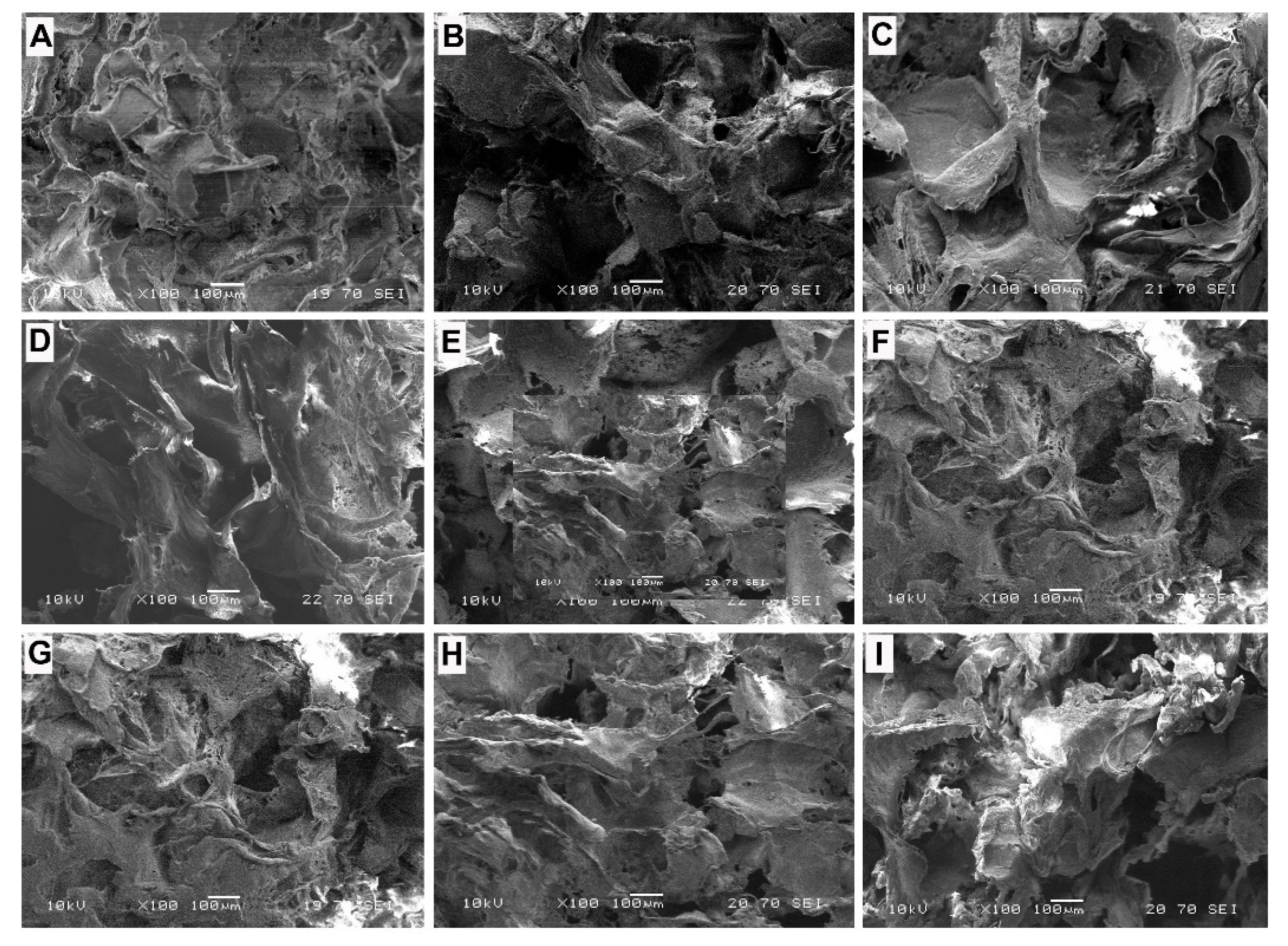
3.1. SEM Analysis
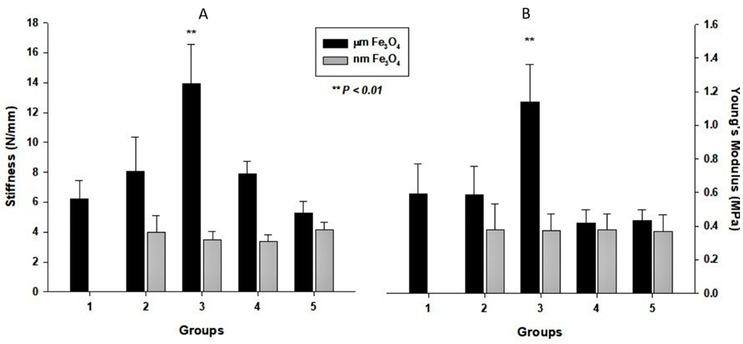
3.2. Mechanical Properties of the Scaffolds
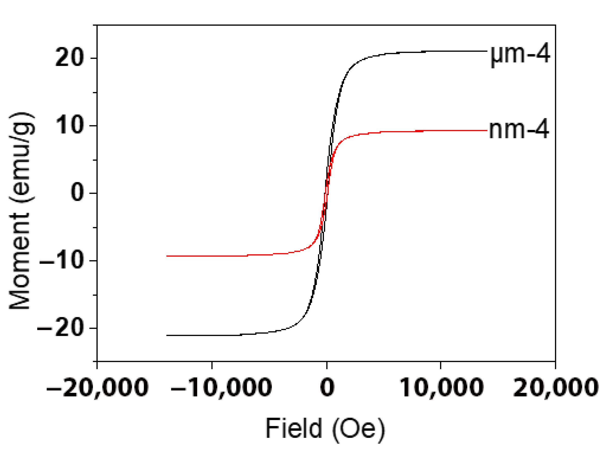
3.3. Magnetic Properties of the Scaffolds
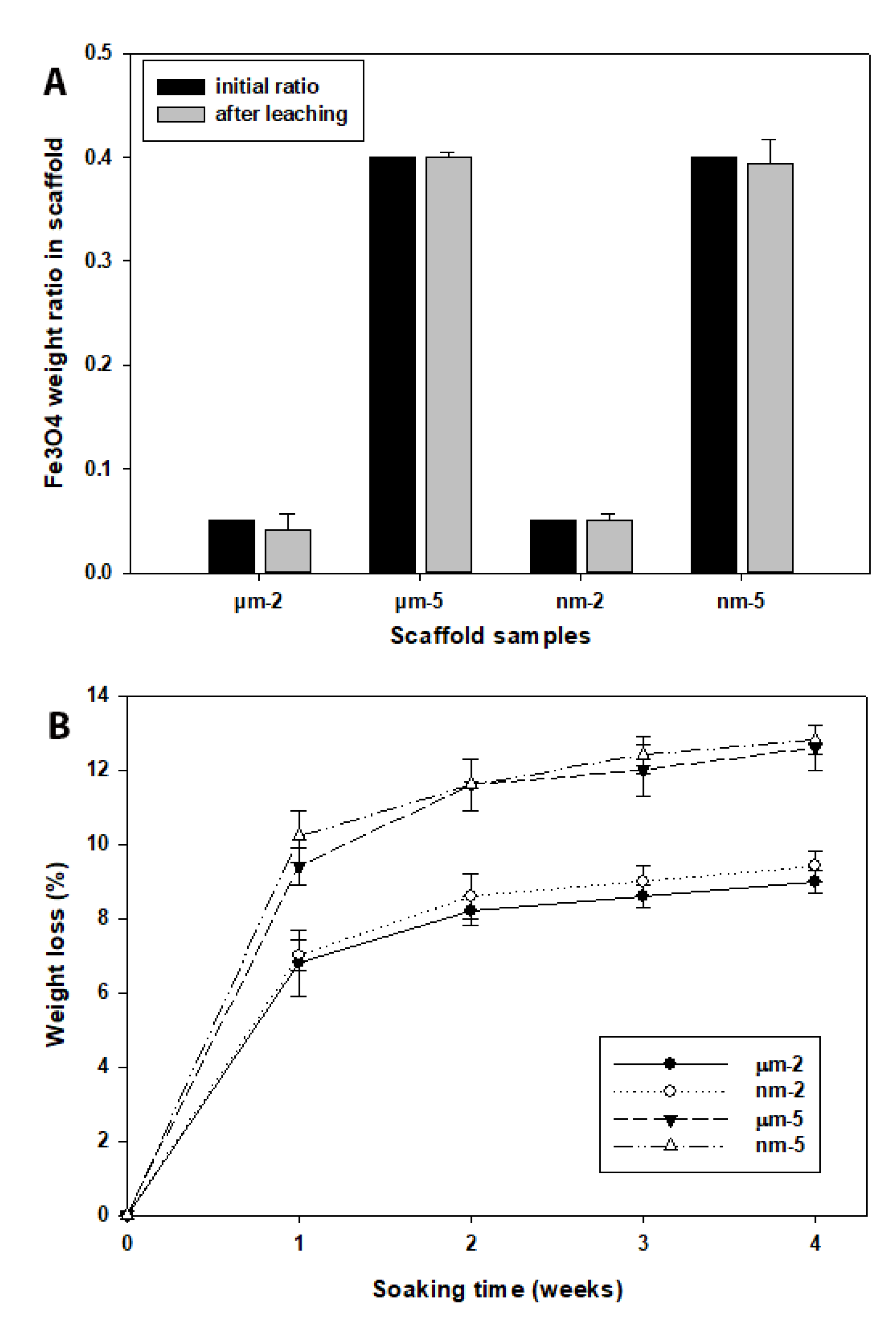
3.4. Magnetic Particle Retention and Degradation Performance of the Fe3O4/PCL Scaffolds
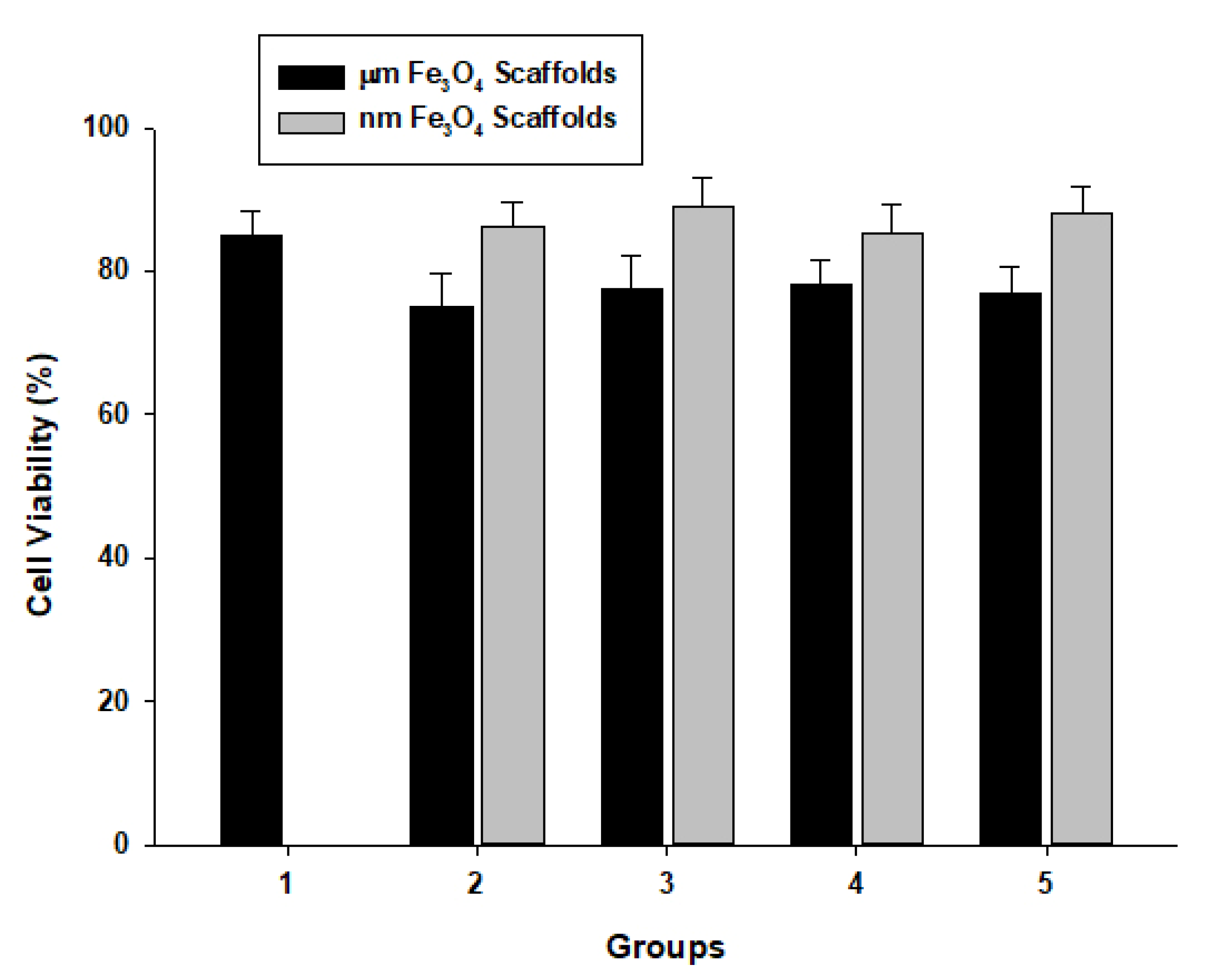
3.5. Evaluation of Cell Biocompatibility on the Fe3O4/PCL Scaffolds
3.6. Cytotoxicity Assay of the Fe3O4/PCL Scaffolds
4. Discussion
5. Conclusions
Author Contributions
Funding
Institutional Review Board Statement
Informed Consent Statement
Data Availability Statement
Acknowledgments
Conflicts of Interest
References
- Tan, P.H.S.; Aung, K.Z.; Toh, S.L.; Goh, J.C.H.; Nathan, S.S. Three-dimensional porous silk tumor constructs in the approximation of in vivo osteosarcoma physiology. Biomaterials 2011, 32, 7. [Google Scholar] [CrossRef] [PubMed]
- Gallego, D.; Ferrell, N.; Sun, Y.; Hansford, D.J. Multilayer micromolding of degradable polymer tissue engineering scaffolds. Mater. Sci. Eng. C 2008, 28, 6. [Google Scholar] [CrossRef]
- Sutthiphong, S.; Pavasant, P.; Supaphol, P. Electrospun 1,6-diisocyanatohexane-extended poly(1,4-butylene succinate) fiber mats and their potential for use as bone scaffolds. Polymer 2009, 50, 11. [Google Scholar] [CrossRef]
- Turhani, D.; Weißenböck, M.; Stein, E.; Wanschitz, F.; Ewers, R. Exogenous Recombinant Human BMP-2 Has Little Initial Effects on Human Osteoblastic Cells Cultured on Collagen Type I Coated/Noncoated Hydroxyapatite Ceramic Granules. J. Oral Maxillofac. Surg. 2007, 65, 9. [Google Scholar] [CrossRef]
- Newell, D.R. How to develop a successful cancer drug—Molecules to medicines or targets to treatments? Eur. J. Cancer 2005, 41, 7. [Google Scholar] [CrossRef]
- Womack, C.; Clack, G. Tissue collection in drug discovery and development research. Eur. J. Cancer Suppl. 2007, 5, 4. [Google Scholar] [CrossRef]
- De Jong, F.A.; Sparreboom, A.; Verweij, J.; Mathijssen, R.H.J. Lifestyle habits as a contributor to anti-cancer treatment failure. Eur. J. Cancer 2008, 44, 9. [Google Scholar] [CrossRef]
- Alymani, N.A.; Smith, M.D.; Williams, D.J.; Petty, R.D. Predictive biomarkers for personalised anti-cancer drug use: Discovery to clinical implementation. Eur. J. Cancer 2010, 46, 11. [Google Scholar] [CrossRef]
- Furlani, E.J. A model for predicting magnetic targeting of multifunctional particles in the microvasculature. J. Magn. Magn. Mater. 2007, 312, 7. [Google Scholar] [CrossRef] [Green Version]
- Pouponneau, P.; Leroux, J.-C.; Soulez, G.; Gaboury, L.; Martel, S. Co-encapsulation of magnetic nanoparticles and doxorubicin into biodegradable microcarriers for deep tissue targeting by vascular MRI navigation. Biomaterials 2011, 32, 6. [Google Scholar] [CrossRef]
- Dandamudi, S.; Campbell, R.B. The drug loading, cytotoxicty and tumor vascular targeting characteristics of magnetite in magnetic drug targeting. Biomaterials 2007, 28, 11. [Google Scholar] [CrossRef] [PubMed]
- Shubayev, V.I.; Pisanic, T.R., II; Jin, S. Magnetic nanoparticles for theragnostics. Adv. Drug Deliv. Rev. 2009, 61, 11. [Google Scholar] [CrossRef] [PubMed] [Green Version]
- Mahmoudi, M.; Sant, S.; Wang, B.; Laurent, S.; Sen, T. Superparamagnetic iron oxide nanoparticles (SPIONs): Development, surface modification and applications in chemotherapy. Adv. Drug Deliv. Rev. 2011, 63, 23. [Google Scholar] [CrossRef] [PubMed] [Green Version]
- Bell, G.; Marino, A.; Chesson, A.; Struve, F. Human sensitivity to weak magnetic fields. Lancet 1991, 338, 2. [Google Scholar] [CrossRef]
- Schenck, J.F. Physical interactions of static magnetic fields with living tissues. Prog. Biophys. Mol. Biol. 2005, 87, 20. [Google Scholar] [CrossRef]
- Lübbe, A.S.; Alexiou, C.; Bergemann, C. Clinical Applications of Magnetic Drug Targeting. J. Surg. Res. 2001, 95, 7. [Google Scholar] [CrossRef]
- Rudge, S.; Peterson, C.; Vessely, C.; Koda, J.; Stevens, S.; Catterall, L. Adsorption and desorption of chemotherapeutic drugs from a magnetically targeted carrier (MTC). J. Control. Release 2001, 74, 6. [Google Scholar] [CrossRef]
- Fannin, P.C.; Scaife, B.K.; Charles, S.W. On the permittivity of magnetic colloids subject to a strong external magnetic field over the frequency range 50 kHz to 1 MHz J. Magn. Magn. Mater. 1993, 122, 4. [Google Scholar] [CrossRef]
- Heinemann, A.; Hoell, A.; Wiedenmann, A.; Pop, L.M. Small-angle scattering of orientated magnetic structures and applications to magnetic colloids. Phys. B Condens. Matter 2006, 385–386, 4. [Google Scholar] [CrossRef]
- Kemppainen, J.M.; Hollister, S.J. Differential effects of designed scaffold permeability on chondrogenesis by chondrocytes and bone marrow stromal cells. Biomaterials 2010, 31, 9. [Google Scholar] [CrossRef]
- Kim, H.-J.; Lee, J.-H.; Im, G.-I. Chondrogenesis using mesenchymal stem cells and PCL scaffolds. J. Biomed. Mater. Res. A 2010, 92, 7. [Google Scholar] [CrossRef] [PubMed]
- Izquierdo, R.; Garcia-Giralt, N.; Rodriguez, M.; Cáceres, E.; García, S.; Ribelles, J.G.; Monleón, M.; Monllau, J.C.; Suay, J. Biodegradable PCL scaffolds with an interconnected spherical pore network for tissue engineering. J. Biomed. Mater. Res. A 2008, 85, 11. [Google Scholar] [CrossRef] [PubMed]
- Li, W.-J.; Tuli, R.; Okafor, C.; Derfoul, A.; Danielson, K.G.; Hall, D.J.; Tuan, R.S. A threedimensional nanofibrous scaffold for cartilage tissue engineering using human mesenchymal stem cells. Biomaterials 2005, 36, 11. [Google Scholar]
- Jeong, C.G.; Hollister, S.J. A comparison of the influence of material on in vitro cartilage tissue engineering with PCL, PGS, and POC 3D scaffold architecture seeded with chondrocytes. Biomaterials 2010, 31, 9. [Google Scholar] [CrossRef] [PubMed] [Green Version]
- Yu, H.; Matthew, H.W.; Wooley, P.H.; Yang, S.Y. Effect of porosity and pore size on microstructures and mechanical properties of poly-epsilon-caprolactone- hydroxyapatite composites. J. Biomed. Mater. Res. B Appl. Biomater. 2008, 86, 541–547. [Google Scholar] [CrossRef] [PubMed]
- Rizzi, S.C.; Heath, D.J.; Coombes, A.G.A.; Bock, N.; Textor, M.; Downes, S. Biodegradable polymer/hydroxyapatite composites: Surface analysis and initial attachment of human osteoblasts. J. Biomed. Mater. Res. A 2001, 55, 12. [Google Scholar] [CrossRef]
- Calandrelli, L.; Immirzi, B.; Malinconico, M.; Luessenheide, S.; Passaro, I.; di Pasquale, R.; Oliva, A. Natural and synthetic hydroxyapatite filled PCL: Mechanical properties and biocompatibility analysis. J. Bioact. Compat. Polym. 2004, 19, 13. [Google Scholar] [CrossRef]
- Ciapetti, G.; Ambrosio, L.; Savarino, L.; Granchi, D.; Cenni, E.; Baldini, N.; Pagani, S.; Guizzardi, S.; Causa, F.; Giunti, A. Osteoblast growth and function in porous poly e-caprolactone matrices for bone repair: A preliminary study. Biomaterials 2003, 24, 10. [Google Scholar] [CrossRef]
- Kim, H.-W.; Knowles, J.C.; Kim, H.-E. Hydroxyapatite/poly(ε-caprolactone) composite coatings on hydroxyapatite porous bone scaffold for drug delivery. Biomaterials 2004, 25, 9. [Google Scholar] [CrossRef]
- Yu, H.; VandeVord, P.J.; Mao, L.; Matthew, H.W.; Wooley, P.H.; Yang, S.-Y. Improved tissue-engineered bone regeneration by endothelial cell mediated vascularization. Biomaterials 2009, 30, 10. [Google Scholar] [CrossRef]
- Asmatulu, R.; Misak, H.; Yang, S.-y.; Wooley, P. Composite Magnetic Nanoparticle Drug Delivery System. U.S. Patent 9,782,342, 10 October 2017. [Google Scholar]
- Ju, H.-Y.; Kuo, C.-H.; Too, J.-R.; Huang, H.-Y.; Twu, Y.-K.; Chang, C.-M.J.; Liu, Y.-C.; Shieh, C.-J. Optimal covalent immobilization of α-chymotrypsin on Fe3O4-chitosan nanoparticles. J. Mol. Catal. B: Enzym. 2012, 78, 7. [Google Scholar] [CrossRef]
- Chu, L.; Jiang, G.; Hu, X.L.; James, T.D.; He, X.P.; Li, Y.; Tang, T. Biodegradable macroporous scaffold with nano-crystal surface microstructure for highly effective osteogenesis and vascularization. J. Mater. Chem B 2018, 6, 1658–1667. [Google Scholar] [CrossRef] [PubMed] [Green Version]
- Ge, J.; Li, M.; Zhang, Q.; Yang, C.Z.; Wooley, P.H.; Chen, X.; Yang, S.Y. Silica Aerogel Improves the Biocompatibility in a Poly-Caprolactone Composite Used as a Tissue Engineering Scaffold. Int. J. Polym. Sci. 2013, 2013, 7. [Google Scholar] [CrossRef] [Green Version]
- Ma, H.-l.; Qi, X.-r.; Maitani, Y.; Nagai, T. Preparation and characterization of superparamagnetic iron oxide nanoparticles stabilized by alginate. Int. J. Pharm. 2007, 333, 10. [Google Scholar] [CrossRef] [PubMed]
- Feng, J.; Mao, J.; Wen, X.; Tu, M. Ultrasonic-assisted in situ synthesis and characterization of superparamagnetic Fe3O4 nanoparticles. J. Alloy. Compd. 2011, 509, 5. [Google Scholar] [CrossRef]
- Ge, J.; Zhai, M.; Zhang, Y.; Bian, J.; Wu, J. Biocompatible Fe3O4/chitosan scaffolds with high magnetism. Int. J. Biol. Macromol. 2019, 128, 406–413. [Google Scholar] [CrossRef]
- Yang, T.; Ge, J.; Asmatulu, R.; Yang, S.-Y. Magnetically Inducible Nanocomposite Scaffolds for Improved Bone Regenerations. Adv. Sci. Eng. Med. 2015, 7, 790–796. [Google Scholar] [CrossRef]
- Wong, H.M.; Zhao, Y.; Leung, F.K.L.; Xi, T.; Zhang, Z.; Zheng, Y.; Wu, S.; Luk, K.D.K.; Cheung, K.M.C.; Chu, P.K.; et al. Functionalized Polymeric Membrane with Enhanced Mechanical and Biological Properties to Control the Degradation of Magnesium Alloy. Adv. Healthc. Mater. 2017, 6, 1601269. [Google Scholar] [CrossRef]


| Sample | Fe3O4 Size | Fe3O4 (w/w, %) | PCL (w/w, %) |
|---|---|---|---|
| 1 | - | 0 | 100 |
| μm-2 | <5 μm | 5 | 95 |
| μm-3 | <5 μm | 10 | 90 |
| μm-4 | <5 μm | 20 | 80 |
| μm-5 | <5 μm | 40 | 60 |
| nm-2 | <50 nm | 5 | 95 |
| nm-3 | <50 nm | 10 | 90 |
| nm-4 | <50 nm | 20 | 80 |
| nm-5 | <50 nm | 40 | 60 |
| Days | 1 | 2 | 3 | 4 | 5 | 6 | 7 | |
| Fe3O4 free | 0.98 ± 0.04 | 0.97 ± 0.02 | 0.98 ± 0.01 | 0.88 ± 0.09 | 1.10 ± 0.04 | 1.09 ± 0.05 | 1.04 ± 0.02 | |
| µm Fe3O4 Scaffold | 5% | 0.92 ± 0.03 | 0.95 ± 0.05 | 0.90 ± 0.04 | 0.82 ± 0.05 | 0.94 ± 0.05 | 0.99 ± 0.10 | 0.94 ± 0.05 |
| 10% | 0.94 ± 0.04 | 0.94 ± 0.03 | 0.95 ± 0.02 | 0.88 ± 0.03 | 1.01 ± 0.02 | 0.97 ± 0.04 | 0.95 ± 0.04 | |
| 20% | 1.03 ± 0.04 | 0.98 ± 0.05 | 0.90 ± 0.05 | 0.81 ± 0.04 | 0.99 ± 0.05 | 0.97 ± 0.05 | 0.95 ± 0.05 | |
| 40% | 0.97 ± 0.05 | 0.90 ± 0.04 | 0.89 ± 0.05 | 0.80 ± 0.08 | 0.94 ± 0.04 | 0.94 ± 0.06 | 0.95 ± 0.05 | |
| nm Fe3O4 Scaffold | 5% | 1.01 ± 0.07 | 0.96 ± 0.05 | 0.94 ± 0.05 | 0.81 ± 0.05 | 1.05 ± 0.04 | 1.08 ± 0.04 | 1.02 ± 0.02 |
| 10% | 0.99 ± 0.05 | 0.97 ± 0.06 | 0.90 ± 0.06 | 0.79 ± 0.06 | 0.97 ± 0.05 | 0.98 ± 0.05 | 1.02 ± 0.05 | |
| 20% | 0.92 ± 0.03 | 0.88 ± 0.05 | 0.87 ± 0.05 | 0.79 ± 0.05 | 0.96 ± 0.08 | 0.97 ± 0.04 | 1.00 ± 0.03 | |
| 40% | 0.92 ± 0.05 | 0.89 ± 0.01 | 0.85 ± 0.03 | 0.81 ± 0.03 | 0.95 ± 0.05 | 0.97 ± 0.05 | 0.95 ± 0.06 | |
Publisher’s Note: MDPI stays neutral with regard to jurisdictional claims in published maps and institutional affiliations. |
© 2022 by the authors. Licensee MDPI, Basel, Switzerland. This article is an open access article distributed under the terms and conditions of the Creative Commons Attribution (CC BY) license (https://creativecommons.org/licenses/by/4.0/).
Share and Cite
Ge, J.; Asmatulu, R.; Zhu, B.; Zhang, Q.; Yang, S.-Y. Synthesis and Properties of Magnetic Fe3O4/PCL Porous Biocomposite Scaffolds with Different Sizes and Quantities of Fe3O4 Particles. Bioengineering 2022, 9, 278. https://doi.org/10.3390/bioengineering9070278
Ge J, Asmatulu R, Zhu B, Zhang Q, Yang S-Y. Synthesis and Properties of Magnetic Fe3O4/PCL Porous Biocomposite Scaffolds with Different Sizes and Quantities of Fe3O4 Particles. Bioengineering. 2022; 9(7):278. https://doi.org/10.3390/bioengineering9070278
Chicago/Turabian StyleGe, Jianhua, Ramazan Asmatulu, Bo Zhu, Qiu Zhang, and Shang-You Yang. 2022. "Synthesis and Properties of Magnetic Fe3O4/PCL Porous Biocomposite Scaffolds with Different Sizes and Quantities of Fe3O4 Particles" Bioengineering 9, no. 7: 278. https://doi.org/10.3390/bioengineering9070278