
Dynamic Foam Characteristics during Cultivation of Arthrospira platensis

 ,
,  , and
, and 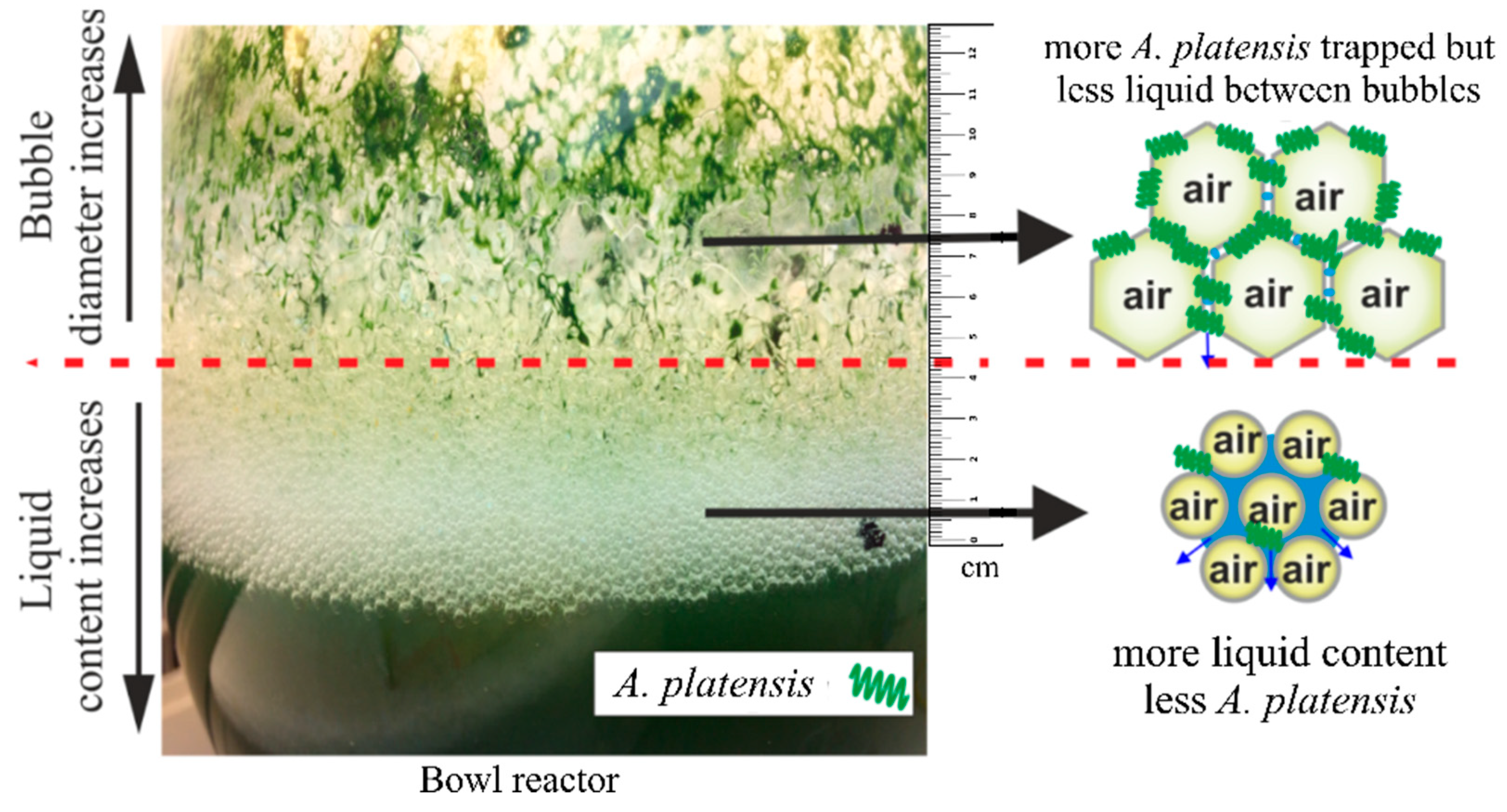
Abstract
:1. Introduction
2. Materials and Methods
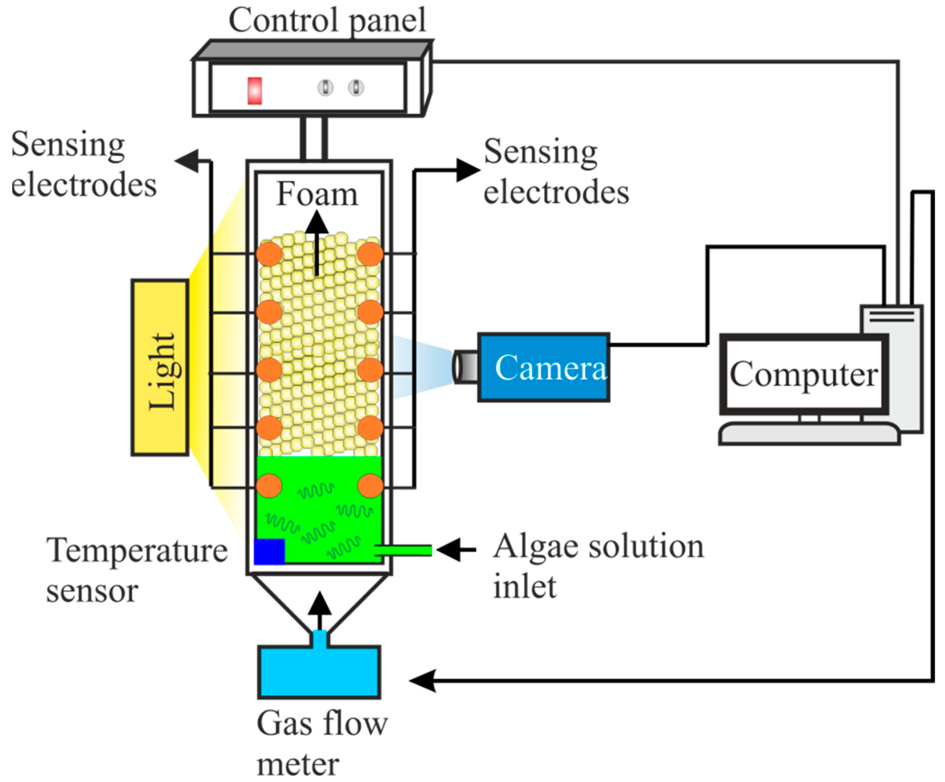
2.1. Instrumental Setup
2.2. Foam Generation and Stability Analysis
2.3. Surface Tension
2.4. Cultivation Conditions
2.5. Microscopic Observation
3. Results and Discussion
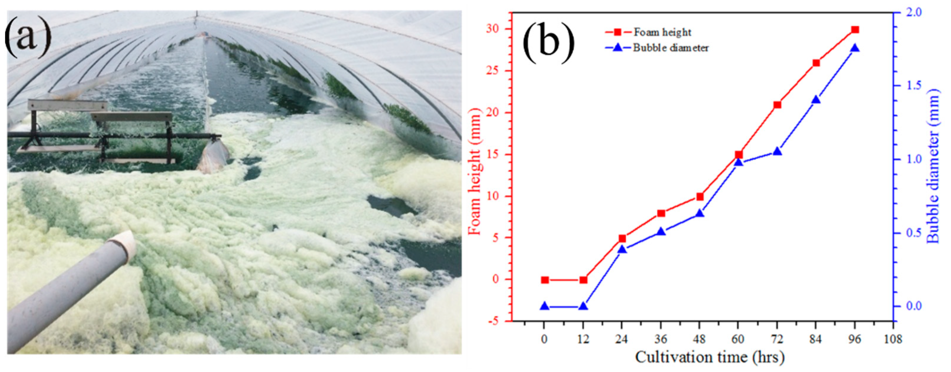
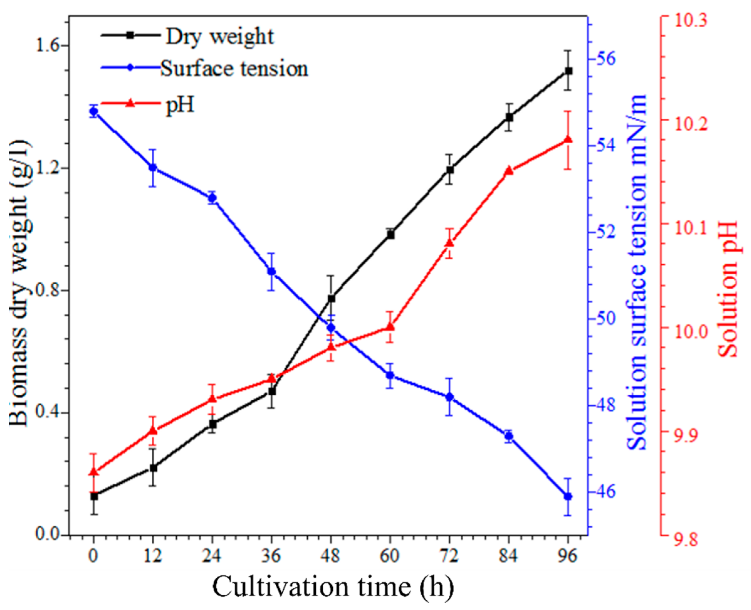
3.1. Foam Morphology during A. platensis Cultivation
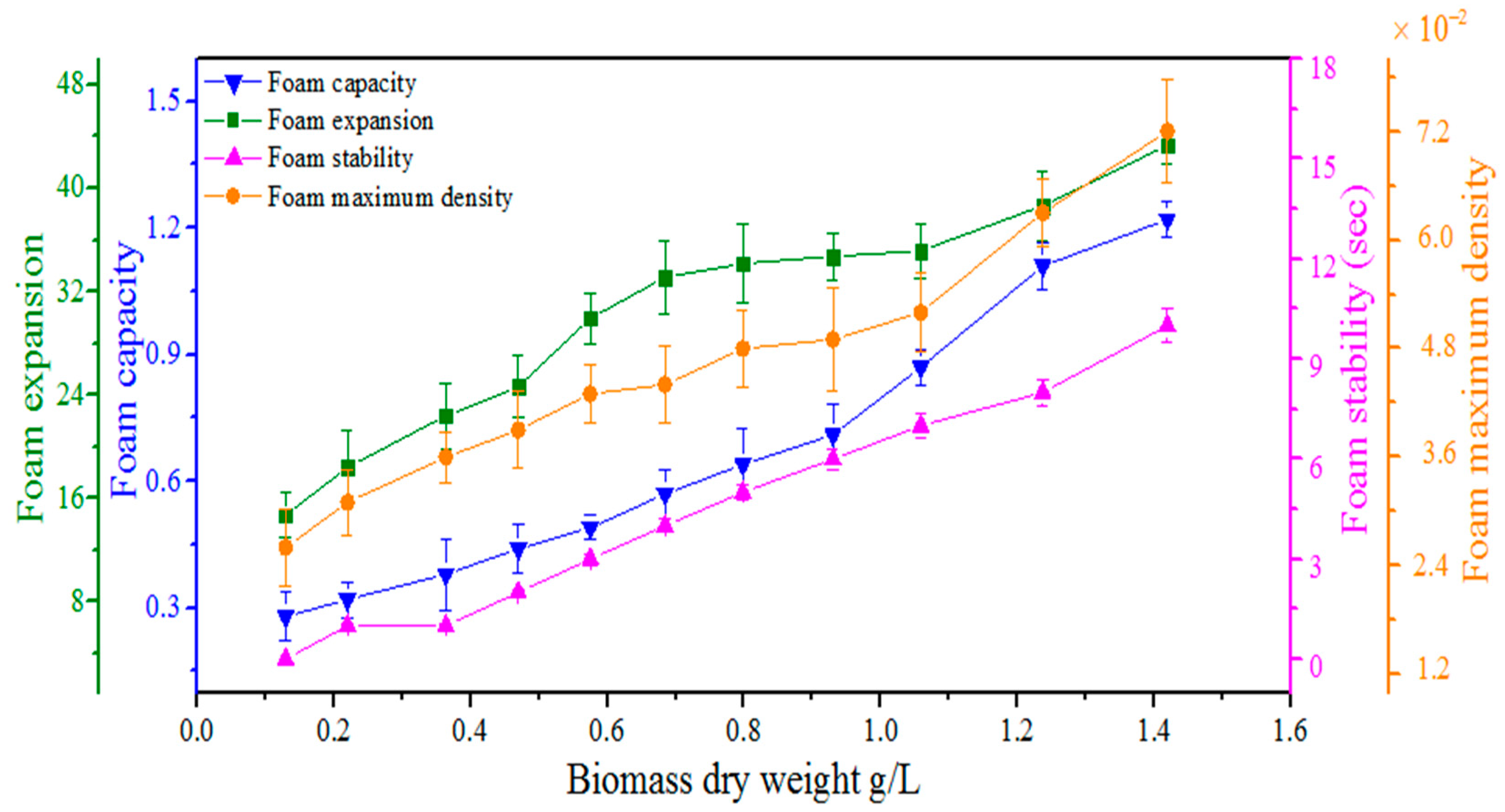
3.2. Stability of Foam during Biomass Production
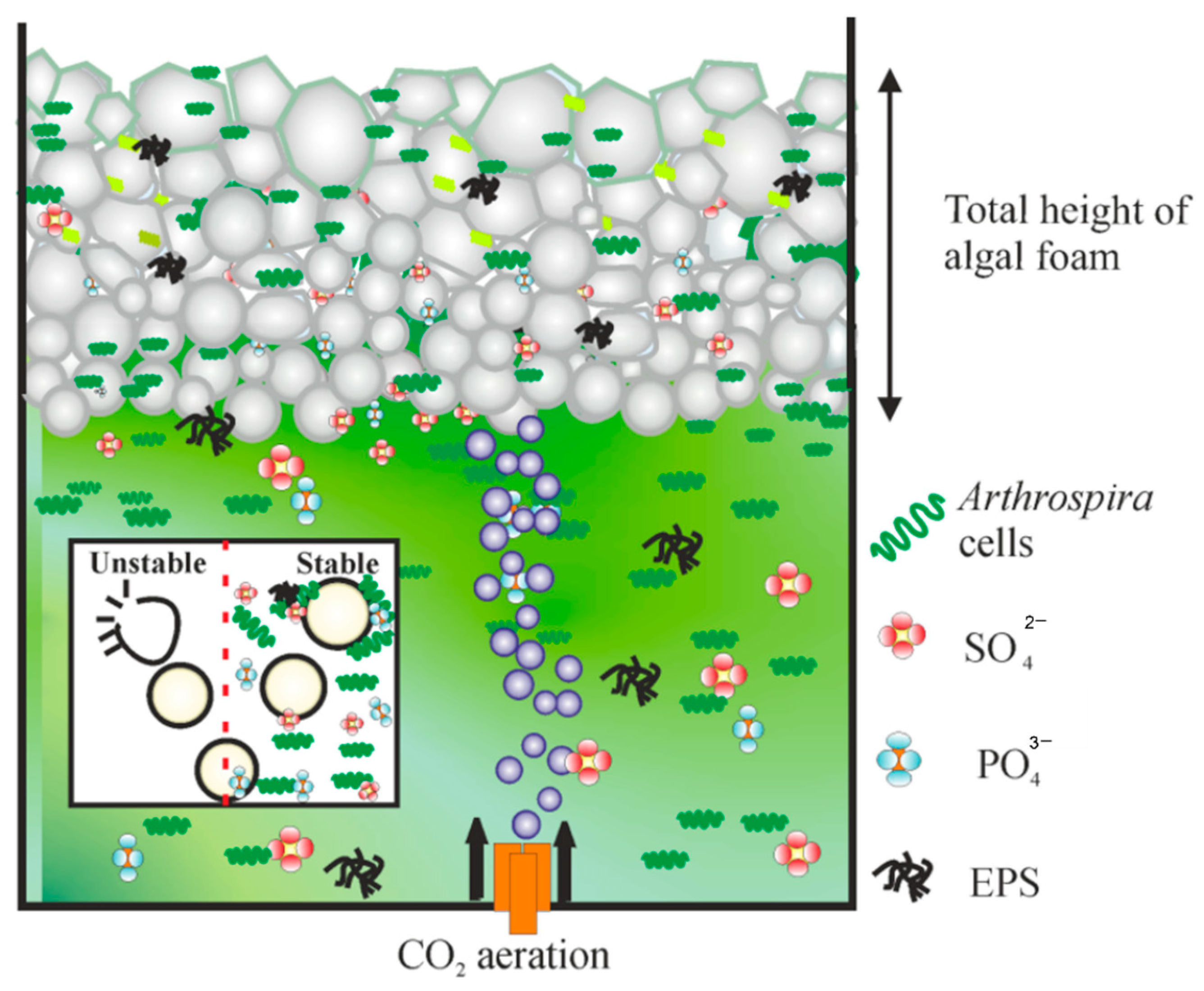
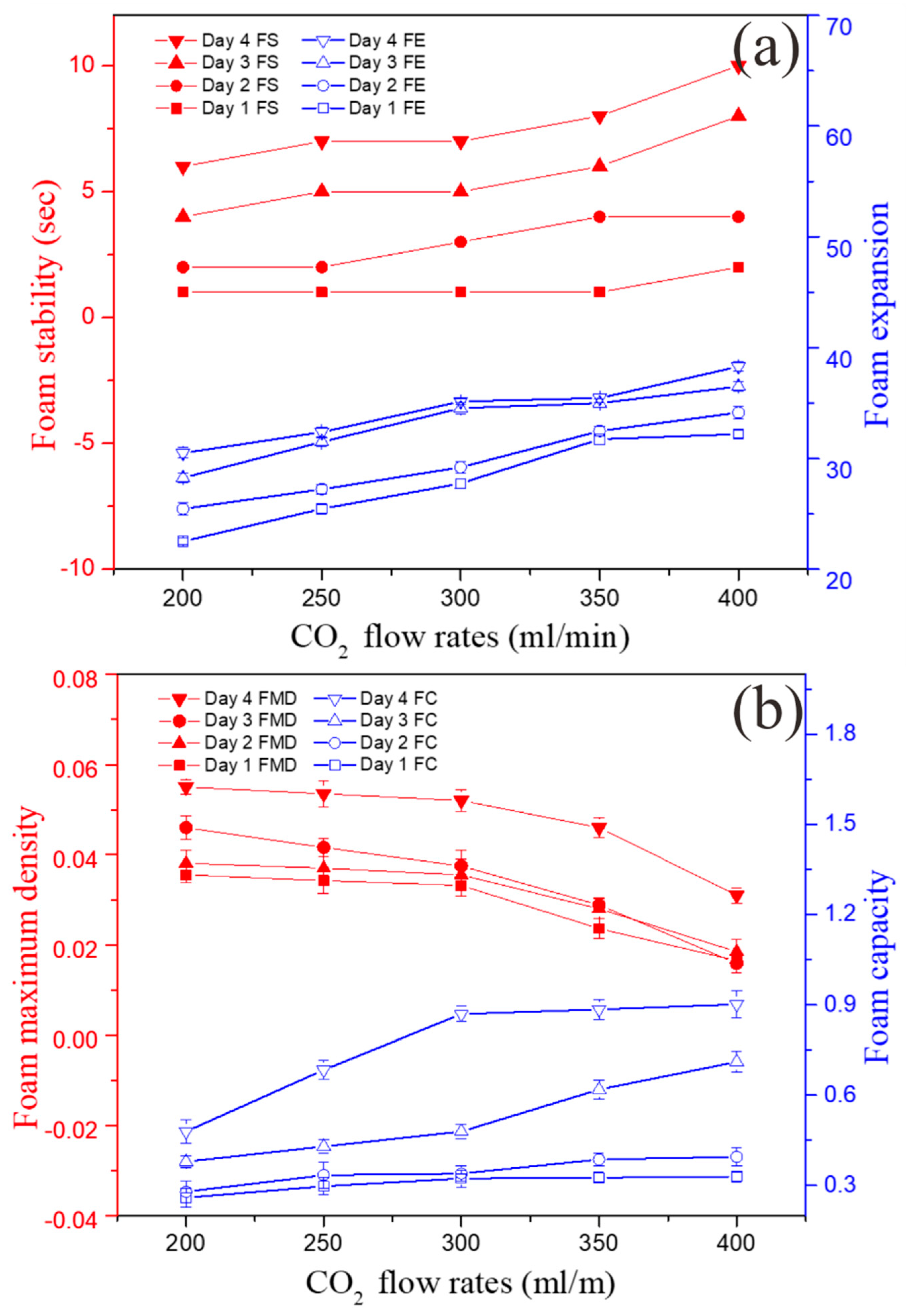
3.3. Effect of CO2 Aeration on Foam Stability
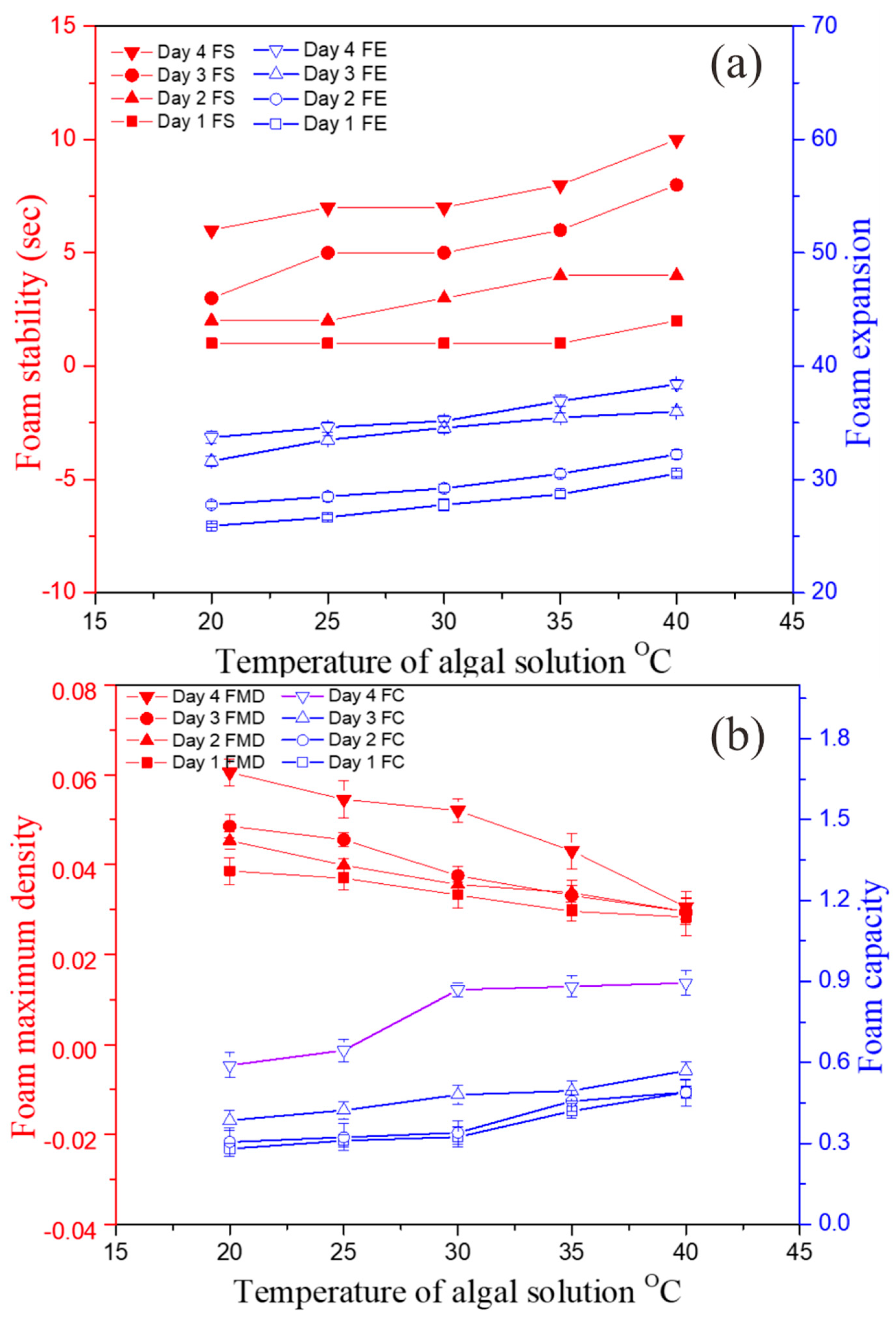
3.4. Effect of Cultivation Temperature on Foaming Stability
4. Conclusions
Author Contributions
Funding
Institutional Review Board Statement
Informed Consent Statement
Data Availability Statement
Acknowledgments
Conflicts of Interest
References
- Zhao, B.; Su, Y.; Zhang, Y.; Cui, G. Carbon Dioxide Fi Xation and Biomass Production from Combustion Fl Ue Gas Using Energy Microalgae. Energy 2015, 89, 347–357. [Google Scholar] [CrossRef]
- Ganesh Saratale, R.; Kumar, G.; Banu, R.; Xia, A.; Periyasamy, S.; Dattatraya Saratale, G. A Critical Review on Anaerobic Digestion of Microalgae and Macroalgae and Co-Digestion of Biomass for Enhanced Methane Generation. Bioresour. Technol. 2018, 262, 319–332. [Google Scholar] [CrossRef] [PubMed]
- Reen, S.; Chyuan, H.; Wayne, K.; Loke, P.; Phang, S.; Chuan, T.; Nagarajan, D.; Lee, D. Sustainable Approaches for Algae Utilisation in Bioenergy Production. Renew. Energy 2017, 129, 838–852. [Google Scholar] [CrossRef]
- Ho, S.; Chen, C.; Lee, D.; Chang, J. Perspectives on Microalgal CO 2 -Emission Mitigation Systems—A Review. Biotechnol. Adv. 2011, 29, 189–198. [Google Scholar] [CrossRef] [PubMed]
- Razzak, S.A.; Hossain, M.M.; Lucky, R.A.; Bassi, A.S.; Lasa, H. De Integrated CO 2 Capture, Wastewater Treatment and Biofuel Production by Microalgae Culturing—A Review. Renew. Sustain. Energy Rev. 2013, 27, 622–653. [Google Scholar] [CrossRef]
- Khan, S.; Siddique, R.; Huanfei, D.; Shereen, M.A.; Nabi, G.; Bai, Q.; Manan, S.; Xue, M.; Ullah, M.W.; Bowen, H. Perspective Applications and Associated Challenges of Using Nanocellulose in Treating Bone-Related Diseases. Front. Bioeng. Biotechnol. 2021, 9, 350. [Google Scholar] [CrossRef] [PubMed]
- Ullah, M.W.; Manan, S.; Kiprono, S.J.; Ul-Islam, M.; Yang, G. Synthesis, Structure, and Properties of Bacterial Cellulose. In Nanocellulose: From Fundamental to Advanced Materials; Huang, J., Lin, N., Dufresne, A., Eds.; Wiley: Weinheim, Germany, 2019; pp. 81–113. [Google Scholar] [CrossRef]
- Pires, J.C.M.; Alvim-ferraz, M.C.M.; Martins, F.G. Photobioreactor Design for Microalgae Production through Computational Fl Uid Dynamics: A Review. Renew. Sustain. Energy Rev. 2017, 79, 248–254. [Google Scholar] [CrossRef]
- Lee, E.; Pruvost, J.; He, X.; Munipalli, R.; Pilon, L. Design Tool and Guidelines for Outdoor Photobioreactors. Chem. Eng. Sci. 2014, 106, 18–29. [Google Scholar] [CrossRef] [Green Version]
- Spolaore, P.; Joannis-Cassan, C.; Duran, E.; Isambert, A. Commercial Applications of Microalgae. J. Biosci. Bioeng. 2006, 101, 87–96. [Google Scholar] [CrossRef] [PubMed] [Green Version]
- Prussi, M.; Buffi, M.; Casini, D.; Chiaramonti, D.; Martelli, F.; Carnevale, M.; Tredici, M.R. ScienceDirect Experimental and Numerical Investigations of Mixing in Raceway Ponds for Algae Cultivation. Biomass Bioenergy 2014, 67, 390–400. [Google Scholar] [CrossRef]
- Pankratz, S.; Oyedun, A.O.; Zhang, X.; Kumar, A. Algae Production Platforms for Canada ’ s Northern Climate. Renew. Sustain. Energy Rev. 2017, 80, 109–120. [Google Scholar] [CrossRef]
- de Godos, I.; Mendoza, J.L.; Acién, F.G.; Molina, E.; Banks, C.J.; Heaven, S.; Rogalla, F. Evaluation of Carbon Dioxide Mass Transfer in Raceway Reactors for Microalgae Culture Using Flue Gases. Bioresour. Technol. 2014, 153, 307–314. [Google Scholar] [CrossRef] [PubMed]
- Wang, J.; Nguyen, A.V.; Farrokhpay, S. A Critical Review of the Growth, Drainage and Collapse of Foams. Adv. Colloid Interface Sci. 2016, 228, 55–70. [Google Scholar] [CrossRef] [PubMed] [Green Version]
- Chen, X.W.; Yang, D.X.; Zou, Y.; Yang, X.Q. Stabilization and Functionalization of Aqueous Foams by Quillaja Saponin-Coated Nanodroplets. Food Res. Int. 2017, 99, 679–687. [Google Scholar] [CrossRef] [PubMed]
- Xu, L.; Xu, G.; Gong, H.; Dong, M.; Li, Y.; Zhou, Y. Colloids and Surfaces A: Physicochemical and Engineering Aspects Foam Properties and Stabilizing Mechanism of Sodium Fatty Alcohol Polyoxyethylene Ether Sulfate-Welan Gum Composite Systems. Colloids Surfaces A Physicochem. Eng. Asp. 2014, 456, 176–183. [Google Scholar] [CrossRef]
- Kougias, P.G.; Boe, K.; Tsapekos, P.; Angelidaki, I. Bioresource Technology Foam Suppression in Overloaded Manure-Based Biogas Reactors Using Antifoaming Agents. Bioresour. Technol. 2014, 153, 198–205. [Google Scholar] [CrossRef]
- Subramanian, B.; Pagilla, K.R. Colloids and Surfaces B: Biointerfaces Mechanisms of Foam Formation in Anaerobic Digesters. Colloids Surfaces B Biointerfaces 2015, 126, 621–630. [Google Scholar] [CrossRef]
- Kougias, P.G.; Boe, K.; Einarsdottir, E.S.; Angelidaki, I. ScienceDirect Counteracting Foaming Caused by Lipids or Proteins in Biogas Reactors Using Rapeseed Oil or Oleic Acid as Antifoaming Agents. Water Res. 2015, 79, 119–127. [Google Scholar] [CrossRef]
- Moeller, L.; Lehnig, M.; Schenk, J.; Zehnsdorf, A. Bioresource Technology Foam Formation in Biogas Plants Caused by Anaerobic Digestion of Sugar Beet. Bioresour. Technol. 2015, 178, 270–277. [Google Scholar] [CrossRef]
- Kubar, A.A.; Cheng, J.; Kumar, S.; Liu, S.; Chen, S.; Tian, J. Strengthening Mass Transfer with the Tesla-Valve Baffles to Increase the Biomass Yield of Arthrospira Platensis in a Column Photobioreactor. Bioresour. Technol. 2021, 320, 124337. [Google Scholar] [CrossRef]
- Cheng, J.; Guo, W.; Song, Y.; Kumar, S.; Ameer Ali, K.; Zhou, J. Enhancing Vorticity Magnitude of Turbulent Flow to Promote Photochemical Efficiency and Trichome Helix Pitch of Arthrospira Platensis in a Raceway Pond with Conic Baffles. Bioresour. Technol. 2018, 269, 1–8. [Google Scholar] [CrossRef] [PubMed]
- Ali Kubar, A.; Cheng, J.; Guo, W.; Kumar, S.; Song, Y. Development of a Single Helical Baffle to Increase CO2 Gas and Microalgal Solution Mixing and Chlorella PY-ZU1 Biomass Yield. Bioresour. Technol. 2020, 307, 123253. [Google Scholar] [CrossRef] [PubMed]
- Kumar, S.; Cheng, J.; Guo, W.; Ali, K.A.; Song, Y. Self-Rotary Propellers with Clockwise/Counterclockwise Blades Create Spiral Flow Fields to Improve Mass Transfer and Promote Microalgae Growth. Bioresour. Technol. 2019, 286, 121384. [Google Scholar] [CrossRef] [PubMed]
- Jones, S.A.; Laskaris, G.; Vincent-Bonnieu, S.; Farajzadeh, R.; Rossen, W.R. Effect of Surfactant Concentration on Foam: From Coreflood Experiments to Implicit-Texture Foam-Model Parameters. J. Ind. Eng. Chem. 2016, 37, 268–276. [Google Scholar] [CrossRef] [Green Version]
- Wang, H.; Guo, W.; Zheng, C.; Wang, D.; Zhan, H. Effect of Temperature on Foaming Ability and Foam Stability of Typical Surfactants Used for Foaming Agent. J. Surfactants Deterg. 2017, 20, 615–622. [Google Scholar] [CrossRef]
- Trabelsi, L.; M’sakni, N.H.; Ouada, H.B.; Bacha, H.; Roudesli, S. Partial Characterization of Extracellular Polysaccharides Produced by Cyanobacterium Arthrospira Platensis. Biotechnol. Bioprocess Eng. 2009, 14, 27–31. [Google Scholar] [CrossRef]
- Chentir, I.; Hamdi, M.; Doumandji, A.; HadjSadok, A.; Ouada, H.B.; Nasri, M.; Jridi, M. Enhancement of Extracellular Polymeric Substances (EPS) Production in Spirulina (Arthrospira Sp.) by Two-Step Cultivation Process and Partial Characterization of Their Polysaccharidic Moiety. Int. J. Biol. Macromol. 2017, 105, 1412–1420. [Google Scholar] [CrossRef]
- Liu, Q.; Zhang, S.; Sun, D.; Xu, J. Foams Stabilized by Laponite Nanoparticles and Alkylammonium Bromides with Different Alkyl Chain Lengths. Colloids Surfaces A Physicochem. Eng. Asp. 2010, 355, 151–157. [Google Scholar] [CrossRef]
- Georgieva, D.; Cagna, A.; Langevin, D. Link between Surface Elasticity and Foam Stability. Soft Matter 2009, 5, 2063–2071. [Google Scholar] [CrossRef]
- Saint-Jalmes, A. Physical Chemistry in Foam Drainage and Coarsening. Soft Matter 2006, 2, 836. [Google Scholar] [CrossRef]
- Roth, A.E.; Jones, C.D.; Durian, D.J. Bubble Statistics and Coarsening Dynamics for Quasi-Two-Dimensional Foams with Increasing Liquid Content. Phys. Rev. E-Stat. Nonlinear Soft Matter Phys. 2013, 87, 1–14. [Google Scholar] [CrossRef] [PubMed] [Green Version]
- Bernaerts, T.M.M.; Gheysen, L.; Foubert, I.; Hendrickx, M.E.; Van Loey, A.M. The Potential of Microalgae and Their Biopolymers as Structuring Ingredients in Food: A Review. Biotechnol. Adv. 2019, 37, 107419. [Google Scholar] [CrossRef] [PubMed]
- Singh, S.P.; Singh, P. Effect of Temperature and Light on the Growth of Algae Species: A Review. Renew. Sustain. Energy Rev. 2015, 50, 431–444. [Google Scholar] [CrossRef]
- Ahmed, M.; Moerdijk-Poortvliet, T.C.W.; Wijnholds, A.; Stal, L.J.; Hasnain, S. Isolation, Characterization and Localization of Extracellular Polymeric Substances from the Cyanobacterium Arthrospira Platensis Strain MMG-9. Eur. J. Phycol. 2014, 49, 143–150. [Google Scholar] [CrossRef] [Green Version]
- Renaud, S.M.; Thinh, L.-V.; Lambrinidis, G.; Parry, D.L. Effect of Temperature on Growth, Chemical Composition and Fatty Acid Composition of Tropical Australian Microalgae Grown in Batch Cultures. Aquaculture 2002, 211, 195–214. [Google Scholar] [CrossRef]
- Mancuso Nichols, C.A.; Nairn, K.M.; Glattauer, V.; Blackburn, S.I.; Ramshaw, J.A.M.; Graham, L.D. Screening Microalgal Cultures in Search of Microbial Exopolysaccharides with Potential as Adhesives. J. Adhes. 2009, 85, 97–125. [Google Scholar] [CrossRef]
- Muñoz, R.; Guieysse, B. Algal-Bacterial Processes for the Treatment of Hazardous Contaminants: A Review. Water Res. 2006, 40, 2799–2815. [Google Scholar] [CrossRef]
- Kapetas, L.; Vincent Bonnieu, S.; Danelis, S.; Rossen, W.R.; Farajzadeh, R.; Eftekhari, A.A.; Mohd Shafian, S.R.; Kamarul Bahrim, R.Z. Effect of Temperature on Foam Flow in Porous Media. J. Ind. Eng. Chem. 2016, 36, 229–237. [Google Scholar] [CrossRef] [Green Version]


Publisher’s Note: MDPI stays neutral with regard to jurisdictional claims in published maps and institutional affiliations. |
© 2022 by the authors. Licensee MDPI, Basel, Switzerland. This article is an open access article distributed under the terms and conditions of the Creative Commons Attribution (CC BY) license (https://creativecommons.org/licenses/by/4.0/).
Share and Cite
Kubar, A.A.; Ali, A.; Kumar, S.; Huo, S.; Ullah, M.W.; Alabbosh, K.F.S.; Ikram, M.; Cheng, J. Dynamic Foam Characteristics during Cultivation of Arthrospira platensis. Bioengineering 2022, 9, 257. https://doi.org/10.3390/bioengineering9060257
Kubar AA, Ali A, Kumar S, Huo S, Ullah MW, Alabbosh KFS, Ikram M, Cheng J. Dynamic Foam Characteristics during Cultivation of Arthrospira platensis. Bioengineering. 2022; 9(6):257. https://doi.org/10.3390/bioengineering9060257
Chicago/Turabian StyleKubar, Ameer Ali, Amjad Ali, Santosh Kumar, Shuhao Huo, Muhammad Wajid Ullah, Khulood Fahad Saud Alabbosh, Muhammad Ikram, and Jun Cheng. 2022. "Dynamic Foam Characteristics during Cultivation of Arthrospira platensis" Bioengineering 9, no. 6: 257. https://doi.org/10.3390/bioengineering9060257