
Implant Periapical Lesion: Clinical and Histological Analysis of Two Case Reports Carried Out with Two Different Approaches

 ,
,  , ,
, , 
Abstract
:1. Introduction
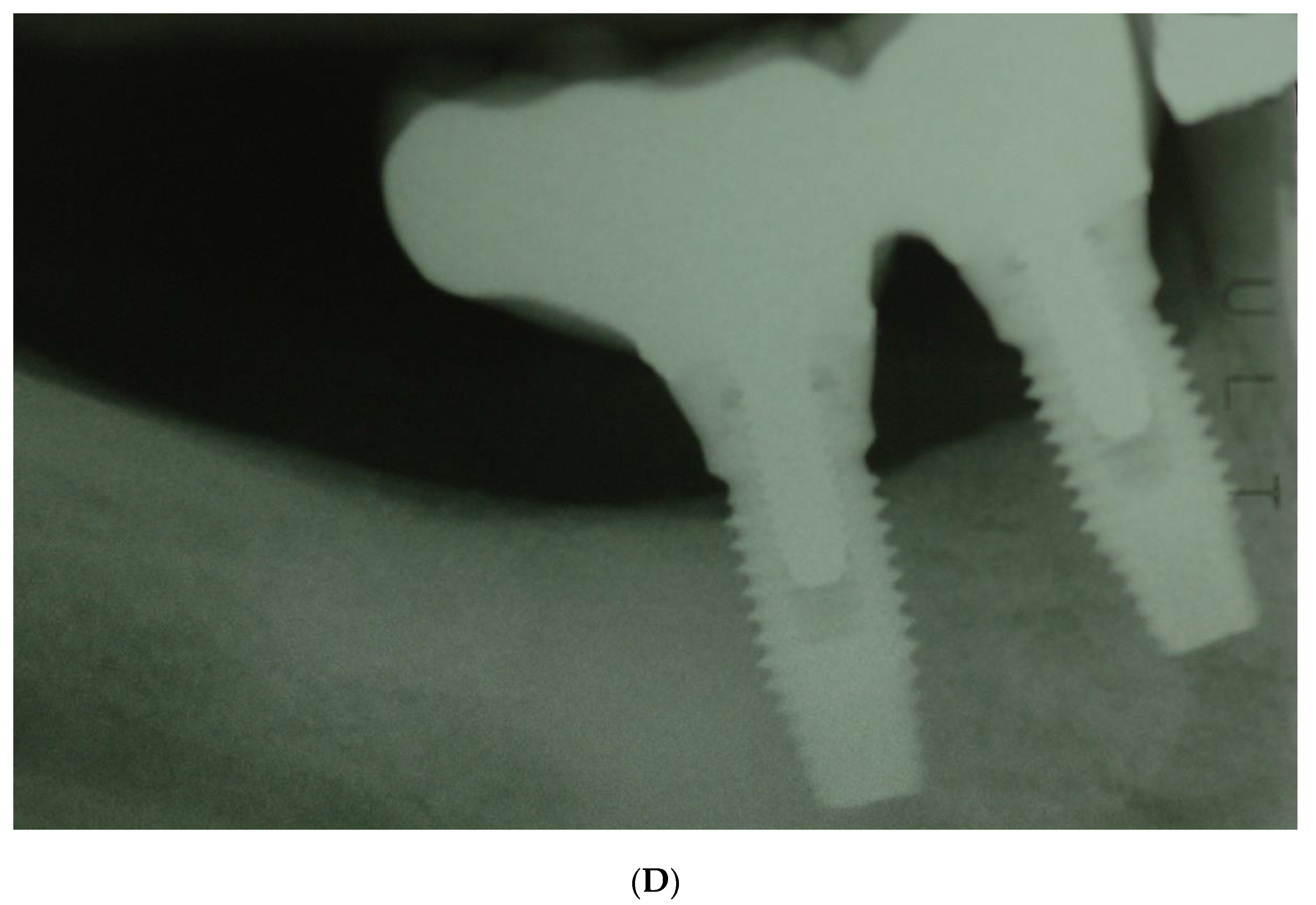
2. Case Report n.1
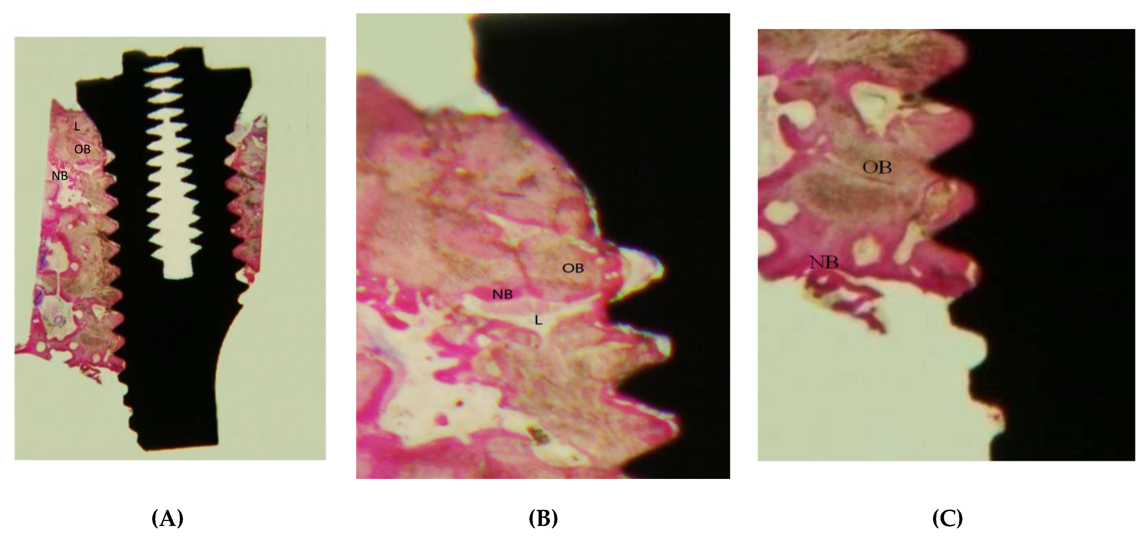
2.1. Histological Preparation
2.2. Histological Analysis
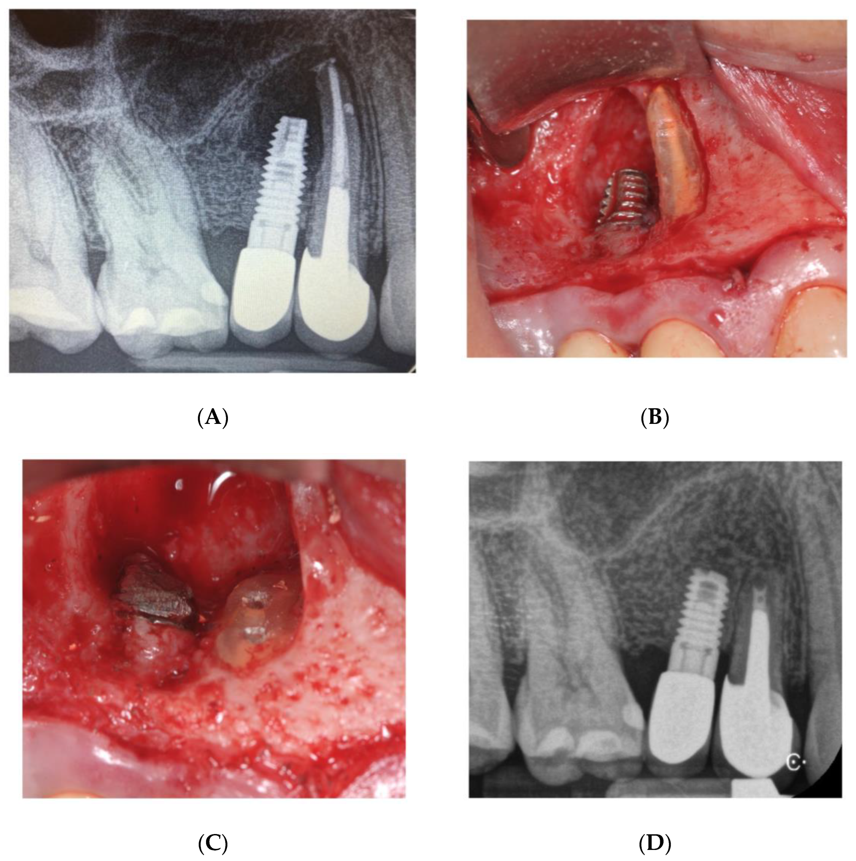
3. Case Report n. 2
4. Discussion
5. Conclusions
Author Contributions
Funding
Institutional Review Board Statement
Informed Consent Statement
Data Availability Statement
Conflicts of Interest
References
- Esposito, M.; Hirsch, J.; Lekholm, U.; Thomsen, P. Differential diagnosis and treatment strategies for biologic complications and failing oral implants: A review of the literature. Int. J. Oral Maxillofac. Implants 1999, 14, 473–490. [Google Scholar] [PubMed]
- Esposito, M.; Hirsch, J.M.; Lekholm, U.; Thomsen, P. Biological factors contributing to failures of osseointegrated oral implants. I: Success criteria and epidemiology. Eur. J. Oral Sci. 1998, 106, 527–551. [Google Scholar] [CrossRef]
- Esposito, M.; Hirsch, J.M.; Lekholm, U.; Thomsen, P. Biological factors contributing to failures of osseointegrated oral implants. II: Etiopathogenesis. Eur. J. Oral Sci. 1998, 106, 721–764. [Google Scholar] [CrossRef]
- Esposito, M. On Biological Failures of Osseointegrated Oral Implants. Ph.D. Thesis, University of Gothenburg, Gothenburg, Sweden, 1999. [Google Scholar]
- El Askary, A.S.; Meffert, R.M.; Griffin, T. Why do implants fail? Part I. Implant Dent. 1999, 8, 173–185. [Google Scholar] [CrossRef] [PubMed]
- Meffert, R.M. Periodontitis and peri-implantitis: One and the same? Pract. Periodontics Aesthet. Dent. 1993, 5, 79–80, 82. [Google Scholar]
- McAllister, B.S.; Masters, D.; Meffert, R.M. Treatment of implants demonstrating periapical radiolucencies. Pract. Periodontics Aesthet. Dent. 1992, 4, 37–41. [Google Scholar] [PubMed]
- Park, S.H.; Sorensen, W.P.; Wang, H.L. Management and prevention of retrograde peri-implant infection from retained root tips: Two case reports. Int. J. Periodontics Restor. Dent. 2004, 24, 422–433. [Google Scholar] [CrossRef] [Green Version]
- Reiser, G.M.; Nevins, M. The implant periapical lesion: Etiology, prevention, and treatment. Compend. Contin. Educ. Dent. 1995, 16, 768, 770, 772. [Google Scholar]
- Quirynen, M.; Vogels, R.; Alsaa, G.; Naert, I.; Jacobs, R.; van Steenberghe, D. Predisposing conditions for retrograde peri-implantitis, and treatment suggestions. Clin. Oral Implants Res. 2005, 16, 599–608. [Google Scholar] [CrossRef]
- Sussman, H.I. Periapical implant pathology. J. Oral Implantol. 1998, 24, 133–138. [Google Scholar] [CrossRef]
- Sussman, H.I. Implant pathology associated with loss of periapical seal of adjacent tooth: Clinical report. Implant Dent. 1997, 6, 33–37. [Google Scholar] [CrossRef] [PubMed]
- Piattelli, A.; Scarano, A.; Piattelli, M.; Podda, G. Implant periapical lesions: Clinical, histological, and histochemical aspects. A case report. Int. J. Periodontics Restor. Dent. 1998, 18, 181–187. [Google Scholar]
- Piattelli, A.; Scarano, A.; Balleri, P.; Favero, G.A. Clinical and histological evaluation of an active “implant periapical lesion”: A case report. Int. J. Oral Maxillofac. Implants 1998, 13, 713–716. [Google Scholar]
- Piattelli, A.; Scarano, A.; Piattelli, M. Abscess formation around the apex of a maxillary root form implant: Clinical and microscopical aspects. A case report. J Periodontol. 1995, 66, 899–903. [Google Scholar] [CrossRef]
- Scarano, A.; Di Domizio, P.; Petrone, G.; Iezzi, G.; Piattelli, A. Implant periapical lesion: A clinical and histological case report. J. Oral Implantol. 2000, 26, 109–113. [Google Scholar] [CrossRef]
- Chaffee, N.R.; Lowden, K.; Tiffee, J.C.; Cooper, L.F. Periapical abscess formation and resolution adjacent to dental implants: A clinical report. J. Prosthet. Dent. 2001, 85, 109–112. [Google Scholar] [CrossRef]
- Nedir, R.; Bischof, M.; Pujol, O.; Houriet, R.; Samson, J.; Lombardi, T. Starch-induced implant periapical lesion: A case report. Int. J. Oral Maxillofac. Implant. 2007, 22, 1001–1006. [Google Scholar]
- Flanagan, D. Apical (retrograde) peri-implantitis: A case report of an active lesion. J. Oral Implantol. 2002, 28, 92–96. [Google Scholar] [CrossRef]
- Jalbout, Z.N.; Tarnow, D.P. The implant periapical lesion: Four case reports and review of the literature. Pract. Periodontic Aesthet. Dent. 2001, 13, 107–112. [Google Scholar]
- Oh, T.J.; Yoon, J.; Wang, H.L. Management of the implant periapical lesion: A case report. Implant Dent. 2003, 12, 41–46. [Google Scholar] [CrossRef]
- Ayangco, L.; Sheridan, P.J. Development and treatment of retrograde peri-implantitis involving a site with a history of failed endodontic and apicoectomy procedures: A series of reports. Int. J. Oral Maxillofac. Implants 2001, 16, 412–417. [Google Scholar] [PubMed]
- Talacko, A.A.; Aldred, M.J.; Abbott, P.V.; Smith, A.C.; Nerwich, A.H. Periapical biopsy? Oral Surg. Oral Med. Oral Pathol. Oral Radiol. Endod. 2000, 89, 532–534. [Google Scholar] [CrossRef]
- Abou-Rass, M.; Bogen, G. Microorganisms in closed periapical lesions. Int. Endod. J. 1998, 31, 39–47. [Google Scholar] [CrossRef] [PubMed]
- Brisman, D.L.; Brisman, A.S.; Moses, M.S. Implant failures associated with asymptomatic endodontically treated teeth. J. Am. Dent. Assoc. 2001, 132, 191–195. [Google Scholar] [CrossRef]
- Balshi, S.F.; Wolfinger, G.J.; Balshi, T.J. A retrospective evaluation of a treatment procedure for dental implant periapical lesions: Long-term results of 39 implant apicoectomies. Int. J. Oral Maxillofac. Implants 2007, 22, 267–272. [Google Scholar] [PubMed]
- Tseng, C.C.; Chen, Y.H.; Pang, I.C.; Weber, H.P. Peri-implant pathology caused by periapical lesion of an adjacent natural tooth: A case report. Int. J. Oral Maxillofac. Implants 2005, 20, 632–635. [Google Scholar] [PubMed]
- Shaffer, M.D.; Juruaz, D.A.; Haggerty, P.C. The effect of periradicular endodontic pathosis on the apical region of adjacent implants. Oral Surg. Oral Med. Oral Pathol. Oral Radiol. Endod. 1998, 86, 578–581. [Google Scholar] [CrossRef]
- Sussman, H. Endodontic pathology leading to implant failure—A case report. J. Oral Implantol. 1997, 23, 112–115. [Google Scholar] [PubMed]
- Haapasalo, M. Bacteroides spp. in dental root canal infections. Endod. Dent. Traumatol. 1989, 5, 1–10. [Google Scholar] [CrossRef]
- Pinheiro, E.T.; Gomes, B.P.; Ferraz, C.C.; Teixeira, F.B.; Zaia, A.A.; Souza Filho, F.J. Evaluation of root canal microorganisms isolated from teeth with endodontic failure and their antimicrobial susceptibility. Oral Microbiol. Immunol. 2003, 18, 100–103. [Google Scholar] [CrossRef] [PubMed]
- Sundqvist, G.; Johansson, E.; Sjögren, U. Prevalence of black-pigmented bacteroides species in root canal infections. J. Endod. 1989, 15, 13–19. [Google Scholar] [CrossRef]
- van Winkelhoff, A.J.; van Steenbergen, T.J.; de Graaff, J. Porphyromonas (Bacteroides) endodontalis: Its role in endodontal infections. J. Endod. 1992, 18, 431–434. [Google Scholar] [CrossRef]
- Bogen, G.; Slots, J. Black-pigmented anaerobic rods in closed periapical lesions. Int. Endod. J. 1999, 32, 204–210. [Google Scholar] [CrossRef] [PubMed]
- Mombelli, A. Etiology, diagnosis, and treatment considerations in peri-implantitis. Curr. Opin. Periodontol. 1997, 4, 127–136. [Google Scholar] [PubMed]
- Blaya-Tárraga, J.A.; Cervera-Ballester, J.; Peñarrocha-Oltra, D.; Peñarrocha-Diago, M. Periapical implant lesion: A systematic review. Med. Oral Patol. Oral Cir. Bucal. 2017, 22, e737. [Google Scholar] [CrossRef]
- Qu, C.; Meng, H.; Han, J. Implant periapical lesion—A review and a case report with histological evaluation. Clin. Oral Implant. Res. 2014, 25, 1099–1104. [Google Scholar] [CrossRef] [PubMed]
- Ramanauskaite, A.; Juodzbalys, G.; Tözüm, T.F. Apical/Retrograde Periimplantitis/Implant Periapical Lesion: Etiology, Risk Factors, and Treatment Options: A Systematic Review. Implant Dent. 2016, 25, 684–697. [Google Scholar] [CrossRef] [PubMed]
- Bsoul, S.A.; Alborz, S.; Terezhalmy, G.T.; Moore, W.S. Idiopathic osteosclerosis (enostosis, dense bone islands, focal periapical osteopetrosis). Quintessence Int. 2004, 35, 590–591. [Google Scholar] [PubMed]
- Sisman, Y.; Ertas, E.T.; Ertas, H.; Sekerci, A.E. The frequency and distribution of idiopathic osteosclerosis of the jaw. Eur. J. Dent. 2011, 5, 409–414. [Google Scholar] [CrossRef] [Green Version]
- Araki, M.; Matsumoto, N.; Matsumoto, K.; Ohnishi, M.; Honda, K.; Komiyama, K. Asymptomatic radiopaque lesions of the jaws: A radio-graphic study using cone-beam computed tomography. J. Oral Sci. 2011, 53, 439–444. [Google Scholar] [CrossRef] [PubMed] [Green Version]
- Yonetsu, K.; Yuasa, K.; Kanda, S. Idiopathic osteosclerosis of the jaws: Panoramic radiographic and computed tomographic findings. Oral Surg. Oral Med. Oral Pathol. Oral Radiol. Endod. 1997, 83, 517. [Google Scholar] [CrossRef]
- Hurzeler, M.B.; Zuhr, O.; Schupbach, P.; Rebele, S.F.; Emmanouilidis, N.; Fickl, S. The socket-shield technique: A proof-of-principle report. J. Clin. Periodontol. 2010, 37, 855–862. [Google Scholar] [CrossRef] [PubMed]
- Gandhi, Y.; Bhatavadekar, N. Inappropriate socket shield protocol as a probable cause of peri-implant bone resorption: A case report. J. Maxillofac. Oral Surg. 2020, 19, 359–363. [Google Scholar] [CrossRef]


Publisher’s Note: MDPI stays neutral with regard to jurisdictional claims in published maps and institutional affiliations. |
© 2022 by the authors. Licensee MDPI, Basel, Switzerland. This article is an open access article distributed under the terms and conditions of the Creative Commons Attribution (CC BY) license (https://creativecommons.org/licenses/by/4.0/).
Share and Cite
Luongo, R.; Faustini, F.; Vantaggiato, A.; Bianco, G.; Traini, T.; Scarano, A.; Pedullà, E.; Bugea, C. Implant Periapical Lesion: Clinical and Histological Analysis of Two Case Reports Carried Out with Two Different Approaches. Bioengineering 2022, 9, 145. https://doi.org/10.3390/bioengineering9040145
Luongo R, Faustini F, Vantaggiato A, Bianco G, Traini T, Scarano A, Pedullà E, Bugea C. Implant Periapical Lesion: Clinical and Histological Analysis of Two Case Reports Carried Out with Two Different Approaches. Bioengineering. 2022; 9(4):145. https://doi.org/10.3390/bioengineering9040145
Chicago/Turabian StyleLuongo, Roberto, Fabio Faustini, Alessandro Vantaggiato, Giuseppe Bianco, Tonino Traini, Antonio Scarano, Eugenio Pedullà, and Calogero Bugea. 2022. "Implant Periapical Lesion: Clinical and Histological Analysis of Two Case Reports Carried Out with Two Different Approaches" Bioengineering 9, no. 4: 145. https://doi.org/10.3390/bioengineering9040145