Article

Genomic Insight into Shimazuella Soli Sp. Nov. Isolated from Soil and Its Putative Novel Class II Lasso Peptide

1
Cell Factory Research Center, Korea Research Institute of Bioscience and Biotechnology (KRIBB), 125 Gwahak-ro, Yuseong-gu, Daejeon 34141, Republic of Korea
2
Department of Biotechnology, Pukyong National University, 45 Yongso-ro, Nam-gu, Busan 48513, Republic of Korea
3
Industrial Biomaterial Research Center, Korea Research Institute of Bioscience and Biotechnology (KRIBB), 125 Gwahak-ro, Yuseong-gu, Daejeon 34141, Republic of Korea
*
Authors to whom correspondence should be addressed.
These authors contributed equally to this work.
Bioengineering 2022, 9(12), 812; https://doi.org/10.3390/bioengineering9120812
Submission received: 15 September 2022 / Revised: 30 November 2022 / Accepted: 12 December 2022 / Published: 16 December 2022

Abstract

:
The strain designated as AN120528T was isolated from farmland soil in South Korea. This strain grows well on R2A medium at 28 °C. The cells are an off-white colour and have no hyphae. The phylogenetic analysis indicated that the strain is a member of the genus Shimazuella with a 98.11% similarity to Shimazuella alba KC615T and a 97.05% similarity to S. kribbensis KCTC 9933T, respectively. The strain AN120528T shares common chemotaxonomic features with the other two type strains in the genus. It has MK-9 (H4) and MK-10 (H4) as its predominant menaquinones. The major fatty acids are iso-C14:0, iso-C15:0, anteiso-C15:0 and iso-C16:0. Diphosphatidylglycerol (DPG), phosphatidylethanolamine (PE), phosphatidylglycerol (PG), lipids (L), and aminolipids (AL) were identified as the major cellular polar lipids. Analysis of the peptidoglycan showed the presence of meso-diaminopimelic acid. Whole-genome sequencing revealed that the genome of the strain is approximately 3.3 Mbp in size. The strain showed a 77.5% average nucleotide identity (ANI) with S. alba KC615T. The genomic DNA (gDNA) G + C content is 39.0%. Based on polyphasic taxonomy analysis, it is proposed that this strain, AN120528T, represents a novel species in the genus Shimazuella, designated as Shimazuella soli sp. nov. The type stain is AN120528T (=KCTC 39810T = DSM 103571T). Furthermore, shimazuellin I, a new 15-amino-acid peptide, was discovered in the AN120528T through genome mining; it has the features of a lasso peptide, containing eight amino acids (-G-Q-G-G-S-N-N-D-) that form a macrolactam ring and seven amino acids (-D-G-W-Y-H-S-K-) that form a tail.

1. Introduction

The genus Shimazuella belongs to the family Thermoactinomycetaceae, order Caryophanales, class Bacilli and phylum Bacillota [1,2]. The genus only had two valid published species [3], with Shimazuella kribbensis as the type species and Shimazuella alba, which was proposed as a new member. Shimazuella spp. are all gram-positive, aerobic, mesophilic and have white-coloured cells [2,4]. The two species, S. kribbensis and S. alba, are both isolated from soil. The cells can grow on International Streptomyces Project medium number 2 (ISP 2), 3 (ISP 3) and nutrient media. Based on chemotaxonomic study, Shimazuella contains MK-9 (H4) and MK-10 (H4) as predominant menaquinones and PE as a major polar lipid. The gDNA G+C contents are between 38.5 and 39.4%, and the genome size is about 3.98 Mbp. The major fatty acids are anteiso-C15:0 for both, iso-C16:0, C16:0, iso-C15:0 and anteiso-C17:0 for S. kribbensis and C20:0 and C18:0 for S. alba [2,4]. Both species have ribose and glucose in their cell-wall hydrolysates. In addition, Shimazuella spp. can grow on media with a pH of 6.0–8.0 at temperature range of 28–37 °C and can tolerate NaCl up to 1%.
Bioactive microbial peptides are short fragments of proteins produced by microorganisms and have been revealed to have substantial potential as drugs that maintain physiological homeostasis, such as antimicrobials, antioxidants, gut homeostasis therapies and immunomodulators [5]. Lasso peptides in bacteria are ribosomally synthesised and post-translationally modified peptides (RiPPs) with a unique N-terminal macrolactam ring structure and a C-terminal linear tail [6]. The peptides can be classified into four types according to the number of disulfide bonds. Among them, class II lasso peptides feature no disulfide bridges, and the topology is stabilised by steric interactions. Class II lasso peptides are known for their notable biological activities, including their antimicrobial, peptide antagonist, protease inhibitory and anti-cancer activities [7]. This wide range of application potential motivates researchers to find novel lasso peptides using genome mining. However, the detailed mechanism of maturation of the peptide has remained elusive due to the lack of structural information about the enzymes involved [6].
During our investigation on the microbial diversity of soil in Korea, we isolated a novel species belonging to the genus Shimazuella. In this study, we describe the characteristics of a novel species in the genus Shimazuella identified through polyphasic taxonomy analysis. We also report a novel class II lasso peptide, biosynthetic gene clusters (BGCs) and the inferred biosynthesis mechanism revealed using genome mining.

2. Materials and Methods

2.1. The Media and Reagents

All media used for bacterial growth and chemotaxonomy analysis—Bennett’s agar, ISP2, ISP4, Luria-Bertani (LB), marine agar (MA), nutrient agar (NA), potato dextrose agar (PDA), Reasoner’s 2A agar (R2A) and trypticase soy agar (TSA)—were provided by BD Difco (Becton, Dickinson and Co., Sparks, MD, USA). Other than those mentioned specifically, all chemicals were purchased as analytical grade from Sigma-Aldrich (Sigma-Aldrich, St. Louis, MO, USA).

2.2. Bacteria and Culture Condition

AN120528T was isolated in 2012 from soil at Goesan-gun, Chungcheongbuk-do, Republic of Korea (36°44′0.6″ N, 127°51′30.1″ E) by spreading on R2A at 28 °C and with a five-day incubation. The isolate was maintained as glycerol (20%) stock solution at −75 °C.

2.3. Phylogenetic Analysis

The gDNA was isolated using a FastDNA SPIN Kit (MP Biomedicals Korea, Seoul, Korea). The 16S rRNA gene was amplified with a 27F-1492R primer set and sequenced using BigDye(R) Terminator v3.1 Cycle Sequencing Kits (Applied Biosystems, Foster City, CA, USA). The sequence similarities were compared using the GenBank database and EzBioCloud (https://www.ezbiocloud.net (accessed on 5 January 2022)) server. Phylogenetic distances were calculated by using the neighbour-joining [8,9], maximum-likelihood [10] and maximum-parsimony methods [11] in the MEGA 7.0 program, with bootstrap support values based on 1000 replications [12].

2.4. Genome Sequencing and Genomic Analysis

The preparation of gDNA for sequencing was performed by MGIEasy DNA Library Prep Kit (BGI, Shenzhen, China) on the de novo MGI platform. The resulting reads were quality trimmed to the Q30 confidence level. The read sequences (12× coverage) were assembled with the CLC Assembly Cell 5.1.1 (Qiagen Inc, Cambridge, MA, USA) using default parameters. The sequences were deposited in the National Center for Biotechnology Information (NCBI) GenBank under accession numbers JAKWBN000000000. The draft genomes were annotated by Rapid Annotation using Subsystem Technology 2.0 (RAST; https://rast.nmpdr.org (accessed on 10 May 2022)) [13]. The circular map was constructed using the PATRIC 3.5.43 online server for bacteria [14]. ANI values were calculated using OrthoANI software [15].
Based on the genome sequence, the gDNA G + C content and the estimated digital DNA–DNA hybridisation (dDDH) values were calculated using the Genome-to-Genome Distance Calculator (GGDC 2.1, https://ggdc.dsmz.de/ggdc.php (accessed on 10 June 2022)) [16]. The functional metabolite BGCs were analysed with the bacterial version of the antiSMASH v5.1.0 software (Technical University of Denmark, Kongens Lyngby, Denmark) [17]. The antibiotic resistance genes were analysed by performing a BLAST search against the Comprehensive Antibiotic Resistance Database (CARD; https://card.mcmaster.ca (accessed on 10 June 2022)). All the microbe protein (FAA) data were retrieved from the NCBI and the phylogenomic trees were constructed using the CVTree 3.0 (http://cvtree.online/v3/cvtree/ (accessed on 20 June 2022)).

2.5. Morphological, Phenotypic and Physiological Analysis

Gram-staining was performed using a Gram-stain kit according to the manufacturer’s instructions (bioMérieux, Lyon, France). Cell morphology was observed by scanning electron microscopy (SEM; JSM-6490LV, JEOL, Tokyo, Japan). For the carbon utilisation test, the basal medium consisted of the following: 2.64 g (NH4)2SO4, 5.65 g/L K2HPO4, 2.38 g/L KH2PO4, 1.0 g/L MgSO4·7H2O, 0.0079 g/L MnCl2·4H2O, 0.0064 g/L CuSO4·5H2O, 0.0015 g/L ZnSO4·7H2O, 0.0011 g/L FeSO4·7H2O and pH 7.2–7.4. The tested carbon sources included lactose, saccharose, mannitol, inositol, xylose, arabinose, glucose, fructose, rhamnose, galactose, mannose and starch. They were added to the basal medium at 0.5%, respectively. For the nitrogen source utilisation test, the basal medium consisted of the following: 1 g D-glucose, 5.65 g/L K2HPO4, 2.38 g/L KH2PO4, 1.0 g/L MgSO4·7H2O, 0.0079 g/L MnCl2·4H2O, 0.0064 g/L CuSO4·5H2O, 0.0015 g/L ZnSO4·7H2O, 0.0011 g/L FeSO4·7H2O and pH 7.2–7.4. Then, 0.5% of each nitrogen source was added to the basal medium. The tested nitrogen sources included arginine, L-glutamic acid, L-aspartic acid, L-cysteine, L-lysine, L-tyrosine, D-valine, L-isoleucine, L-serine, glycine, L-cysteine, L-alanine and L-threonine. After inoculation of the cells, observation continued for four weeks. The development of spores was examined as described previously using SEM in the aerial mycelium on the tested media after cells had grown on R2A for 30 days [18,19]. Anaerobic growth was examined on the R2A agar medium using a GasPak EZ Anaerobe Pouch System (Beckton Dickinson and Company, Sparks, MD, USA) at 28 °C. Cell motility was observed by using a phase-contrast microscope (Nikon SMZ-U, Tokyo, Japan), following the hanging-drop technique [20]. The tests included growth temperature range (4–55 °C), different concentration of NaCl (0–10%, w/v) and pH range (pH 4.0–11.0). The media pH was adjusted using 1 M Na2HPO4/NaH2PO4 or Na2CO3/NaHCO3 buffers. Catalase activity was examined with 3% (v/v) hydrogen peroxide solution and evaluated by observation of bubble formation. Oxidase activity was tested using oxidase reagent from an API 20E kit (bioMérieux, Lyon, France) under the manufacturer’s instructions. The antibiotic susceptibility was determined on R2A utilising paper discs containing the following antibiotics (µg per disc): amikacin (30), ampicillin/sulbactam (20), chloramphenicol (30), erythromycin (30) gentamicin (30), kanamycin (30), lincomycin (15), rifampicin (30), spectinomycin (25), streptomycin (25), teicoplanin (30), tetracycline (30) and vancomycin (30) [21,22]. Other metabolic properties were analysed using API 50CH and API ZYM kits (bioMérieux, Lyon, France) according to the manufacturer’s instructions. Coagulation and peptonization on skimmed milk were observed for 30 days after microbe inoculation. The test medium contained 200 g of skimmed milk and 0.2 g of CaCO3 and was autoclaved twice at 115 °C for 10 min [23]. Cells were inoculated into a medium containing gelatin (20%) and gelatin liquefaction was observed for 30 days. Starch degradation was tested on media containing soluble starch (1%) by halo zone detection after being staining with I2-KI solution (0.15% I2 in 1.5% KI).

2.6. Chemotaxonomy

The cells of strain AN120528T were harvested from the culture broth and grown on TSA medium at 28 °C for three days. The fatty acids were extracted and methylated according to the instructions of the Microbial Identification System (MIDI) [24] and analysed with a gas chromatography (Model 6890; Hewlett Packard Co., Wilmington, DE, USA). Menaquinones were analysed by high-performance liquid chromatography (HPLC) according to Tamaoka et al., (1983) [25]. Cell wall amino acids and sugars were identified by following the method described in Staneck and Roberts (1974) [26]. Polar lipids were examined by two-dimensional thin-layer chromatography (TLC, silica, 20 × 20 cm, Merck, Darmstadt, Germany). For the first and second dimensional separation, a mixture of chloroform:methanol:DW (65:25:4, v/v) and another mixture of chloroform:acetic acid:methanol:DW (80:18:12:5, v/v) were used, respectively. Cell harvests were hydrolysed in 1.0 M sulfuric acid at 100 °C for 6 h and used for whole-cell sugar component analysis. The extracts were loaded onto cellulose TLC plates and developed by a mixture solution of n-butanol:DW:pyridine:toluene (10:6:6:1, v/v), twice [27].

2.7. Molecular Modelling and Docking

The molecular modelling and docking based on amino acid sequences were performed with reference to the previous study [28]. Briefly, amino acid sequences of putative biosynthesis-related enzymes were applied to the protein structure homology-modelling server (https://swissmodel.expasy.org/ (accessed on 11 July 2022)) [29]. Then, TfuA-Leader (PDB ID: 6jx3.1.B), hypothetical protein Atu2299 (2hly.1.A), asparagine synthetase (6gq3.1.A), TfuB1 lasso peptide synthetase B1 (6jx3.1.A) and uncharacterised ABC transporter ATP-binding protein TM_0288 (6quz.1.B) were used as templates for crystal structures. The three-dimensional structures were further analysed using PyMOL (DeLano Scientific, San Carlos, CA, USA, https://www.pymol.org (accessed on 11 July 2022)). Open Babel 2.4.0 (OpenEye Scientific Software, Inc., Santa Fe, NM, USA) was used to optimise the geometry and to minimise the energy of molecules [30]. Each three-dimensional structure was further modified and visualised using the molecular modelling program (https://www.cgl.ucsf.edu/chimera (accessed on 12 July 2022)). For docking analysis, the partial structures of the lasso peptides were sketched using ACD/ChemSketch 12.01 software (www.acdlabs.com (accessed on 12 July 2022), Toronto, ON, Canada), and then the sketched peptides were optimised using gradient optimisation after addition of explicit H atoms. Possible interactions of processing enzymes with the peptide were evaluated using the SwissDock server (SIB Swiss Institute of Bioinformatics, Lausanne, Switzerland) [31]. Intramolecular interactions were estimated by the Protein Interactions Calculator (PIC; (https://crick.mbu.iisc.ernet (accessed on 13 July 2022). in/∼PIC) [32].

3. Results

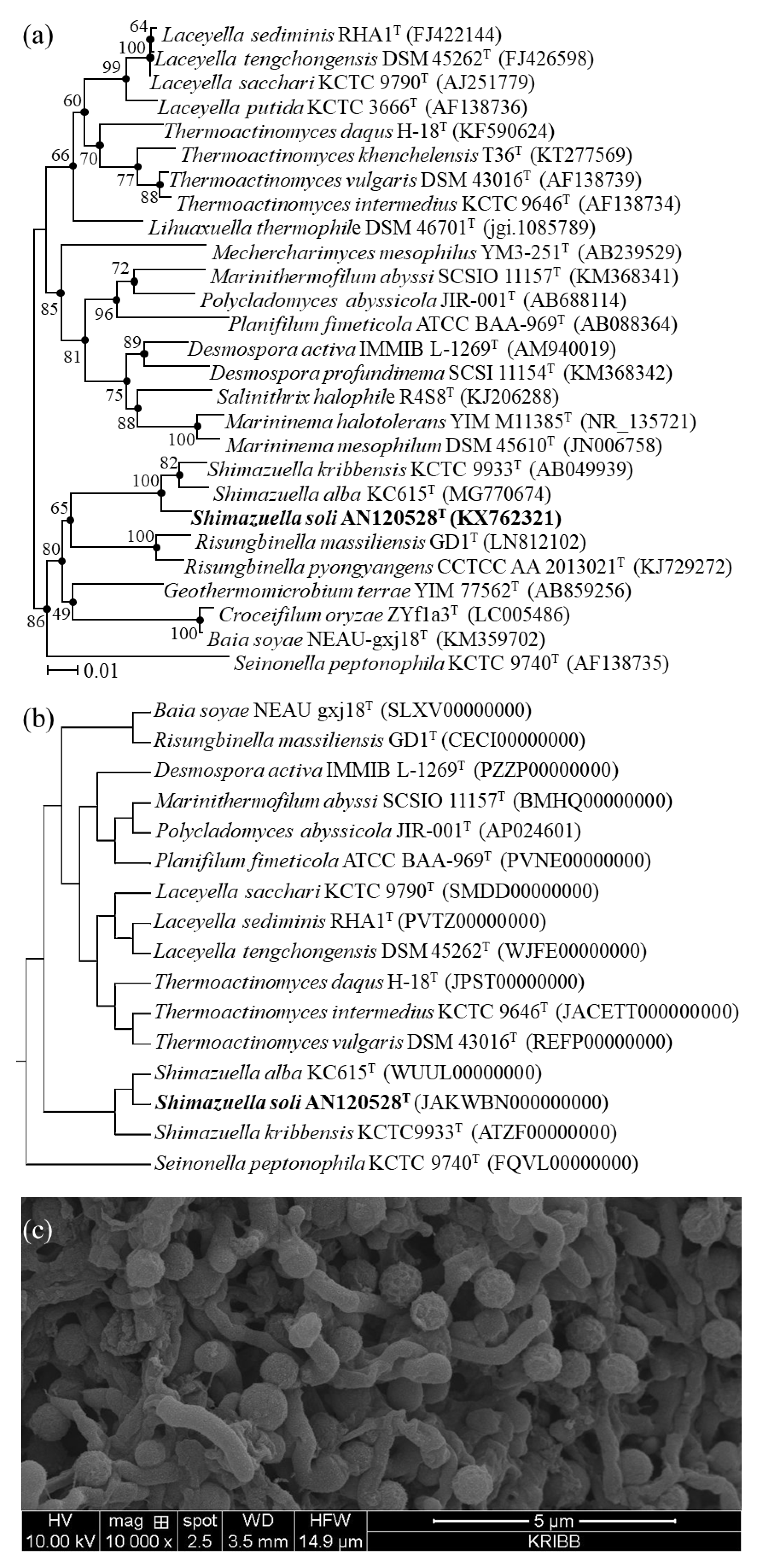
3.1. Phylogenetic Analysis

Phylogenetic and phylogenomic analysis based on the 16S rRNA gene (1470 bp; GenBank accession number KX762321) and genome sequence revealed that the strain AN120528T was clearly a member of the genus Shimazuella, having the highest 16S rRNA gene sequence similarity to S. alba KC615T (98.11%; accession no. MG770674) [4], followed by S. kribbensis KCTC 9933T (97.05%; AB049939) [2] (Figure 1a,b). In addition, the neighbour-joining, maximum-parsimony and maximum-likelihood tree-making algorithms showed that the strain AN120528T formed a monophyletic group with the two species of the genus Shimazuella.

3.2. Morphological, Physiological and Biochemical Characteristics

The strain AN120528T could grow well on Bennett’s agar ISP2, PDA and R2A. The strain showed a white-coloured colony and a 1.0 × 1.2 μm cell size on R2A agar medium (Figure 1c). Cells were aerobic and non-motile with a white colour. The strain AN120528T was gram-positive, aerobic, spore-forming and non-motile. Colonies were circular, opaque and creamy white on the R2A medium. AN120528T was able to grow in the range of 20–45 °C, with optimum growth at 28–40 °C, and at a pH of 6.0–7.0 with an optimum pH of 7.0, and it could tolerate up to 1.0% NaCl. Anaerobic growth was not observed. For cell growth, the strain utilised D-galactose and D-mannose as carbon sources and L-tyrosine and L-cysteine as nitrogen sources. It was positive for Tween 40 and 80 and negative for Tween 20. Cells were susceptible to all antibiotics tested. Table 1 shows several characteristics that distinguish the strain AN120528T from the phylogenetically closely related strains.

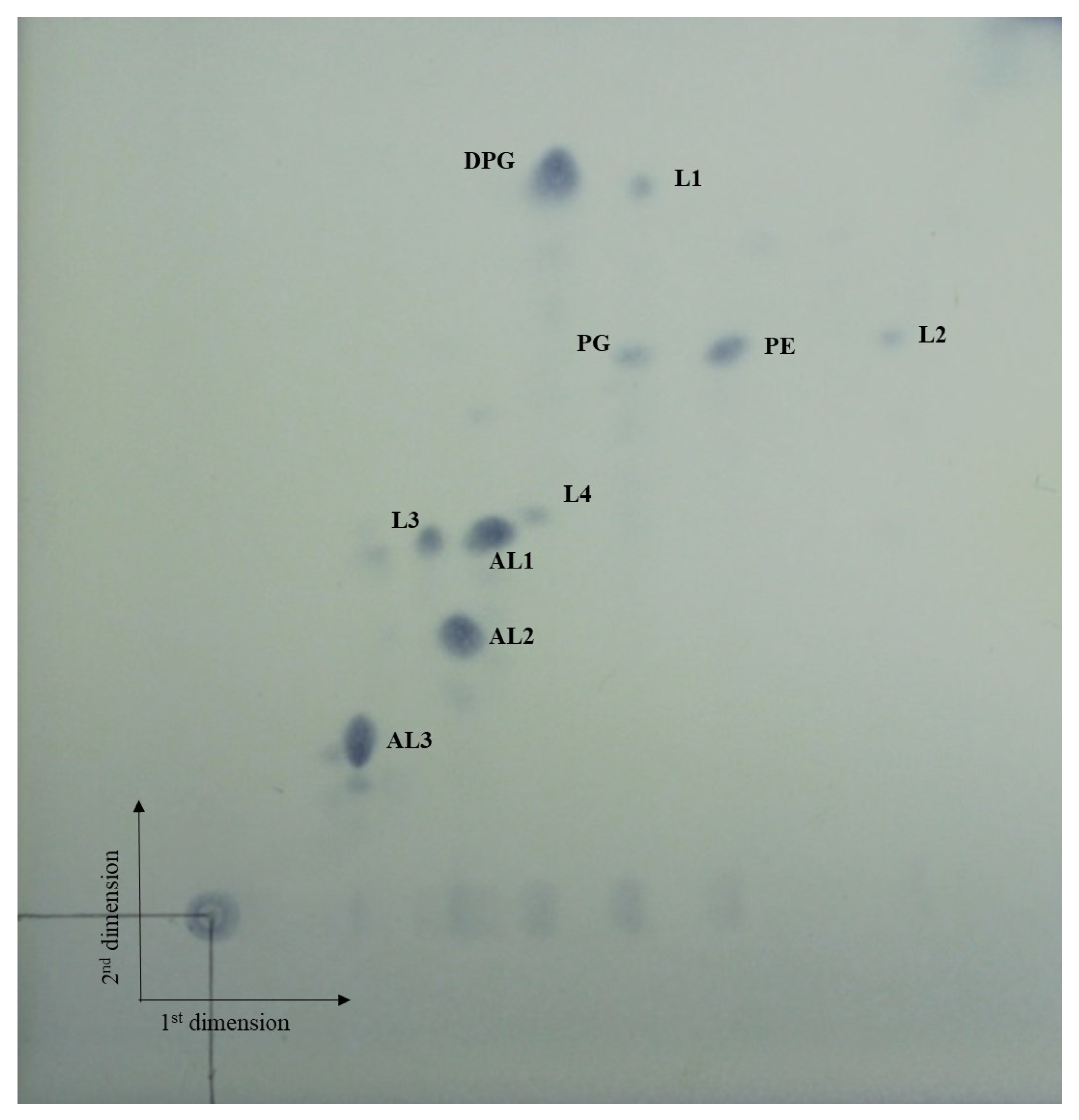
3.3. Chemotaxonomic Characteristics

Table 2 lists the chemotaxonomic characteristics of the strain AN120528T and its related strains. The major fatty acids (>30%) of AN120528T were anteiso-C15:0 (32.3%) and iso-C15:0 (31.8%). Furthermore, the strain possessed MK-9 (H4) and MK-10 (H4), as the predominant menaquinones, and meso-diaminopimelic acid in the cell-wall peptidoglycan. The major polar lipids were DPG, PG, PE, AL and L (Figure 2). The major cell-wall sugars were ribose and glucose.

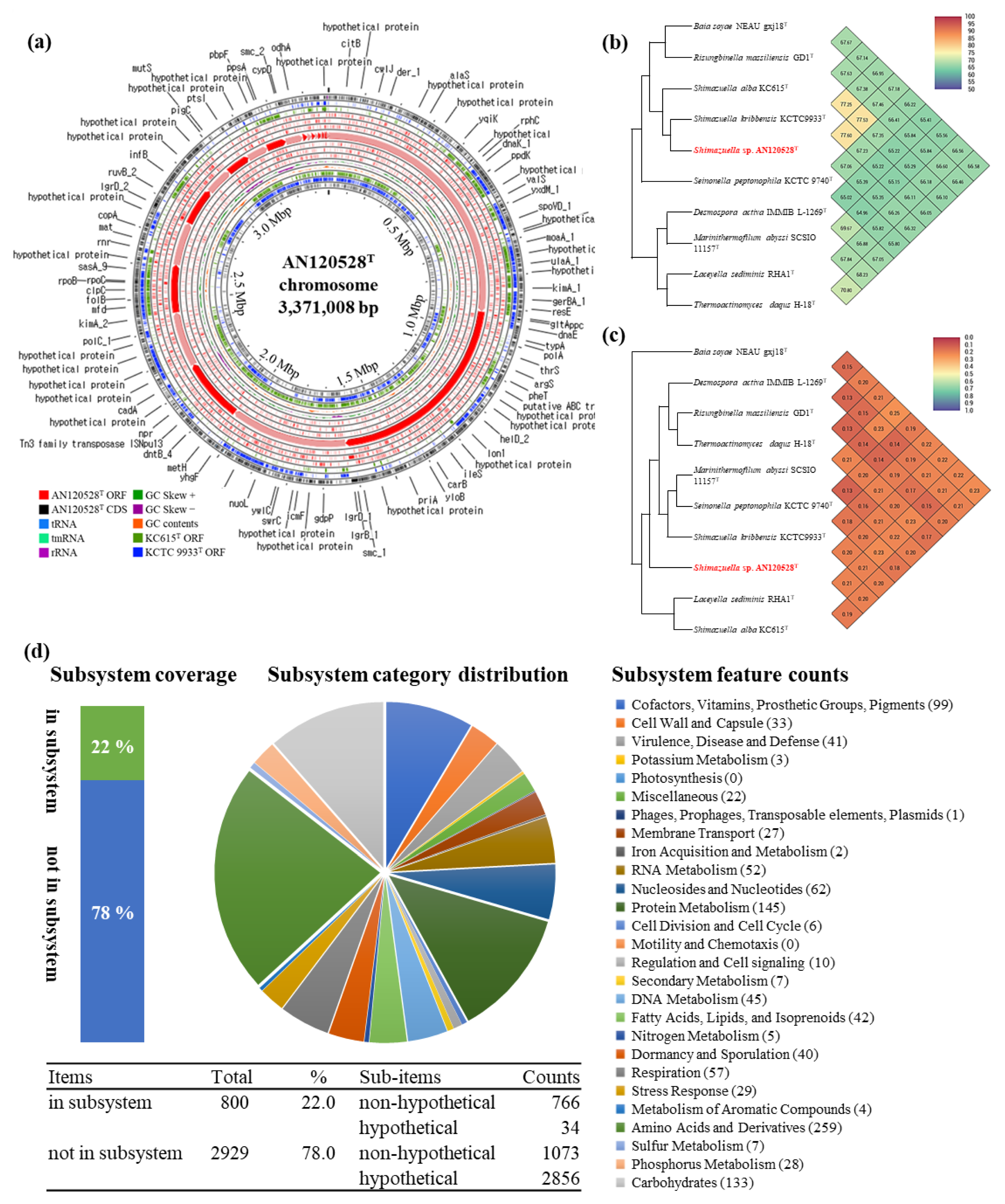
3.4. Genome Analysis

The assembled draft genome of AN120528T was 3.37 Mbp, containing 25 contigs with an N50 length of 408,672 bp, 3408 coding sequences, 10 rRNA and 52 tRNA (Table 3). The gDNA G + C content was revealed to be 39.0%. The GenBank accession number for the genome sequences of the AN120528T strain is JAKWBN000000000. Figure 3a shows the comparative genomic circular map. The OrthoANI values between AN120528T and its related species S. alba KC615T (accession no. WUUL00000000) and S. kribbensis KCTC 9933T (ATZF01000001) were 77.53 and 77.60%, respectively (Figure 3b). Furthermore, the dDDH values of S. alba KC615T and S. kribbensis KCTC 9933T were 20.60 and 21.13%, respectively (Figure 3c). Based on the genome analysis on the RAST webserver, around 22% of detected genes—a total of 800 genes—were annotated in the subsystem (Figure 3d). The antiSMASH results on the functional metabolites showed BGCs for the one lasso peptide, three terpenes, one type I polyketide synthase (T1PKS), one type III polyketide synthase (T3PKS), three non-ribosomal peptide synthetases and one azol(in)e-containing peptide.

3.5. In Silico Analysis of the Novel Lasso Peptide Shimazuellin of AN120528T

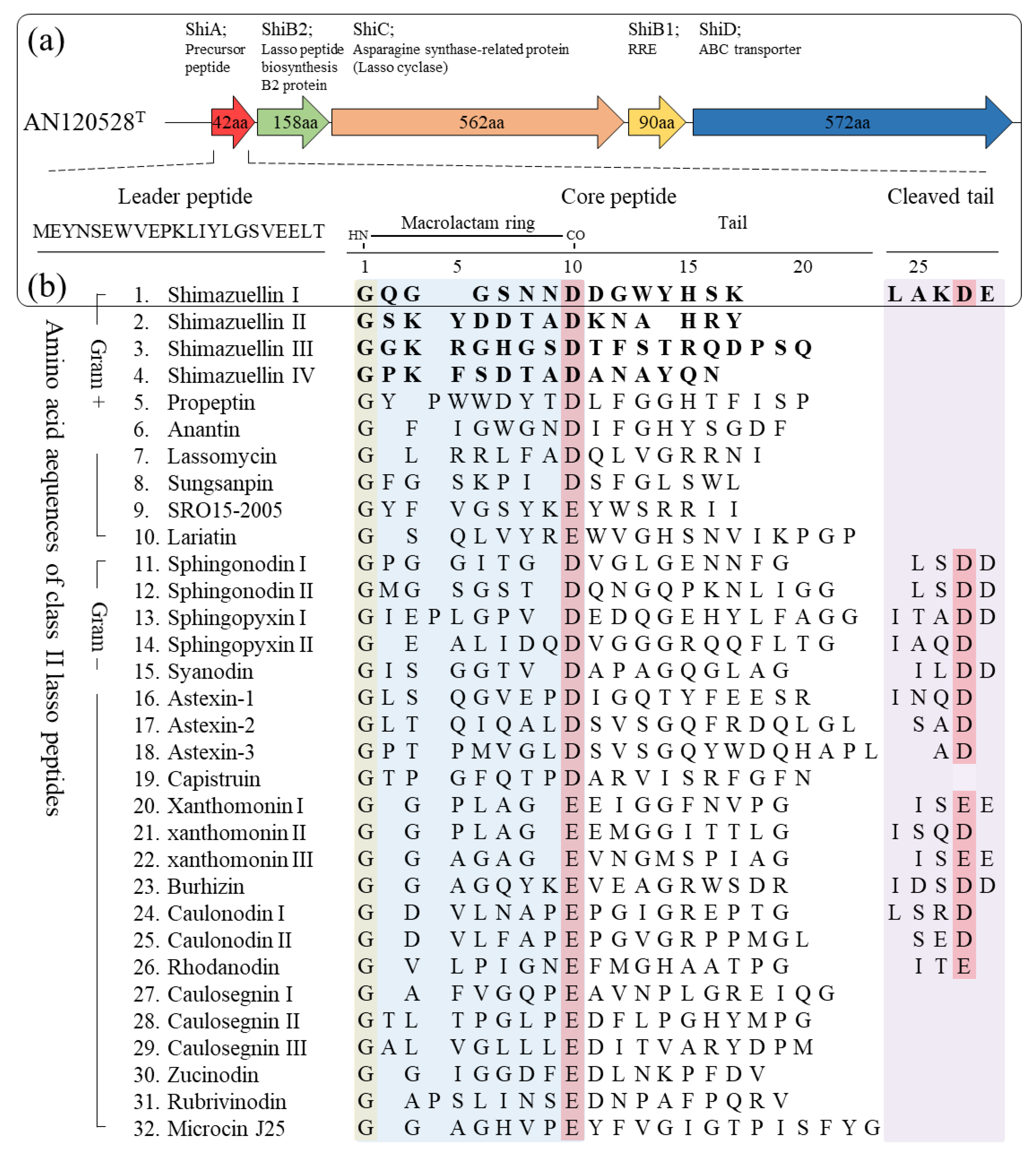
3.5.1. Genome Mining and Identification of Shimazuellin BGCs

Based on the precursor peptide sequence (protein ID; 00326) predicted from antiSMASH, shimazuellin was identified as a class II lasso peptide, and was called shimazuellin due to it being the first lasso peptide discovered in the genus Shimazuella. Adjacent proteins (00327, 00328, 00329 and 00330) of the precursor estimated to be essential enzymes in the biosynthetic pathway were selected as queries, and a BLAST homology search was performed (Table 4). The putative precursor peptide sequence was shown to have low levels of similarity with hypothetical protein PPOP_1752 (GAC42395.1) and hypothetical protein PPOP 1273 (GAC41916.1) of Paenibacillus popilliae ATCC 14706 at 47.06% and 45.45%, respectively. The putative lasso peptide-related proteins, which were annotated as uncharacterized proteins, had the highest amino acid sequence similarity to the lasso peptide biosynthesis protein (67.09%; accession no. WP_028776449.1), the asparagine synthase-related protein (65.54%; WP_028776448.1) and the hypothetical protein (60.70%; WP_028776447.1) of the strain KCTC 9933T. Although they had low sequence similarities with even closely related proteins, lasso-related enzymes were identified by the local presence of gene encoding proteins matching the pivotal motif and domain for the precursor peptide, the lasso protease, the lasso cyclase, the RiPP recognition element (RRE) and the ABC transporter. The shimazuellin BGC housed the five major genes involved in peptide precursor, biosynthesis, maturation and secretion. Although the sequences and functions of the peptides can vary markedly, the BGC for shimazuellin has a typical lasso peptide biosynthetic gene locus encoding a linear precursor peptide without disulfide bonds, three conserved proteins for peptide maturation and a transporter to export the matured peptide (Figure 4a). ShiA consists of 22 amino acids for the leader peptide, 16 amino acids for the core peptide region, and five amino acids for the truncated C-terminal tail, respectively [34]. The shiC encodes for amidotransferase, ATP pyrophosphatase and asparagine synthetase-like protein domain, which is responsible for formation of both isopeptide bonds and subsequent macrocyclisation [35]. ShiB1, containing a pyrroloquinoline quinone protein domain D (PqqD), also known as the RRE, binds to the leader region and transfers the precursor peptide to the protease ShiB2 for further processing [34,36]. The ShiD is an ABC transporter which secretes shimazuelin from the cytoplasm to the extracellular space, and the presence of the ShiD indicates that the biosynthesised lasso peptides could exhibit antimicrobial activity. The putative leader region, MEYNSEWVEPKLIYLGSVEELT, was shown to have a VXPXLXXXG conserved motif, which is commonly found in lasso leader peptides [37]. The leader and core regions are separated by the emblematic TG motif needed to remove the leader peptide during maturation [38]. Residues G and D were identified in the core peptide which can form macrolactam rings [39] (Figure 4b). Furthermore, a cleaved tail containing D residues was identified. This is not commonly found in gram-positive bacteria. In addition, in this study, class II lasso peptides that do not have a cleaved tail were found in S. alba KC615T (shimazuellin II and IV) and S. kribbensis KCTC 9933T (shimazuellin II and shimazuellin III) using comparative genome analysis.

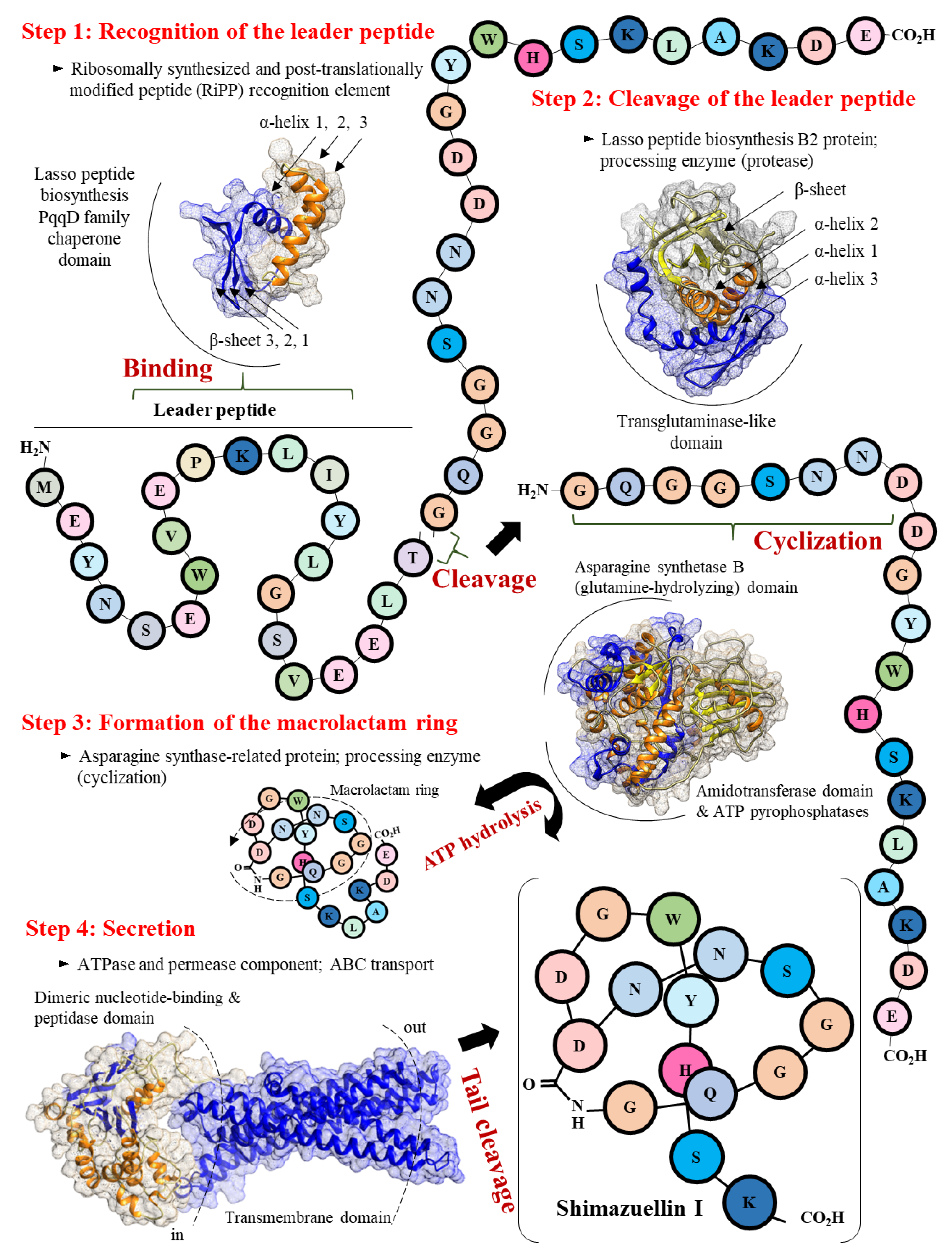
3.5.2. Scheme of the Putative Biosynthetic Mechanism of Shimazuellin in AN120528T

Figure 5 shows the proposed mechanism of shimazuellin biosynthesis and secretion. Four steps seem to be necessary for the biosynthesis and secretion of shimazuellin based on the gene clusters encoding separate ShiA, ShiB2, ShiC, ShiB1 and ShiD. First, after a precursor peptide of shimazuellin is translated from mRNA, ShiB1 binds to the VXPXLXXXG region of the leader peptide in ShiA to recognise ShiA. Second, the ShiB2 protein removes the leader peptide via proteolysis of the TG region and thereby releases the core peptide of shimazuellin. Next, ShiC, the lasso cyclase, activates the Asp carboxylic acid in the form of an adenosine monophosphate ester before catalysing the macrolactam formation via condensation with the α-amino group. Finally, the ShiD-encoded ABC transporter performs cleavage in the tail (-LAKDE-) and exports the mature form of shimazuellin out of the cells.

3.6. Description of Shimazuella Soli Sp. Nov.

Shimazuella soli (so’li. L. neut. gen. n. soli of soil, referring to the source of the strain) cells are gram-positive, spore-forming, non-motile and white coloured. Growth occurs at 20–45 °C (optimum, 28–40), with a pH of 6.0–7.0 (optimum, 7.0) and in the presence of 0–1% (w/v) (optimum, 0.5) NaCl. The strain utilises D-galactose and D-mannose as carbon sources and L-tyrosine and L-cysteine as nitrogen sources for growth. It is susceptible to amikacin, ampicillin/sulbactam, chloramphenicol, erythromycin, gentamicin, kanamycin, lincomycin, rifampicin, spectinomycin, streptomycin, teicoplanin, tetracycline and vancomycin. It can degrade Tween 40 and 80, but not Tween 20, starch, cellulose or gelatine. It can solidify and peptonise milk. It is positive for β-glucosidase. Its major fatty acids are iso-C14:0, iso-C15:0, anteiso-C15:0 and iso-C16:0. The respiratory quinones from the cell wall are MK-9 (H4) and MK-10 (H4). Its polar lipids are diphosphatidylglycerol, phosphatidylglycerol, phosphatidylethanolamine, unidentified aminolipid and unidentified lipids. The G+C content of the gDNA is 39.0%. The type strain, AN120528T (=KCTC 39810T = DSM 103571T), was isolated from soil in South Korea.

4. Discussion

The genus Shimazuella, represented by S. kribbensis KCTC 9933T, was first proposed in 2007 [2]. However, the Shimazuella spp. have consisted of only two recognized species so far. Based on our observation, along with previously published results, Shimazuella spp. prefer extremely limited or unique carbon sources for metabolism and take about 7 days or more to reach the stable late log phase or early stationary phase [2,4]. Shimazuella is considered to have evolved a metabolism that prefers other sugars and does not have a glucose metabolism, which is used as a basic carbon source for most organisms to survive in a community consisting of various microbes in soil. Because of these physiological characteristics, it could be difficult to isolate by antagonistic actions in the microbial community during the screening process of a single strain from the environment. Furthermore, the limitations of Shimazuella isolation were supported by an antibiotic susceptibility test and genome analysis in this study. These analyses found that Shimazuella spp. do not contain any antibiotic resistance genes except for one putative glycopeptide resistance gene cluster common to all strains and two antibiotic efflux-related genes in the strain KC615T. In this study, we successfully isolated AN120528T from soil using extreme serial dilution to obtain rare bacteria and investigate their functionality. Comparative genome analysis showed that the ANI and dDDH values between the strain AN120528T and its related species were lower than the cut-off of 95–96% and were 70% for the delineation of a novel species, respectively [40,41]. Moreover, the results of the carbon utilisation and fatty acid composition allowed for the systematic differentiation of AN120528T from related species. Therefore, the strain AN120528T represents a novel species of the genus Shimazuella, for which we propose the name Shimazuella soli sp. nov. In addition, the description of the genus Shimazuella is as given previously [2,4], with the following modifications: its diagnostic polar lipids are DPG, PE and PG; its major fatty acids are anteiso-C15:0, iso-C14:0, iso-C15:0, iso-C16:0 and C16:0; and the G+C content is around 38.4–39.0%.
Shimazuellin I, a new lasso peptide belonging to class II in S. soli AN120528T, was discovered, and lasso peptides with different sequences were also found in KC615T (shimazuellin II and IV) and in KCTC 9933T (shimazuellin II and shimazuellin III). Although its sequence homology with known lasso peptide biosynthetic enzymes was significantly low, sequence-based protein 3D modelling, comparative structure analysis, conserved domain and in silico molecular docking analysis demonstrated that the ShiA, ShiB1, ShiB2, ShiC and ShiD enzymes could be involved in lasso peptide biosynthesis. Shimazuellin I of S. soli AN120528T was identified for the first time in this study, as it has a C-terminal cleavage tail unlike lasso peptides generally reported in gram-positive bacteria. In addition to shimazuellin I–IV, the following putative BGCs for various antimicrobial peptides were identified: enniatin, micrococcin P1, non-ribosomal tripeptide (D-Phe-D-Ala-Trp) and carnocyclin A in S. soli AN120528T; xenematide, lanthipeptide, micrococcin P1, sevadicin and xenotetrapeptide in KC615T; and micrococcin P1 and massetolide A in KCTC 9933T. It is inferred that Shimazuella possesses various antimicrobial peptides for the stable uptake of nutrients after it germinates in the presence of sufficiently preferred sugars, even though it maintains a spore state under unfavourable conditions. This is similar to how Shimazuella has evolved a unique and limited carbohydrate metabolism to survive.
The trend in peptide science has changed from simply finding and applying natural peptides derived from organisms to the rational design of peptides with desirable physiological functions [42]. Major innovations in genomics, bioinformatics, and sequencing technology have enabled the rational design of excellent peptides with desirable biochemical activities. As a result, approximately 20 new therapeutic peptides have been released in the last 10 years, and dozens of peptides are in clinical development [43]. Therefore, the continuous discovery of new peptides that can inspire us makes peptides applicable to a wide range of diseases. At present, the existence of many lasso peptide gene clusters has been identified from a variety of bacteria with the development of bioinformatics and next-generation sequencing technology, resulting in revealed amino acid sequence diversity of precursor peptides [44]. Based on their biological functions, the peptides may be useful tools for the treatment of metabolic syndrome, autoimmune disease, microbial infections and cancer [7,45,46]. Furthermore, the macrocyclic forms of lasso peptides are an appropriate backbone for epitope grafting due to their proteolytic stability and thermostability [47]. These redesigned peptides can be applied as molecular probes and drug carriers for therapeutics [48,49,50]. Therefore, in addition to determining the extent of their physiological function, the major motivations of genomic efforts are to mine new examples of lasso peptides and discover novel classes, such as shimazuellin found in S. soli AN120528T in our study. Considering the wide selection of active candidates, we suggest that the mining of shimazuellin can both contribute to an expansion in the scope of peptide therapeutics and be used in basic research that advances our peptide design capabilities. Taken together, we believe that the S. soli AN120528T and shimazuellin we report here could be utilised as useful information to boost peptide research in the postgenomic era.

Author Contributions

Conceptualization, C.-Z.J., J.M.L. and K.-S.S.; methodology, C.-Z.J. and J.M.L.; software, C.-Z.J. and J.M.L.; validation, C.-Z.J. and J.M.L.; formal analysis, C.-Z.J. and J.M.L.; investigation, C.-Z.J. and J.M.L.; resources, C.-J.K.; data curation, C.-Z.J. and J.M.L.; writing—original draft preparation, C.-Z.J. and J.M.L.; writing—review and editing, C.-Z.J., J.M.L. and K.-S.S.; visualization, J.M.L.; supervision, H.-G.L. and K.-S.S.; project administration, C.-J.K., H.-G.L. and K.-S.S.; funding acquisition, C.-J.K., H.-G.L. and K.-S.S. All authors have read and agreed to the published version of the manuscript.

Funding

This work was supported by IPET through the Agricultural Machinery/Equipment Localization Technology Development Program funded by MAFRA (321056) and National Research Foundation of Korea (NRF) grants (NRF-2013M3A9A5076603) and the Korea Environment Industry & Technology Institute (KEITI) project to develop eco-friendly new materials and processing technology derived from wildlife, funded by the Korea Ministry of Environment (MOE) (2021003240004).

Data Availability Statement

The raw data supporting the conclusions of this article will be made available by the authors, without undue reservation.

Acknowledgments

The authors gratefully acknowledge Dong-Jin Park (Industrial Biomaterial Research Center, Korea Research Institute of Bioscience and Biotechnology, Daejeon, Republic of Korea) for providing useful information on Shimazuella spp.

Conflicts of Interest

The authors declare no conflict of interest.

References

  1. Oren, A.; Garrity, G.M. Valid publication of the names of forty-two phyla of prokaryotes. Int. J. Syst. Evol. Microbiol. 2021, 71, 005056. [Google Scholar] [CrossRef] [PubMed]
  2. Park, D.-J.; Dastager, S.G.; Lee, J.-C.; Yeo, S.-H.; Yoon, J.-H.; Kim, C.-J. Shimazuella kribbensis gen. nov., sp. nov., a mesophilic representative of the family Thermoactinomycetaceae. Int. J. Syst. Evol. Microbiol. 2007, 57, 2660–2664. [Google Scholar] [CrossRef] [PubMed] [Green Version]
  3. Parte, A.C.; Sarda Carbasse, J.; Meier-Kolthoff, J.P.; Reimer, L.C.; Göker, M. List of Prokaryotic names with Standing in Nomenclature (LPSN) moves to the DSMZ. Int. J. Syst. Evol. Microbiol. 2020, 70, 5607–5612. [Google Scholar] [CrossRef] [PubMed]
  4. Saygin, H.; Ay, H.; Sahin, N. Shimazuella alba sp. nov. isolated from desert soil and emended description of the genus Shimazuella Park et al. 2007. Arch. Microbiol. 2020, 202, 1831–1838. [Google Scholar] [CrossRef] [PubMed]
  5. Singh, A. Materials modulate immunity and gut microbiome. Nat. Mater. 2020, 19, 3–4. [Google Scholar] [CrossRef]
  6. Sumida, T.; Dubiley, S.; Wilcox, B.; Severinov, K.; Tagami, S. Structural basis of leader peptide recognition in lasso peptide biosynthesis pathway. ACS Chem. Biol. 2019, 14, 1619–1627. [Google Scholar] [CrossRef]
  7. Cheng, C.; Hua, Z.-C. Lasso Peptides: Heterologous production and potential medical application. Front. Bioeng. Biotechnol. 2020, 8, 571165. [Google Scholar] [CrossRef]
  8. Saitou, N.; Nei, M. The neighbor-joining method: A new method for reconstructing phylogenetic trees. Mol. Biol. Evol. 1987, 4, 406–425. [Google Scholar] [CrossRef]
  9. Kimura, M. A simple method for estimating evolutionary rates of base substitutions through comparative studies of nucleotide sequences. J. Mol. Evol. 1980, 16, 111–120. [Google Scholar] [CrossRef]
  10. Felsenstein, J. Evolutionary trees from DNA sequences: A maximum likelihood approach. J. Mol. Evol. 1981, 17, 368–376. [Google Scholar] [CrossRef]
  11. Fitch, W.M. Toward defining the course of evolution: Minimum change for a specific tree topology. Syst. Biol. 1971, 20, 406–416. [Google Scholar] [CrossRef]
  12. Kumar, S.; Stecher, G.; Tamura, K. MEGA7: Molecular evolutionary genetics analysis version 7.0 for bigger datasets. Mol. Biol. Evol. 2016, 33, 1870–1874. [Google Scholar] [CrossRef] [PubMed] [Green Version]
  13. Brettin, T.; Davis, J.J.; Disz, T.; Edwards, R.A.; Gerdes, S.; Olsen, G.J.; Olson, R.; Overbeek, R.; Parrello, B.; Pusch, G.D.; et al. RASTtk: A modular and extensible implementation of the RAST algorithm for building custom annotation pipelines and annotating batches of genomes. Sci. Rep. 2015, 5, 8365. [Google Scholar] [CrossRef] [PubMed] [Green Version]
  14. Stothard, P.; Wishart, D.S. Circular genome visualization and exploration using CGView. Bioinformatics 2005, 21, 537–539. [Google Scholar] [CrossRef] [PubMed] [Green Version]
  15. Lee, I.; Kim, Y.O.; Park, S.-C.; Chun, J. OrthoANI: An improved algorithm and software for calculating average nucleotide identity. Int. J. Syst. Evol. Microbiol. 2016, 66, 1100–1103. [Google Scholar] [CrossRef] [PubMed]
  16. Auch, A.F.; Von Jan, M.; Klenk, H.-P.; Göker, M. Digital DNA-DNA hybridization for microbial species delineation by means of genome-to-genome sequence comparison. Stand. Genom. Sci. 2010, 2, 117–134. [Google Scholar] [CrossRef] [Green Version]
  17. Blin, K.; Shaw, S.; Kloosterman, A.M.; Charlop-Powers, Z.; van Wezel, G.P.; Medema, M.H.; Weber, T. antiSMASH 6.0: Improving cluster detection and comparison capabilities. Nucleic Acids Res. 2021, 49, W29–W35. [Google Scholar] [CrossRef]
  18. Lee, J.M.; Jin, C.-Z.; Kang, M.-K.; Park, S.H.; Park, D.-J.; Kim, D.-G.; Kim, C.-J. Nocardioides humilatus sp. nov., isolated from farmland soil in the Republic of Korea. Int. J. Syst. Evol. Microbiol. 2022, 72, 004928. [Google Scholar] [CrossRef]
  19. Lee, J.M.; Jin, C.-Z.; Park, S.H.; Kang, M.-K.; Park, D.-J.; Kim, C.-J. Nocardioides antri sp. nov., Isolated from Soil in a Rock Cave. Curr. Microbiol. 2021, 78, 2130–2135. [Google Scholar] [CrossRef]
  20. Skerman, V.B.D. A Guide to the Identification of the Genera of Bacteria. J. Med. Educ. 1960, 35, 92. [Google Scholar]
  21. Jin, C.-Z.; Jin, L.; Liu, M.-J.; Lee, J.-M.; Park, D.-J.; Kim, C.-J. Solihabitans fulvus gen. nov., sp. nov., a member of the family Pseudonocardiaceae isolated from soil. Int. J. Syst. Evol. Microbiol. 2022, 72, 005110. [Google Scholar] [CrossRef]
  22. Jin, C.-Z.; Jin, L.; Kang, D.H.; Liu, M.-J.; Lee, J.M.; Park, D.-J.; Kim, C.-J. Description of desferrioxamine-producing bacterium Chitinophaga agrisoli sp. nov., isolated from soil. Antonie van Leeuwenhoek 2021, 114, 741–750. [Google Scholar] [CrossRef]
  23. Gonzalez, C.; Gutierrez, C.; Ramirez, C. Halobacterium vallismortis sp. nov. An amylolytic and carbohydrate-metabolizing, extremely halophilic bacterium. Can. J. Microbiol. 1978, 24, 710–715. [Google Scholar] [CrossRef]
  24. Kämpfer, P.; Kroppenstedt, R.M. Numerical analysis of fatty acid patterns of coryneform bacteria and related taxa. Can. J. Microbiol. 1996, 42, 989–1005. [Google Scholar] [CrossRef]
  25. Tamaoka, J.; Katayama-Fujimura, Y.; Kuraishi, H. Analysis of bacterial menaquinone mixtures by high performance liquid chromatography. J. Appl. Bacteriol. 1983, 54, 31–36. [Google Scholar] [CrossRef]
  26. Staneck, J.L.; Roberts, G.D. Simplified approach to identification of aerobic actinomycetes by thin-layer chromatography. Appl. Environ. Microbiol. 1974, 28, 226–231. [Google Scholar] [CrossRef]
  27. Komagata, K.; Suzuki, K.-I. 4 Lipid and cell-wall analysis in bacterial systematics. In Current Methods for Classification and Identification of Microorganisms; Colwell, R.R., Grigorova, R., Eds.; Methods in Microbiology; Academic Press: Cambridge, MA, USA, 1988; Volume 19, pp. 161–207. [Google Scholar]
  28. Lee, J.M.; Moon, S.Y.; Kim, Y.-R.; Kim, K.W.; Lee, B.-J.; Kong, I.-S. Improvement of thermostability and halostability of β-1,3-1,4-glucanase by substituting hydrophobic residue for Lys 48. Int. J. Biol. Macromol. 2017, 94, 594–602. [Google Scholar] [CrossRef]
  29. Schwede, T.; Kopp, J.; Guex, N.; Peitsch, M.C. SWISS-MODEL: An automated protein homology-modeling server. Nucleic Acids Res. 2003, 31, 3381–3385. [Google Scholar] [CrossRef] [Green Version]
  30. O’Boyle, N.M.; Banck, M.; James, C.A.; Morley, C.; Vandermeersch, T.; Hutchison, G.R. Open Babel: An open chemical toolbox. J. Cheminform. 2011, 3, 33. [Google Scholar] [CrossRef] [Green Version]
  31. Grosdidier, A.; Zoete, V.; Michielin, O. SwissDock, a protein-small molecule docking web service based on EADock DSS. Nucleic Acids Res. 2011, 39 (Suppl. 2), W270–W277. [Google Scholar] [CrossRef] [Green Version]
  32. Tina, K.G.; Bhadra, R.; Srinivasan, N. PIC: Protein interactions calculator. Nucleic Acids Res. 2007, 35 (Suppl. 2), W473–W476. [Google Scholar] [CrossRef] [PubMed]
  33. Kim, M.; Kim, T.; Ri, S.; Jiang, F.; Chang, X.; Liu, Y.; Ren, L.; Qiu, X.; Deng, S.; Fang, C.; et al. Risungbinella pyongyangensis gen. nov., sp. nov., a mesophilic member of the family Thermoactinomycetaceae isolated from an agricultural soil sample. Int. J. Syst. Evol. Microbiol. 2015, 65, 2726–2733. [Google Scholar] [CrossRef] [PubMed]
  34. Burkhart, B.J.; Hudson, G.A.; Dunbar, K.L.; Mitchell, D.A. A prevalent peptide-binding domain guides ribosomal natural product biosynthesis. Nat. Chem. Biol. 2015, 11, 564–570. [Google Scholar] [CrossRef] [PubMed] [Green Version]
  35. Maksimov, M.O.; Link, A.J. Discovery and characterization of an isopeptidase that linearizes lasso peptides. J. Am. Chem. Soc. 2013, 135, 12038–12047. [Google Scholar] [CrossRef] [PubMed]
  36. Zhu, S.; Fage, C.D.; Hegemann, J.D.; Mielcarek, A.; Yan, D.; Linne, U.; Marahiel, M.A. The B1 Protein Guides the Biosynthesis of a Lasso Peptide. Sci. Rep. 2016, 6, 35604. [Google Scholar] [CrossRef] [Green Version]
  37. Cortés-Albayay, C.; Jarmusch, S.A.; Trusch, F.; Ebel, R.; Andrews, B.A.; Jaspars, M.; Asenjo, J.A. Downsizing class II lasso peptides: Genome mining-guided isolation of huascopeptin containing the first Gly1-Asp7 macrocycle. J. Org. Chem. 2020, 85, 1661–1667. [Google Scholar] [CrossRef]
  38. Oman, T.J.; Van Der Donk, W.A. Follow the leader: The use of leader peptides to guide natural product biosynthesis. Nat. Chem. Biol. 2010, 6, 9–18. [Google Scholar] [CrossRef] [Green Version]
  39. Zirah, S.; Afonso, C.; Linne, U.; Knappe, T.A.; Marahiel, M.A.; Rebuffat, S.; Tabet, J.-C. Topoisomer differentiation of molecular knots by FTICR MS: Lessons from class II lasso peptides. J. Am. Soc. Mass Spectrom. 2011, 22, 467–479. [Google Scholar] [CrossRef] [Green Version]
  40. Goris, J.; Konstantinidis, K.T.; Klappenbach, J.A.; Coenye, T.; Vandamme, P.; Tiedje, J.M. DNA–DNA hybridization values and their relationship to whole-genome sequence similarities. Int. J. Syst. Evol. Microbiol. 2007, 57, 81–91. [Google Scholar] [CrossRef] [Green Version]
  41. Richter, M.; Rosselló-Móra, R. Shifting the genomic gold standard for the prokaryotic species definition. Proc. Natl. Acad. Sci. USA 2009, 106, 19126–19131. [Google Scholar] [CrossRef] [Green Version]
  42. Sheehan, F.; Sementa, D.; Jain, A.; Kumar, M.; Tayarani-Najjaran, M.; Kroiss, D.; Ulijn, R.V. Peptide-based supramolecular systems chemistry. Chem. Rev. 2021, 121, 13869–13914. [Google Scholar] [CrossRef]
  43. Wang, L.; Wang, N.; Zhang, W.; Cheng, X.; Yan, Z.; Shao, G.; Wang, X.; Wang, R.; Fu, C. Therapeutic peptides: Current applications and future directions. Signal Transduct. Target. Ther. 2022, 7, 48. [Google Scholar] [CrossRef]
  44. Boolchandani, M.; D’Souza, A.W.; Dantas, G. Sequencing-based methods and resources to study antimicrobial resistance. Nat. Rev. Genet. 2019, 20, 356–370. [Google Scholar] [CrossRef]
  45. Sakai, K.; Passioura, T.; Sato, H.; Ito, K.; Furuhashi, H.; Umitsu, M.; Takagi, J.; Kato, Y.; Mukai, H.; Warashina, S.; et al. Macrocyclic peptide-based inhibition and imaging of hepatocyte growth factor. Nat. Chem. Biol. 2019, 15, 598–606. [Google Scholar] [CrossRef]
  46. Vinogradov, A.A.; Yin, Y.; Suga, H. Macrocyclic peptides as drug candidates: Recent progress and remaining challenges. J. Am. Chem. Soc. 2019, 141, 4167–4181. [Google Scholar] [CrossRef]
  47. Hegemann, J.D. Factors governing the thermal stability of lasso peptides. Chembiochem 2019, 21, 7–18. [Google Scholar] [CrossRef]
  48. Knappe, T.A.; Manzenrieder, F.; Mas-Moruno, C.; Linne, U.; Sasse, F.; Kessler, H.; Xie, X.; Marahiel, M.A. Introducing lasso peptides as molecular scaffolds for drug design: Engineering of an integrin antagonist. Angew. Chem. Int. Ed. 2011, 50, 8714–8717. [Google Scholar] [CrossRef] [Green Version]
  49. Mihara, E.; Watanabe, S.; Bashiruddin, N.K.; Nakamura, N.; Matoba, K.; Sano, Y.; Maini, R.; Yin, Y.; Sakai, K.; Arimori, T.; et al. Lasso-grafting of macrocyclic peptide pharmacophores yields multi-functional proteins. Nat. Commun. 2021, 12, 1543. [Google Scholar] [CrossRef]
  50. Rodríguez, V. Insights into post-translational modification enzymes from RiPPs: A toolkit for applications in peptide synthesis. Biotechnol. Adv. 2022, 56, 107908. [Google Scholar] [CrossRef]
Figure 1. Phylogenetic tree, phylogenomic tree and SEM of the strain AN120528T. (a) Neighbour-joining phylogenetic tree showing the relationships of the strain AN120528T with S. alba KC615T, S. KCTC 9933T and other related species in the Thermoactinomycetaceae family. Bootstrap percentages (50%) based on 1000 re-samplings are given at the nodes. Dots indicate branches that were also recovered using the maximum-parsimony and maximum-likelihood algorithms. Bar, 0.01 substitutions per nucleotide position. (b) Phylogenomic tree of the genus Shimazuella and related genera. (c) SEM of the strain AN120528T grown on an R2A agar plate for 5 days at 28 °C.
Bioengineering 09 00812 g001
Figure 2. TLC of the polar lipids of strain AN120528T. Spots were stained with 5% ethanolic molybdophosphoric acid.
Bioengineering 09 00812 g002
Figure 3. Graphic representation of genomic features of AN120528T. (a) Circular plot of the comparison genomes of AN120528T, KC615T and KCTC 9933T. (b) OrthoANI analysis with other related strains. (c) GGDC analysis. (d) Genome annotation results of AN120528T on the RAST webserver.
Bioengineering 09 00812 g003
Figure 4. Gene organisation in the lasso peptide cluster of AN120528T and other core peptide sequences of class II lasso peptides. (a) Shimazuellin biosynthetic gene locus. (b) The putative leader and core regions of Shimazuellin with other related lasso peptides.
Bioengineering 09 00812 g004
Figure 5. Proposed mechanism of shimazuellin biosynthesis and secretion, involving four steps.
Bioengineering 09 00812 g005
Table 1. Physiological and biochemical properties of the strain AN120528T and related strains. All strains were gram-positive, aerobic and non-motile, and anaerobic growth was not observed. All strains also have MK-9 (H4) and MK-10 (H4) as respiratory quinones, meso-diaminopimelic acid as the diamino acid peptidoglycan and ribose and glucose as the major cell-wall sugars.
CharacteristicsAN120528TKC615TKCTC 9933T
Spores (μm)1.1–1.20.6–0.91.0–1.4
Growth conditions
Temperature range (°C)20–4528–3720–50
pH range6.0–7.06.0–8.06.0–9.0
NaCl tolerance (%)0–10–10–2
Degradation of
Starch+
Gelatin
Tween 40++
Tween 80++
Carbon utilisation
D-Galactose+
D-Mannose+
D-Raffinose+
Adonitol+
Nitrogen utilisation
L-Alanine+
L-Arginine+
L-Asparagine+
L-Cysteine++
L-Methionine+
L-Tyrosine++
L-Valine+
Enzymatic assay
Arbutin hydrolysis+
β-glucosidase+
Major polar lipids *DPG, PE, PG, 3AL, 4LDPG, PE, OH-PE, AL, GL, LDPG, PE, PG, PME, APL, 4AL, 2L
DPG, diphosphatidylglycerol; PG, phosphatidylglycerol; PME, phosphatidyl-N-methylethanolamine; PE, phosphatidylethanolamine; AL, aminolipid; OH-PE, hydroxy-phosphatidylethanolamine; APL, unknown aminophospholipid; GL, glycolipid; L, unknown lipid. * The polar lipids of KC615T and KCTC 9933T were obtained from Saygin et al. (2020) and Kim et al. (2015) [4,33].
Table 2. Fatty acid compositions of strain AN120528T and another related strains.
Fatty acid (%)AN120528TKC615TKCTC 9933T
Saturated
C13:0tr--
C14:01.11.41.4
C16:04.91.76.3
C17:01.4-tr
C18:01.110.1-
C19:01.11.1-
C20:0-15.8-
Branched
iso C13:0tr-tr
iso C14:09.55.25.5
iso C15:031.86.213.2
iso C16:09.32.44.4
iso C17:01.52.71.1
iso C18:0-2.5-
iso C19:0-4.9-
iso C17:1 ω10ctr -
anteiso-C13:0tr--
anteiso-C15:032.337.9159.1
anteiso-C17:01.55.13.2
anteiso-C19:0-3.3-
C16:1 ω11c1.8-3.4
C16:1 ω7c alcohol1.9-5.5
C18:1 ω9cTr--
Summed feature 4--tr
-, not detected. Summed feature 4 contains iso-IC17:1/anteiso-B or anteiso-C17:1 B/iso-I. tr, trace amount <1%. The fatty acids of KC615T and KCTC 9933T were obtained from Saygin et al. (2020) and Kim et al. (2015) [2,33].
Table 3. General genomic and functional features of AN120528T and related members of the genus Shimazuella.
FeaturesAN120528TKC615TKCTC 9933T
Genome size (bp)3,371,0083,989,5834,185,101
Contigs254442
N50 (bp)408,672278,159244,454
L50contig 3contig 6contig 6
Total genes354840544273
Pseudogene74203109
CDSs340837964087
rRNA10320
tRNA524853
G+C content (%)39.038.538.4
Antibiotic
resistance gene clusters
1
(glycopeptide resistance)
3
(one glycopeptide resistance and two antibiotic efflux)
1
(glycopeptide resistance)
CRISPR system
(Number of spacers)
Type III-B (13)Type I-C (30)Type I-C (17)
Number of BGCs101116
Table 4. Proposed functions of the open reading frames in the putative lasso peptide biosynthetic-related proteins of AN120528T.
In the Genome of AN120528TNCBI Blast Putative
Protein
ID
Locus
(Contig 1)
AnnotationDescription
(Accession No.)
Scientific
Name
Identity
(%)
Protein
(Functions)
00326295741_
295869
hypothetical
protein
hypothetical
protein
PPOP_1752
(GAC42395.1)
Paenibacillus popilliae ATCC 1470647.06ShiA
(precursor peptide)
00327295926_
296402
hypothetical
protein
lasso peptide biosynthesis
protein
(WP_028776449.1)
S. kribbensis KCTC 9933T67.09ShiB2
(B2 element; protease)
00328296415_
298103
hypothetical
protein
asparagine
synthase-related
protein
(WP_028776448.1)
S. kribbensis KCTC 9933T65.54ShiC
(cyclase)
00329298066_
298338
hypothetical
protein
hypothetical
protein
(WP_028776447.1)
S. kribbensis KCTC 9933T60.70ShiB1
(B1 element; RRE)
00330298360_
300078
putative
ATP-binding
protein
ABC transporter ATP-binding protein/
permease
(WP_028776446.1)
S. kribbensis KCTC 9933T74.17ShiD
(ABC transporter)
Publisher’s Note: MDPI stays neutral with regard to jurisdictional claims in published maps and institutional affiliations.

