
The Use of Microfabrication Techniques for the Design and Manufacture of Artificial Stem Cell Microenvironments for Tissue Regeneration

 , and
, and
Abstract
:1. Introduction
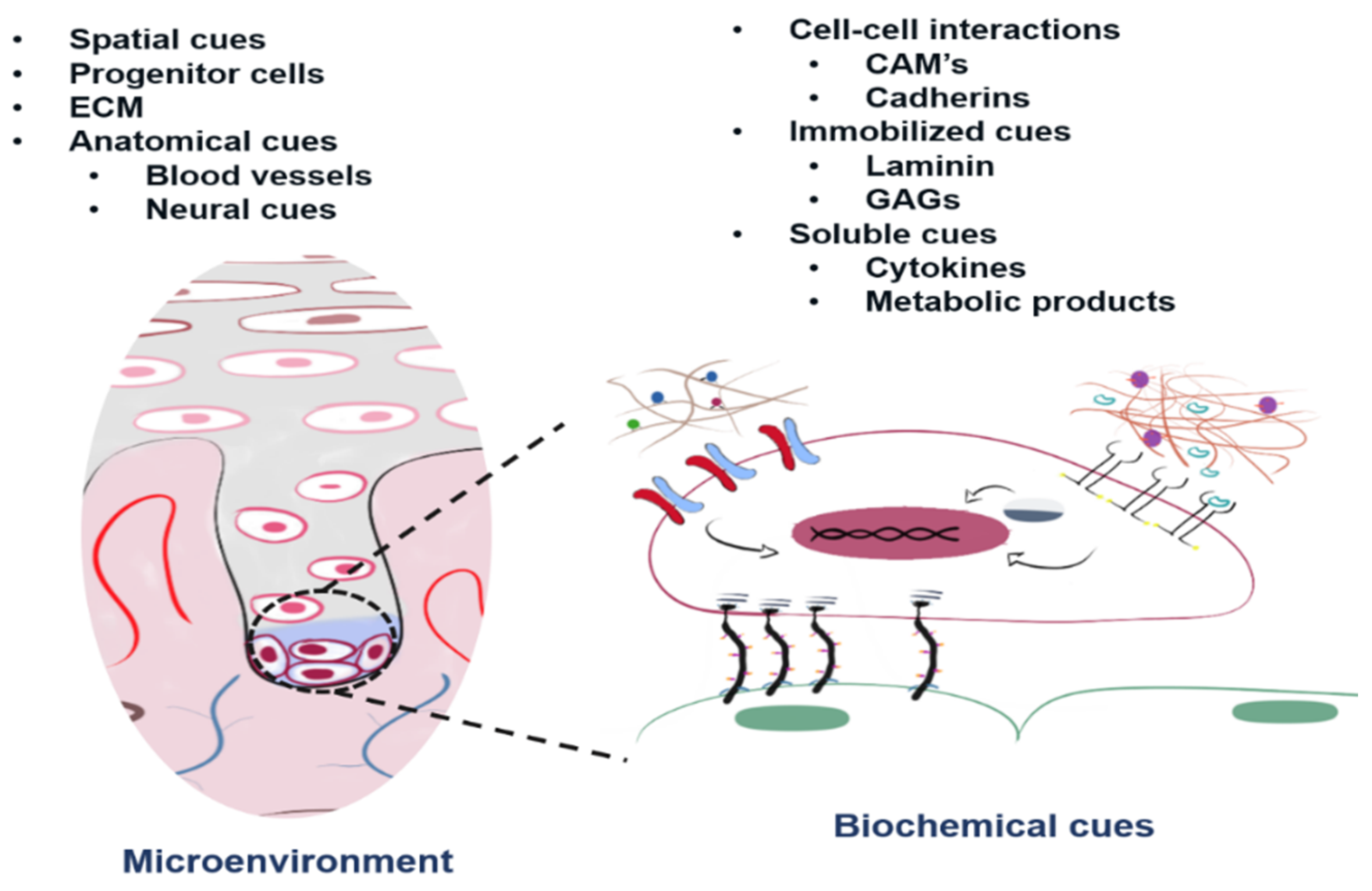
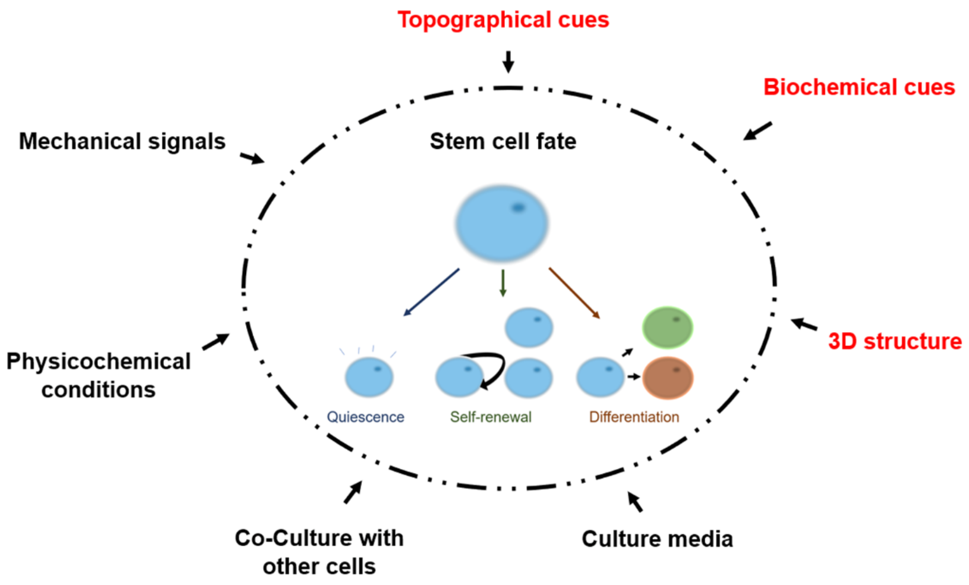
2. The Adult Stem Cell Microenvironment
3. Mimicking Biological Components of the Stem Cell Microenvironment
4. Mimicking the Spatial and Physical Components of the Stem Cell Microenvironment
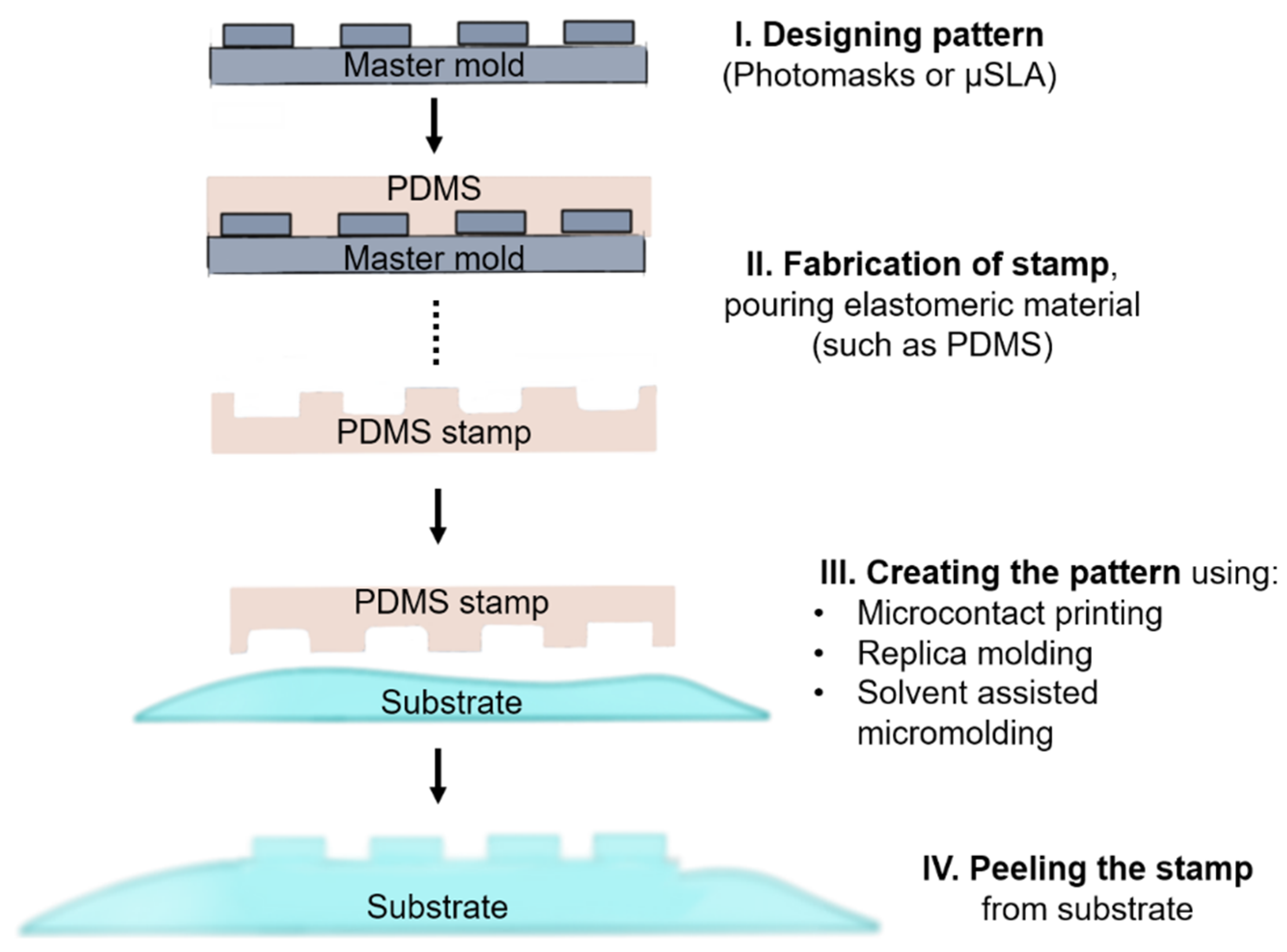
4.1. Soft-Lithographic Methods
4.1.1. Patterned Hydrogels
4.1.2. Microfluidic Devices
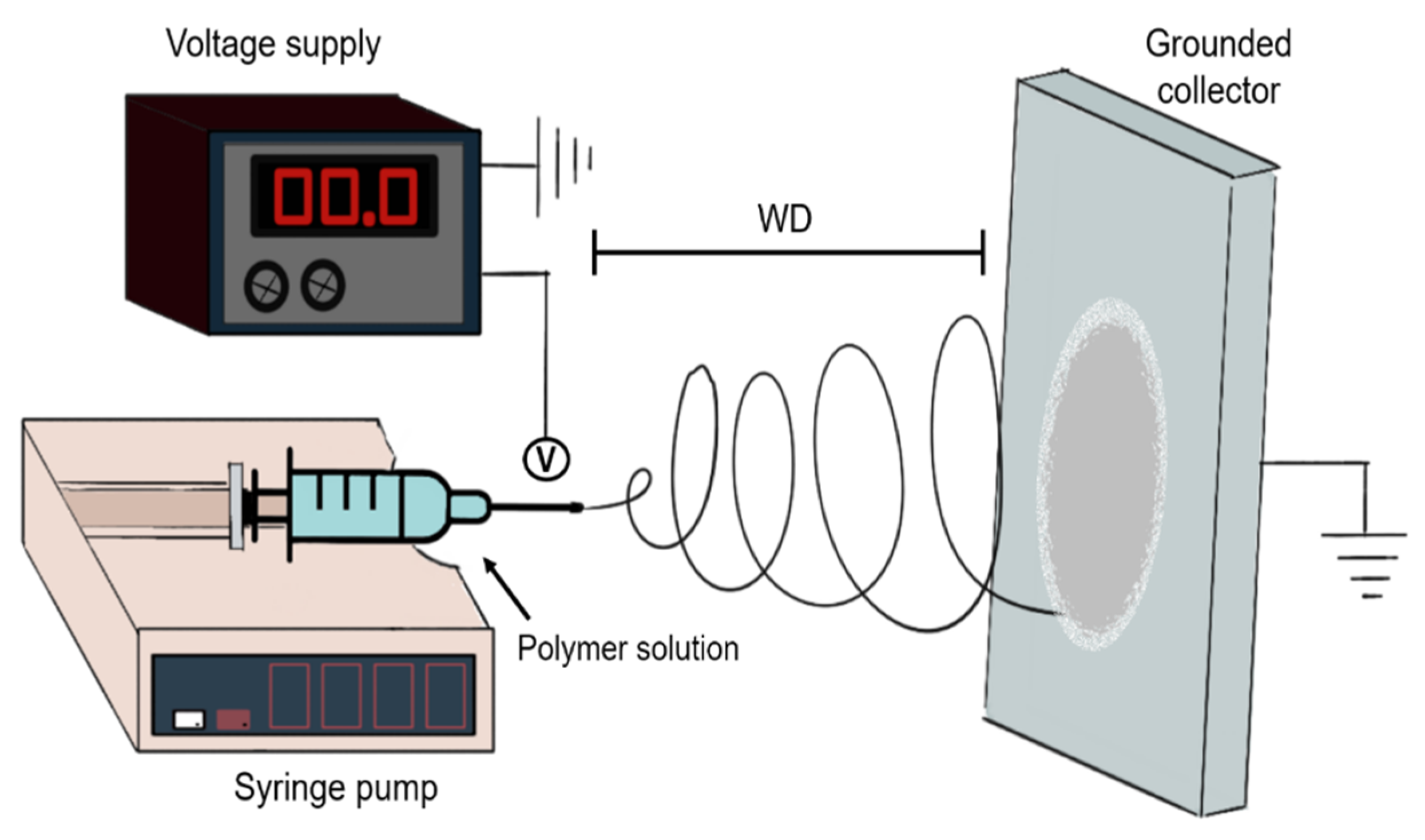
4.2. Electrospinning-Based Methods
4.3. Other Fabrication Methods
4.4. Combining Micro and Nanofabrication Techniques
5. Future Perspective and Concluding Remarks
Author Contributions
Funding
Conflicts of Interest
References
- Stice, S.L.; Boyd, N.L.; Dhara, S.K.; Gerwe, B.A.; Machacek, D.W.; Shin, S. Human embryonic stem cells: Challenges and opportunities. Reprod. Fertil. Dev. 2006, 18, 839–846. [Google Scholar] [CrossRef] [Green Version]
- Lo, B.; Parham, L. Ethical Issues in Stem Cell Research. Endocr. Rev. 2009, 30, 204–213. [Google Scholar] [CrossRef]
- Banks, J.M.; Harley, B.A.C.; Bailey, R.C. Tunable, Photoreactive Hydrogel System to Probe Synergies between Mechanical and Biomolecular Cues on Adipose-Derived Mesenchymal Stem Cell Differentiation. ACS Biomater. Sci. Eng. 2015, 1, 718–725. [Google Scholar] [CrossRef]
- Alsberg, E.; Von Recum, H.A.; Mahoney, M.J. Environmental cues to guide stem cell fate decision for tissue engineering applications. Expert Opin. Biol. Ther. 2006, 6, 847–866. [Google Scholar] [CrossRef]
- Zhang, Y.; Gordon, A.; Qian, W.; Chen, W. Engineering Nanoscale Stem Cell Niche: Direct Stem Cell Behavior at Cell-Matrix Interface. Adv. Health Mater. 2015, 4, 1900–1914. [Google Scholar] [CrossRef] [PubMed] [Green Version]
- McNamara, L.E.; McMurray, R.J.; Biggs, M.J.P.; Kantawong, F.; Oreffo, R.O.C.; Dalby, M.J. Nanotopographical Control of Stem Cell Differentiation. J. Tissue Eng. 2010, 1, 120623. [Google Scholar] [CrossRef]
- Morrison, S.J.; Kimble, J. Asymmetric and symmteric stem cell divisions in development cancer. Nature 2006, 441, 1068–1074. [Google Scholar] [CrossRef] [PubMed]
- Li, L.; Xie, T. STEM CELL NICHE: Structure and Function. Annu. Rev. Cell Dev. Biol. 2005, 21, 605–631. [Google Scholar] [CrossRef] [Green Version]
- Lutolf, M.P.; Blau, H.M. Artificial Stem Cell Niches. Adv. Mater. 2009, 21, 3255–3268. [Google Scholar] [CrossRef] [Green Version]
- Loeffler, M.; Roeder, I. Tissue stem cells: Definition, plasticity, heterogeneity, self-organization and models—A conceptual approach. Cells Tissues Organs 2002, 171, 8–26. [Google Scholar] [CrossRef]
- Schofield, R. The relationship between the spleen colony-forming cell and the haemopoietic stem cell. Blood Cells 1978, 4, 7–25. [Google Scholar]
- Crittenden, S.L.; Eckmann, C.R.; Wang, L.; Bernstein, D.S.; Wickens, M.; Kimble, J. Regulation of the mitosis/meiosis decision in the Caenorhabditis elegans germline. Philos. Trans. R. Soc. B Biol. Sci. 2003, 358, 1359–1362. [Google Scholar] [CrossRef] [Green Version]
- Lin, H.; Spradling, A.C. Germline Stem Cell Division and Egg Chamber Development in Transplanted Drosophila Germaria. Dev. Biol. 1993, 159, 140–152. [Google Scholar] [CrossRef]
- Xie, T.; Spradling, A.C. A Niche Maintaining Germ Line Stem Cells in the Drosophila ovary. Science 2000, 290, 328–330. [Google Scholar] [CrossRef]
- Jiang, J.; Papoutsakis, E.T. Stem-Cell Niche Based Comparative Analysis of Chemical and Nano-mechanical Material Properties Impacting Ex Vivo Expansion and Differentiation of Hematopoietic and Mesenchymal Stem Cells. Adv. Health Mater. 2012, 2, 25–42. [Google Scholar] [CrossRef]
- Rezza, A.; Sennett, R.; Rendl, M. Adult Stem Cell Niches. Curr. Top. Dev. Biol. 2014, 107, 333–372. [Google Scholar] [CrossRef]
- Jones, D.L.; Wagers, A.J. No place like home: Anatomy and function of the stem cell niche. Nat. Rev. Mol. Cell Biol. 2008, 9, 11–21. [Google Scholar] [CrossRef]
- Conboy, I.M.; Conboy, M.J.; Wagers, A.J.; Girma, E.R.; Weissman, I.L.; Rando, T.A. Rejuvenation of aged progenitor cells by exposure to a young systemic environment. Nature 2005, 433, 760–764. [Google Scholar] [CrossRef]
- Buom-Yong, R.; Orwig, K.E.; Oatley, J.M.; Avarbock, M.R.; Brinster, R.L. Effects of Aging and Niche Microenvironment on Spermatogonial Stem Cell Self Renewal. Stem Cells 2006, 24, 1505–1511. [Google Scholar] [CrossRef]
- Greco, V.; Guo, S. Compartmentalized organization: A common and required feature of stem cell niches? Development 2010, 137, 1586–1594. [Google Scholar] [CrossRef] [Green Version]
- Lavker, R.M.; Sun, T.-T. Epidermal stem cells: Properties, markers, and location. Proc. Natl. Acad. Sci. USA 2000, 97, 13473–13475. [Google Scholar] [CrossRef] [Green Version]
- Pincelli, C.; Marconi, A. Keratinocyte stem cells: Friends and foes. J. Cell. Physiol. 2010, 225, 310–315. [Google Scholar] [CrossRef] [Green Version]
- Lawlor, K.T.; Kaur, P. Dermal Contributions to Human Interfollicular Epidermal Architecture and Self-Renewal. Int. J. Mol. Sci. 2015, 16, 28098–28107. [Google Scholar] [CrossRef] [Green Version]
- Webb, A.; Li, A.; Kaur, P. Location and phenotype of human adult keratinocyte stem cells of the skin. Differentiation 2004, 72, 387–395. [Google Scholar] [CrossRef]
- Morrison, S.J.; Scadden, D.T. The bone marrow niche for haematopoietic stem cells. Nature 2014, 505, 327–334. [Google Scholar] [CrossRef] [Green Version]
- Ehninger, A.; Trumpp, A. The bone marrow stem cell niche grows up: Mesenchymal stem cells and macrophages move in. J. Exp. Med. 2011, 208, 421–428. [Google Scholar] [CrossRef] [Green Version]
- Santos, A.J.; Lo, Y.-H.; Mah, A.T.; Kuo, C.J. The Intestinal Stem Cell Niche: Homeostasis and Adaptations. Trends Cell Biol. 2018, 28, 1062–1078. [Google Scholar] [CrossRef]
- Leri, A.; Rota, M.; Hosoda, T.; Goichberg, P.; Anversa, P. Cardiac stem cell niches. Stem Cell Res. 2014, 13, 631–646. [Google Scholar] [CrossRef] [Green Version]
- Jayasuriya, C.T.; Chen, Y.; Liu, W.; Chen, Q. The influence of tissue microenvironment on stem cell-based cartilage repair. Ann. N. Y. Acad. Sci. 2016, 1383, 21–33. [Google Scholar] [CrossRef] [Green Version]
- Shortt, A.J.; Secker, G.A.; Munro, P.M.; Khaw, P.T.; Tuft, S.J.; Daniels, J.T. Characterization of the Limbal Epithelial Stem Cell Niche: Novel Imaging Techniques Permit In Vivo Observation and Targeted Biopsy of Limbal Epithelial Stem Cells. Stem Cells 2007, 25, 1402–1409. [Google Scholar] [CrossRef]
- Cheng, L.-C.; Pastrana, E.; Tavazoie, M.; Doetsch, F. miR-124 regulates adult neurogenesis in the SVZ stem cell niche. Nat. Neurosci. 2009, 12, 399–408. [Google Scholar] [CrossRef] [Green Version]
- Doetsch, F. A niche for adult neural stem cells. Curr. Opin. Genet. Dev. 2003, 13, 543–550. [Google Scholar] [CrossRef]
- Parent, J.M. Adult neurogenesis in the intact and epileptic dentate gyrus. Prog. Brain Res. 2007, 163, 529–817. [Google Scholar] [CrossRef]
- Volckaert, T.; De Langhe, S. Lung epithelial stem cells and their niches: Fgf10 takes center stage. Fibrogenes. Tissue Repair 2014, 7, 8. [Google Scholar] [CrossRef] [Green Version]
- Borthwick, D.W.; Shahbazian, M.; Krantz, Q.T.; Dorin, J.R.; Randell, S.H. Evidence for Stem-Cell Niches in the Tracheal Epithelium. Am. J. Respir. Cell Mol. Biol. 2001, 24, 662–670. [Google Scholar] [CrossRef] [Green Version]
- Graziano, A.; d’Aquino, R.; Laino, G.; Papaccio, G. Dental pulp stem cells: A promising tool for bone regeneration. Stem Cell Rev. 2008, 4, 21–26. [Google Scholar] [CrossRef] [Green Version]
- Mitsiadis, T.; Feki, A.; Papaccio, G.; Catón, J. Dental Pulp Stem Cells, Niches, and Notch Signaling in Tooth Injury. Adv. Dent. Res. 2011, 23, 275–279. [Google Scholar] [CrossRef]
- Crane, G.M.; Jeffery, E.; Morrison, S.J. Adult haematopoietic stem cell niches. Nat. Rev. Immunol. 2017, 17, 573–590. [Google Scholar] [CrossRef] [PubMed]
- Moore, K.A. Stem Cells and Their Niches. Science 2006, 311, 1880–1885. [Google Scholar] [CrossRef] [Green Version]
- Li, W.; Hayashida, Y.; Chen, Y.-T.; Tseng, S.C.G. Niche regulation of corneal epithelial stem cells at the limbus. Cell Res. 2007, 17, 26–36. [Google Scholar] [CrossRef] [Green Version]
- Riether, C.; Schurch, C.; Ochsenbein, A.F. Regulation of hematopoietic and leukemic stem cells by the immune system. Cell Death Differ. 2015, 22, 187–198. [Google Scholar] [CrossRef] [PubMed] [Green Version]
- Li, L.; Clevers, H. Coexistence of Quiescent and Active Adult Stem Cells in Mammals. Science 2010, 327, 542–545. [Google Scholar] [CrossRef] [PubMed] [Green Version]
- Chacón-Martínez, C.A.; Koester, J.; Wickström, S.A. Signaling in the stem cell niche: Regulating cell fate, function and plasticity. Development 2018, 145, dev165399. [Google Scholar] [CrossRef] [Green Version]
- Boyle, M.; Wong, C.; Rocha, M.; Jones, D.L. Decline in Self-Renewal Factors Contributes to Aging of the Stem Cell Niche in the Drosophila Testis. Cell Stem Cell 2007, 1, 470–478. [Google Scholar] [CrossRef] [Green Version]
- Hsu, H.-J.; Drummond-Barbosa, D. Insulin signals control the competence of the Drosophila female germline stem cell niche to respond to Notch ligands. Dev. Biol. 2011, 350, 290–300. [Google Scholar] [CrossRef] [Green Version]
- Dellatore, S.M.; Garcia, A.S.; Miller, W.M. Mimicking stem cell niches to increase stem cell expansion. Curr. Opin. Biotechnol. 2008, 19, 534–540. [Google Scholar] [CrossRef] [Green Version]
- Garcion, E.; Halilagic, A.; Faissner, A.; Ffrench-Constant, C. Generation of an environmental niche for neural stem cell development bythe extracellular matrix molecule tenascin C. Development 2004, 131, 3423–3432. [Google Scholar] [CrossRef] [Green Version]
- Hynes, R.O. Extracellular matrix: Not just pretty fibrils. Science 2009, 326, 1216–1219. [Google Scholar] [CrossRef] [Green Version]
- Abdelmoneim, D.; Alhamdani, G.M.; Paterson, T.E.; Romero, M.E.S.; Monteiro, B.J.C.; Hatton, P.V.; Asencio, I.O. Bioactive and Topographically-Modified Electrospun Membranes for the Creation of New Bone Regeneration Models. Process 2020, 8, 1341. [Google Scholar] [CrossRef]
- Scadden, D.T. The stem-cell niche as an entity of action. Nat. Cell Biol. 2006, 441, 1075–1079. [Google Scholar] [CrossRef]
- Ruoslahti, E.; Pierschbacher, M.D. Arg-Gly-Asp: A versatile cell recognition signal. Cell 1986, 44, 517–518. [Google Scholar] [CrossRef]
- Smith-Berdan, S.; Nguyen, A.; Hassanein, D.; Zimmer, M.; Ugarte, F.; Ciriza, J.; Li, D.; García-Ojeda, M.E.; Hinck, L.; Forsberg, E.C. Robo4 Cooperates with Cxcr4 to Specify Hematopoietic Stem Cell Localization to Bone Marrow Niches. Cell Stem Cell 2011, 8, 72–83. [Google Scholar] [CrossRef] [PubMed] [Green Version]
- Avigdor, A.; Goichberg, P.; Shivtiel, S.; Dar, A.; Peled, A.; Samira, S.; Kollet, O.; Hershkoviz, R.; Alon, R.; Hardan, I.; et al. CD44 and hyaluronic acid cooperate with SDF-1 in the traf cking of human CD34+ stem/progenitor cells to bone marrow. Blood 2004, 103, 2981–2989. [Google Scholar] [CrossRef] [PubMed]
- Gattazzo, F.; Urciuolo, A.; Bonaldo, P. Extracellular matrix: A dynamic microenvironment for stem cell niche. Biochim. Acta 2014, 1840, 2506–2519. [Google Scholar] [CrossRef]
- Ellis, S.J.; Tanentzapf, G. Integrin-mediated adhesion and stem-cell-niche interactions. Cell Tissue Res. 2009, 339, 121–130. [Google Scholar] [CrossRef]
- Sharma, M.B.; Limaye, L.S.; Kale, V.P. Mimicking the functional hematopoietic stem cell niche in vitro: Recapitulation of marrow physiology by hydrogel-based three-dimensional cultures of mesenchymal stromal cells. Haematologica 2011, 97, 651–660. [Google Scholar] [CrossRef] [Green Version]
- Kobel, S.; Lutolf, M. High-throughput methods to define complex stem cell niches. Biotechniques 2010, 48, ix–xxii. [Google Scholar] [CrossRef]
- Weiss, P.; Garber, B. Shape and Movement of Mesenchyme Cells as Functions of the Physical Structure of the Medium: Contributions to a Quantitative Morphology. Proc. Natl. Acad. Sci. USA 1952, 38, 264–280. [Google Scholar] [CrossRef] [Green Version]
- Curtis, A.S.; Varde, M. Control of cell behavior: Topological factors. J. Natl. Cancer Inst. 1964, 33, 15–26. [Google Scholar]
- Yim, E.K.; Darling, E.M.; Kulangara, K.; Guilak, F.; Leong, K.W. Nanotopography-induced changes in focal adhesions, cytoskeletal organization, and mechanical properties of human mesenchymal stem cells. Biomaterials 2010, 31, 1299–1306. [Google Scholar] [CrossRef] [Green Version]
- Murphy, W.L.; McDevitt, T.C.; Engler, A.J. Materials as stem cell regulators. Nat. Mater. 2014, 13, 547–557. [Google Scholar] [CrossRef]
- Farrukh, A.; Zhao, S.; Del Campo, A. Microenvironments Designed to Support Growth and Function of Neuronal Cells. Front. Mater. 2018, 5, 1–22. [Google Scholar] [CrossRef]
- Tran, K.T.; Nguyen, T.D. Lithography-based methods to manufacture biomaterials at small scales. J. Sci. Adv. Mater. Devices 2017, 2, 1–14. [Google Scholar] [CrossRef]
- Khademhosseini, A.; Langer, R.; Borenstein, J.; Vacanti, J.P. Microscale technologies for tissue engineering and biology. Proc. Natl. Acad. Sci. USA 2006, 103, 2480–2487. [Google Scholar] [CrossRef] [Green Version]
- Qin, D.; Xia, Y.; Whitesides, G.M. Soft lithography for micro- and nanoscale patterning. Nat. Protoc. 2010, 5, 491–502. [Google Scholar] [CrossRef] [Green Version]
- Acikgoz, C.; Hempenius, M.A.; Huskens, J.; Vancso, G.J. Polymers in conventional and alternative lithography for the fabrication of nanostructures. Eur. Polym. J. 2011, 47, 2033–2052. [Google Scholar] [CrossRef] [Green Version]
- Tanaka, N.; Ota, H.; Fukumori, K.; Miyake, J.; Yamato, M.; Okano, T. Micro-patterned cell-sheets fabricated with stamping-force-controlled micro-contact printing. Biomaterials 2014, 35, 9802–9810. [Google Scholar] [CrossRef] [PubMed]
- Ungrin, M.D.; Joshi, C.; Nica, A.; Bauwens, C.; Zandstra, P.W. Reproducible, Ultra High-Throughput Formation of Multicellular Organization from Single Cell Suspension-Derived Human Embryonic Stem Cell Aggregates. PLoS ONE 2008, 3, e1565. [Google Scholar] [CrossRef] [Green Version]
- Müller, E.; Pompe, T.; Freudenberg, U.; Werner, C. Solvent-Assisted Micromolding of Biohybrid Hydrogels to Maintain Human Hematopoietic Stem and Progenitor Cells Ex Vivo. Adv. Mater. 2017, 29, 1703489. [Google Scholar] [CrossRef] [PubMed]
- Ye, F.; Jiang, J.; Chang, H.; Xie, L.; Deng, J.; Ma, Z.; Yuan, W. Improved single-cell culture achieved using micromolding in capillaries technology coupled with poly (HEMA). Biomicrofluidics 2015, 9, 044106. [Google Scholar] [CrossRef] [PubMed] [Green Version]
- Gadegaard, N.; Dalby, M.J.; Martines, E.; Seunarine, K.; Riehle, M.O.; Curtis, A.S.; Wilkinson, C.D. Nano Patterned Surfaces for Biomaterial Applications. Adv. Sci. Technol. 2006, 53, 107–115. [Google Scholar] [CrossRef]
- McMurray, R.J.; Gadegaard, N.; Tsimbouri, P.M.; Burgess, K.V.; McNamara, L.E.; Tare, R.S.; Murawski, K.; Kingham, E.; Oreffo, R.O.C.; Dalby, M.J. Nanoscale surfaces for the long-term maintenance of mesenchymal stem cell phenotype and multipotency. Nat. Mater. 2011, 10, 637–644. [Google Scholar] [CrossRef] [PubMed]
- Donnelly, H.; Salmeron-Sanchez, M.; Dalby, M.J. Designing stem cell niches for differentiation and self-renewal. J. R. Soc. Interface 2018, 15, 20180388. [Google Scholar] [CrossRef] [PubMed]
- Dalby, M.J.; Gadegaard, N.; Tare, R.; Andar, A.; Riehle, M.O.; Herzyk, P.; Wilkinson, C.D.W.; Oreffo, R.O.C. The control of human mesenchymal cell differentiation using nanoscale symmetry and disorder. Nat. Mater. 2007, 6, 997–1003. [Google Scholar] [CrossRef]
- Sahin, O.; AshokKumar, M.; Ajayan, P.M. Micro- and nanopatterning of biomaterial surfaces. In Fundamental Biomaterials: Metals; Elsevier: Amsterdam, The Netherlands, 2018; pp. 67–78. [Google Scholar]
- Gates, B.D.; Xu, Q.; Stewart, M.; Ryan, D.; Willson, C.G.; Whitesides, G.M. New Approaches to Nanofabrication: Molding, Printing, and Other Techniques. Chem. Rev. 2005, 105, 1171–1196. [Google Scholar] [CrossRef]
- Schmid, H.; Michel, B. Siloxane Polymers for High-Resolution, High-Accuracy Soft Lithography. Macromolecules 2000, 33, 3042–3049. [Google Scholar] [CrossRef]
- Cordey, M.; Limacher, M.; Kobel, S.; Taylor, V.; Lutolf, M.P. Enhancing the Reliability and Throughput of Neurosphere Culture on Hydrogel Microwell Arrays. Stem Cells 2008, 26, 2586–2594. [Google Scholar] [CrossRef]
- Boldrin, L.; Elvassore, N.; Malerba, A.; Flaibani, M.; Cimetta, E.; Piccoli, M.; Baroni, M.D.; Gazzola, M.V.; Messina, C.; Gamba, P.; et al. Satellite Cells Delivered by Micro-Patterned Scaffolds: A New Strategy for Cell Transplantation in Muscle Diseases. Tissue Eng. 2007, 13, 253–262. [Google Scholar] [CrossRef]
- McUsic, A.C.; Lamba, D.A.; Reh, T.A. Guiding the morphogenesis of dissociated newborn mouse retinal cells and hES cell-derived retinal cells by soft lithography-patterned microchannel PLGA scaffolds. Biomaterials 2012, 33, 1396–1405. [Google Scholar] [CrossRef] [Green Version]
- Qian, Z.; Ross, D.; Jia, W.; Xing, Q.; Zhao, F. Bioactive polydimethylsiloxane surface for optimal human mesenchymal stem cell sheet culture. Bioact. Mater. 2018, 3, 167–173. [Google Scholar] [CrossRef]
- Ghaemi, S.R.; Harding, F.J.; Delalat, B.; Gronthos, S.; Voelcker, N.H. Exploring the mesenchymal stem cell niche using high throughput screening. Biomaterials 2013, 34, 7601–7615. [Google Scholar] [CrossRef] [PubMed]
- Green, A.M.; Jansen, J.A.; Van Der Waerden, J.P.; Von Recum, A.F. Fibroblast response to microtextured silicone surfaces: Texture orientation into or out of the surface. J. Biomed. Mater. Res. 1994, 28, 647–653. [Google Scholar] [CrossRef] [PubMed]
- Meyle, J.; Gültig, K.; Nisch, W. Variation in contact guidance by human cells on a microstructured surface. J. Biomed. Mater. Res. 1995, 29, 81–88. [Google Scholar] [CrossRef]
- Chen, S.; Lewallen, M.; Xie, T. Adhesion in the stem cell niche: Biological roles and regulation. Development 2012, 140, 255–265. [Google Scholar] [CrossRef] [PubMed] [Green Version]
- Hwang, N.S.; Varghese, S.; Elisseeff, J. Controlled differentiation of stem cells. Adv. Drug Deliv. Rev. 2008, 60, 199–214. [Google Scholar] [CrossRef] [Green Version]
- Wood, A. Contact guidance on microfabricated substrata: The response of teleost fin mesenchyme cells to repeating topographical patterns. J. Cell Sci. 1988, 90, 667–681. [Google Scholar] [PubMed]
- Zhang, W.; Choi, D.S.; Nguyen, Y.H.; Chang, J.; Qin, L. Studying Cancer Stem Cell Dynamics on PDMS Surfaces for Microfluidics Device Design. Sci. Rep. 2013, 3, srep02332. [Google Scholar] [CrossRef] [Green Version]
- Watari, S.; Hayashi, K.; Wood, J.A.; Russell, P.; Nealey, P.F.; Murphy, C.J.; Genetos, D.C. Modulation of osteogenic differentiation in hMSCs cells by submicron topographically-patterned ridges and grooves. Biomaterials 2012, 33, 128–136. [Google Scholar] [CrossRef] [Green Version]
- Clement, A.L.; Moutinho, T.J.; Pins, G.D. Micropatterned dermal-epidermal regeneration matrices create functional niches that enhance epidermal morphogenesis. Acta Biomater. 2013, 9, 9474–9484. [Google Scholar] [CrossRef] [Green Version]
- Magin, C.M.; Neale, D.B.; Drinker, M.C.; Willenberg, B.J.; Reddy, S.T.; La Perle, K.M.; Schultz, G.S.; Brennan, A.B. Evaluation of a bilayered, micropatterned hydrogel dressing for full-thickness wound healing. Exp. Biol. Med. 2016, 241, 986–995. [Google Scholar] [CrossRef] [Green Version]
- You, M.-H.; Kwak, M.K.; Kim, D.-H.; Kim, K.; Levchenko, A.; Kim, D.-Y.; Suh, K.-Y. Synergistically Enhanced Osteogenic Differentiation of Human Mesenchymal Stem Cells by Culture on Nanostructured Surfaces with Induction Media. Biomacromolecules 2010, 11, 1856–1862. [Google Scholar] [CrossRef] [PubMed] [Green Version]
- Bush, K.A.; Pins, G.D. Development of Microfabricated Dermal Epidermal Regenerative Matrices to Evaluate the Role of Cellular Microenvironments on Epidermal Morphogenesis. Tissue Eng. Part A 2012, 18, 2343–2353. [Google Scholar] [CrossRef] [PubMed] [Green Version]
- Sayin, E.; Baran, E.T.; Hasirci, V. Osteogenic differentiation of adipose derived stem cells on high and low aspect ratio micropatterns. J. Biomater. Sci. Polym. Ed. 2015, 26, 1402–1424. [Google Scholar] [CrossRef] [PubMed]
- Yu, J.Z.; Korkmaz, E.; Berg, M.I.; LeDuc, P.R.; Ozdoganlar, O.B. Biomimetic scaffolds with three-dimensional undulated microtopographies. Biomaterials 2017, 128, 109–120. [Google Scholar] [CrossRef] [PubMed]
- Viswanathan, P.; Guvendiren, M.; Chua, W.; Telerman, S.B.; Liakath-Ali, K.; Burdick, J.A.; Watt, F.M. Mimicking the topography of the epidermal–dermal interface with elastomer substrates. Integr. Biol. 2015, 8, 21–29. [Google Scholar] [CrossRef] [PubMed] [Green Version]
- Braber, E.T.D.; De Ruijter, J.E.; Smits, H.T.J.; Ginsel, L.A.; Von Recum, A.F.; Jansen, J.A. Effect of parallel surface microgrooves and surface energy on cell growth. J. Biomed. Mater. Res. 1995, 29, 511–518. [Google Scholar] [CrossRef] [PubMed] [Green Version]
- Khadpekar, A.J.; Khan, M.; Sose, A.; Majumder, A. Low Cost and Lithography-free Stamp fabrication for Microcontact Printing. Sci. Rep. 2019, 9, 1024. [Google Scholar] [CrossRef] [PubMed]
- Kim, Y.S.; Lee, H.H.; Hammond, P.T. High density nanostructure transfer in soft molding using polyurethane acrylate molds and polyelectrolyte multilayers. Nanotechnology 2003, 14, 1140–1144. [Google Scholar] [CrossRef] [Green Version]
- Nagamine, K.; Hirata, T.; Okamoto, K.; Abe, Y.; Kaji, H.; Nishizawa, M. Portable Micropatterns of Neuronal Cells Supported by Thin Hydrogel Films. ACS Biomater. Sci. Eng. 2015, 1, 329–334. [Google Scholar] [CrossRef]
- Chai, Q.; Jiao, Y.; Yu, X. Hydrogels for Biomedical Applications: Their Characteristics and the Mechanisms behind Them. Gels 2017, 3, 6. [Google Scholar] [CrossRef] [Green Version]
- Ahmed, E.M. Hydrogel: Preparation, characterization, and applications: A review. J. Adv. Res. 2015, 6, 105–121. [Google Scholar] [CrossRef] [Green Version]
- Cuchiara, M.L.; Coşkun, S.; Banda, O.A.; Horter, K.L.; Hirschi, K.K.; West, J.L. Bioactive poly(ethylene glycol) hydrogels to recapitulate the HSC niche and facilitate HSC expansion in culture. Biotechnol. Bioeng. 2015, 113, 870–881. [Google Scholar] [CrossRef] [PubMed]
- Beckstead, B.L.; Santosa, D.M.; Giachelli, C.M. Mimicking cell–cell interactions at the biomaterial–cell interface for control of stem cell differentiation. J. Biomed. Mater. Res. Part A 2006, 79, 94–103. [Google Scholar] [CrossRef]
- Paul, A.; Stührenberg, M.; Chen, S.; Rhee, D.; Lee, W.-K.; Odom, T.W.; Heilshorn, S.C.; Enejder, A. Micro- and nano-patterned elastin-like polypeptide hydrogels for stem cell culture. Soft Matter 2017, 13, 5665–5675. [Google Scholar] [CrossRef] [Green Version]
- Egawa, E.Y.; Kato, K.; Hiraoka, M.; Nakaji-Hirabayashi, T.; Iwata, H. Enhanced proliferation of neural stem cells in a collagen hydrogel incorporating engineered epidermal growth factor. Biomaterials 2011, 32, 4737–4743. [Google Scholar] [CrossRef]
- Khetan, S.; Guvendiren, M.; Legant, W.R.; Cohen, D.M.; Chen, C.S.; Burdick, J.A. Degradation-mediated cellular traction directs stem cell fate in covalently crosslinked three-dimensional hydrogels. Nat. Mater. 2013, 12, 458–465. [Google Scholar] [CrossRef] [PubMed] [Green Version]
- Shin, H. Fabrication methods of an engineered microenvironment for analysis of cell–biomaterial interactions. Biomaterials 2007, 28, 126–133. [Google Scholar] [CrossRef]
- Pelham, R.J.; Wang, Y.-L. Cell locomotion and focal adhesions are regulated by substrate flexibility. Proc. Natl. Acad. Sci. USA 1997, 94, 13661–13665. [Google Scholar] [CrossRef] [Green Version]
- Antoine, E.E.; Vlachos, P.P.; Rylander, M.N. Review of Collagen I Hydrogels for Bioengineered Tissue Microenvironments: Characterization of Mechanics, Structure, and Transport. Tissue Eng. Part B Rev. 2014, 20, 683–696. [Google Scholar] [CrossRef] [Green Version]
- Antoine, E.E.; Vlachos, P.P.; Rylander, M.N. Tunable Collagen I Hydrogels for Engineered Physiological Tissue Micro-Environments. PLoS ONE 2015, 10, e0122500. [Google Scholar] [CrossRef] [PubMed] [Green Version]
- Lee, K.Y.; Mooney, D.J. Hydrogels for Tissue Engineering. Chem. Rev. 2001, 101, 1869–1880. [Google Scholar] [CrossRef] [PubMed]
- Zhu, J. Bioactive modification of poly(ethylene glycol) hydrogels for tissue engineering. Biomaterials 2010, 31, 4639–4656. [Google Scholar] [CrossRef] [PubMed] [Green Version]
- Muth, C.A.; Steinl, C.B.; Klein, G.; Lee-Thedieck, C. Regulation of Hematopoietic Stem Cell Behavior by the Nanostructured Presentation of Extracellular Matrix Components. PLoS ONE 2013, 8, e54778. [Google Scholar] [CrossRef] [Green Version]
- Kratzer, D.; Ludwig-Husemann, A.; Junges, K.; Geckle, U.; Lee-Thedieck, C. Nanostructured Bifunctional Hydrogels as Potential Instructing Platform for Hematopoietic Stem Cell Differentiation. Front. Mater. 2019, 5, 1–16. [Google Scholar] [CrossRef]
- Cuchiara, M.P.; Allen, A.C.; Chen, T.M.; Miller, J.S.; West, J.L. Multilayer microfluidic PEGDA hydrogels. Biomaterials 2010, 31, 5491–5497. [Google Scholar] [CrossRef]
- Levis, H.J.; Massie, I.; Dziasko, M.A.; Kaasi, A.; Daniels, J.T. Rapid tissue engineering of biomimetic human corneal limbal crypts with 3D niche architecture. Biomaterials 2013, 34, 8860–8868. [Google Scholar] [CrossRef] [Green Version]
- Massie, I.; Kureshi, A.K.; Schrader, S.; Shortt, A.J.; Daniels, J.T. Optimization of optical and mechanical properties of real architecture for 3-dimensional tissue equivalents: Towards treatment of limbal epithelial stem cell deficiency. Acta Biomater. 2015, 24, 241–250. [Google Scholar] [CrossRef] [Green Version]
- Kim, D.-H.; Smith, R.R.; Kim, P.; Ahn, E.H.; Kim, H.-N.; Marbán, E.; Suh, K.-Y.; Levchenko, A. Nanopatterned cardiac cell patches promote stem cell niche formation and myocardial regeneration. Integr. Biol. 2012, 4, 1019–1033. [Google Scholar] [CrossRef]
- Yang, J.; Li, Y.; Liu, Y.; Li, D.; Zhang, L.; Wang, Q.; Xiao, Y.; Zhang, X. Influence of hydrogel network microstructures on mesenchymal stem cell chondrogenesis in vitro and in vivo. Acta Biomater. 2019, 91, 159–172. [Google Scholar] [CrossRef]
- Cuchiara, M.L.; Horter, K.L.; Banda, O.A.; West, J.L. Covalent immobilization of stem cell factor and stromal derived factor 1α for in vitro culture of hematopoietic progenitor cells. Acta Biomater. 2013, 9, 9258–9269. [Google Scholar] [CrossRef] [PubMed] [Green Version]
- Jeon, O.; Alsberg, E. Regulation of Stem Cell Fate in a Three-Dimensional Micropatterned Dual-Crosslinked Hydrogel System. Adv. Funct. Mater. 2013, 23, 4765–4775. [Google Scholar] [CrossRef]
- Vignesh, S.; Gopalakrishnan, A.; Poorna, M.R.; Nair, S.V.; Jayakumar, R.; Mony, U.; Poorna, M. Fabrication of micropatterned alginate-gelatin and k-carrageenan hydrogels of defined shapes using simple wax mould method as a platform for stem cell/induced Pluripotent Stem Cells (iPSC) culture. Int. J. Biol. Macromol. 2018, 112, 737–744. [Google Scholar] [CrossRef] [PubMed]
- Kang, D.H.; Kim, S.M.; Lee, B.; Yoon, H.; Suh, K.-Y. Stimuli-responsive hydrogel patterns for smart microfluidics and microarrays. Analyst 2013, 138, 6230–6242. [Google Scholar] [CrossRef] [PubMed]
- Tanaka, M.; Nakahata, M.; Linke, P.; Kaufmann, S. Stimuli-responsive hydrogels as a model of the dynamic cellular microenvironment. Polym. J. 2020, 52, 861–870. [Google Scholar] [CrossRef]
- Huang, Y.; Agrawal, B.; Sun, D.; Kuo, J.S.; Williams, J.C. Microfluidics-based devices: New tools for studying cancer and cancer stem cell migration. Biomicrofluidics 2011, 5, 013412. [Google Scholar] [CrossRef] [PubMed] [Green Version]
- Zhang, J.; Wei, X.; Zeng, R.; Xu, F.; Li, X. Stem cell culture and differentiation in microfluidic devices toward organ-on-a-chip. Futur. Sci. OA 2017, 3, FSO187. [Google Scholar] [CrossRef] [Green Version]
- Lee, S.; Jin, S.-P.; Kim, Y.K.; Sung, G.Y.; Chung, J.H.; Sung, J.H. Construction of 3D multicellular microfluidic chip for an in vitro skin model. Biomed. Microdevices 2017, 19, 22. [Google Scholar] [CrossRef] [PubMed]
- Krishna, L.; Dhamodaran, K.; Jayadev, C.; Chatterjee, K.; Shetty, R.; Khora, S.S.; Das, D. Nanostructured scaffold as a determinant of stem cell fate. Stem Cell Res. Ther. 2016, 7, 188. [Google Scholar] [CrossRef] [Green Version]
- Coluccio, M.L.; Perozziello, G.; Malara, N.; Parrotta, E.; Zhang, P.; Gentile, F.; Limongi, T.; Raj, P.M.; Cuda, G.; Candeloro, P.; et al. Microfluidic platforms for cell cultures and investigations. Microelectron. Eng. 2019, 208, 14–28. [Google Scholar] [CrossRef]
- Zhang, Q.; Austin, R.H. Applications of Microfluidics in Stem Cell Biology. BioNanoScience 2012, 2, 277–286. [Google Scholar] [CrossRef]
- Choi, N.W.; Cabodi, M.; Held, B.; Gleghorn, J.P.; Bonassar, L.J.; Stroock, A.D. Microfluidic scaffolds for tissue engineering. Nat. Mater. 2007, 6, 908–915. [Google Scholar] [CrossRef]
- Peeni, B.A.; Lee, M.L.; Hawkins, A.R.; Woolley, A.T. Sacrificial Layer Microfluidic Device Fabrication Methods Bridget. Electrophoresis 2006, 27, 4888–4895. [Google Scholar] [CrossRef]
- Allazetta, S.; Lutolf, M.P. Stem cell niche engineering through droplet microfluidics. Curr. Opin. Biotechnol. 2015, 35, 86–93. [Google Scholar] [CrossRef] [PubMed]
- Golden, A.P.; Tien, J. Fabrication of microfluidic hydrogels using molded gelatin as a sacrificial element. Lab Chip 2007, 7, 720–725. [Google Scholar] [CrossRef]
- Kim, J.A.; Hong, S.; Rhee, W.J. Microfluidic three-dimensional cell culture of stem cells for high-throughput analysis. World J. Stem Cells 2019, 11, 803–816. [Google Scholar] [CrossRef]
- Chin, V.I.; Taupin, P.; Sanga, S.; Scheel, J.; Gage, F.H.; Bhatia, S.N. Microfabricated platform for studying stem cell fates. Biotechnol. Bioeng. 2004, 88, 399–415. [Google Scholar] [CrossRef] [PubMed]
- Shin, Y.; Yang, K.; Han, S.; Park, H.-J.; Heo, Y.S.; Cho, S.-W.; Chung, S. Reconstituting Vascular Microenvironment of Neural Stem Cell Niche in Three-Dimensional Extracellular Matrix. Adv. Health Mater. 2014, 3, 1457–1464. [Google Scholar] [CrossRef] [PubMed]
- Allazetta, S.; Hausherr, T.C.; Lutolf, M.P. Microfluidic Synthesis of Cell-Type-Specific Artificial Extracellular Matrix Hydrogels. Biomacromolecules 2013, 14, 1122–1131. [Google Scholar] [CrossRef]
- Tumarkin, E.; Tzadu, L.; Csaszar, E.; Seo, M.; Zhang, H.; Lee, A.; Peerani, R.; Purpura, K.; Zandstra, P.W.; Kumacheva, E. High-throughput combinatorial cell co-culture using microfluidics. Integr. Biol. 2011, 3, 653–662. [Google Scholar] [CrossRef] [PubMed]
- Brandenberg, N.; Lutolf, M.P. In Situ Patterning of Microfluidic Networks in 3D Cell-Laden Hydrogels. Adv. Mater. 2016, 28, 7450–7456. [Google Scholar] [CrossRef]
- Hsiao, A.Y.; Torisawa, Y.-S.; Tung, Y.-C.; Sud, S.; Taichman, R.S.; Pienta, K.J.; Takayama, S. Microfluidic system for formation of PC-3 prostate cancer co-culture spheroids. Biomaterials 2009, 30, 3020–3027. [Google Scholar] [CrossRef] [Green Version]
- Bhardwaj, N.; Kundu, S.C. Electrospinning: A fascinating fiber fabrication technique. Biotechnol. Adv. 2010, 28, 325–347. [Google Scholar] [CrossRef] [PubMed]
- Hu, X.; Liu, S.; Zhou, G.; Huang, Y.; Xie, Z.; Jing, X. Electrospinning of polymeric nanofibers for drug delivery applications. J. Control. Release 2014, 185, 12–21. [Google Scholar] [CrossRef]
- Sill, T.J.; Von Recum, H.A. Electrospinning: Applications in drug delivery and tissue engineering. Biomaterials 2008, 29, 1989–2006. [Google Scholar] [CrossRef] [PubMed]
- Dias, J.; Granja, P.; Bártolo, P. Advances in electrospun skin substitutes. Prog. Mater. Sci. 2016, 84, 314–334. [Google Scholar] [CrossRef]
- Blackwood, K.A.; Mckean, R.; Canton, I.; Freeman, C.O.; Franklin, K.L.; Cole, D.; Brook, I.; Farthing, P.; Rimmer, S.; Haycock, J.W.; et al. Development of biodegradable electrospun scaffolds for dermal replacement. Biomaterials 2008, 29, 3091–3104. [Google Scholar] [CrossRef] [PubMed]
- Repanas, A.; Andriopoulou, S.; Glasmacher, B. The significance of electrospinning as a method to create fibrous scaffolds for biomedical engineering and drug delivery applications. J. Drug Deliv. Sci. Technol. 2016, 31, 137–146. [Google Scholar] [CrossRef]
- Li, M.; Mondrinos, M.J.; Gandhi, M.R.; Ko, F.K.; Weiss, A.S.; Lelkes, P.I. Electrospun protein fibers as matrices for tissue engineering. Biomaterials 2005, 26, 5999–6008. [Google Scholar] [CrossRef] [PubMed]
- Zhang, X.; Reagan, M.R.; Kaplan, D.L. Electrospun silk biomaterial scaffolds for regenerative medicine. Adv. Drug Deliv. Rev. 2009, 61, 988–1006. [Google Scholar] [CrossRef] [Green Version]
- Liu, X.; Baldursdottir, S.G.; Aho, J.; Qu, H.; Christensen, L.P.; Rantanen, J.; Yang, M. Electrospinnability of Poly Lactic-co-glycolic Acid (PLGA): The Role of Solvent Type and Solvent Composition. Pharm. Res. 2017, 34, 738–749. [Google Scholar] [CrossRef]
- Zha, Z.; Teng, W.; Markle, V.; Dai, Z.; Wu, X. Fabrication of gelatin nanofibrous scaffolds using ethanol/phosphate buffer saline as a benign solvent. Biopolymers 2012, 97, 1026–1036. [Google Scholar] [CrossRef] [PubMed]
- Song, J.-H.; Kim, H.-E.; Kim, H.-W. Production of electrospun gelatin nanofiber by water-based co-solvent approach. J. Mater. Sci. Mater. Med. 2008, 19, 95–102. [Google Scholar] [CrossRef] [PubMed]
- Jiang, Q.; Reddy, N.; Zhang, S.; Roscioli, N.; Yang, Y. Water-stable electrospun collagen fibers from a non-toxic solvent and crosslinking system. J. Biomed. Mater. Res. Part A 2012, 101, 1237–1247. [Google Scholar] [CrossRef]
- Liu, S.-J.; Kau, Y.-C.; Chou, C.-Y.; Chen, J.-K.; Wu, R.-C.; Yeh, W.-L. Electrospun PLGA/collagen nanofibrous membrane as early-stage wound dressing. J. Membr. Sci. 2010, 355, 53–59. [Google Scholar] [CrossRef]
- Meng, Z.; Wang, Y.; Ma, C.; Zheng, W.; Li, L.; Zheng, Y. Electrospinning of PLGA/gelatin randomly-oriented and aligned nanofibers as potential scaffold in tissue engineering. Mater. Sci. Eng. C 2010, 30, 1204–1210. [Google Scholar] [CrossRef]
- Swindle-Reilly, K.E.; Paranjape, C.S.; Miller, C.A. Electrospun poly(caprolactone)-elastin scaffolds for peripheral nerve regeneration. Prog. Biomater. 2014, 3, 20. [Google Scholar] [CrossRef] [Green Version]
- He, J.; Qin, Y.; Cui, S.; Gao, Y.; Wang, S. Structure and properties of novel electrospun tussah silk fibroin/poly(lactic acid) composite nanofibers. J. Mater. Sci. 2011, 46, 2938–2946. [Google Scholar] [CrossRef]
- Chua, K.-N.; Chai, C.; Lee, P.-C.; Tang, Y.-N.; Ramakrishna, S.; Leong, K.W.; Mao, H.-Q. Surface-aminated electrospun nanofibers enhance adhesion and expansion of human umbilical cord blood hematopoietic stem/progenitor cells. Biomaterials 2006, 27, 6043–6051. [Google Scholar] [CrossRef]
- Christopherson, G.T.; Song, H.; Mao, H.-Q. The influence of fiber diameter of electrospun substrates on neural stem cell differentiation and proliferation. Biomaterials 2009, 30, 556–564. [Google Scholar] [CrossRef]
- Lim, S.H.; Liu, X.Y.; Song, H.; Yarema, K.J.; Mao, H.-Q. The effect of nanofiber-guided cell alignment on the preferential differentiation of neural stem cells. Biomaterials 2010, 31, 9031–9039. [Google Scholar] [CrossRef] [Green Version]
- Xie, S.-Y.; Peng, L.-H.; Shan, Y.-H.; Niu, J.; Xiong, J.; Gao, J.-Q. Adult Stem Cells Seeded on Electrospinning Silk Fibroin Nanofiberous Scaffold Enhance Wound Repair and Regeneration. J. Nanosci. Nanotechnol. 2016, 16, 5498–5505. [Google Scholar] [CrossRef] [PubMed]
- Tan, S.-H.; Inai, R.; Kotaki, M.; Ramakrishna, S. Systematic parameter study for ultra-fine fiber fabrication via electrospinning process. Polymer 2005, 46, 6128–6134. [Google Scholar] [CrossRef]
- Fridrikh, S.V.; Yu, J.H.; Brenner, M.P.; Rutledge, G.C. Controlling the Fiber Diameter during Electrospinning. Phys. Rev. Lett. 2003, 90, 144502. [Google Scholar] [CrossRef] [PubMed] [Green Version]
- Paterson, T.E.; Beal, S.N.; Santocildes-Romero, M.E.; Sidambe, A.T.; Hatton, P.V.; Asencio, I.O. Selective laser melting–enabled electrospinning: Introducing complexity within electrospun membranes. Proc. Inst. Mech. Eng. Part H J. Eng. Med. 2017, 231, 565–574. [Google Scholar] [CrossRef] [Green Version]
- Asencio, I.O.; Mittar, S.; Sherborne, C.; Raza, A.; Claeyssens, F.; MacNeil, S. A methodology for the production of microfabricated electrospun membranes for the creation of new skin regeneration models. J. Tissue Eng. 2018, 9, 204173141879985. [Google Scholar] [CrossRef] [Green Version]
- Ortega, Í.; Ryan, A.J.; Deshpande, P.; MacNeil, S.; Claeyssens, F. Combined microfabrication and electrospinning to produce 3-D architectures for corneal repair. Acta Biomater. 2013, 9, 5511–5520. [Google Scholar] [CrossRef] [PubMed]
- Kikutani, T.; Radhakrishnan, J.; Arikawa, S.; Takaku, A.; Okui, N.; Jin, X.; Niwa, F.; Kudo, Y. High-speed melt spinning of bicomponent fibers: Mechanism of fiber structure development in poly(ethylene terephthalate)/polypropylene system. J. Appl. Polym. Sci. 1996, 62, 1913–1924. [Google Scholar] [CrossRef]
- Chung, S.; Ingle, N.P.; Montero, G.A.; Kim, S.H.; King, M.W. Bioresorbable elastomeric vascular tissue engineering scaffolds via melt spinning and electrospinning. Acta Biomater. 2010, 6, 1958–1967. [Google Scholar] [CrossRef]
- Pelto, J.; Björninen, M.; Pälli, A.; Talvitie, E.; Hyttinen, J.; Mannerström, B.; Seppanen, R.S.; Kellomäki, M.; Miettinen, S.; Haimi, S. Novel Polypyrrole-Coated Polylactide Scaffolds Enhance Adipose Stem Cell Proliferation and Early Osteogenic Differentiation. Tissue Eng. Part A 2013, 19, 882–892. [Google Scholar] [CrossRef] [Green Version]
- Irvine, S.A.; Venkatraman, S.S. Bioprinting and Differentiation of Stem Cells. Molecules 2016, 21, 1188. [Google Scholar] [CrossRef]
- Jakab, K.; Norotte, C.; Marga, F.; Murphy, K.; Vunjak-Novakovic, G.; Forgacs, G. Tissue engineering by self-assembly and bio-printing of living cells. Biofabrication 2010, 2, 022001. [Google Scholar] [CrossRef] [PubMed]
- Skeldon, G.; Lucendo-Villarin, B.; Shu, W. Three-dimensional bioprinting of stem-cell derived tissues for human regenerative medicine. Philos. Trans. R. Soc. B Biol. Sci. 2018, 373, 20170224. [Google Scholar] [CrossRef] [PubMed] [Green Version]
- Gao, G.; Schilling, A.F.; Yonezawa, T.; Wang, J.; Dai, G.; Cui, X. Bioactive nanoparticles stimulate bone tissue formation in bioprinted three-dimensional scaffold and human mesenchymal stem cells. Biotechnol. J. 2014, 9, 1304–1311. [Google Scholar] [CrossRef]
- Ong, C.S.; Yesantharao, P.; Huang, C.Y.; Mattson, G.; Boktor, J.; Fukunishi, T.; Zhang, H.; Hibino, N. 3D bioprinting using stem cells. Pediatr. Res. 2018, 83, 223–231. [Google Scholar] [CrossRef] [PubMed] [Green Version]
- Ovsianikov, A.; Schlie, S.; Ngezahayo, A.; Haverich, A.; Chichkov, B.N. Two-photon polymerization technique for microfabrication of CAD-designed 3D scaffolds from commercially available photosensitive materials. J. Tissue Eng. Regen. Med. 2007, 1, 443–449. [Google Scholar] [CrossRef] [PubMed]
- Gill, A.A.; Ortega, Í.; Kelly, S.; Claeyssens, F. Towards the fabrication of artificial 3D microdevices for neural cell networks. Biomed. Microdevices 2015, 17, 27. [Google Scholar] [CrossRef] [PubMed]
- Raimondi, M.T.; Eaton, S.M.; Laganà, M.; Aprile, V.; Nava, M.M.; Cerullo, G.; Osellame, R. Three-dimensional structural niches engineered via two-photon laser polymerization promote stem cell homing. Acta Biomater. 2013, 9, 4579–4584. [Google Scholar] [CrossRef] [PubMed]
- Lee, H.J.; Koh, W.-G. Hydrogel Micropattern-Incorporated Fibrous Scaffolds Capable of Sequential Growth Factor Delivery for Enhanced Osteogenesis of hMSCs. ACS Appl. Mater. Interfaces 2014, 6, 9338–9348. [Google Scholar] [CrossRef] [PubMed]
- Tallawi, M.; Dippold, D.; Rai, R.; D’Atri, D.; Roether, J.; Schubert, D.; Rosellini, E.; Engel, F.; Boccaccini, A. Novel PGS/PCL electrospun fiber mats with patterned topographical features for cardiac patch applications. Mater. Sci. Eng. C 2016, 69, 569–576. [Google Scholar] [CrossRef]
- Lee, K.H.; Kwon, G.H.; Shin, S.J.; Baek, J.-Y.; Han, D.K.; Park, Y.; Lee, S.H. Hydrophilic electrospun polyurethane nanofiber matrices for hMSC culture in a microfluidic cell chip. J. Biomed. Mater. Res. Part A 2009, 90, 619–628. [Google Scholar] [CrossRef]
- Ma, B.; Xie, J.; Jiang, J.; Wu, J. Sandwich-type fiber scaffolds with square arrayed microwells and nanostructured cues as microskin grafts for skin regeneration. Biomaterials 2014, 35, 630–641. [Google Scholar] [CrossRef] [Green Version]
- Brunelle, A.R.; Horner, C.B.; Low, K.; Ico, G.; Nam, J. Electrospun thermosensitive hydrogel scaffold for enhanced chondrogenesis of human mesenchymal stem cells. Acta Biomater. 2018, 66, 166–176. [Google Scholar] [CrossRef] [PubMed] [Green Version]
- Hennink, W.; Van Nostrum, C. Novel crosslinking methods to design hydrogels. Adv. Drug Deliv. Rev. 2012, 64, 223–236. [Google Scholar] [CrossRef]
- Jorge-Herrero, E.; Fernández, P.; Turnay, J.; Olmo, N.; Calero, P.; García, R.; Freile, I.; Castillo-Olivares, J. Influence of different chemical cross-linking treatments on the properties of bovine pericardium and collagen. Biomaterials 1999, 20, 539–545. [Google Scholar] [CrossRef]
- Charulatha, V. Influence of different crosslinking treatments on the physical properties of collagen membranes. Biomaterials 2003, 24, 759–767. [Google Scholar] [CrossRef]
- Eichholz, K.F.; Hoey, D.A. Mediating human stem cell behaviour via defined fibrous architectures by melt electrospinning writing. Acta Biomater. 2018, 75, 140–151. [Google Scholar] [CrossRef]
- Jahani, H.; Kaviani, S.; Hassanpour-Ezatti, M.; Soleimani, M.; Kaviani, Z.; Zonoubi, Z. The Effect of Aligned and Random Electrospun Fibrous Scaffolds on Rat Mesenchymal Stem Cell Proliferation. Cell J. 2012, 14, 31–38. [Google Scholar]
- Ventre, M.; Coppola, V.; Natale, C.F.; Netti, P.A. Aligned fibrous decellularized cell derived matrices for mesenchymal stem cell amplification. J. Biomed. Mater. Res. Part A 2019, 107, 2536–2546. [Google Scholar] [CrossRef]
- Vanderpoorten, O.; Peter, Q.; Challa, P.K.; Keyser, U.F.; Baumberg, J.; Kaminski, C.F.; Knowles, T.P.J. Scalable integration of nano-, and microfluidics with hybrid two-photon lithography. Microsyst. Nanoeng. 2019, 5, 40. [Google Scholar] [CrossRef] [Green Version]


| Tissue/System | Location | Stem Cell Population | Ref |
| Skin | Hair follicles | Melanocyte stem cells, Hair follicle dermal papilla cells | [21,22] |
| Rete ridges | Epidermal stem cells | [23] | |
| Keratinocyte stem cells | [24] | ||
| Hematopoietic system | Bone marrow | Hematopoietic stem cells (HSCs) | [25] |
| Mesenchymal stem cells (MSC) | [26] | ||
| Small intestine | Epithelium of the small intestine | Intestinal stem cells, non-epithelial stromal cells, myofibroblasts. | [27] |
| Heart | Epicardial lining | Cardiac stem cells (CSC) | [28] |
| Myocardium | |||
| Cartilage | Articular cartilage | Bone marrow Mesenchymal stem cells (BMSC), Cartilage-derived mesenchymal progenitors | [29] |
| Eye | Corneal limbus/Palisades of Vogt | Limbal epithelial stem cells | [30] |
| Neural system | Subventricular zone | Neuronal stem cells (NSC) | [31,32] |
| Hilus of the dentate gyrus | Radial neural stem cells, Dentate gyrus neural stem cells | [33] | |
| Lung | Lung epithelium/tracheal submucosal glands | Basal cells, club cells, and alveolar epithelial cells type II cells. | [34,35] |
| Primary or permanent teeth | Dental pulp tissue | Human dental pulp stem cells, MSCs, BMSCs. | [36,37] |
| Application | Polymer | Outcome | Ref |
|---|---|---|---|
| Study MSC fate and neurosphere formation. | PDMS mold cast on PEG hydrogel. | 96-well plate structure. Each well is composed of 33 × 33 microwells of 100 µm diameter. | [78] |
| Observe retinal progenitor cell behavior. | PDMS mold cast on PLGA 75:25 substrate. | Microchannels of 15 µm diameter and 40 µm height. | [80] |
| Assess the effects of ridges and grooves on hMSCs differentiation and proliferation. | PDMS stamp cast on NOA81 polyurethane | Microgrooves of 300 nm in depth and 400, 1400, or 4000 nm pitch. | [89] |
| Study keratinocyte stem cell niches of the dermal-epidermal junction | Collagen type I pour on PDMS mold. Collagen was then conjugated with fibronectin | Multilayer constructs with a series of 200 µm deep channels with variable widths of 50, 100, 200, and 400 µm. | [90] |
| Analyze the response of hHSC and progenitor cells to specific spatial and biochemical cues. | PDMS stamp cast on starPEG–heparin hydrogels. | Grooves, rings, and cubes from 2–500 µm. | [69] |
| Create a bilayered hydrogel dressing to induce revascularization and re-epithelialization. | Platinum-catalyzed PDMS cast on gelatin hydrogels. | SharkletTM micropatterns of 1 µm H–10 µm W and 10 µm H–50 µm H. | [91] |
| Study the effects of nanotopograhical cues on hMSCs osteogenesis. | UV curable polyurethane acrylate coated with gelatin. | Nanoscale dots of 150, 400, and 600 nm diameter and lines of 150, 400, and 600 nm width. | [92] |
| Create dermal-epidermal regeneration matrices with microfeatures to mimic the DEJ and to study their effect on basal keratinocyte functions. | PDMS mold on stamped on collagen I—GAG gel, conjugated with fibronectin. | Micro channels with a depth of 200 µm and widths of 50, 100, 200, and 400 µm. | [93] |
| Investigate the effects of micro spatial cues on adipose-derived stem cells differentiation. | PDMS molds on a collagen—silk fibroin substrate. | Microchannel and micropillar patterns of 10 µm and 8 µm respectively. | [94] |
| To culture neonatal human fibroblasts (NHFs) to study the dermal papillae. | PDMS mold cast on Gelatin-chondroitin-6-sulfate-hyaluronic acid substrate. | Undulated microtopographies that range from 150–450 µm height and 364–1062 µm width. | [95] |
| Characterize the effects of topographical cues on primary human keratinocytes. | PDMS patterns coated with collagen type I. | Patterned substrates with undulations that range from 100 to 300 µm. | [96] |
| Study the effects of surface treatment and microgrooves on rat dermal fibroblasts. | PDMS molds treated with UV, RFGD, or a combination of both. | Square grooved surface with features of 2, 5, or 10 µm width and 0.5 µm depth. | [97] |
| Study the effect of surface topography on abdomen fibroblasts. | PDMS mold. | Square wells with micro topographical cues of 2, 5, or 10 µm. | [83] |
| Application | Polymer | Outcome | Ref |
|---|---|---|---|
| Mimic the ECM 3D structured of BMSCs to study cell-matrix interactions. | Photocrosslinked collagen hydrogel. | Porous network collagen hydrogels. Average pore size of 0.3–0.7 µm and average fiber size <100 nm. | [120] |
| Replicate the structural and biochemical cues of the bone marrow microenvironment in vitro. | PEG- diacrylate hydrogel loaded with relevant niche biomolecules. | Functionalized microwells of 500 μm depth and 5.34 mm in diameter. | [103,121] |
| Develop a platform to study how hHSC behaves when exposed to ligands expressed in their microenvironment. | PEG-diacrylate and RGD modified PEG acrylate hydrogel. | Hexagonally ordered arrays of homogeneously distributed gold nanoparticles. An interparticle distance of 40 and 90 nm. | [115] |
| Observe the cellular behavior of hASCs exposed to a 3D micropattern environment. | Dual-Crosslinked oxidized methacrylated alginate-PEG hydrogel using a photomask to create the micropattern. | Micro checkerboard tile patterns with dimensions of 25, 50, 100, or 200 μm. | [122] |
| Create an in vitro platform that mimics the native myocardial matrix of the cardiac stem cell niche. | A UV curable polyurethane acrylate mold cast on a PEG hydrogel using CFL. | An array of ridges with 400 nm width and 500 nm height, and grooves of 400 nm width. | [119] |
| Create a platform to study the effects of topographical cues on 3D substrates for hMSCs and hiPSCs | Alginate-gelatin and κ-carrageenan hydrogels created using micropatterned wax molds | 1000 μm circular projections with 400 μm channels and 1500 μm circular projections with 600 μm channels, and square grids of 620 μm, ridges of 330 μm and channels of 270 μm. | [123] |
| Study the interactions of limbal epithelial stem cells inside bioengineered limbal crypts | Hydrophilic porous absorbers with microtopographies on collagen I hydrogels using RAFTTM | Micro ridges of equal depths and widths of 100, 150, 200, or 250 µm. | [117] |
| Application | Polymer | Outcome | Ref |
|---|---|---|---|
| Create a microfabrication platform to study adult NSC fate | SU-8 photoresist material coated with poly-ornithine and laminin, placed on oxygen plasma treated glass coverslips | An array of microwells with dimensions that ranged from 20 to 500 µm in diameter and 10–500 µm in height. | [137] |
| Study the effects of 3D microenvironment for NSCs on self-renewal and differentiation | PDMS surface coated with COL I fabricated with a SU-8 pattern master. A COL I hydrogel was used as a cell carrier | 3D collagen-coated microchannels of 140–160 μm height. | [138] |
| New fabrication approach to recreate stem cell niches using hydrogel engineering with droplet microfluidic technology | PDMS microfluidic bonded to glass coverslips using oxygen plasma. Chips were loaded with functionalized PEG hydrogels. | Microchannels array of 100 μm deep with three different channel widths of 100, 200, and 300 μm. | [139] |
| Generate a high-throughput platform to study the stem cell microenvironment with a tunable ratio of encapsulated species. | Cell-laden agarose microgels loaded into a functionalized PDMS surface. | An array of micro agarose gels of 70 to 110 µm. | [140] |
| Build functional networks that can be modified during the experiment to manipulate hMSC behavior in situ. | PDMS mount to cast crosslinked PED hydrogels | Artificial blood-vessel microfluidic network within cell-containing hydrogels. Channel diameter can be controlled in situ. | [141] |
| Create a two-layer microfluidic system to culture 3D multi-cell type spheroids to study cancer stem cell microenvironment. | PDMS device separated by a polycarbonate membrane and treated with 1% w/v Pluronic F108 | A microfluidic system with a lower channel of 100 μm H and 2 mm in W, and a central microchannel of 200 μm H and 50 μm in W. | [142] |
| Application | Polymer | Outcome | Ref |
|---|---|---|---|
| Study adhesion and expansion of hHSCs | Polyethersulfone (PES) aminated using acrylic acid | Non-woven PES nanofiber meshes of 529 ± 114 nm in diameter. | [159] |
| Study the effects of fiber diameter on NSC differentiation and proliferation | Laminin-coated PES mats | Electrospun fiber meshes with average diameters of 283 ± 45 nm, 749 ± 153 nm, and 1452 ± 312 nm | [160] |
| Observe the sensibility of NSCs when exposed to an aligned topography | PCL fibrous mats coated with polyornithine and laminin | Aligned electrospun fibers with average diameters of 251, 472, 923 nm, and random fibers of 269, 481, 934 nm. | [161] |
| Study the influence of transplanting MSCs and ESCs in re-epithelization | Silk fibroin protein/gelatin polymer solution | Random or aligned uniform bead-less fibers with diameters of 63.1 ± 2.7 nm | [162] |
| Application | Polymer | Outcome | Ref |
|---|---|---|---|
| Develop fibrous membranes with controlled microenvironments to study MSC behavior | SLM metallic collectors used as templates for PCL fibers | Three different topographies were tested with dimensions 667, 1038, and 1168 µm. Average fiber diameter of 1.8–2.2 µm | [165] |
| Study of osteogenesis of hMSCs using sequential delivery of multiple growth factors | PCL/gelatin fibers incorporated into PEG-diacrylate hydrogels | PCL/gelatin microfibers of 1.32 ± 0.11 μm in diameter loaded into square pattern arrays of 1 × 1 mm | [179] |
| Design artificial limbal stem cell niches using biodegradable electrospun rings containing microfeatures | Polyethylene glycol diacrylate (PEGDA) collectors used with PLGA 50:50 fibers | Constructs of 1.2 cm diameter and 0.36 mm thickness containing U-shaped micro pockets of 150–300 µm diameter made of microfibers of ~3.5 µm in diameter. | [167] |
| Create patterned scaffolds to simulate the anisotropic and multiscale architecture of cardiac tissue, to promote cardiac cell alignment | Teflon-coated silicon wafer patterned collector to use with a blend of poly(glycerol sebacate) (PGS) and PCL | Fibrous constructs with an average fiber diameter of 1.2 µ and three patterns tested: Two arrays of parallel grooves of 10 μm, and square shaped features of 100 μm. | [180] |
| Develop a new in vitro model in which to study epithelial stem cell behavior | Poly(3-hydroxybu- tyrate-co-3-hydroxyvalerate (PHBV) fibers patterned using a PEGDA template | Fibrous bilayer constructs with an average fiber diameter of 750 nm. The micropattern layer was made of square or rectangular features of 200–1000 µm in width and 200–500 µm in depth. | [166] |
| Create a platform to mimic the cellular microenvironment of hMSCs | Oxygen plasma treated PDMS microfluidic device with carboxyl group modified PU fibers | Microfluidic chip with randomly orientated nanofibers of 200–500 nm diameter. | [181] |
| Study the use of a sandwich-type scaffold to promote re-epithelialization | Stainless steel collector coated with plasma treated PCL polymer fibers | Random and aligned fibers with microwells of 200–280 µm in depth. No fiber diameter was reported. | [182] |
| Develop a hybrid scaffold to study chondrogenic differentiation of hMSCs based on protein and gene expression | Composite of a thermosensitive PEG-PNIPAAm gel and PCL fibers | An electrospun scaffold of ~11 µm fiber diameter encapsulated in a mold-less hydrogel. | [183] |
Publisher’s Note: MDPI stays neutral with regard to jurisdictional claims in published maps and institutional affiliations. |
© 2021 by the authors. Licensee MDPI, Basel, Switzerland. This article is an open access article distributed under the terms and conditions of the Creative Commons Attribution (CC BY) license (https://creativecommons.org/licenses/by/4.0/).
Share and Cite
Ramos-Rodriguez, D.H.; MacNeil, S.; Claeyssens, F.; Asencio, I.O. The Use of Microfabrication Techniques for the Design and Manufacture of Artificial Stem Cell Microenvironments for Tissue Regeneration. Bioengineering 2021, 8, 50. https://doi.org/10.3390/bioengineering8050050
Ramos-Rodriguez DH, MacNeil S, Claeyssens F, Asencio IO. The Use of Microfabrication Techniques for the Design and Manufacture of Artificial Stem Cell Microenvironments for Tissue Regeneration. Bioengineering. 2021; 8(5):50. https://doi.org/10.3390/bioengineering8050050
Chicago/Turabian StyleRamos-Rodriguez, David H., Sheila MacNeil, Frederik Claeyssens, and Ilida Ortega Asencio. 2021. "The Use of Microfabrication Techniques for the Design and Manufacture of Artificial Stem Cell Microenvironments for Tissue Regeneration" Bioengineering 8, no. 5: 50. https://doi.org/10.3390/bioengineering8050050