Article

Mendelian Randomisation Analysis of Causal Association between Lifestyle, Health Factors, and Keratoconus

1
National Engineering Research Center of Ophthalmology and Optometry, Eye Hospital, Wenzhou Medical University, Wenzhou 325027, China
2
National Clinical Research Center for Ocular Diseases, Eye Hospital, Wenzhou Medical University, Wenzhou 325027, China
3
NMPA Key Laboratory for Clinical Research and Evaluation of Medical Devices and Drug for Ophthalmic Diseases, Eye Hospital, Wenzhou Medical University, Wenzhou 325027, China
4
Department of Ophthalmology, Sichuan Mental Health Center, Mianyang 621054, China
*
Authors to whom correspondence should be addressed.
These authors contributed equally to this work.
Bioengineering 2024, 11(3), 221; https://doi.org/10.3390/bioengineering11030221
Submission received: 31 January 2024 / Revised: 22 February 2024 / Accepted: 23 February 2024 / Published: 26 February 2024
(This article belongs to the Special Issue Ophthalmic Engineering 2.0)

Abstract

:
Keratoconus (KC), a leading cause of vision impairment, has an unclear aetiology. This study used Mendelian randomization (MR) to explore the causal links between various factors (smoking, asthma, Down syndrome, inflammatory bowel disease, atopic dermatitis, and serum 25-hydroxyvitamin D levels) and KC. A two-sample MR design, grounded in genome-wide association study (GWAS) summary statistics, was adopted using data from FinnGen, UK Biobank, and other GWAS-related articles. The inverse-variance weighted (IVW) method was employed, complemented by the Wald ratio method for factors with only one single-nucleotide polymorphism (SNP). Sensitivity and stability were assessed through Cochrane’s Q test, the MR-Egger intercept test, MR-PRESSO outlier test, and the leave-one-out analysis. The IVW results for the ORA (Ocular Response Analyzer) biomechanical parameters indicated significant associations between tobacco smoking (CH: p < 0.001; CRF: p = 0.009) and inflammatory bowel disease (CH: p = 0.032; CRF: p = 0.001) and corneal biomechanics. The Wald ratio method showed tobacco smoking was associated with a lower risk of KC (p = 0.024). Conversely, asthma (p = 0.009), atopic dermatitis (p = 0.012), inflammatory bowel disease (p = 0.017), and serum 25-hydroxyvitamin D levels (p = 0.039) were associated with a higher risk of KC by IVW, and the same applied to Down syndrome (p = 0.004) using the Wald ratio. These results underscore the role of corneal biomechanics as potential mediators in KC risk, warranting further investigation using Corvis ST and Brillouin microscopy. The findings emphasise the importance of timely screening for specific populations in KC prevention and management.

1. Introduction

Keratoconus (KC) is a progressive, noninflammatory, and asymmetric disease, which leads to irregular astigmatism, corneal thinning, and eventually visual impairments. Early in the disease, patients are usually asymptomatic. However, as the disease progresses, visual acuity decreases and eventually the patient will notice distorted vision and a significant loss of vision [1]. In KC, the corneal stroma progressively thinning and losing structural integrity eventually leads to corneal bulging. Corneal stromal injury has been the main cause of corneal stromal thinning [2,3]. Meanwhile KC has become the most common cause of corneal transplants in both developed and developing countries [4,5] and its prevalence is reported to be between 0.2 and 4790 per 100,000 persons [6]. KC typically occurs and progresses rapidly in the second to fourth decade of life, but early symptoms of the disease can be subtle. However, if the disease is undiagnosed or untreated, the cornea may undergo local thinning and protrude into a conical shape. Therefore, the early diagnosis of KC and identification of its causal risk factors are critical for preventing the onset and progression of the disease.
The diagnosis of KC relies primarily on corneal tomography but now increasingly on corneal biomechanical parameters, given the fact that localised biomechanical decompensation often precedes diagnosable abnormality in corneal tomography [7]. Ocular Response Analyzer (ORA) and Corvis ST (CVS) are the only two clinical devices that facilitate in vivo biomechanical evaluations of the cornea and numerous studies have demonstrated enhanced diagnostic efficacy with biomechanical indices provided by these devices. The ORA applies an air puff to the anterior cornea, then records and measures the pressures at the two corneal applanation times [8]. CH (corneal hysteresis) and CRF (corneal resistance factor) are two important parameters measured by ORA. CH is an indicator of corneal viscous resistance, and CRF mainly reflects the combined effect of viscous and elastic resistance during corneal deformation. However, the causes of the biomechanical weakening in KC is undetermined, and in fact, the aetiology of KC, believed to be multifactorial with genetic and environmental factors, still remains elusive [1]. Observational epidemiological and cross-sectional studies have investigated the associations of smoking [9], asthma [1], Down syndrome [10,11], inflammatory bowel disease [12], atopic dermatitis [13,14], and serum 25-hydroxyvitamin D levels [15] with KC, but the findings bear uncertainties in the true causal relationship owing to limitations introduced by confounding factors and reverse causal associations.
The randomised control trial (RCT) is the gold standard in medical and scientific research. It is a research design that aims to evaluate the effects of a specific treatment, intervention, or strategy on a study group. RCT attempts to exclude confounding factors by randomly assigning participants to experimental and control groups, thereby producing evidence of a strong causal relationship [16]. But RCT experiments can be difficult due to time, cost, and ethical constraints. Meanwhile, Mendelian randomisation (MR) analysis is frequently considered analogous to a natural RCT [16]. It follows Mendel’s law of inheritance that “parental alleles are randomly assigned to the offspring” and genetic variation is unaffected by traditional confounding factors, such as environmental exposure, socioeconomic status, behavioural factors, or biomarkers [17]. Such characteristics mean that the effect of confounding factors and also the potential impact of reverse causation can be avoided with the MR analysis [18]. Therefore, the current study adopted a two-sample MR analysis approach to explore whether there was a causal association between aforementioned risk factors and KC. The study further included ORA parameters to investigate the roles of corneal biomechanics in KC development.

2. Methods

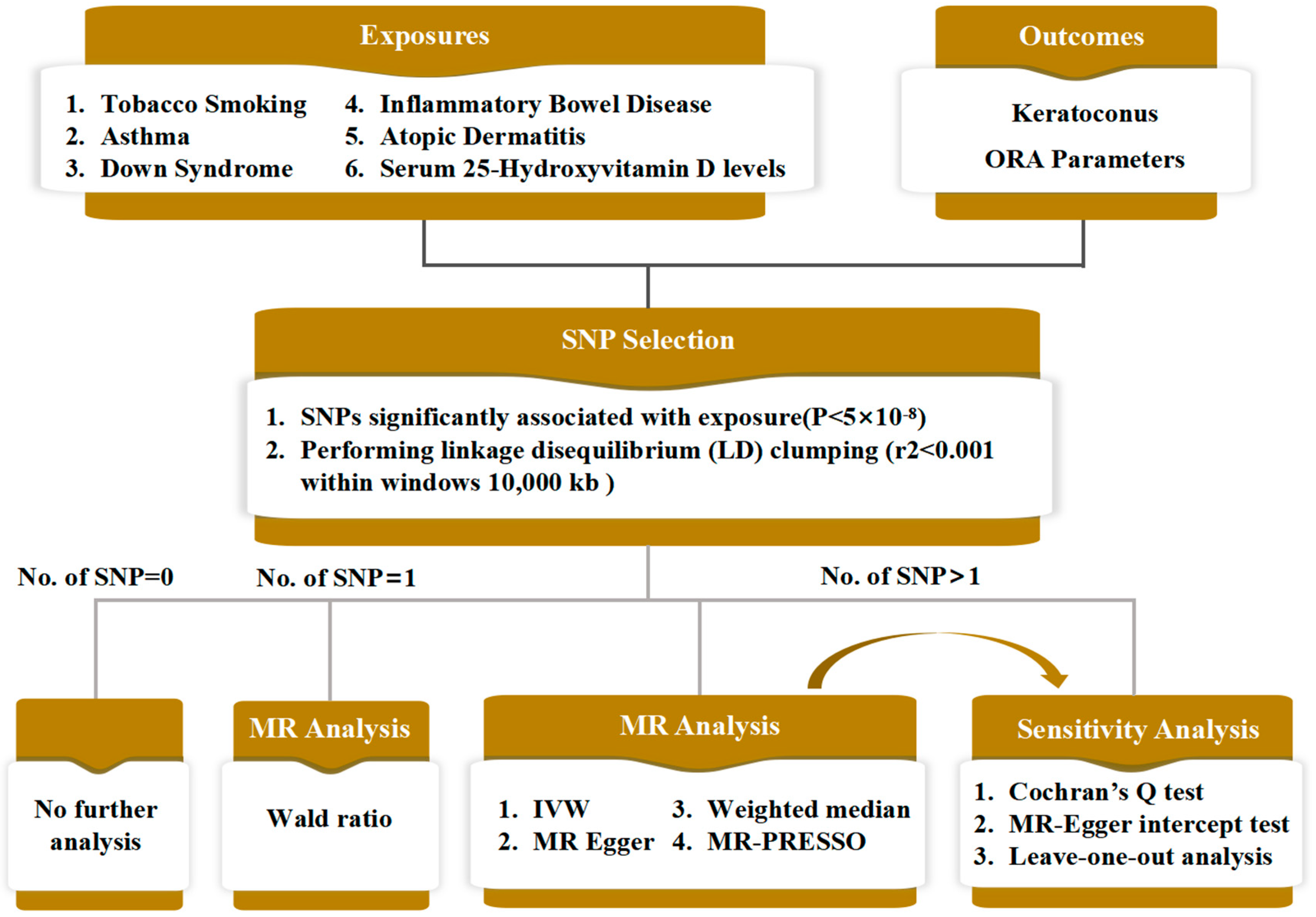
2.1. Study Design and Data Source

Due to the evidenced link between corneal biomechanical changes and KC development, the MR analysis of this study was divided into two parts: (1) explore the effects of exposure factors on corneal biomechanics by selecting ORA parameters (CH and CRF) as the outcomes; (2) explore the effects of these factors on KC directly by using KC itself as outcome. The exposure factors considered in this study included smoking, asthma, Down syndrome, inflammatory bowel disease, atopic dermatitis, and serum 25-hydroxyvitamin D levels (Figure 1).
All data used in the study were from the IEU Open GWAS project (https://gwas.mrcieu.ac.uk/, accessed on 3 July 2023). Genetic instruments obtained from the FinnGen database as exposure or outcome included keratoconus (311 cases and 209,287 controls; GWAS ID “finn-b-H7_CORNEALDEFORM”) and atopic dermatitis (7024 cases and 198,740 controls; GWAS ID” finn-b-L12_ATOPIC”). Summary statistics from UK Biobank were used as the data for current tobacco smoking, including 462,434 individuals of European (GWAS ID “ukb-b-223”) and 6572 individuals of African American or Afro-Caribbean (GWAS ID “ukb-e-1239_AFR”), ORA biomechanical parameters with corneal hysteresis (GWAS ID “ukb-b-11650”) and corneal resistance factors (GWAS ID “ukb-b-4717”) of the left eyes for 97,465 individuals of European ancestry, and asthma (GWAS ID “ukb-b-18113”) for 462,933 individuals of European ancestry. Genome-wide data for inflammatory bowel disease were from the International Inflammatory Bowel Disease Genetics Consortium (IIBDGC) published in 2012 (75,000 individuals of European ancestry; GWAS ID “ieu-a-292”) [19]. The data for the Down syndrome cell adhesion molecule were from a genome-wide association study of 3301 participants conducted by the Cardiovascular Epidemiology Unit (CEU) team at the University of Cambridge in 2018 (GWAS ID “prot-a-868”) [20]. Serum 25-hydroxyvitamin D levels were obtained from a UK Biobank genome-wide association study involving 417580 European individuals, and the median, mean, and interquartile ranges of serum 25-hydroxyvitamin D levels were 47.9, 49.6, and 33.5–63.2 nmol/L−1 (GWAS ID “ebi-a-GCST90000617”) [15].
Since all data involved in this study were obtained from the GWAS public database, no ethical approval or informed consent was required.

2.2. Data Analysis

An important step in the MR method is the determination of instrumental variables (IV) to carry out the analysis, and the validity of causal estimates with MR are dependent on three key assumptions: (1) the IV is unrelated to the typical confounding factors, (2) the IV is (reliably) associated with the exposure, and (3) the IV affects the outcome only through the risk factor [21]. The MR method thus uses genetic variants as IVs for assessing causal relationships from observational data [22]; meanwhile, single nucleotide polymorphisms (SNPs) are the most common genetic variant in the human genome. The development of high-throughput genomic technologies and genome-wide association studies (GWAS) has helped identify SNPs and determine genetic factors associated with complex diseases [23]. Therefore, SNPs that strongly related to each exposure factor were used as IVs in this study.
Figure 1 highlights the main steps for data analysis, which started with SNP selection, based on which various MR analyses was carried out, followed by sensitivity analyses. To ensure that the obtained SNPs were independent of exposure, linkage disequilibrium (LD) clumping (r2 < 0.001 within windows 10,000 kb for variants in the same locus) was used. SNPs were considered significantly related to the exposures if p < 5 × 10⁻⁸, and these SNPs were then adopted as IVs. To assess the association strength between the IV and the exposure factor, the F value was calculated for each IV in the form of R 2 × ( N - 2 ) / ( 1 - R 2 ) when N, the sample size of the GWAS for each exposure, was available and β exposure 2 / se exposure 2 when N was not available; here, R 2 was estimated as 2 × β exposure 2 × EAF exposure × ( 1 - EAF exposure ) , where EAF exposure was the effect allele frequency and β exposure and se exposure were the estimated genetic effects on the exposure factor in question [24,25]. IVs with F values < 10 were excluded for further analysis.
MR analysis methods were applied as follows [26]: IVW, MR-Egger, weighted median, and MR-PRESSO were applied and scatter plots of the analysis results were also plotted for visualisation when the numbers of SNPs were >1. The Wald ratio was used if only a single SNP was identified, whereas no further analysis was carried out for exposure factors with no identified SNP.
Because genetic variants may affect the outcome through pathways other than through the risk factor of interest (so called horizontal pleiotropic effects). The intercept from the MR-Egger analysis can be interpreted as the average pleiotropic effect of a genetic variant included in the analysis [27,28]. P > 0.05 for the MR-Egger intercept indicates that no evidence of horizontal pleiotropy exists. Cochran’s Q statistic was used to assess heterogeneity of the IVs and p > 0.05 means that no significant heterogeneity exists for these associations. The MR-PRESSO analysis was used for outlier detection [29], and the MR analysis, heterogeneity testing, and horizontal testing were performed again after removing outliers if detected (Figure 1). The leave-one-out analysis was used to test the stability of the MR results. For IVs containing only a single SNP, the sensitivity analysis outlined herein could not be performed.
All analysis was conducted using the TwoSampleMR package (version 0.5.7) in R language (version 4.3.0). p < 0.05 was considered statistically significant.

3. Results

3.1. Mendelian Randomisation Analysis with ORA Parameters as the Outcomes

The results of the two-sample MR analysis examining the causal associations of tobacco smoking and inflammatory bowel disease with the ORA parameters are listed in Table 1 and Table 2. The analysis indicated no associations between other exposures and the ORA parameters (i.e., 0 SNP).
Outliers for inflammatory bowel disease (CH: rs6920220 and rs7240004; CRF: rs7240004 and rs12142199) were detected by MR-PRESSO; therefore, the results in the tables are based on analysis after removing the outliers.
MR estimates indicated that tobacco smoking was significantly associated with ORA parameters susceptibility using the IVW (CH: OR = 1.572, p < 0.001; CRF: OR = 1.380, p = 0.009). Such causal association was also presented in the scatter plot (Supplementary Figures S1a and S2a). The leave-one-out analysis suggested that the causal effect results of tobacco smoking and ORA parameters in the IVW analysis were not driven by any single SNP (Supplementary Figures S1b and S2b), indicating the stability of the analysis. Similarly, the IVW (CH: OR = 0.989, p = 0.032; CRF: OR = 0.982, p = 0.001) showed a causal relationship between inflammatory bowel disease and ORA parameters (Supplementary Figures S3a and S4a). Moreover, in the MR-Egger intercept test, no horizontal pleiotropy was found for SNPs used in tobacco smoking (CH: P-pleiotropy = 0.192; CRF: P-pleiotropy = 0.624) and inflammatory bowel disease (CH: P-pleiotropy = 0.722; CRF: P-pleiotropy = 0.879). Collectively, these findings suggest that tobacco smoking and inflammatory bowel disease affect corneal biomechanical properties.

3.2. Mendelian Randomisation Analysis with Keratoconus as the Outcome

Table 3 showed the results for the causal associations between KC and other risk factors, including current tobacco smoking, Down syndrome, asthma, atopic dermatitis, inflammatory bowel disease, and serum 25-hydroxyvitamin D levels. The Wald ratio (OR = 0.055, p = 0.024) method showed that tobacco smoking is associated with a lower risk of KC. Instead, asthma (OR = 39.901, p = 0.009), atopic dermatitis (OR = 1.452, p = 0.012), inflammatory bowel disease (OR = 1.206, p = 0.017), and serum 25-hydroxyvitamin D levels (OR = 2.146, p = 0.039) were associated with a higher risk of KC by the method of IVW. The Wald ratio (OR = 3.276, p = 0.004) showed statistical significance between Down syndrome and higher risk of KC. The MR-Egger intercept test did not yield any indication of directional pleiotropy for the association between these factors and KC (all Ps > 0.05), and Cochran’s Q test revealed no significant heterogeneity exists for these associations (all Ps > 0.05), indicating the reliability of the causal results. The causal associations were presented in the scatter plots, and the leave-one-out analysis suggested that the causal effect results of these risk factors and KC in the IVW analysis were not driven by any single SNP (Supplementary Figures S5–S8).

4. Discussion

This study examined causal associations between several potential influencing factors and KC using a broad set of variables obtained from the largest genetic databases currently available. Two sample MR analysis showed that there was a causal relationship between tobacco smoking and lower risk of KC, while Down syndrome, asthma, atopic dermatitis, inflammatory bowel disease, and Serum 25-Hydroxyvitamin D levels had causal associations with higher risk of KC. Causal associations of tobacco smoking and inflammatory bowel disease with the ORA parameters was also observed. Collectively, it can be speculated that changes in corneal biomechanical properties may be an intermediate factor that prevents or leads to the development of KC.
In recent years, MR analysis has been gradually gaining research attentions with its new perspective of inferring the causal relationship between exposure and outcome [16,18]. A number of MR studies have identified potential causal associations between lifestyle or biological exposures and ocular diseases, thus providing an opportunity for further pathological studies and interventional development [30,31,32]. To the best of the authors’ knowledge, this was the first study to estimate potential risk factors for KC by using the GWAS database.
The study indicates that there is potential genetic evidence for a causal relationship between tobacco smoking and the lower risk of KC, after excluding confounding factors and reverse causal associations, which are consistent with the observational studies. Hafezi et al. showed a statistically significant increase in CH and CRF in smokers, which suggested that chronic smoking might have a beneficial effect on corneal biomechanics [33], because conversely the reduction in corneal biomechanical properties were thought to play an important role in the progression of KC [34], and studies indicated a lower CH and CRF in KC [8,35]. A study by Sahebjada et al. suggested KC was associated with smoking using univariate regression analysis but that association became statistically insignificant following a multivariate regression analysis [9]. While these observational studies showed partially significant associations between smoking and KC, they had their own limitations related to potential uncontrolled confounders that might mask the true associations. The design of the current study could otherwise overcome these limitations.
Although KC is traditionally considered a non-inflammatory disease, some inflammatory molecules have been found in the tear fluid of patients with KC, suggesting that immune-related diseases factors may be associated with the pathogenesis of KC [36]. In a study among Dutch residents, KC appeared to be positively associated with a variety of immune-mediated diseases, including asthma and inflammatory skin conditions [37]. Sahebjada et al. have also highlighted a significant association between asthma and an increased risk of severe KC [9]. An observational study involving of 4272 adults with severe atopic dermatitis demonstrated a notably high hazard ratio (HR) of 10.01 (95% confidence interval [CI], 5.02–19.96) for developing KC [38]. In contrast, a recent MR analysis, based on a different database other than the one in the current study, did not identify a significant causal effect of atopic dermatitis on KC [39]. Nevertheless, the current study aligns with previous observational findings, indicating that asthma, inflammatory bowel disease, and atopic dermatitis were positively associated with KC risk.
The main clinical manifestation of KC is corneal thin and ectasia, phenomena largely ascribed to the degradation of the extracellular matrix (ECM) [40]. MMPs (matrix metalloproteinases), which are zinc-dependent endopeptidases secreted by epithelial cells, stromal cells, and neutrophils, play a critical role in this process by cleaving components of the ECM, such as collagen and elastin [41]. Current evidence indicates that KC patients exhibit elevated levels of inflammatory molecules, including interleukin-6 (IL-6) and tumour necrosis factor-α (TNF-α), in their tears [42,43]. Furthermore, several studies have established that IL and TNF regulate the expression of MMPs [14,40,42]. This suggests that chronic inflammatory processes are integral to the pathogenesis of KC.
Meanwhile, inflammatory bowel disease, including Crohn’s disease and ulcerative colitis, is a chronic inflammatory disorder of the intestines of unknown aetiology. In inflammatory bowel disease, tissue damage is primarily driven by adaptive immunity, and the adaptive response to specific antigens is influenced by a combination of resident and recruited cell populations. This includes the release of cytokines such as IL-1, IL-6, and TNF-α by macrophages [12,44]. Furthermore, Vitamin D plays a role in modulating both innate and adaptive immune systems, and its deficiency is associated with allergic disorders [45,46,47].
These findings collectively elucidate the causal relationship between KC and immune-related factors, including asthma, atopic dermatitis, inflammatory bowel disease, and serum 25-hydroxyvitamin D levels.
The study also indicated a causal association of Down syndrome with higher risk of KC. Concurrently, Maria et al. found that patients with Down syndrome had more than six times higher odds of KC than those without this condition [13]. A study, which reported the corneal morphologic characteristics in a large series of patients with Down syndrome, showed that patients with Down syndrome had steeper and thinner corneas and more corneal aberrations than those without genetic alterations and normal corneas [48]. Some studies have shown that Down syndrome was associated with more frequent eye rubbing, which may contribute to the high prevalence of KC among persons with Down syndrome [49,50].
Although MR has many advantages in causal inference and can provide a complement to traditional observational studies, several limitations of the current study need to be noted. Firstly, the study was limited to individuals of European ancestry and the conclusions may not be fully applicable to other populations. Secondly, the robustness of the results for two exposures (namely, smoking and Down syndrome) was constrained because these exposures only had a single SNP. Thirdly, the KC data included in this study did not distinguish between gender and age. However, the recent studies suggest that sex hormones can maintain corneal structural integrity by influencing wound healing and corneal stromal thickness, and that prolactin-induced protein is an important hormonally regulated biomarker in KC [51,52]. Similarly, KC usually progresses rapidly in youth. Therefore, the further division of KC population according to gender and age before MR analysis will yield more accurate results. Meanwhile, the purpose of this study was to investigate the risk factors for KC and to use this as a reference for clinical prevention and screening. Currently, the main treatments for KC are rigid gas permeable, corneal cross-linking, etc. [53]. These treatments can control the progression of KC, so early diagnosis of KC and effective treatment measures will reduce the risk of corneal transplantation in patients. Lastly, the mechanism behind the causal association between smoking and KC was not directly proven. Instead, the inference was made through the ORA parameters, the only in vivo corneal biomechanical parameters available in the database. However, they may not be the most accurate representation of corneal biomechanics, and in fact, the study did not find any other exposure factors besides tobacco smoking and inflammatory bowel disease to be causally associated with the parameters of ORA. Although the CH and CRF of KC are lower than those of healthy corneas, it has been observed that there is considerable overlap in the distribution of the two parameters, and therefore, the sensitivity and specificity of the KC diagnosis are relatively weak and do not accurately predict the changes in corneal biomechanics [54]. Different from ORA, Corvis ST analyses corneal deformation parameters based on the dynamic examination of the corneal response. It uses an ultra-high-speed Scheimpflug camera to take 140 horizontal 8 mm frames over a period of 33 ms. In particular, Corvis ST provides a more reliable representation of parameters that characterise corneal biomechanics, for example, SSI, SP-A1, CBI, etc, which have important applications in the diagnosis and treatment of KC [8]. Further MR studies with biomechanical data from, e.g., Corvis ST [55] and Brillouin optical microscopy [56] may be desirable as these technologies were shown to provide more accurate biomechanical indices to reflect the biomechanical abnormalities in KC [8] and, therefore, may promising for better explaining the bridging roles of corneal biomechanics between KC development and its exposure factors.
In conclusion, this MR study suggests that in European populations, asthma, Down syndrome, inflammatory bowel disease, atopic dermatitis, and serum 25-hydroxyvitamin D levels are associated with a higher risk of KC, while smoking is associated with a lower risk. These findings highlight the multifactorial nature of KC involving both environmental and genetic factors and emphasise the importance of timely screening in specific populations for effective KC prevention. Further MR analysis is warranted to include more representative biomechanical measures with more SNPs and KC cases.

Supplementary Materials

The following supporting information can be downloaded at: https://www.mdpi.com/article/10.3390/bioengineering11030221/s1, Figure S1: Scatter and leave-one-out plots of tobacco smoking with Corneal Hysteresis (left eye); Figure S2: Scatter and leave-one-out plots of tobacco smoking with Corneal Resistance Factor (left eye); Figure S3: Scatter and leave-one-out plots of inflammatory bowel disease with Corneal Hysteresis (left eye); Figure S4: Scatter and leave-one-out plots of inflammatory bowel disease with Corneal Resistance Factor (left eye); Figure S5: Scatter and leave-one-out plots of asthma with keratoconus. a: Scatter plot demonstrating the effect of each asthma-associated genetic variant on keratoconus on the log-odds scale; Figure S6: Supplementary Figure S6 Scatter and leave-one-out plots of atopic dermatitis with KC; Figure S7: Scatter and leave-one-out plots of inflammatory bowel disease with KC; Figure S8: Supplementary Figure S8 Scatter and leave-one-out plots of serum 25−hydroxyvitamin D levels with KC.

Author Contributions

Conceptualization, J.W.; methodology, J.C.; software, J.C.; validation, L.Y. and L.H.; formal analysis, J.C. and L.H.; investigation, Y.Y.; data curation, J.C., L.Y. and Y.Y.; writing—original draft preparation, J.C.; writing—review and editing, J.C., L.Y., S.C. and J.W.; supervision, S.C. and J.W.; project administration, S.C.; funding acquisition, J.W. All authors have read and agreed to the published version of the manuscript.

Funding

This work was supported by the National Natural Science Foundation of China (82001924) and the Zhejiang Provincial Natural Science Foundation of China (LY22H180005).

Institutional Review Board Statement

Not applicable.

Informed Consent Statement

Not applicable.

Data Availability Statement

Publicly available datasets were analysed in this study. This data can be found here: [https://gwas.mrcieu.ac.uk/] (accessed on 22 February 2024).

Conflicts of Interest

The authors declare no conflict of interest.

Abbreviations

KCKeratoconus
MRMendelian randomisation
IVWInverse variance weighted
SNPSingle-nucleotide polymorphism
ORAOcular Response Analyzer
CVSCorvis ST
CHCorneal hysteresis
CRFCorneal resistance factor
RCTRandomised control trial
GWASGenome-wide association study
SSIStress–stain index
CBISorneal biomechanical index
CEUCardiovascular Epidemiology Unit
IVInstrumental variables
LDLinkage disequilibrium
MR EggerMendelian randomisation–Egger
CIConfidence interval
OROdds ratio
ECMExtracellular matrix
MMPsMatrix metalloproteinases
IL-6Interleukin-6
TNF-αTumor necrosis factor-α
SP-A1Corneal stiffness parameter at first applanation time

References

  1. Gordon-Shaag, A.; Millodot, M.; Shneor, E.; Liu, Y. The Genetic and Environmental Factors for Keratoconus. BioMed Res. Int. 2015, 2015, 795738. [Google Scholar] [CrossRef]
  2. Veerappa, A.M. Cascade of interactions between candidate genes reveals convergent mechanisms in keratoconus disease pathogenesis. Ophthalmic Genet. 2021, 42, 114–131. [Google Scholar] [CrossRef]
  3. Utsunomiya, T.; Hanada, K.; Muramatsu, O.; Ishibazawa, A.; Nishikawa, N.; Yoshida, A. Wound Healing Process After Corneal Stromal Thinning Observed With Anterior Segment Optical Coherence Tomography. Cornea 2014, 33, 1056–1060. [Google Scholar] [CrossRef]
  4. Godefrooij, D.A.; de Wit, G.A.; Uiterwaal, C.S.; Imhof, S.M.; Wisse, R.P.L. Age-specific Incidence and Prevalence of Keratoconus: A Nationwide Registration Study. Am. J. Ophthalmol. 2017, 175, 169–172. [Google Scholar] [CrossRef]
  5. Hashemi, H.; Heydarian, S.; Hooshmand, E.; Saatchi, M.; Yekta, A.; Aghamirsalim, M.; Valadkhan, M.; Mortazavi, M.; Hashemi, A.; Khabazkhoob, M. The Prevalence and Risk Factors for Keratoconus: A Systematic Review and Meta-Analysis. Cornea 2020, 39, 263–270. [Google Scholar] [CrossRef]
  6. Santodomingo-Rubido, J.; Carracedo, G.; Suzaki, A.; Villa-Collar, C.; Vincent, S.J.; Wolffsohn, J.S. Keratoconus: An updated review. Contact Lens Anterior Eye 2022, 45, 101559. [Google Scholar] [CrossRef]
  7. Roberts, C.J. Biomechanics of corneal ectasia and biomechanical treatments. J. Cataract Refract. Surg. 2014, 40, 991–998. [Google Scholar] [CrossRef] [PubMed]
  8. Esporcatte, L.P.G.; Salomão, M.Q.; Lopes, B.T.; Sena, N.; Ferreira, É.; Filho, J.B.R.F.; Machado, A.P.; Ambrósio, R. Biomechanics in Keratoconus Diagnosis. Curr. Eye Res. 2023, 48, 130–136. [Google Scholar] [CrossRef]
  9. Sahebjada, S.; Chan, E.; Xie, J.; Snibson, G.R.; Daniell, M.; Baird, P.N. Risk factors and association with severity of keratoconus: The Australian study of Keratoconus. Int. Ophthalmol. 2021, 41, 891–899. [Google Scholar] [CrossRef] [PubMed]
  10. Mathan, J.J.; Simkin, S.K.; Gokul, A.; McGhee, C.N.J. Down syndrome and the eye: Ocular characteristics and ocular assessment. Surv. Ophthalmol. 2022, 67, 1631–1646. [Google Scholar] [CrossRef] [PubMed]
  11. Kristianslund, O.; Drolsum, L. Prevalence of keratoconus in persons with Down syndrome: A review. BMJ Open Ophth. 2021, 6, e000754. [Google Scholar] [CrossRef] [PubMed]
  12. Tréchot, F.; Angioi, K.; Latarche, C.; Conroy, G.; Beaujeux, P.; Andrianjafy, C.; Portier, M.; Batta, B.; Conart, J.-B.; Cloché, V.; et al. Keratoconus in Inflammatory Bowel Disease Patients: A Cross-sectional Study. J Crohns Colitis. 2015, 9, 1108–1112. [Google Scholar] [CrossRef] [PubMed]
  13. Woodward, M.A.; Blachley, T.S.; Stein, J.D. The Association Between Sociodemographic Factors, Common Systemic Diseases, and Keratoconus. Ophthalmology 2016, 123, 457–465.e2. [Google Scholar] [CrossRef] [PubMed]
  14. Chang, Y.; Weng, S.; Wang, J.; Tseng, S.; Wang, J.; Jan, H.; Ko, S.; Tsai, W.; Chen, H.; Liou, C.; et al. Association between keratoconus and the risk of adolescent- or adult-onset atopic dermatitis. Allergy 2020, 75, 2946–2948. [Google Scholar] [CrossRef]
  15. Revez, J.A.; Lin, T.; Qiao, Z.; Xue, A.; Holtz, Y.; Zhu, Z.; Zeng, J.; Wang, H.; Sidorenko, J.; Kemper, K.E.; et al. Genome-wide association study identifies 143 loci associated with 25 hydroxyvitamin D concentration. Nat. Commun. 2020, 11, 1647. [Google Scholar] [CrossRef] [PubMed]
  16. Cornish, A.J.; Tomlinson, I.P.M.; Houlston, R.S. Mendelian randomisation: A powerful and inexpensive method for identifying and excluding non-genetic risk factors for colorectal cancer. Mol. Asp. Med. 2019, 69, 41–47. [Google Scholar] [CrossRef] [PubMed]
  17. Li, J.; Li, C.; Huang, Y.; Guan, P.; Huang, D.; Yu, H.; Yang, X.; Liu, L. Mendelian randomization analyses in ocular disease: A powerful approach to causal inference with human genetic data. J. Transl. Med. 2022, 20, 621. [Google Scholar] [CrossRef]
  18. Burgess, S.; Dudbridge, F.; Thompson, S.G. Combining information on multiple instrumental variables in Mendelian randomization: Comparison of allele score and summarized data methods. Statist. Med. 2016, 35, 1880–1906. [Google Scholar] [CrossRef]
  19. The International IBD Genetics Consortium (IIBDGC); Jostins, L.; Ripke, S.; Weersma, R.K.; Duerr, R.H.; McGovern, D.P.; Hui, K.Y.; Lee, J.C.; Philip Schumm, L.; Sharma, Y.; et al. Host–microbe interactions have shaped the genetic architecture of inflammatory bowel disease. Nature 2012, 491, 119–124. [Google Scholar] [CrossRef]
  20. Sun, B.B.; Maranville, J.C.; Peters, J.E.; Stacey, D.; Staley, J.R.; Blackshaw, J.; Burgess, S.; Jiang, T.; Paige, E.; Surendran, P.; et al. Genomic atlas of the human plasma proteome. Nature 2018, 558, 73–79. [Google Scholar] [CrossRef]
  21. Sheehan, N.A.; Didelez, V.; Burton, P.R.; Tobin, M.D. Mendelian Randomisation and Causal Inference in Observational Epidemiology. PLoS Med. 2008, 5, e177. [Google Scholar] [CrossRef]
  22. Yavorska, O.O.; Burgess, S. MendelianRandomization: An R package for performing Mendelian randomization analyses using summarized data. Int. J. Epidemiol. 2017, 46, 1734–1739. [Google Scholar] [CrossRef]
  23. Li, P.; Guo, M.; Wang, C.; Liu, X.; Zou, Q. An overview of SNP interactions in genome-wide association studies. Brief. Funct. Genom. 2015, 14, 143–155. [Google Scholar] [CrossRef]
  24. Wood, A.T.A. An F Approximation to the Distribution of a Linear Combination of Chi-squared Variables. Commun. Stat.-Simul. Comput. 1989, 18, 1439–1456. [Google Scholar] [CrossRef]
  25. Zhang, H.; Yao, Y.; Zhong, X.; Meng, F.; Hemminki, K.; Qiu, J.; Shu, X. Association between intake of the n-3 polyunsaturated fatty acid docosahexaenoic acid (n-3 PUFA DHA) and reduced risk of ovarian cancer: A systematic Mendelian Randomization study. Clin. Nutr. 2023, 42, 1379–1388. [Google Scholar] [CrossRef]
  26. Bowden, J.; Del Greco, M.F.; Minelli, C.; Davey Smith, G.; Sheehan, N.; Thompson, J. A framework for the investigation of pleiotropy in two-sample summary data Mendelian randomization: A framework for two-sample summary data MR. Statist. Med. 2017, 36, 1783–1802. [Google Scholar] [CrossRef]
  27. Davies, N.M.; Holmes, M.V.; Davey Smith, G. Reading Mendelian randomisation studies: A guide, glossary, and checklist for clinicians. BMJ 2018, 362, k601. [Google Scholar] [CrossRef]
  28. Burgess, S.; Thompson, S.G. Interpreting findings from Mendelian randomization using the MR-Egger method. Eur. J. Epidemiol. 2017, 32, 377–389. [Google Scholar] [CrossRef]
  29. Verbanck, M.; Chen, C.-Y.; Neale, B.; Do, R. Detection of widespread horizontal pleiotropy in causal relationships inferred from Mendelian randomization between complex traits and diseases. Nat. Genet. 2018, 50, 693–698. [Google Scholar] [CrossRef]
  30. Han, X.; Ong, J.-S.; An, J.; Hewitt, A.W.; Gharahkhani, P.; MacGregor, S. Using Mendelian randomization to evaluate the causal relationship between serum C-reactive protein levels and age-related macular degeneration. Eur. J. Epidemiol. 2020, 35, 139–146. [Google Scholar] [CrossRef]
  31. Cuellar-Partida, G.; Lu, Y.; Kho, P.F.; Hewitt, A.W.; Wichmann, H.-E.; Yazar, S.; Stambolian, D.; Bailey-Wilson, J.E.; Wojciechowski, R.; Wang, J.J.; et al. Assessing the Genetic Predisposition of Education on Myopia: A Mendelian Randomization Study: Assessing the Genetic Predisposition of Education on Myopia. Genet. Epidemiol. 2016, 40, 66–72. [Google Scholar] [CrossRef]
  32. Cuellar-Partida, G.; Williams, K.M.; Yazar, S.; Guggenheim, J.A.; Hewitt, A.W.; Williams, C.; Wang, J.J.; Kho, P.-F.; Saw, S.M.; Cheng, C.-Y.; et al. Genetically low vitamin D concentrations and myopic refractive error: A Mendelian randomization study. Int. J. Epidemiol. 2017, 46, 1882–1890. [Google Scholar] [CrossRef]
  33. Hafezi, F. Smoking and Corneal Biomechanics. Ophthalmology 2009, 116, 2259.e1. [Google Scholar] [CrossRef]
  34. Zhao, Y.; Shen, Y.; Yan, Z.; Tian, M.; Zhao, J.; Zhou, X. Relationship Among Corneal Stiffness, Thickness, and Biomechanical Parameters Measured by Corvis ST, Pentacam and ORA in Keratoconus. Front. Physiol. 2019, 10, 740. [Google Scholar] [CrossRef]
  35. Shah, S.; Laiquzzaman, M.; Bhojwani, R.; Mantry, S.; Cunliffe, I. Assessment of the Biomechanical Properties of the Cornea with the Ocular Response Analyzer in Normal and Keratoconic Eyes. Investig. Ophthalmol. Vis. Sci. 2007, 48, 3026. [Google Scholar] [CrossRef]
  36. Balasubramanian, S.A.; Mohan, S.; Pye, D.C.; Willcox, M.D.P. Proteases, proteolysis and inflammatory molecules in the tears of people with keratoconus. Acta Ophthalmol. 2012, 90, e303–e309. [Google Scholar] [CrossRef]
  37. Claessens, J.L.J.; Godefrooij, D.A.; Vink, G.; Frank, L.E.; Wisse, R.P.L. Nationwide epidemiological approach to identify associations between keratoconus and immune-mediated diseases. Br. J. Ophthalmol. 2022, 106, 1350–1354. [Google Scholar] [CrossRef]
  38. Thyssen, J.P.; Toft, P.B.; Halling-Overgaard, A.-S.; Gislason, G.H.; Skov, L.; Egeberg, A. Incidence, prevalence, and risk of selected ocular disease in adults with atopic dermatitis. J. Am. Acad. Dermatol. 2017, 77, 280–286.e1. [Google Scholar] [CrossRef]
  39. Zhou, W.; Cai, J.; Li, Z.; Lin, Y. Association of atopic dermatitis with conjunctivitis and other ocular surface diseases: A bidirectional two-sample Mendelian randomization study. Acad. Dermatol. Venereol. 2023, 37, 1642–1648. [Google Scholar] [CrossRef]
  40. Balasubramanian, S.A.; Pye, D.C.; Willcox, M.D.P. Are Proteinases the Reason for Keratoconus? Curr. Eye Res. 2010, 35, 185–191. [Google Scholar] [CrossRef]
  41. Cui, N.; Hu, M.; Khalil, R.A. Biochemical and Biological Attributes of Matrix Metalloproteinases. In Progress in Molecular Biology and Translational Science; Elsevier: Amsterdam, The Netherlands, 2017; Volume 147, pp. 1–73. [Google Scholar] [CrossRef]
  42. Lema, I.; Duran, J. Inflammatory Molecules in the Tears of Patients with Keratoconus. Ophthalmology 2005, 112, 654–659. [Google Scholar] [CrossRef]
  43. Lema, I.; Sobrino, T.; Duran, J.A.; Brea, D.; Diez-Feijoo, E. Subclinical keratoconus and inflammatory molecules from tears. Br. J. Ophthalmol. 2009, 93, 820–824. [Google Scholar] [CrossRef]
  44. Peyrin-Biroulet, L.; Desreumaux, P.; Sandborn, W.J.; Colombel, J.-F. Crohn’s disease: Beyond antagonists of tumour necrosis factor. Lancet 2008, 372, 67–81. [Google Scholar] [CrossRef]
  45. Akkaya, S.; Ulusoy, D.M. Serum Vitamin D Levels in Patients with Keratoconus. Ocul. Immunol. Inflamm. 2020, 28, 348–353. [Google Scholar] [CrossRef]
  46. Deluca, H.F.; Cantorna, M.T. Vitamin D: Its role and uses in immunology. FASEB J. 2001, 15, 2579–2585. [Google Scholar] [CrossRef]
  47. Ao, T.; Kikuta, J.; Ishii, M. The Effects of Vitamin D on Immune System and Inflammatory Diseases. Biomolecules 2021, 11, 1624. [Google Scholar] [CrossRef]
  48. Alio, J.L.; Vega-Estrada, A.; Sanz, P.; Osman, A.A.; Kamal, A.M.; Mamoon, A.; Soliman, H. Corneal Morphologic Characteristics in Patients With Down Syndrome. JAMA Ophthalmol. 2018, 136, 971–978. [Google Scholar] [CrossRef]
  49. Kristianslund, O.; Drolsum, L. Prevalence of Keratoconus in Persons With Down Syndrome in a National Registry in Norway. JAMA Netw. Open 2021, 4, e210814. [Google Scholar] [CrossRef]
  50. Haugen, O.H.; Høvding, G.; Eide, G.E. Biometric measurements of the eyes in teenagers and young adults with Down syndrome. Acta Ophthalmol. Scand. 2001, 79, 616–625. [Google Scholar] [CrossRef]
  51. McKay, T.B.; Priyadarsini, S.; Karamichos, D. Sex Hormones, Growth Hormone, and the Cornea. Cells 2022, 11, 224. [Google Scholar] [CrossRef]
  52. Sharif, R.; Bak-Nielsen, S.; Hjortdal, J.; Karamichos, D. Pathogenesis of Keratoconus: The intriguing therapeutic potential of Prolactin-inducible protein. Prog. Retin. Eye Res. 2018, 67, 150–167. [Google Scholar] [CrossRef]
  53. Mazzotta, C.; Ferrise, M.; Gabriele, G.; Gennaro, P.; Meduri, A. Chemically-Boosted Corneal Cross-Linking for the Treatment of Keratoconus through a Riboflavin 0.25% Optimized Solution with High Superoxide Anion Release. JCM 2021, 10, 1324. [Google Scholar] [CrossRef]
  54. Esporcatte, L.P.G. Biomechanical diagnostics of the cornea. Eye Vis. 2020, 7, 9. [Google Scholar] [CrossRef]
  55. Tejwani, S.; Shetty, R.; Kurien, M.; Dinakaran, S.; Ghosh, A.; Roy, A.S. Biomechanics of the Cornea Evaluated by Spectral Analysis of Waveforms from Ocular Response Analyzer and Corvis-ST. PLoS ONE 2014, 9, e97591. [Google Scholar] [CrossRef] [PubMed]
  56. Yun, S.H.; Chernyak, D. Brillouin microscopy: Assessing ocular tissue biomechanics. Curr. Opin. Ophthalmol. 2018, 29, 299–305. [Google Scholar] [CrossRef]
Figure 1. The schematic flow of the Mendelian randomisation analysis adopted in this study.
Bioengineering 11 00221 g001
Table 1. MR results of tobacco smoking on CH and CRF.
OutcomeMR MethodsNo. of SNPsOR (95%CI)p-ValueP-HeterogeneityP-Pleiotropy
CH 20 0.3600.192
IVW 1.572
(1.216–2.033)
<0.001 *
Weighted median 1.345
(0.955–1.893)
0.090
MR-Egger
0.874
(0.360–2.121)
0.769
CRF 20 0.5250.624
IVW 1.380
(1.086–1.755)
0.009 *
Weighted median 1.251
(0.881–1.777)
0.211
MR-Egger 1.122
(0.481–2.616)
0.792
IVW = inverse variance weighted; MR-Egger = Mendelian randomisation–Egger; CI = confidence interval; OR = odds ratio; MR = Mendelian randomisation; SNP = single-nucleotide polymorphism; CH= corneal hysteresis; CRF= corneal resistance factor; * represents the correlation is significant at 0.05 level.
Table 2. MR results of inflammatory bowel disease on CH and CRF.
OutcomeMR MethodsNo. of SNPsOR (95%CI)p-ValueP-HeterogeneityP-Pleiotropy
CH
(Outlier removed)
110 <0.001 *0.722
IVW 0.989
(0.978–0.999)
0.032 *
Weighted median 0.994
(0.980–1.009)
0.446
MR-Egger 0.984
(0.960–1.010)
0.226
CRF
(Outlier removed)
108 <0.001 *0.879
IVW 0.982
(0.971–0.993)
0.001 *
Weighted median 0.983
(0.971–0.996)
0.010 *
MR-Egger 0.980
(0.955–1.006)
0.137
IVW = inverse variance weighted; MR-Egger = Mendelian randomisation–Egger; CI = confidence interval; OR = odds ratio; MR = Mendelian randomisation; SNP = single-nucleotide polymorphism; CH = corneal hysteresis; CRF = corneal resistance factor; * represents the correlation is significant at 0.05 level.
Table 3. MR results of smoking, asthma, down syndrome, inflammatory bowel disease, atopic dermatitis and serum 25-hydroxyvitamin D levels on the risk of KC.
ExposuresMR MethodsNo. of SNPsOR (95%CI)p-ValueP-HeterogeneityP-Pleiotropy
Current tobacco smokingWald ratio10.055
(0.004–0.677)
0.024 *NANA
Down syndromeWald ratio13.276
(1.453–7.388)
0.004 *NANA
Asthma 98 0.6830.255
IVW 39.901
(2.522–631.169)
0.009 *
Weighted median 3.273
(0.043–251.786)
0.593
MR-Egger 1.020
(0.001–966.600)
0.996
Inflammatory bowel disease 111 0.3030.723
IVW 1.206
(1.034–1.407)
0.017 *
Weighted median 1.163
(0.921–1.469)
0.203
MR-Egger 1.132
(0.770–1.663)
0.531
Atopic dermatitis 20 0.6060.504
IVW 1.452
(1.085–1.944)
0.012 *

Weighted median 1.302
(0.864–1.961)
0.207
MR-Egger 1.142
(0.539–2.417)
0.733
Serum 25-Hydroxyvitamin D levels 104 0.3270.930
IVW 2.146
(1.040–4.429)
0.039 *
Weighted median 2.365
(0.770–7.264)
0.142
MR-Egger 2.218
(0.673–7.317)
0.195
IVW = inverse variance weighted; MR-Egger = Mendelian randomisation–Egger; CI = confidence interval; OR = odds ratio; MR = Mendelian randomisation; SNP = single-nucleotide polymorphism; * represents the correlation is significant at 0.05 level; NA = not applicable.
Disclaimer/Publisher’s Note: The statements, opinions and data contained in all publications are solely those of the individual author(s) and contributor(s) and not of MDPI and/or the editor(s). MDPI and/or the editor(s) disclaim responsibility for any injury to people or property resulting from any ideas, methods, instructions or products referred to in the content.

