
Histopathological Analysis for Detecting Lung and Colon Cancer Malignancies Using Hybrid Systems with Fused Features
 and
and
Abstract
:1. Introduction
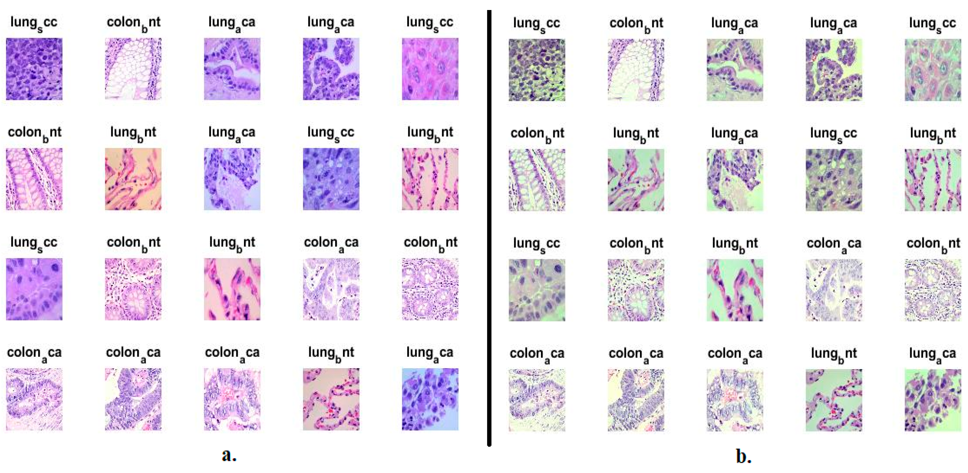
- Improving histological images in an overlapping manner between an average filter and the CLAHE method.
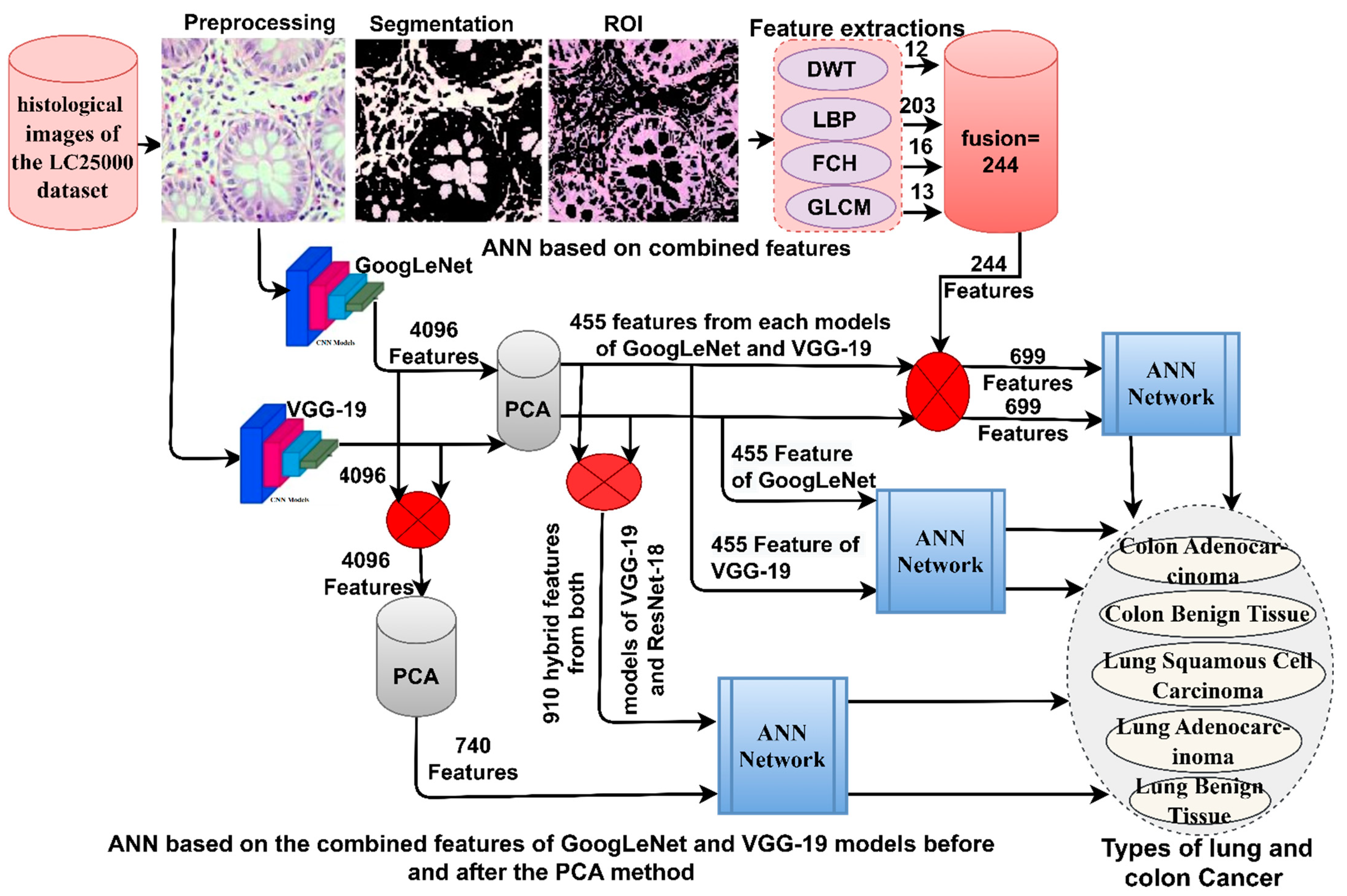
- Eliminating redundant and unnecessary features produced from GoogLeNet and VGG-19 models and save essential features by the PCA method.
- Extracting handcrafted features by integrating DWT, LBP, FCH and GLCM methods features
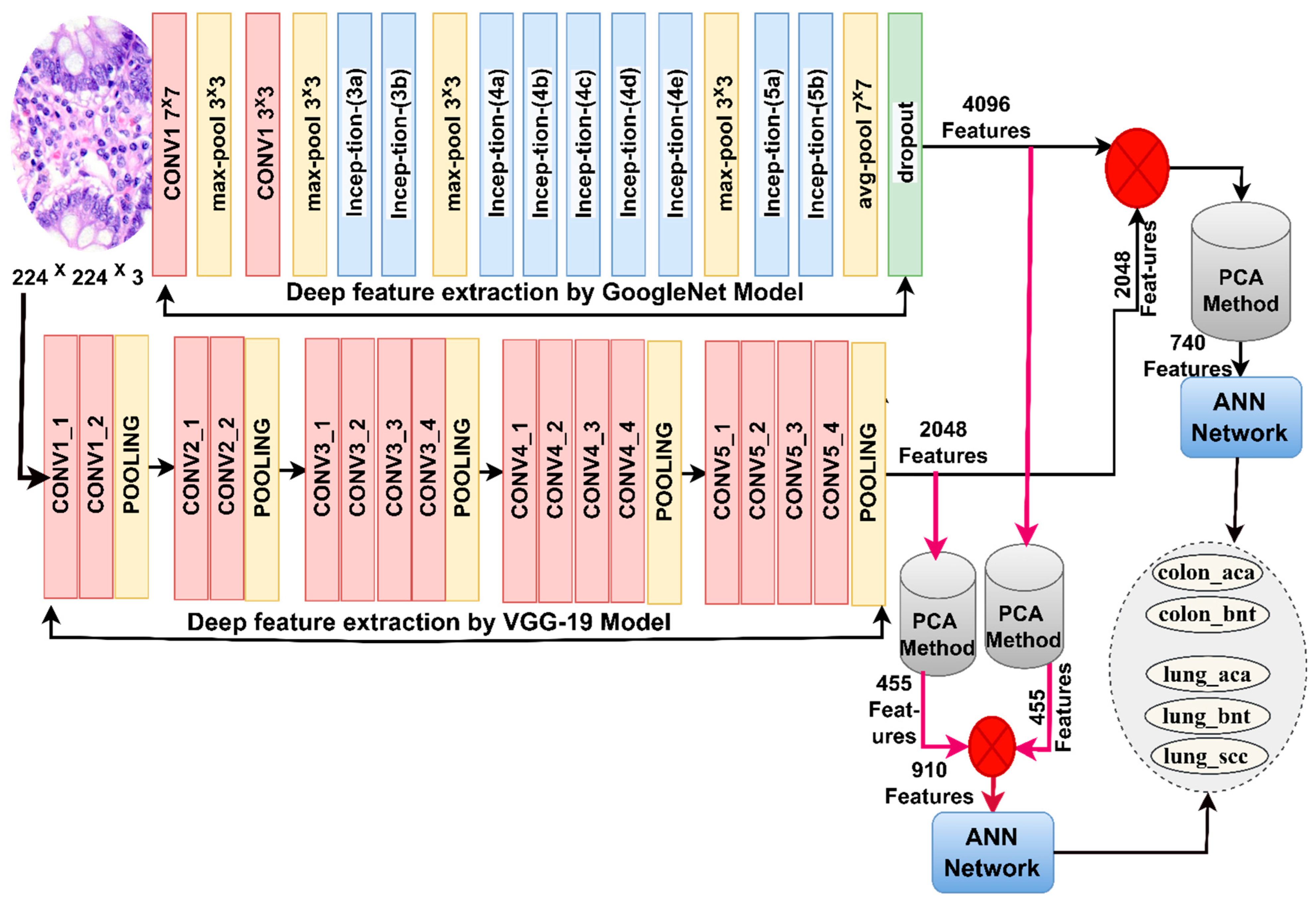
- Combining the features of the GoogLeNet and VGG-19 models after and before reducing their high dimensions.
- Generating fusion feature vectors by integrating the features of the GoogLeNet and VGG-19 models with the handcrafted features.
- Developing effective systems to help physicians and pathologists diagnose histological images and support their decisions.
2. Related Work
3. Materials and Methods
3.1. Description of the LC25000 Dataset
3.2. Improving Histological Images for the LC25000 Dataset
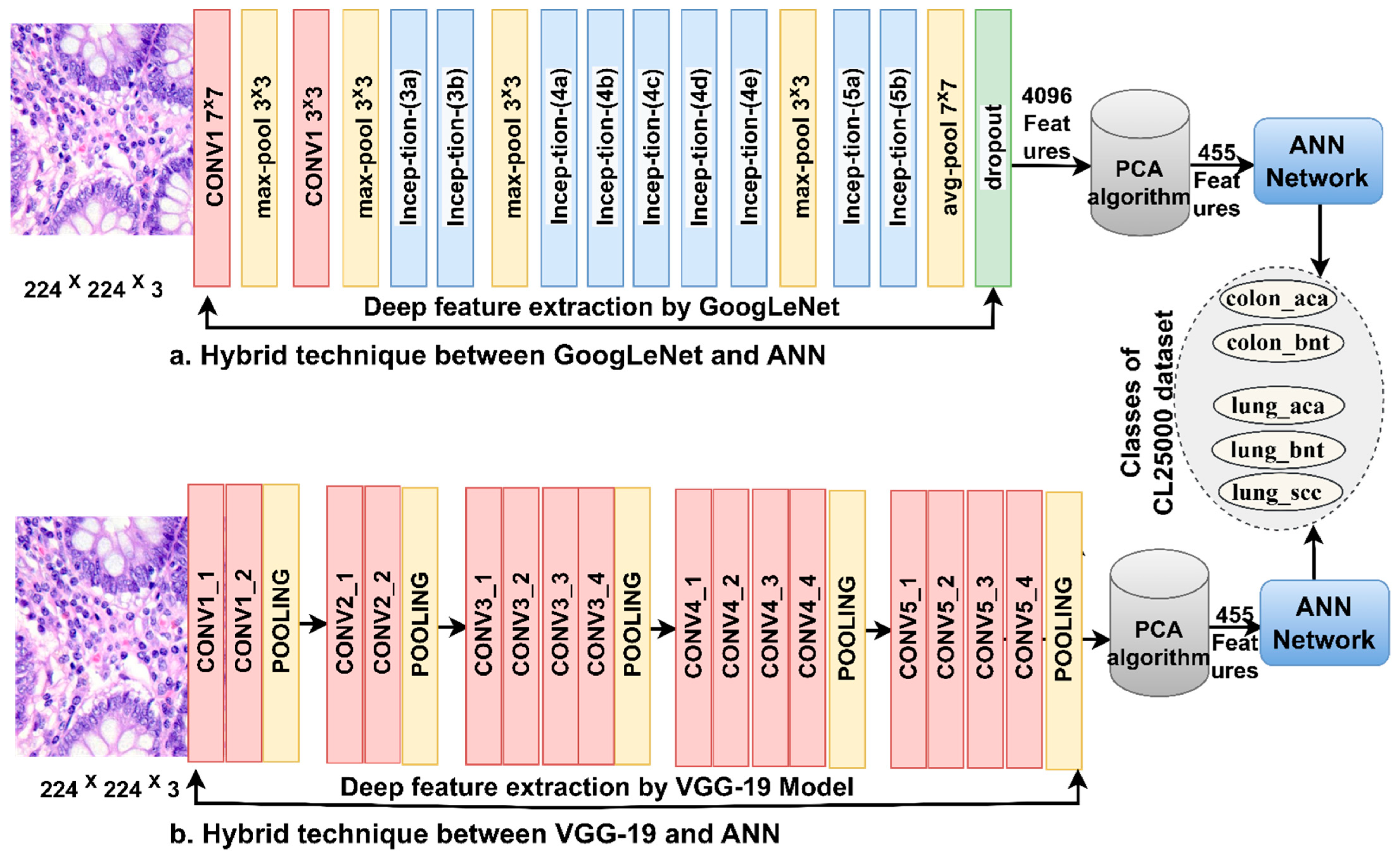
3.3. ANN Network with CNN Features
3.3.1. CNN Features
3.3.2. The ANN Network
3.4. ANN with Fusion Features of CNN
3.5. ANN with Fusion Features of CNN and Handcrafted
4. The Results of the System Execution
4.1. Split of LC25000 Dataset
4.2. Evaluation Metrics
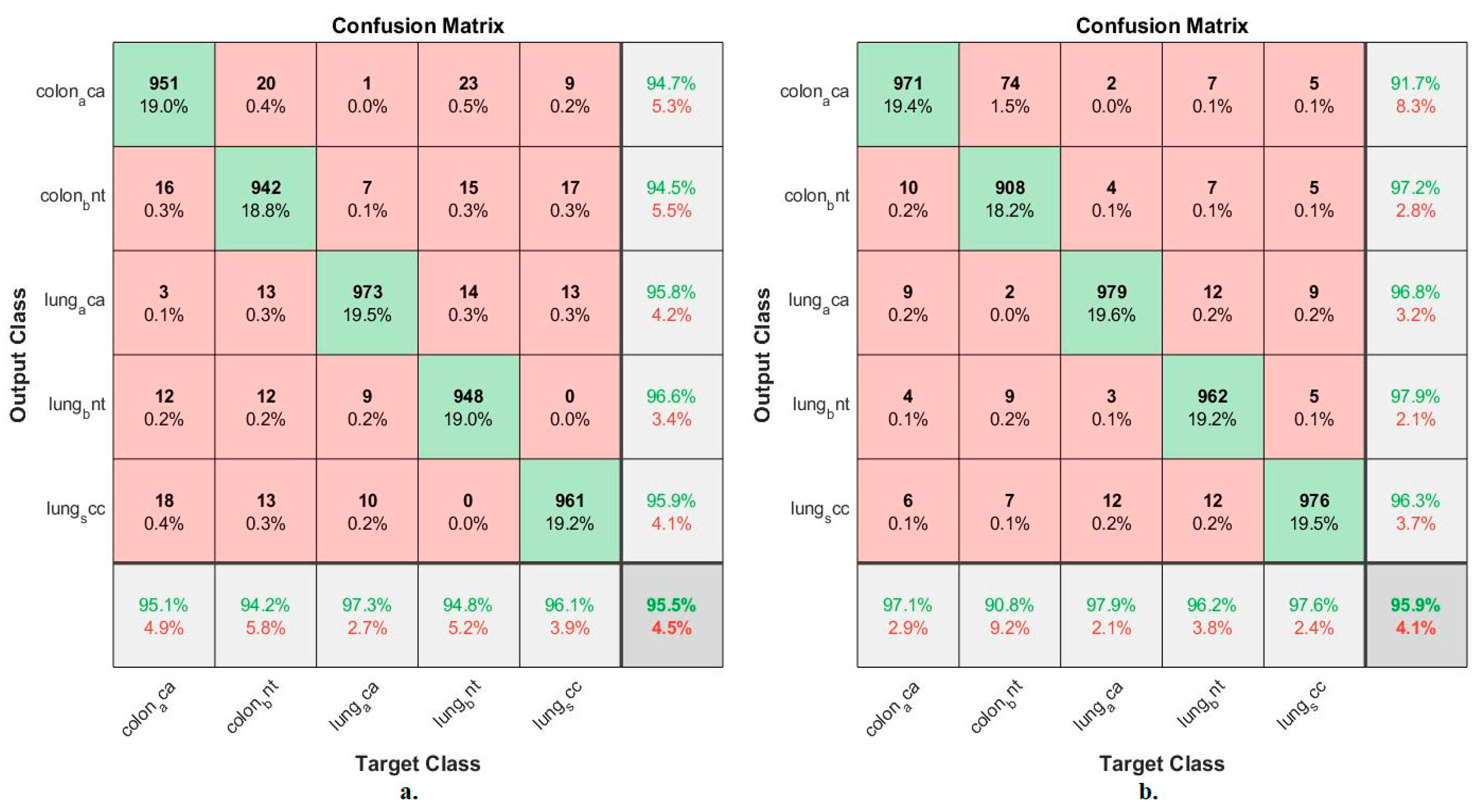
4.3. Results of ANN with CNN Features
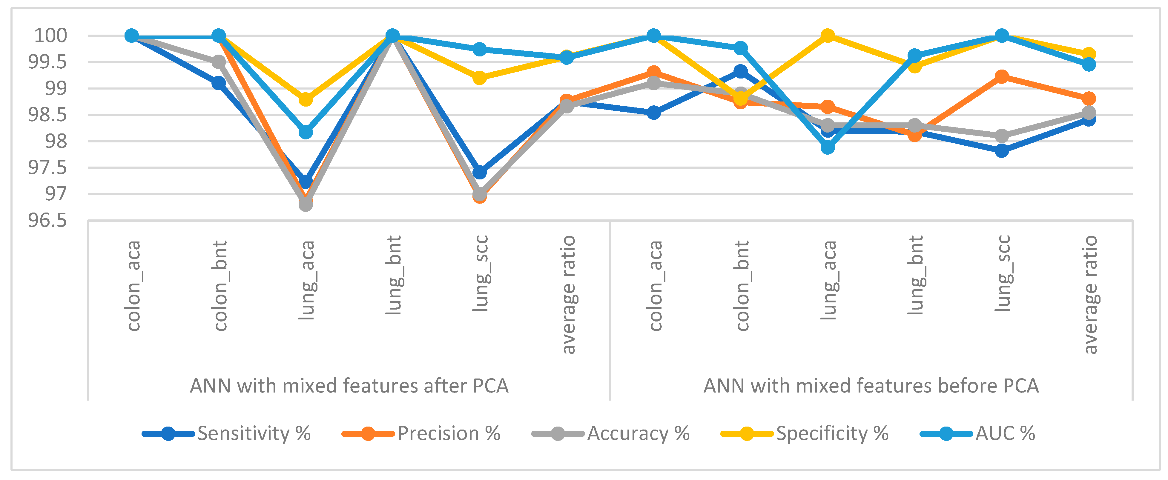
4.4. Results of ANN with Fusion Features of CNN
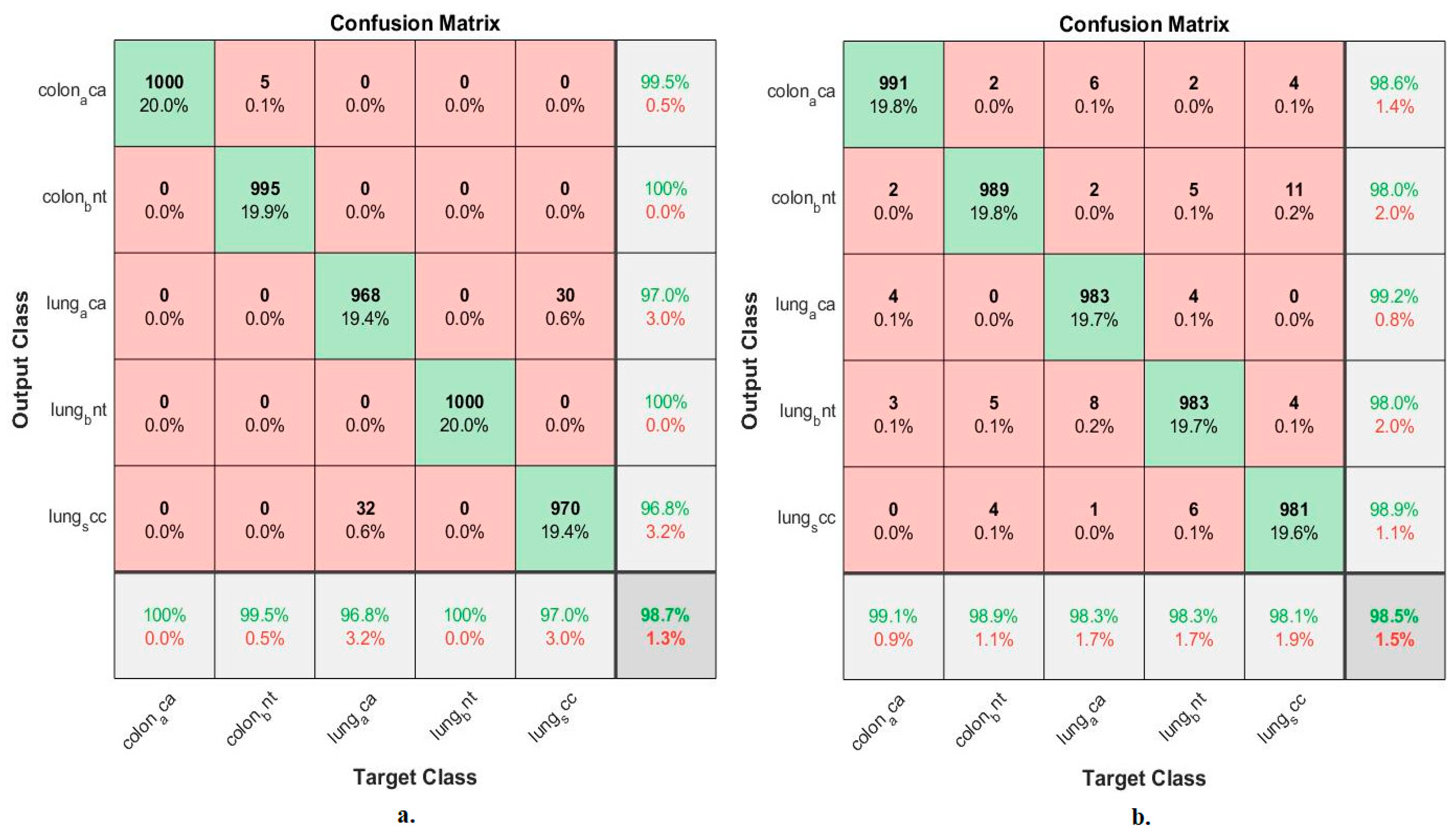
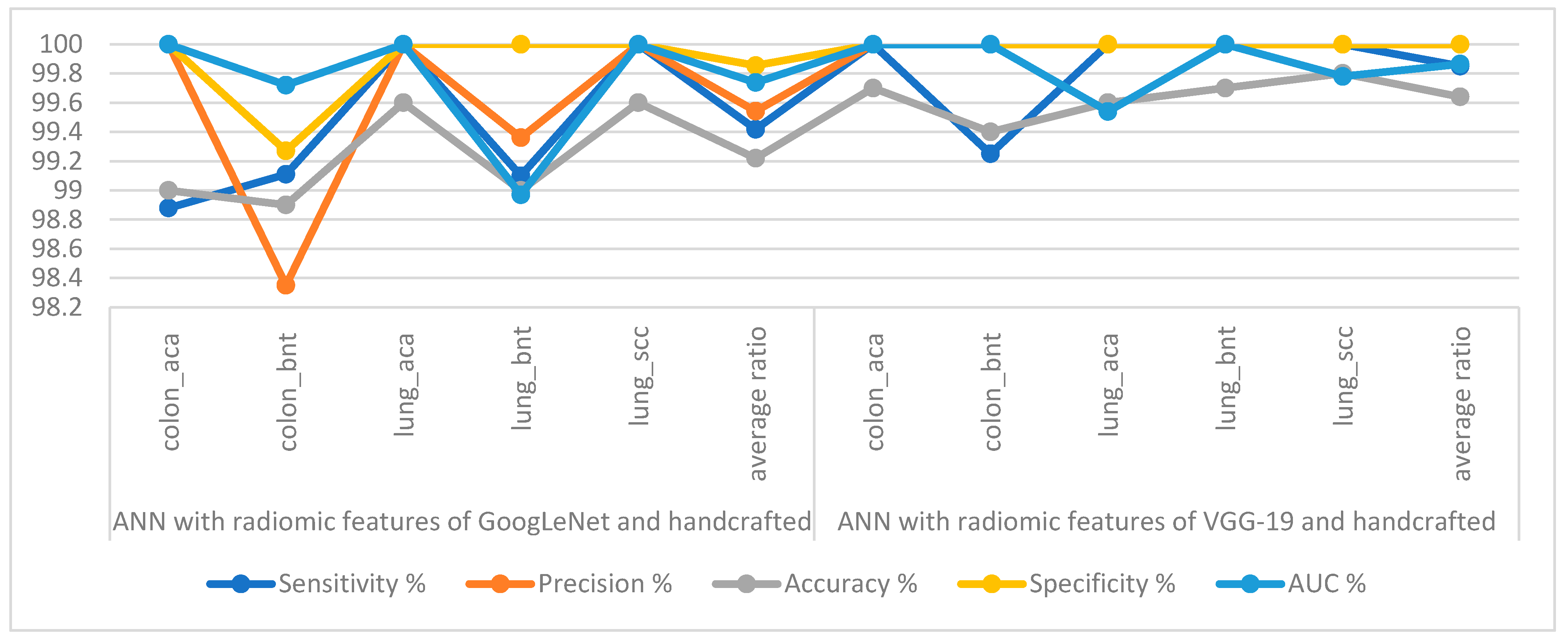
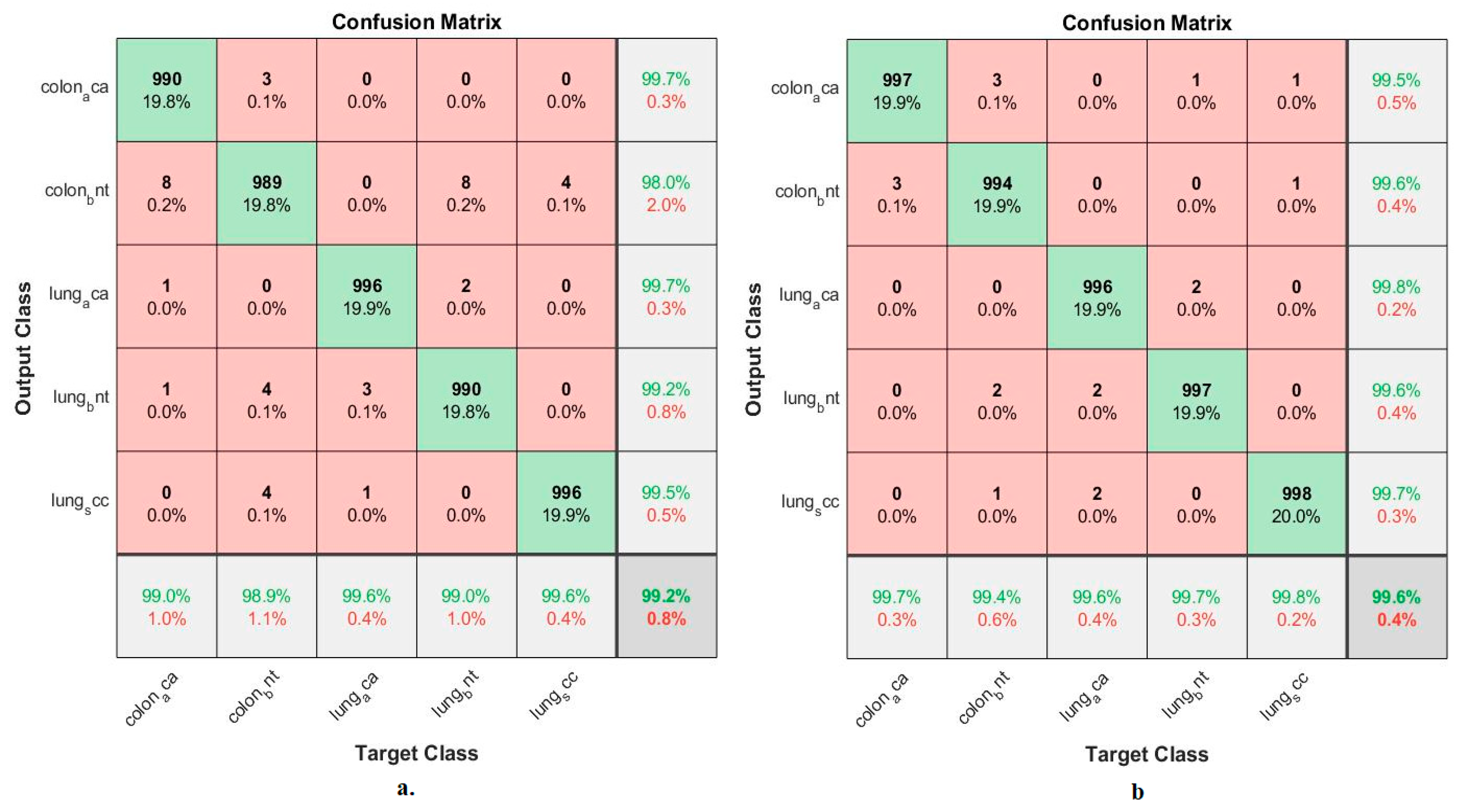
4.5. Results of ANN with Fusion Features of CNN and Handcrafted
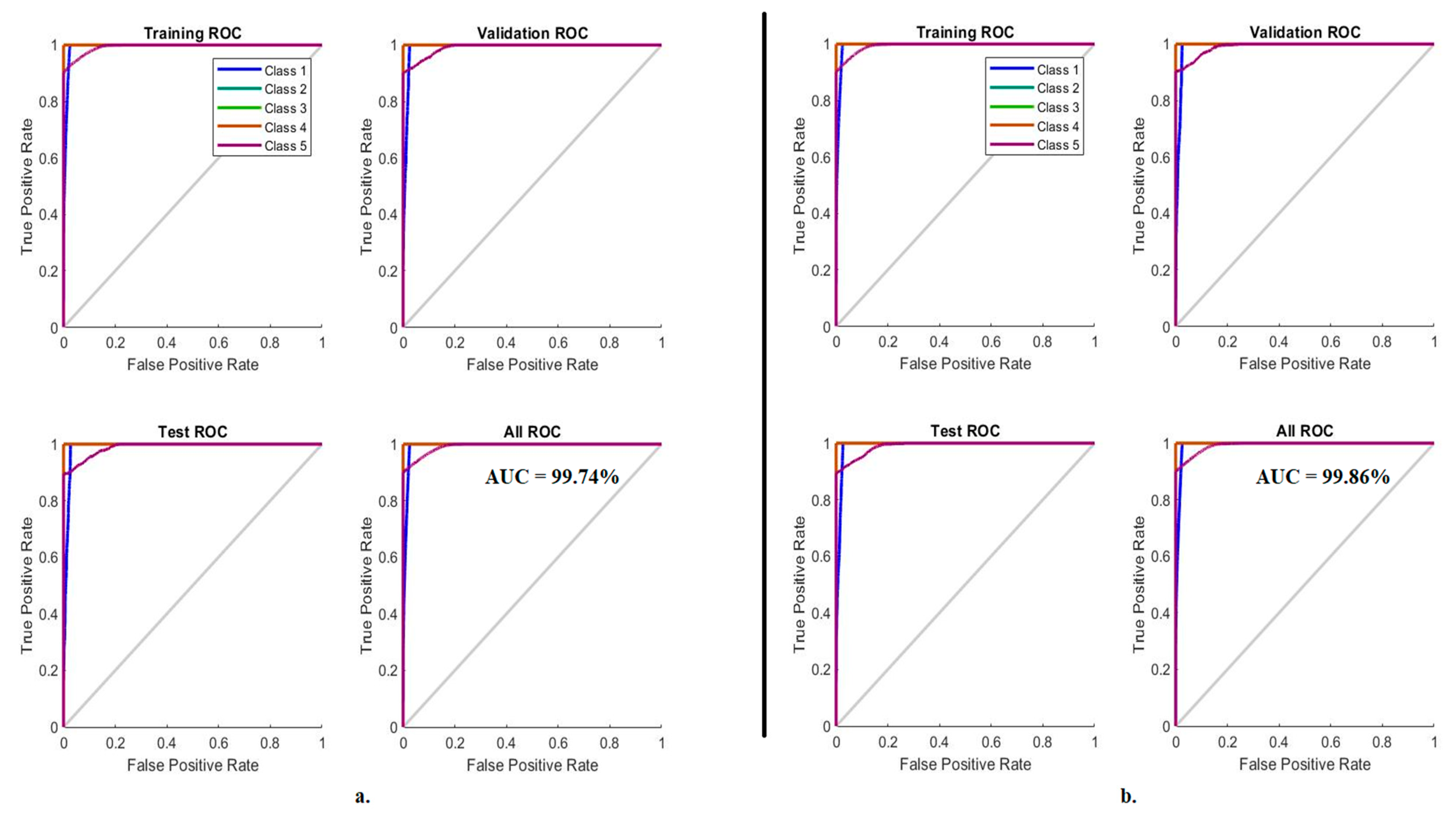
4.5.1. Receiver Operating Characteristic (ROC)
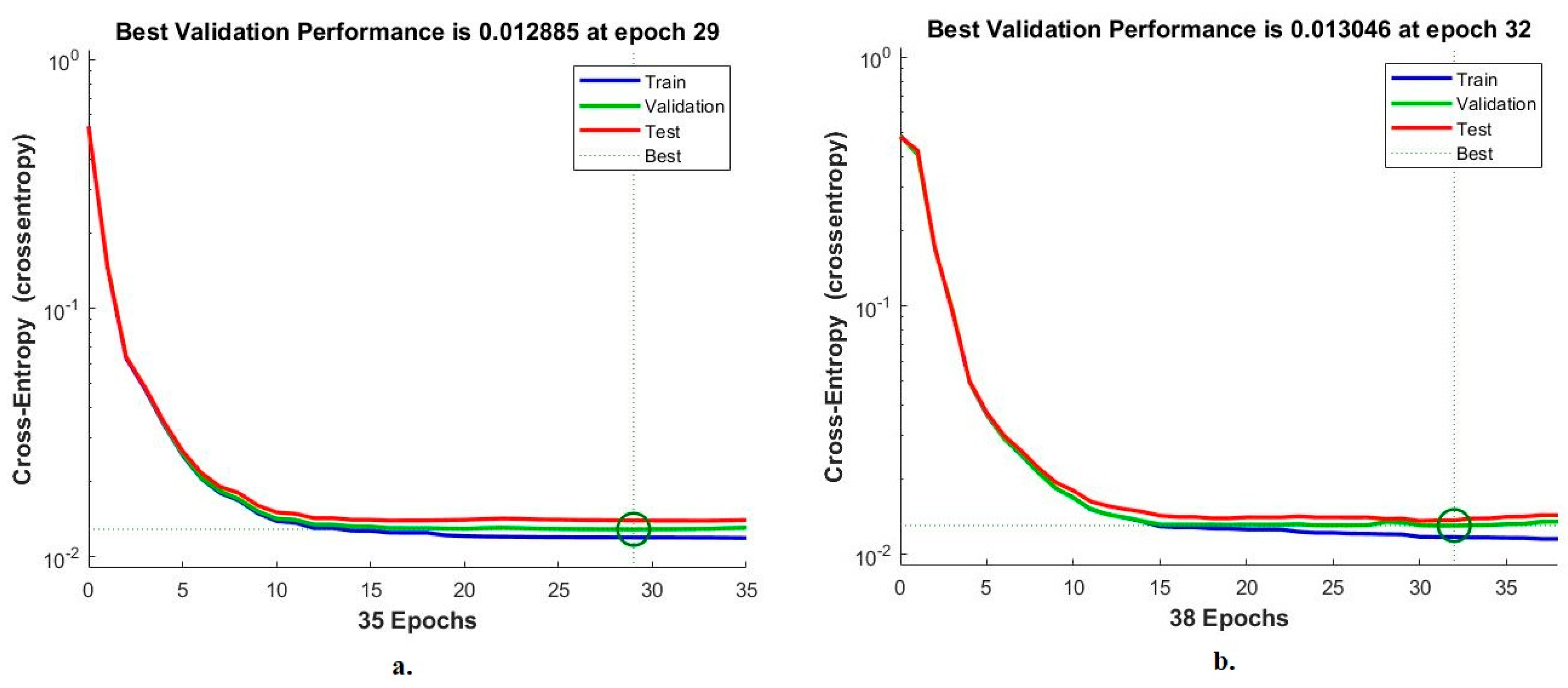
4.5.2. Cross-Entropy
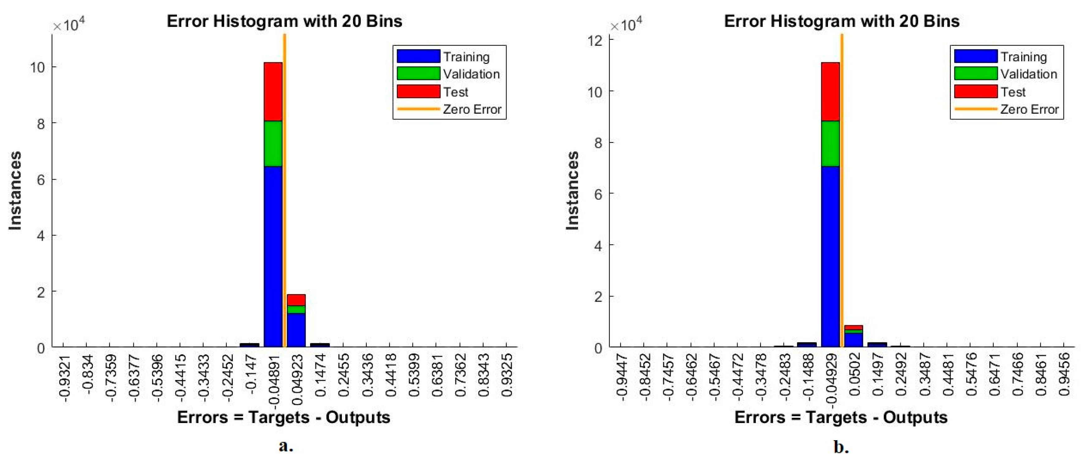
4.5.3. Error Histogram
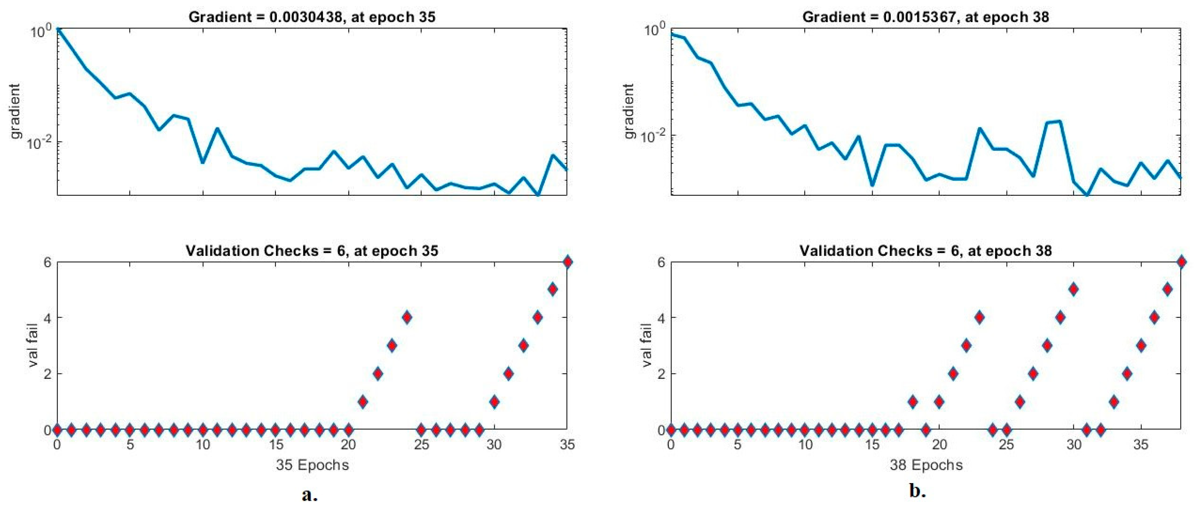
4.5.4. Validation Checks and Gradient
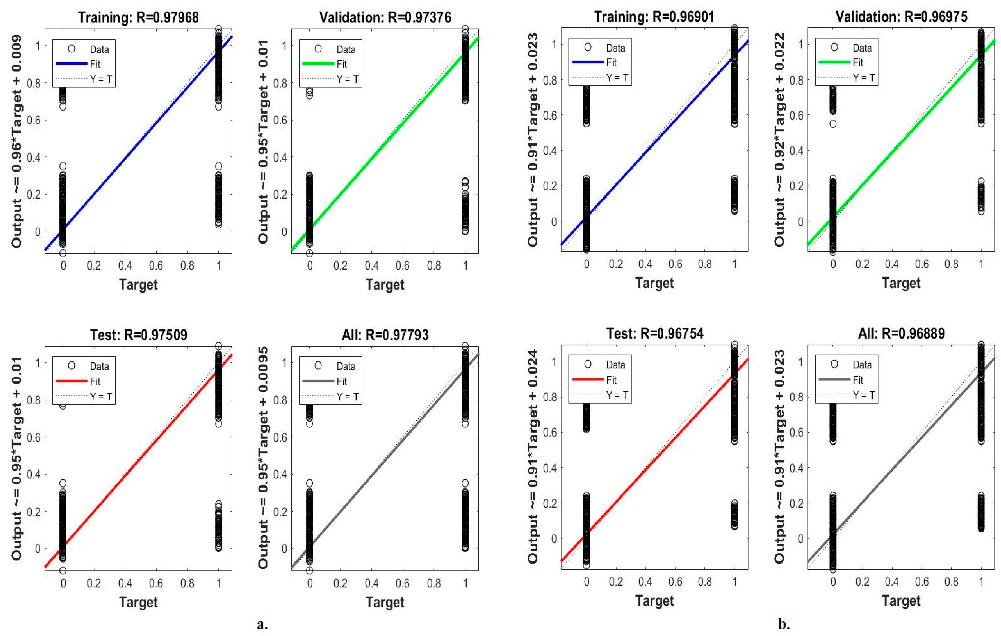
4.5.5. Regression
5. Discussion of the Performance of the Systems
6. Conclusions
Author Contributions
Funding
Institutional Review Board Statement
Informed Consent Statement
Data Availability Statement
Acknowledgments
Conflicts of Interest
Abbreviation
| Abbreviation | Full Form |
| LC25000 | Lung-Colon-25000 |
| ANN | Artificial Neural Networks |
| CNN | Convolutional Neural Network |
| CLAHE | Contrast limited adaptive histogram equalization |
| GLCM | Gray Level Co-occurrence Matrix |
| FCH | Fuzzy Color Histogram |
| LBP | Local Binary Pattern |
| DWT | Discrete Wavelet Transform |
| PCA | Principal Component Analysis |
| colon_aca | Colon Adenocarcinoma |
| colon_bnt | Colon Benign Tissue |
| lung_aca | Lung Adenocarcinoma |
| lung_bnt | Lung Benign Tissue |
| lung_scc | Lung Squamous Cell Carcinoma |
| MSE | Minimum Square Error |
References
- Cancer. Available online: https://www.who.int/news-room/fact-sheets/detail/cancer (accessed on 26 October 2022).
- How Do Cancer Cells Grow and Spread? June 2019. Available online: https://www.ncbi.nlm.nih.gov/books/NBK279410/ (accessed on 27 October 2022).
- Pettit, R.W.; Byun, J.; Han, Y.; Ostrom, Q.T.; Edelson, J.; Walsh, K.M.; Amos, C.I. The shared genetic architecture between epidemiological and behavioral traits with lung cancer. Sci. Rep. 2021, 11, 17559. [Google Scholar] [CrossRef] [PubMed]
- Kurishima, K.; Miyazaki, K.; Watanabe, H.; Shiozawa, T.; Ishikawa, H.; Satoh, H.; Hizawa, N. Lung cancer patients with synchronous colon cancer. Mol. Clin. Oncol. 2018, 8, 137–140. [Google Scholar] [CrossRef] [PubMed] [Green Version]
- Del Re, M.; Rofi, E.; Restante, G.; Crucitta, S.; Arrigoni, E.; Fogli, S.; Danesi, R. Implications of KRAS mutations in acquired resistance to treatment in NSCLC. Oncotarget 2018, 9, 6630. [Google Scholar] [CrossRef] [PubMed] [Green Version]
- Testing for Colorectal Cancer | How Is Colorectal Cancer Diagnosed? Available online: https://www.cancer.org/cancer/colon-rectal-cancer/detection-diagnosis-staging/how-diagnosed.html (accessed on 27 October 2022).
- Van der Velden, B.H.; Kuijf, H.J.; Gilhuijs, K.G.; Viergever, M.A. Explainable artificial intelligence (XAI) in deep learning-based medical image analysis. Med. Image Anal. 2022, 79, 102470. [Google Scholar] [CrossRef]
- Chakraborty, S.; Mali, K. An overview of biomedical image analysis from the deep learning perspective. In Anthology on Improving Medical Imaging Techniques for Analysis and Intervention; IGI Global: Hershey, PA, USA, 2023; pp. 43–59. [Google Scholar] [CrossRef]
- Barragán-Montero, A.; Javaid, U.; Valdés, G.; Nguyen, D.; Desbordes, P.; Macq, B.; Lee, J.A. Artificial intelligence and machine learning for medical imaging: A technology review. Phys. Med. 2021, 83, 242–256. [Google Scholar] [CrossRef]
- Adu, K.; Yu, Y.; Cai, J.; Owusu-Agyemang, K.; Twumasi, B.A.; Wang, X. DHS-CapsNet: Dual horizontal squash capsule networks for lung and colon cancer classification from whole slide histopathological images. Int. J. Imaging Syst. Technol. 2021, 31, 2075–2092. [Google Scholar] [CrossRef]
- Mangal, S.; Chaurasia, A.; Khajanchi, A. Convolution neural networks for diagnosing colon and lung cancer histopathological images. arXiv 2020, arXiv:2009.03878. [Google Scholar]
- Ali, M.; Ali, R. Multi-Input Dual-Stream Capsule Network for Improved Lung and Colon Cancer Classification. Diagnostics 2021, 11, 1485. [Google Scholar] [CrossRef]
- Mehmood, S.; Ghazal, T.M.; Khan, M.A.; Zubair, M.; Naseem, M.T.; Faiz, T.; Ahmad, M. Malignancy detection in lung and colon histopathology images using transfer learning with class selective image processing. IEEE Access 2022, 10, 25657–25668. Available online: https://ieeexplore.ieee.org/abstract/document/9709814/ (accessed on 15 October 2022). [CrossRef]
- Toğaçar, M. Disease type detection in lung and colon cancer images using the complement approach of inefficient sets. Comput. Biol. Med. 2021, 137, 104827. Available online: https://www.sciencedirect.com/science/article/pii/S0010482521006211 (accessed on 15 October 2022). [CrossRef]
- Masud, M.; Sikder, N.; Nahid, A.-A.; Bairagi, A.K.; AlZain, M.A. A Machine Learning Approach to Diagnosing Lung and Colon Cancer Using a Deep Learning-Based Classification Framework. Sensors 2021, 21, 748. [Google Scholar] [CrossRef]
- Hamida, A.B.; Devanne, M.; Weber, J.; Truntzer, C.; Derangère, V.; Ghiringhelli, F.; Wemmert, C. Deep learning for colon cancer histopathological images analysis. Comput. Biol. Med. 2021, 136, 104730. [Google Scholar] [CrossRef] [PubMed]
- Sarker, M.M.K.; Makhlouf, Y.; Craig, S.G.; Humphries, M.P.; Loughrey, M.; James, J.A.; Salto-Tellez, M.; O’Reilly, P.; Maxwell, P. A Means of Assessing Deep Learning-Based Detection of ICOS Protein Expression in Colon Cancer. Cancers 2021, 13, 3825. [Google Scholar] [CrossRef] [PubMed]
- Sarwinda, D.; Paradisa, R.H.; Bustamam, A.; Anggia, P. Deep learning in image classification using residual network (ResNet) variants for detection of colorectal cancer. Procedia Comput. Sci. 2021, 179, 423–431. [Google Scholar] [CrossRef]
- Zhou, C.; Jin, Y.; Chen, Y.; Huang, S.; Huang, R.; Wang, Y.; Liao, J. Histopathology classification and localization of colorectal cancer using global labels by weakly supervised deep learning. Comput. Med. Imaging Graph. 2021, 88, 101861. [Google Scholar] [CrossRef]
- Xu, L.; Walker, B.; Liang, P.I.; Tong, Y.; Xu, C.; Su, Y.C.; Karsan, A. Colorectal cancer detection based on deep learning. J. Pathol. Inform. 2020, 11, 28. [Google Scholar] [CrossRef]
- Moitra, D.; Mandal, R.K. Classification of non-small cell lung cancer using one-dimensional convolutional neural network. Expert Syst. Appl. 2020, 159, 113564. Available online: https://www.sciencedirect.com/science/article/pii/S0957417420303882 (accessed on 15 October 2022). [CrossRef]
- Kumar, V.; Bakariya, B. Classification of malignant lung cancer using deep learning. J. Med. Eng. Technol. 2021, 45, 85–93. [Google Scholar] [CrossRef]
- Shim, W.S.; Yim, K.; Kim, T.-J.; Sung, Y.E.; Lee, G.; Hong, J.H.; Chun, S.H.; Kim, S.; An, H.J.; Na, S.J.; et al. DeepRePath: Identifying the Prognostic Features of Early-Stage Lung Adenocarcinoma Using Multi-Scale Pathology Images and Deep Convolutional Neural Networks. Cancers 2021, 13, 3308. [Google Scholar] [CrossRef]
- Nishio, M.; Nishio, M.; Jimbo, N.; Nakane, K. Homology-Based Image Processing for Automatic Classification of Histopathological Images of Lung Tissue. Cancers 2021, 13, 1192. [Google Scholar] [CrossRef]
- Borkowski, A.A.; Bui, M.M.; Thomas, L.B.; Wilson, C.P.; DeLand, L.A.; Mastorides, S.M. Lung and colon cancer histopathological image dataset (lc25000). arXiv 2019, arXiv:1912.12142. Available online: https://www.kaggle.com/datasets/andrewmvd/lung-and-colon-cancer-histopathological-images (accessed on 15 October 2022).
- Ahmed, I.A.; Senan, E.M.; Rassem, T.H.; Ali, M.A.H.; Shatnawi, H.S.A.; Alwazer, S.M.; Alshahrani, M. Eye Tracking-Based Diagnosis and Early Detection of Autism Spectrum Disorder Using Machine Learning and Deep Learning Techniques. Electronics 2022, 11, 530. [Google Scholar] [CrossRef]
- Fati, S.M.; Senan, E.M.; ElHakim, N. Deep and Hybrid Learning Technique for Early Detection of Tuberculosis Based on X-ray Images Using Feature Fusion. Appl. Sci. 2022, 12, 7092. [Google Scholar] [CrossRef]
- Al-Mekhlafi, Z.G.; Senan, E.M.; Rassem, T.H.; Mohammed, B.A.; Makbol, N.M.; Alanazi, A.A.; Ghaleb, F.A. Deep Learning and Machine Learning for Early Detection of Stroke and Haemorrhage. Comput. Mater. Contin. 2021, 72, 775–796. Available online: http://eprints.bournemouth.ac.uk/36721/ (accessed on 15 October 2022). [CrossRef]
- Abunadi, I.; Senan, E.M. Deep Learning and Machine Learning Techniques of Diagnosis Dermoscopy Images for Early Detection of Skin Diseases. Electronics 2021, 10, 3158. [Google Scholar] [CrossRef]
- Fati, S.M.; Senan, E.M.; Azar, A.T. Hybrid and Deep Learning Approach for Early Diagnosis of Lower Gastrointestinal Diseases. Sensors 2022, 22, 4079. [Google Scholar] [CrossRef]
- Mohammed, B.A.; Senan, E.M.; Al-Mekhlafi, Z.G.; Rassem, T.H.; Makbol, N.M.; Alanazi, A.A.; Almurayziq, T.S.; Ghaleb, F.A.; Sallam, A.A. Multi-Method Diagnosis of CT Images for Rapid Detection of Intracranial Hemorrhages Based on Deep and Hybrid Learning. Electronics 2022, 11, 2460. [Google Scholar] [CrossRef]
- Samee, N.A.; Alhussan, A.A.; Ghoneim, V.F.; Atteia, G.; Alkanhel, R.; Al-antari, M.A.; Kadah, Y.M. A Hybrid Deep Transfer Learning of CNN-Based LR-PCA for Breast Lesion Diagnosis via Medical Breast Mammograms. Sensors 2022, 22, 4938. [Google Scholar] [CrossRef] [PubMed]
- Liu, Y.; Durlofsky, L.J. 3D CNN-PCA: A deep-learning-based parameterization for complex geomodels. Comput. Geosci. 2022, 148, 104676. [Google Scholar] [CrossRef]
- Senan, E.M.; Jadhav, M.E.; Rassem, T.H.; Aljaloud, A.S.; Mohammed, B.A.; Al-Mekhlafi, Z.G. Early diagnosis of brain tumour mri images using hybrid techniques between deep and machine learning. Comput. Math. Methods Med. 2022, 2022, 8330833b. Available online: https://www.hindawi.com/journals/cmmm/2022/8330833/ (accessed on 15 October 2022). [CrossRef]
- Mohammed, B.A.; Senan, E.M.; Rassem, T.H.; Makbol, N.M.; Alanazi, A.A.; Al-Mekhlafi, Z.G.; Almurayziq, T.S.; Ghaleb, F.A. Multi-Method Analysis of Medical Records and MRI Images for Early Diagnosis of Dementia and Alzheimer’s Disease Based on Deep Learning and Hybrid Methods. Electronics 2021, 10, 2860. [Google Scholar] [CrossRef]
- Senan, E.M.; Jadhav, M.E. Techniques for the Detection of Skin Lesions in PH 2 Dermoscopy Images Using Local Binary Pattern (LBP). In Proceedings of the International Conference on Recent Trends in Image Processing and Pattern Recognition, Aurangabad, India, 3–4 January 2020; Springer: Singapore, 2020; pp. 14–25. [Google Scholar] [CrossRef]
- Senan, E.M.; Jadhav, M.E.; Kadam, A. Classification of PH2 images for early detection of skin diseases. In Proceedings of the 2021 6th International Conference for Convergence in Technology (I2CT), Maharashtra, India, 2–4 April 2021; pp. 1–7. [Google Scholar] [CrossRef]
- Senan, E.M.; Jadhav, M.E. Diagnosis of dermoscopy images for the detection of skin lesions using SVM and KNN. In Proceedings of the Third International Conference on Sustainable Computing; Springer: Singapore, 2022; pp. 125–134. [Google Scholar] [CrossRef]
- Senan, E.M.; Abunadi, I.; Jadhav, M.E.; Fati, S.M. Score and Correlation Coefficient-Based Feature Selection for Predicting Heart Failure Diagnosis by Using Machine Learning Algorithms. Comput. Math. Methods Med. 2021, 2021, 8500314. [Google Scholar] [CrossRef] [PubMed]
- Al-Mekhlafi, Z.G.; Senan, E.M.; Mohammed, B.A.; Alazmi, M.; Alayba, A.M.; Alreshidi, A.; Alshahrani, M. Diagnosis of Histopathological Images to Distinguish Types of Malignant Lymphomas Using Hybrid Techniques Based on Fusion Features. Electronics 2022, 11, 2865. [Google Scholar] [CrossRef]
- Mohammed, B.A.; Senan, E.M.; Al-Mekhlafi, Z.G.; Alazmi, M.; Alayba, A.M.; Alanazi, A.A.; Alreshidi, A.; Alshahrani, M. Hybrid Techniques for Diagnosis with WSIs for Early Detection of Cervical Cancer Based on Fusion Features. Appl. Sci. 2022, 12, 8836. [Google Scholar] [CrossRef]
- Fati, S.M.; Senan, E.M.; Javed, Y. Early Diagnosis of Oral Squamous Cell Carcinoma Based on Histopathological Images Using Deep and Hybrid Learning Approaches. Diagnostics 2022, 12, 1899. [Google Scholar] [CrossRef] [PubMed]
- Dabass, M.; Dabass, J.; Vashisth, S.; Vig, R. A hybrid U-Net model with attention and advanced convolutional learning modules for simultaneous gland segmentation and cancer grade prediction in colorectal histopathological images. Intell.-Based Med. 2023, 7, 100094. [Google Scholar] [CrossRef]
- Dabass, M.; Dabass, J. An Atrous Convolved Hybrid Seg-Net Model with residual and attention mechanism for gland detection and segmentation in histopathological images. Comput. Biol. Med. 2023, 155, 106690. [Google Scholar] [CrossRef]
- Bukhari, S.U.K.; Syed, A.; Bokhari, S.K.A.; Hussain, S.S.; Armaghan, S.U.; Shah, S.S.H. The histological diagnosis of colonic adenocarcinoma by applying partial self supervised learning. medRxiv 2020. [Google Scholar] [CrossRef]
- Liu, Y.; Wang, H.; Song, K.; Sun, M.; Shao, Y.; Xue, S.; Li, L.; Li, Y.; Cai, H.; Jiao, Y.; et al. CroReLU: Cross-Crossing Space-Based Visual Activation Function for Lung Cancer Pathology Image Recognition. Cancers 2022, 14, 5181. [Google Scholar] [CrossRef]
- Attallah, O.; Aslan, M.F.; Sabanci, K. A Framework for Lung and Colon Cancer Diagnosis via Lightweight Deep Learning Models and Transformation Methods. Diagnostics 2022, 12, 2926. [Google Scholar] [CrossRef]
- El-Ghany, S.A.; Azad, M.; Elmogy, M. Robustness Fine-Tuning Deep Learning Model for Cancers Diagnosis Based on Histopathology Image Analysis. Diagnostics 2023, 13, 699. [Google Scholar] [CrossRef] [PubMed]


| Phase | (80:20) Training and Validation | Testing 20% | |
|---|---|---|---|
| Classes | Training (80%) | Validation (20%) | |
| colon_aca | 3800 | 800 | 1000 |
| colon_bnt | 3800 | 800 | 1000 |
| lung_aca | 3800 | 800 | 1000 |
| lung_bnt | 3800 | 800 | 1000 |
| lung_scc | 3800 | 800 | 1000 |
| Techniques | Type of Class | Sensitivity % | Precision % | Accuracy % | Specificity % | AUC % |
|---|---|---|---|---|---|---|
| ANN with features of GoogLeNet | colon_aca | 97.2 | 95.35 | 95.1 | 99.41 | 98.74 |
| colon_bnt | 95.84 | 96.89 | 94.2 | 98.98 | 97.82 | |
| lung_aca | 96.19 | 95.36 | 97.3 | 98.52 | 98.1 | |
| lung_bnt | 95.77 | 97.15 | 94.8 | 99.29 | 96.15 | |
| lung_scc | 97.43 | 96.22 | 96.1 | 99.38 | 97.56 | |
| average ratio | 96.49 | 96.19 | 95.50 | 99.12 | 97.67 | |
| ANN with features of VGG-19 | colon_aca | 96.54 | 92.25 | 97.1 | 98.15 | 97.4 |
| colon_bnt | 91.33 | 96.95 | 90.8 | 98.64 | 98.23 | |
| lung_aca | 97.82 | 97.17 | 97.9 | 98.53 | 97.88 | |
| lung_bnt | 95.79 | 98.36 | 96.2 | 99.1 | 98.9 | |
| lung_scc | 97.94 | 96.19 | 97.6 | 97.98 | 97.67 | |
| average ratio | 95.88 | 96.18 | 95.92 | 98.48 | 98.02 |
| Techniques | Type of Class | Sensitivity % | Precision % | Accuracy % | Specificity % | AUC % |
|---|---|---|---|---|---|---|
| ANN with mixed features after PCA | colon_aca | 100 | 100 | 100 | 100 | 100 |
| colon_bnt | 99.1 | 100 | 99.5 | 100 | 100 | |
| lung_aca | 97.23 | 96.87 | 96.8 | 98.79 | 98.17 | |
| lung_bnt | 100 | 100 | 100 | 100 | 100 | |
| lung_scc | 97.41 | 96.95 | 97 | 99.2 | 99.74 | |
| average ratio | 98.75 | 98.76 | 98.66 | 99.60 | 99.58 | |
| ANN with mixed features before PCA | colon_aca | 98.54 | 99.3 | 99.1 | 100 | 100 |
| colon_bnt | 99.32 | 98.74 | 98.9 | 98.81 | 99.76 | |
| lung_aca | 98.2 | 98.65 | 98.3 | 100 | 97.88 | |
| lung_bnt | 98.18 | 98.12 | 98.3 | 99.42 | 99.62 | |
| lung_scc | 97.82 | 99.22 | 98.1 | 100 | 100 | |
| average ratio | 98.41 | 98.81 | 98.54 | 99.65 | 99.45 |
| Techniques | Type of Class | Sensitivity % | Precision % | Accuracy % | Specificity % | AUC % |
|---|---|---|---|---|---|---|
| ANN with fusion features of GoogLeNet and handcrafted | colon_aca | 98.88 | 100 | 99 | 100 | 100 |
| colon_bnt | 99.11 | 98.35 | 98.9 | 99.27 | 99.72 | |
| lung_aca | 100 | 100 | 99.6 | 100 | 100 | |
| lung_bnt | 99.1 | 99.36 | 99 | 100 | 98.97 | |
| lung_scc | 100 | 100 | 99.6 | 100 | 100 | |
| average ratio | 99.42 | 99.54 | 99.22 | 99.85 | 99.74 | |
| ANN with fusion features of VGG-19 and handcrafted | colon_aca | 100 | 100 | 99.7 | 100 | 100 |
| colon_bnt | 99.25 | 100 | 99.4 | 100 | 100 | |
| lung_aca | 99.7 | 100 | 99.6 | 99.6 | 99.54 | |
| lung_bnt | 99.6 | 100 | 99.7 | 99.9 | 100 | |
| lung_scc | 99.6 | 100 | 99.8 | 100 | 99.78 | |
| average ratio | 99.85 | 100.00 | 99.64 | 100.00 | 99.86 |
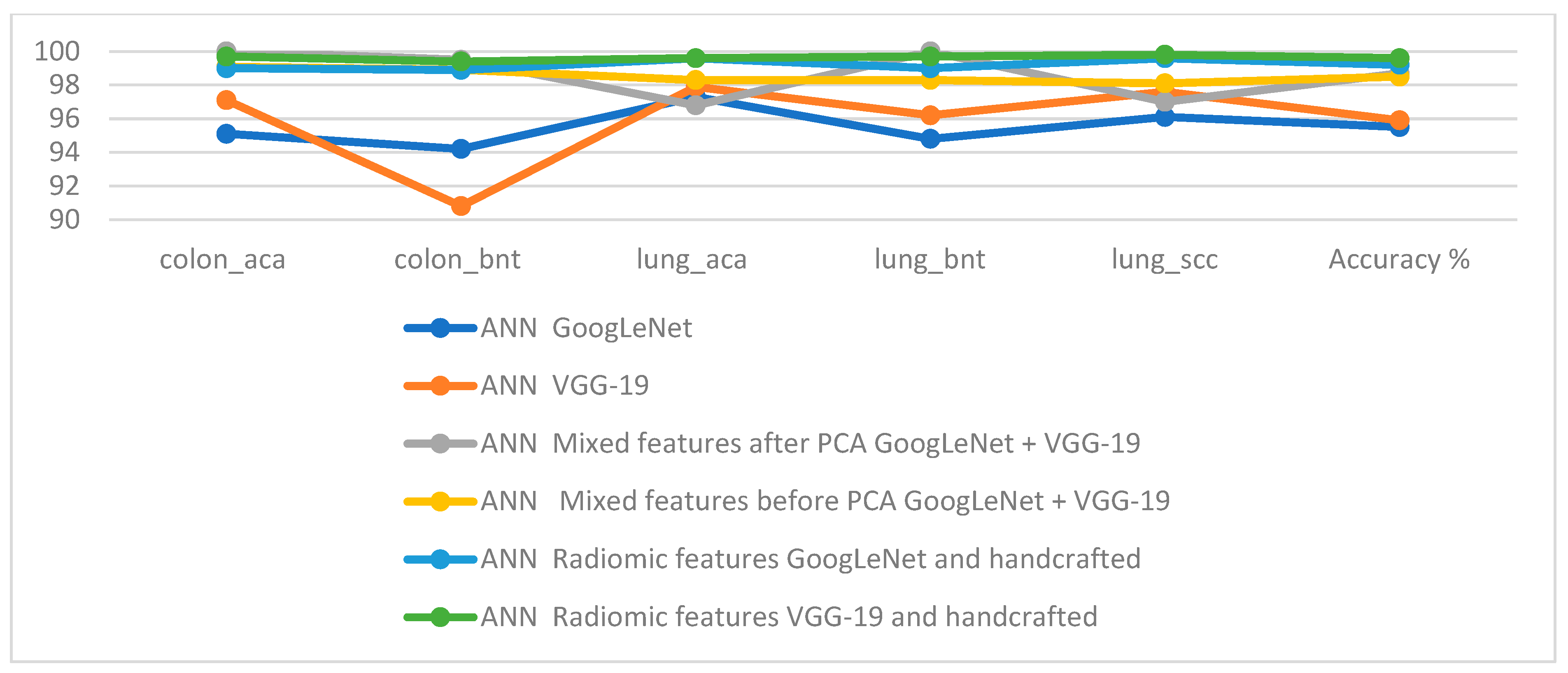
| Techniques | Features | Colon_aca | Colon_bnt | Lung_aca | Lung_bnt | Lung_scc | Accuracy % | |
|---|---|---|---|---|---|---|---|---|
| ANN | GoogLeNet | 95.1 | 94.2 | 97.3 | 94.8 | 96.1 | 95.5 | |
| VGG-19 | 97.1 | 90.8 | 97.9 | 96.2 | 97.6 | 95.9 | ||
| ANN | Mixed features after PCA | GoogLeNet + VGG-19 | 100 | 99.5 | 96.8 | 100 | 97 | 98.7 |
| Mixed features before PCA | GoogLeNet + VGG-19 | 99.1 | 98.9 | 98.3 | 98.3 | 98.1 | 98.5 | |
| Fusion features | GoogLeNet and handcrafted | 99 | 98.9 | 99.6 | 99 | 99.6 | 99.2 | |
| VGG-19 and handcrafted | 99.7 | 99.4 | 99.6 | 99.7 | 99.8 | 99.6 | ||
| Previous Studies | Accuracy % | Sensitivity % | Specificity % | AUC % | Precision % |
|---|---|---|---|---|---|
| Dabass, M. et al. [43] | 97.4 | 97.2 | 97.1 | - | 97.21 |
| Dabass, M. et al. [44] | 95.85 | - | - | - | |
| Bukhari, S. et al. [45] | 93.04 | 94.79 | 84.21 | - | 96.81 |
| Masud et al. [15] | 96.33 | 96.37 | - | - | 96.39 |
| Liu, Y. et al. [46] | 97.01 | 97.04 | - | - | 97.07 |
| Attallah, O. et al. [47] | 99.3 | 98.9 | 99.7 | - | 99 |
| El-Ghany, S. et al. [48] | 98.97 | 97.82 | 99.35 | - | 98.04 |
| Proposed model | 99.64 | 99.85 | 100 | 99.86 | 100 |
Disclaimer/Publisher’s Note: The statements, opinions and data contained in all publications are solely those of the individual author(s) and contributor(s) and not of MDPI and/or the editor(s). MDPI and/or the editor(s) disclaim responsibility for any injury to people or property resulting from any ideas, methods, instructions or products referred to in the content. |
© 2023 by the authors. Licensee MDPI, Basel, Switzerland. This article is an open access article distributed under the terms and conditions of the Creative Commons Attribution (CC BY) license (https://creativecommons.org/licenses/by/4.0/).
Share and Cite
Al-Jabbar, M.; Alshahrani, M.; Senan, E.M.; Ahmed, I.A. Histopathological Analysis for Detecting Lung and Colon Cancer Malignancies Using Hybrid Systems with Fused Features. Bioengineering 2023, 10, 383. https://doi.org/10.3390/bioengineering10030383
Al-Jabbar M, Alshahrani M, Senan EM, Ahmed IA. Histopathological Analysis for Detecting Lung and Colon Cancer Malignancies Using Hybrid Systems with Fused Features. Bioengineering. 2023; 10(3):383. https://doi.org/10.3390/bioengineering10030383
Chicago/Turabian StyleAl-Jabbar, Mohammed, Mohammed Alshahrani, Ebrahim Mohammed Senan, and Ibrahim Abdulrab Ahmed. 2023. "Histopathological Analysis for Detecting Lung and Colon Cancer Malignancies Using Hybrid Systems with Fused Features" Bioengineering 10, no. 3: 383. https://doi.org/10.3390/bioengineering10030383