
Bond Strength and Adhesion Mechanisms of Novel Bone Adhesives

Abstract
:1. Introduction
2. Materials and Methods
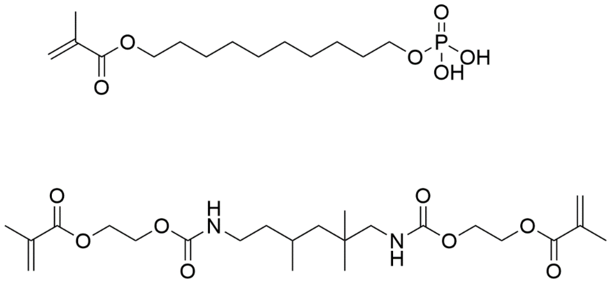
2.1. Preparations of Adhesive Formulations and Primers
2.2. Preparation of Bone Samples and Polymer Discs
2.3. Preparation of Polymer Adhered Bone Samples and Shear Tests
2.4. Shear Bond Strength Tests
2.5. Transmission Electron Microscopy (TEM)
2.6. X-ray Diffraction (XRD)
2.7. Cytotoxicity Testing
2.7.1. Indirect Cytotoxicity Assay
2.7.2. Direct Cytotoxicity Assays
3. Results
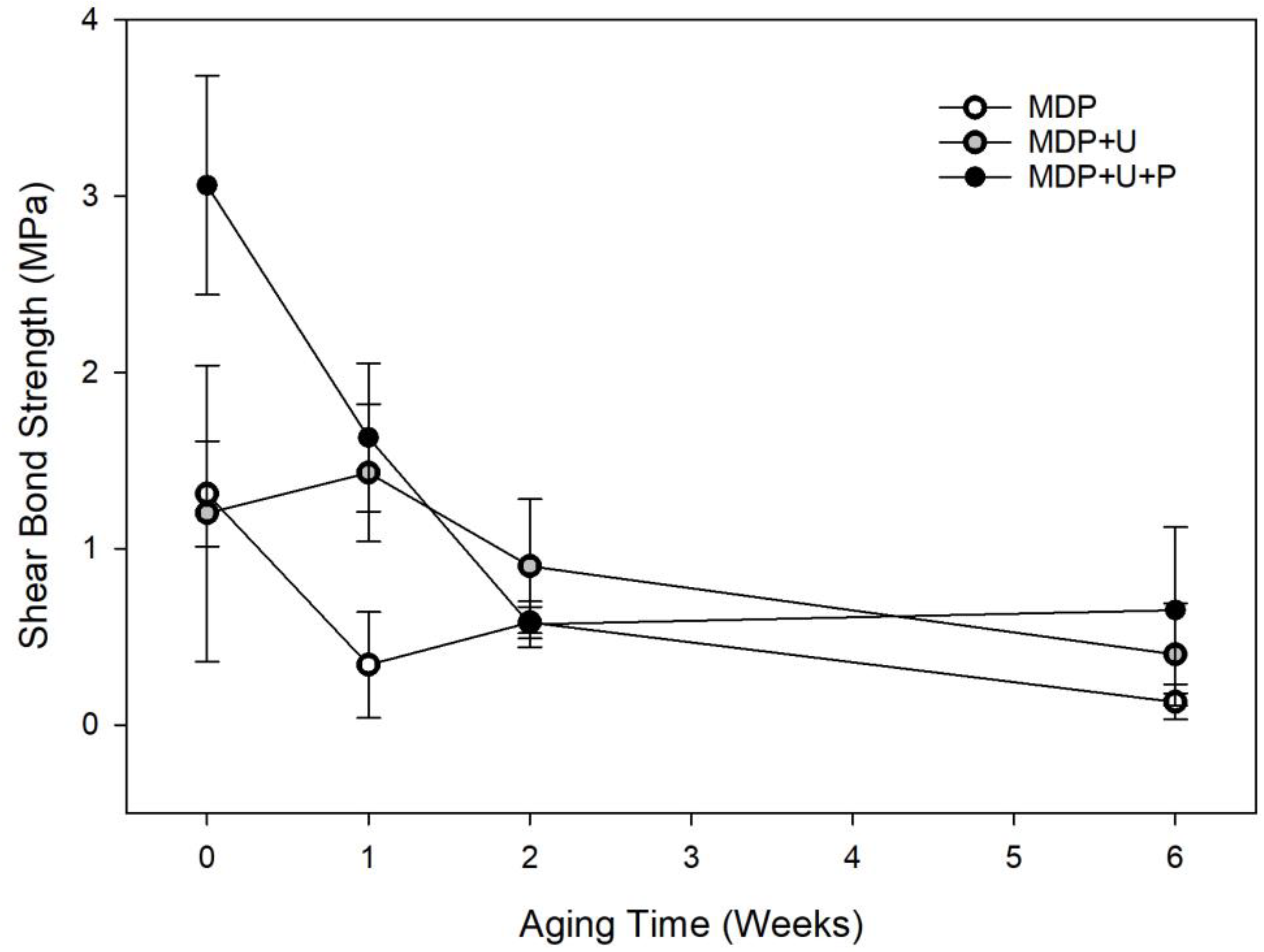
3.1. In Vitro Artificial Aging
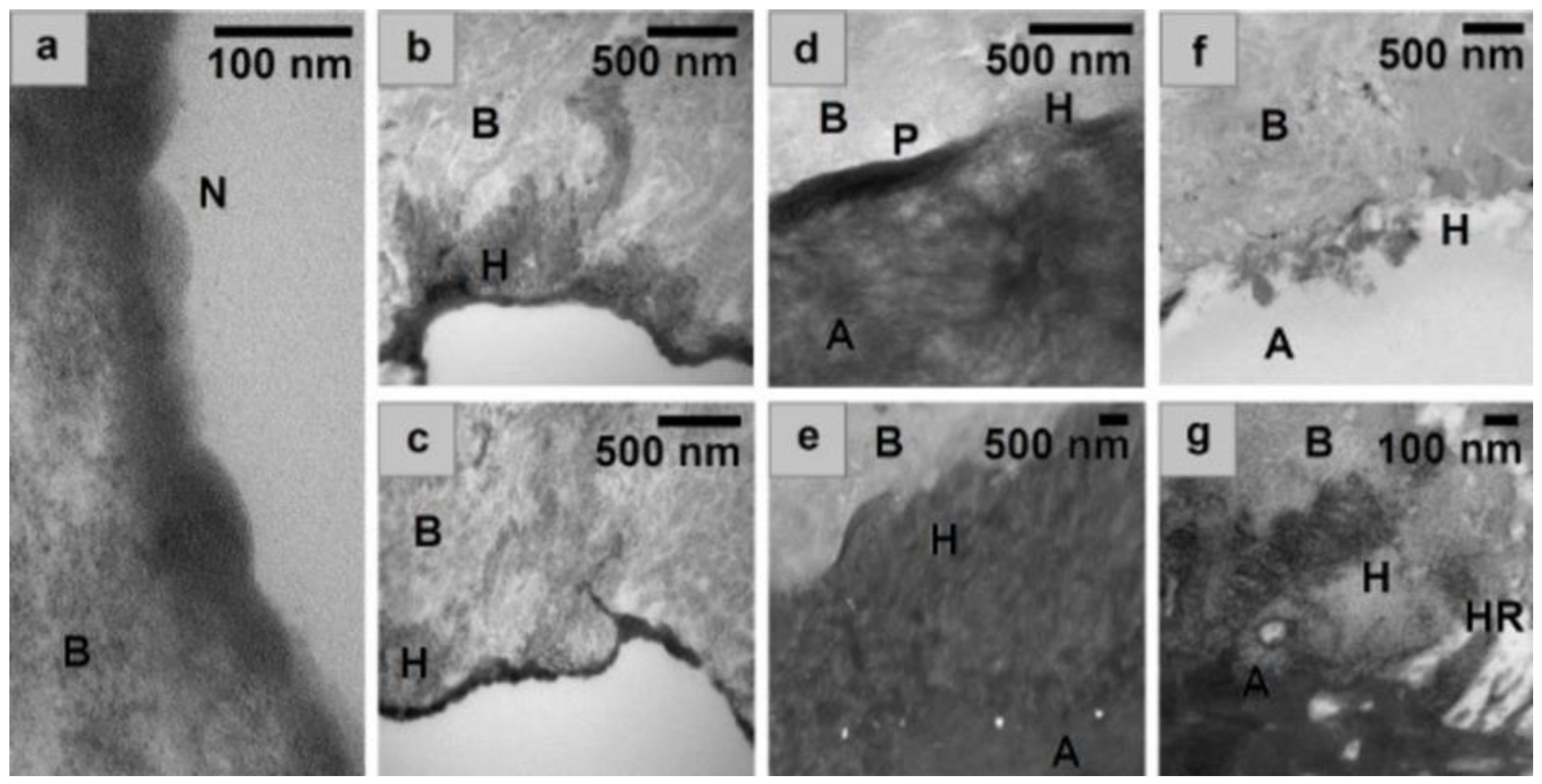
3.2. TEM
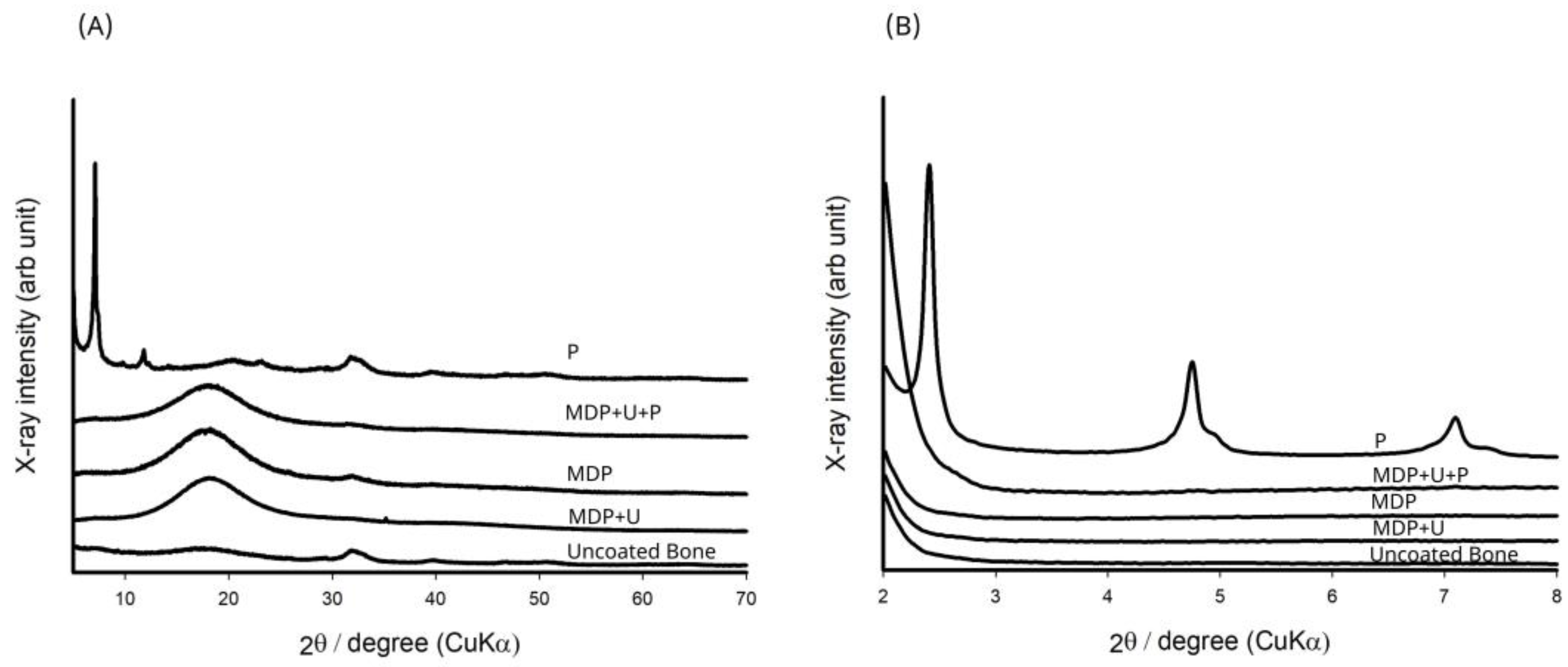
3.3. XRD
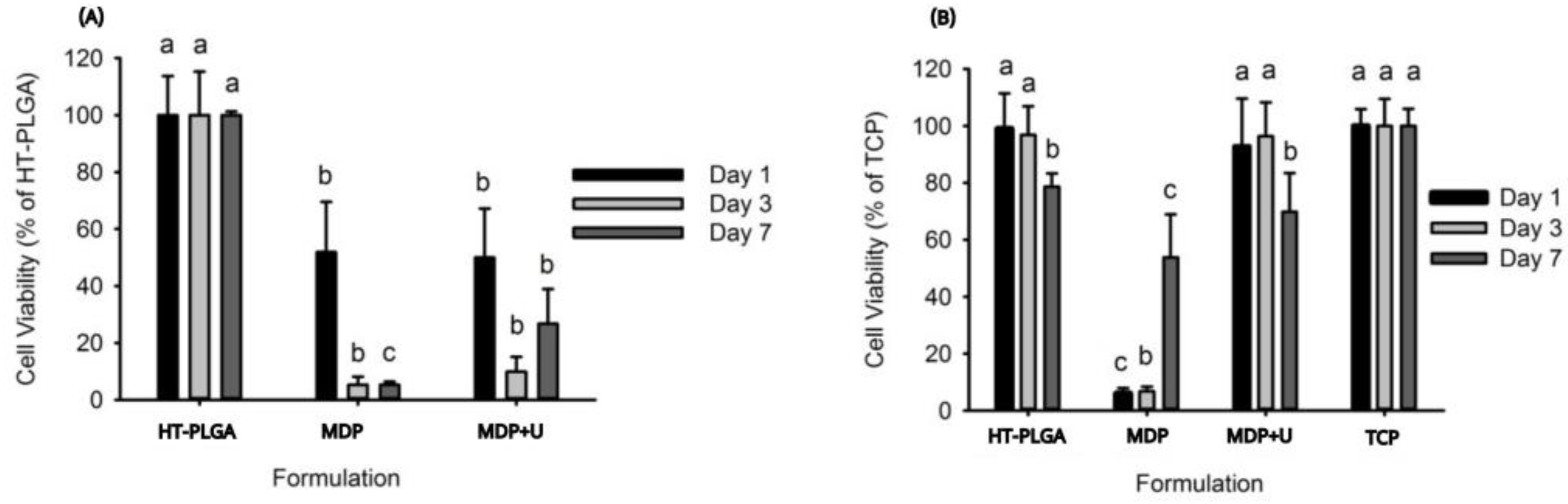
3.4. Cytotoxicity Assays
4. Discussion
5. Conclusions
Author Contributions
Funding
Institutional Review Board Statement
Informed Consent Statement
Data Availability Statement
Acknowledgments
Conflicts of Interest
References
- George, J.; Brahmabhatt, P.; Farboud, A.; Marnane, C. A Retrospective Review of Facial Fractures in Wales. Reports 2018, 1, 24. [Google Scholar] [CrossRef] [Green Version]
- Davies, R.; Hammond, D.; Ridout, F.; Hutchison, I.; Magennis, P. British Association of Oral and Maxillofacial Surgeons’ National Facial Injury Surveys: Hard tissue facial injuries presenting to UK emergency departments. Br. J. Oral Maxillofac. Surg. 2020, 58, 152–157. [Google Scholar] [CrossRef] [PubMed]
- Lalloo, R.; Lucchesi, L.R.; Bisignano, C.; Castle, C.D.; Dingels, Z.V.; Fox, J.T.; Hamilton, E.B.; Liu, Z.; Roberts, N.L.S.; Sylte, D.O.; et al. Epidemiology of facial fractures: Incidence, prevalence and years lived with disability estimates from the Global Burden of Disease 2017 study. Inj. Prev. 2020, 26, i27–i35. [Google Scholar] [CrossRef] [PubMed] [Green Version]
- Abukhder, M.; Mobarak, D. A retrospective cohort study on the aetiology and characteristics of maxillofacial fractures presenting to a tertiary centre in the UK. Ann. Med. Surg. 2022, 77, 103622. [Google Scholar] [CrossRef]
- West, G.H.; Griggs, J.A.; Chandran, R.; Precheur, H.V.; Buchanan, W.; Caloss, R. Treatment outcomes with the use of maxillomandibular fixation screws in the management of mandible fractures. J. Oral Maxillofac. Surg. 2014, 72, 112–120. [Google Scholar] [CrossRef] [PubMed]
- Hashemi, H.M.; Parhiz, A. Complications using intermaxillary fixation screws. J. Oral Maxillofac. Surg. 2011, 69, 1411–1414. [Google Scholar] [CrossRef]
- Smeets, R.; Endres, K.; Stockbrink, G.; Hanken, H.; Hermanns-Sachweh, B.; Marx, R.; Heiland, M.; Blessmann, M.; Wolff, K.D.; Kolk, A. The innovative application of a novel bone adhesive for facial fracture osteosynthesis-in vitro and in vivo results. J. Biomed. Mater. Res. A 2013, 101, 2058–2066. [Google Scholar] [CrossRef]
- Tzagiollari, A.; McCarthy, H.O.; Levingstone, T.J.; Dunne, N.J. Biodegradable and Biocompatible Adhesives for the Effective Stabilisation, Repair and Regeneration of Bone. Bioengineering 2022, 9, 250. [Google Scholar] [CrossRef]
- Hoffmann, B.; Volkmer, E.; Kokott, A.; Augat, P.; Ohnmacht, M.; Sedlmayr, N.; Schieker, M.; Claes, L.; Mutschler, W.; Ziegler, G. Characterisation of a new bioadhesive system based on polysaccharides with the potential to be used as bone glue. J. Mater. Sci. Mater. Med. 2009, 20, 2001–2009. [Google Scholar] [CrossRef]
- Farrar, D.F. Bone adhesives for trauma surgery: A review of challenges and developments. Int. J. Adhes. Adhes. 2012, 33, 89–97. [Google Scholar] [CrossRef]
- Kandalam, U.; Bouvier, A.J.; Casas, S.B.; Smith, R.L.; Gallego, A.M.; Rothrock, J.K.; Thompson, J.Y.; Huang, C.Y.; Stelnicki, E.J. Novel bone adhesives: A comparison of bond strengths in vitro. Int. J. Oral Maxillofac. Surg. 2013, 42, 1054–1059. [Google Scholar] [CrossRef] [PubMed]
- Nanci, A. Ten Cate’s Oral Histology: Development, Structure, and Function; Mosby Elsevier: Maryland Heights, MO, USA, 2008. [Google Scholar]
- Pashley, D.H.; Tay, F.R.; Breschi, L.; Tjaderhane, L.; Carvalho, R.M.; Carrilho, M.; Tezvergil-Mutluay, A. State of the art etch-and-rinse adhesives. Dent. Mater. 2011, 27, 1–16. [Google Scholar] [CrossRef] [PubMed] [Green Version]
- Baidya, P.; Meechan, J.G.; McCabe, J.F. Bonding of composite to bone: Durability and effect of drying time. Br. J. Oral Maxillofac. Surg. 1996, 34, 406–408. [Google Scholar] [CrossRef] [PubMed]
- Heiss, C.; Schettler, N.; Wenisch, S.; Cords, S.; Schilke, F.; Lips, K.S.; Alt, V.; Schnettler, R. Bond strength of an alkylene bis(dilactoyl)-methacrylate bone adhesive: A biomechanical evaluation in sheep. J. Biomater. Sci. Polym. Ed. 2010, 21, 1345–1358. [Google Scholar] [CrossRef] [PubMed]
- Maurer, P.; Bekes, K.; Gernhardt, C.R.; Schaller, H.G.; Schubert, J. Tensile bond strength of different adhesive systems between bone and composite compared: An in vitro study. J. Craniomaxillofac. Surg. 2004, 32, 85–89. [Google Scholar] [CrossRef]
- Maurer, P.; Bekes, K.; Gernhardt, C.R.; Schaller, H.G.; Schubert, J. Comparison of the bond strength of selected adhesive dental systems to cortical bone under in vitro conditions. Int. J. Oral Maxillofac. Surg. 2004, 33, 377–381. [Google Scholar] [CrossRef]
- Ortiz Ruiz, A.J.; Vicente, A.; Camacho Alonso, F.; Lopez Jornet, P. A new use for self-etching resin adhesives: Cementing bone fragments. J. Dent. 2010, 38, 750–756. [Google Scholar] [CrossRef]
- Upson, S.J.; Partridge, S.W.; Tcacencu, I.; Fulton, D.A.; Corbett, I.; German, M.J.; Dalgarno, K.W. Development of a methacrylate-terminated PLGA copolymer for potential use in craniomaxillofacial fracture plates. Mater. Sci. Eng. C Mater. Biol. Appl. 2016, 69, 470–477. [Google Scholar] [CrossRef] [Green Version]
- Asmussen, E.; Peutzfeldt, A. Influence of selected components on crosslink density in polymer structures. Eur. J. Oral Sci. 2001, 109, 282–285. [Google Scholar] [CrossRef]
- Moszner, N.; Hirt, T. New polymer-chemical developments in clinical dental polymer materials: Enamel-dentin adhesives and restorative composites. J. Polym. Sci. Polym. Chem. 2012, 50, 4369–4402. [Google Scholar] [CrossRef]
- Ferracane, J.L. Hygroscopic and hydrolytic effects in dental polymer networks. Dent. Mater. 2006, 22, 211–222. [Google Scholar] [CrossRef]
- Van Landuyt, K.L.; Yoshida, Y.; Hirata, I.; Snauwaert, J.; De Munck, J.; Okazaki, M.; Suzuki, K.; Lambrechts, P.; Van Meerbeek, B. Influence of the Chemical Structure of Functional Monomers on Their Adhesive Performance. J. Dent. Res. 2008, 87, 757–761. [Google Scholar] [CrossRef] [PubMed]
- Yoshihara, K.; Nagaoka, N.; Yoshida, Y.; Van Meerbeek, B.; Hayakawa, S. Atomic level observation and structural analysis of phosphoric-acid ester interaction at dentin. Acta Biomater. 2019, 97, 544–556. [Google Scholar] [CrossRef]
- Yoshihara, K.; Nagaoka, N.; Okihara, T.; Kuroboshi, M.; Hayakawa, S.; Maruo, Y.; Nishigawa, G.; De Munck, J.; Yoshida, Y.; Van Meerbeek, B. Functional monomer impurity affects adhesive performance. Dent. Mater. 2015, 31, 1493–1501. [Google Scholar] [CrossRef] [PubMed]
- Yaguchi, T. Layering mechanism of MDP-Ca salt produced in demineralization of enamel and dentin apatite. Dent. Mater. 2017, 33, 23–32. [Google Scholar] [CrossRef] [PubMed]
- Van Meerbeek, B.; Yoshihara, K.; Van Landuyt, K.; Yoshida, Y.; Peumans, M. From Buonocore’s Pioneering Acid-Etch Technique to Self-Adhering Restoratives. A Status Perspective of Rapidly Advancing Dental Adhesive Technology. J. Adhes. Dent. 2020, 22, 7–34. [Google Scholar] [CrossRef] [PubMed]
- Delgado, A.H.S.; Owji, N.; Ashley, P.; Young, A.M. Varying 10-methacryloyloxydecyl dihydrogen phosphate (10-MDP) level improves polymerisation kinetics and flexural strength in self-adhesive, remineralising composites. Dent. Mater. 2021, 37, 1366–1376. [Google Scholar] [CrossRef] [PubMed]
- Yoshida, Y.; Nagakane, K.; Fukuda, R.; Nakayama, Y.; Okazaki, M.; Shintani, H.; Inoue, S.; Tagawa, Y.; Suzuki, K.; De Munck, J.; et al. Comparative study on adhesive performance of functional monomers. J. Dent. Res. 2004, 83, 454–458. [Google Scholar] [CrossRef]
- Hiraishi, N.; Tochio, N.; Kigawa, T.; Otsuki, M.; Tagami, J. Monomer-collagen interactions studied by saturation transfer difference NMR. J. Dent. Res. 2013, 92, 284–288. [Google Scholar] [CrossRef]
- Wang, R.; Shi, Y.; Li, T.; Pan, Y.; Cui, Y.; Xia, W. Adhesive interfacial characteristics and the related bonding performance of four self-etching adhesives with different functional monomers applied to dentin. J. Dent. 2017, 62, 72–80. [Google Scholar] [CrossRef]
- Zhou, J.; Wurihan; Shibata, Y.; Tanaka, R.; Zhang, Z.; Zheng, K.; Li, Q.; Ikeda, S.; Gao, P.; Miyazaki, T. Quantitative/qualitative analysis of adhesive-dentin interface in the presence of 10-methacryloyloxydecyl dihydrogen phosphate. J. Mech. Behav. Biomed. Mater. 2019, 92, 71–78. [Google Scholar] [CrossRef]
- Hidari, T.; Takamizawa, T.; Imai, A.; Hirokane, E.; Ishii, R.; Tsujimoto, A.; Suzuki, T.; Miyazaki, M. Role of the functional monomer 10-methacryloyloxydecyl dihydrogen phosphate in dentin bond durability of universal adhesives in etch-&-rinse mode. Dent. Mater. J. 2020, 39, 616–623. [Google Scholar] [CrossRef] [PubMed] [Green Version]
- Weber, S.C.; Chapman, M.W. Adhesives in orthopaedic surgery. A review of the literature and in vitro bonding strengths of bone-bonding agents. Clin. Orthop. Relat. Res. 1984, 191, 249–261. [Google Scholar] [CrossRef]
- Yoshihara, K.; Yoshida, Y.; Nagaoka, N.; Fukegawa, D.; Hayakawa, S.; Mine, A.; Nakamura, M.; Minagi, S.; Osaka, A.; Suzuki, K.; et al. Nano-controlled molecular interaction at adhesive interfaces for hard tissue reconstruction. Acta Biomater. 2010, 6, 3573–3582. [Google Scholar] [CrossRef] [PubMed] [Green Version]
- Fukegawa, D.; Hayakawa, S.; Yoshida, Y.; Suzuki, K.; Osaka, A.; Van Meerbeek, B. Chemical interaction of phosphoric acid ester with hydroxyapatite. J. Dent. Res. 2006, 85, 941–944. [Google Scholar] [CrossRef] [PubMed]
- Tsuchiya, K.; Takamizawa, T.; Barkmeier, W.W.; Tsubota, K.; Tsujimoto, A.; Berry, T.P.; Erickson, R.L.; Latta, M.A.; Miyazaki, M. Effect of a functional monomer (MDP) on the enamel bond durability of single-step self-etch adhesives. Eur. J. Oral Sci. 2016, 124, 96–102. [Google Scholar] [CrossRef] [PubMed]
- Cevik, P.; Yildirim, A.Z.; Artvin, Z.; Özcan, M. Microtensile Bond Strength and Failure Type Analysis of Self-Etch Adhesive Systems on Superficial and Deep Dentin after Long-term Water Storage. Braz. Dent. Sci. 2020, 23, 1–12. [Google Scholar] [CrossRef]
- Carrilho, E.; Cardoso, M.; Marques Ferreira, M.; Marto, C.M.; Paula, A.; Coelho, A.S. 10-MDP Based Dental Adhesives: Adhesive Interface Characterization and Adhesive Stability—A Systematic Review. Materials 2019, 12, 790. [Google Scholar] [CrossRef] [Green Version]
- Silva e Souza Junior, M.H.; Carneiro, K.G.K.; Lobato, M.F.; Silva e Souza, P.d.A.R.; Góes, M.F.d. Adhesive systems: Important aspects related to their composition and clinical use. J. Appl. Oral Sci. 2010, 18, 207–214. [Google Scholar] [CrossRef]
- Zhao, Q.; Gao, Y.; Jin, X.; Han, F.; Chen, K.; Chen, C. Influence of Acidic Environment on Hydrolytic Stability of MDP-Ca Salts with Nanolayered and Amorphous Structures. Int. J. Nanomed. 2022, 17, 1695–1709. [Google Scholar] [CrossRef]
- Inoue, S.; Koshiro, K.; Yoshida, Y.; De Munck, J.; Nagakane, K.; Suzuki, K.; Sano, H.; Van Meerbeek, B. Hydrolytic stability of self-etch adhesives bonded to dentin. J. Dent. Res. 2005, 84, 1160–1164. [Google Scholar] [CrossRef] [PubMed]
- Tian, F.C.; Wang, X.Y.; Huang, Q.; Niu, L.N.; Mitchell, J.; Zhang, Z.Y.; Prananik, C.; Zhang, L.; Chen, J.H.; Breschi, L.; et al. Effect of nanolayering of calcium salts of phosphoric acid ester monomers on the durability of resin-dentin bonds. Acta Biomater. 2016, 38, 190–200. [Google Scholar] [CrossRef] [PubMed]
- Van Meerbeek, B.; Yoshihara, K.; Yoshida, Y.; Mine, A.; De Munck, J.; Van Landuyt, K.L. State of the art of self-etch adhesives. Dent. Mater. 2011, 27, 17–28. [Google Scholar] [CrossRef] [PubMed]
- Koulaouzidou, E.A.; Helvatjoglu-Antoniades, M.; Palaghias, G.; Karanika-Kouma, A.; Antoniades, D. Cytotoxicity evaluation of an antibacterial dentin adhesive system on established cell lines. J. Biomed. Mater. Res. B Appl. Biomater. 2008, 84, 271–276. [Google Scholar] [CrossRef]
- Kumler, W.D.; Eiler, J.J. The Acid Strength of Mono and Diesters of Phosphoric Acid. The n-Alkyl Esters from Methyl to Butyl, the Esters of Biological Importance, and the Natural Guanidine Phosphoric Acids. J. Am. Chem. Soc. 1943, 65, 2355–2361. [Google Scholar] [CrossRef]
- Demirci, M.; Hiller, K.A.; Bosl, C.; Galler, K.; Schmalz, G.; Schweikl, H. The induction of oxidative stress, cytotoxicity, and genotoxicity by dental adhesives. Dent. Mater. 2008, 24, 362–371. [Google Scholar] [CrossRef]


Disclaimer/Publisher’s Note: The statements, opinions and data contained in all publications are solely those of the individual author(s) and contributor(s) and not of MDPI and/or the editor(s). MDPI and/or the editor(s) disclaim responsibility for any injury to people or property resulting from any ideas, methods, instructions or products referred to in the content. |
© 2023 by the authors. Licensee MDPI, Basel, Switzerland. This article is an open access article distributed under the terms and conditions of the Creative Commons Attribution (CC BY) license (https://creativecommons.org/licenses/by/4.0/).
Share and Cite
Upson, S.J.; Benning, M.J.; Fulton, D.A.; Corbett, I.P.; Dalgarno, K.W.; German, M.J. Bond Strength and Adhesion Mechanisms of Novel Bone Adhesives. Bioengineering 2023, 10, 78. https://doi.org/10.3390/bioengineering10010078
Upson SJ, Benning MJ, Fulton DA, Corbett IP, Dalgarno KW, German MJ. Bond Strength and Adhesion Mechanisms of Novel Bone Adhesives. Bioengineering. 2023; 10(1):78. https://doi.org/10.3390/bioengineering10010078
Chicago/Turabian StyleUpson, Sarah J., Matthew J. Benning, David A. Fulton, Ian P. Corbett, Kenneth W. Dalgarno, and Matthew J. German. 2023. "Bond Strength and Adhesion Mechanisms of Novel Bone Adhesives" Bioengineering 10, no. 1: 78. https://doi.org/10.3390/bioengineering10010078