
Functionalized Carbon Nanoparticles as Theranostic Agents and Their Future Clinical Utility in Oncology

Abstract
:1. Introduction
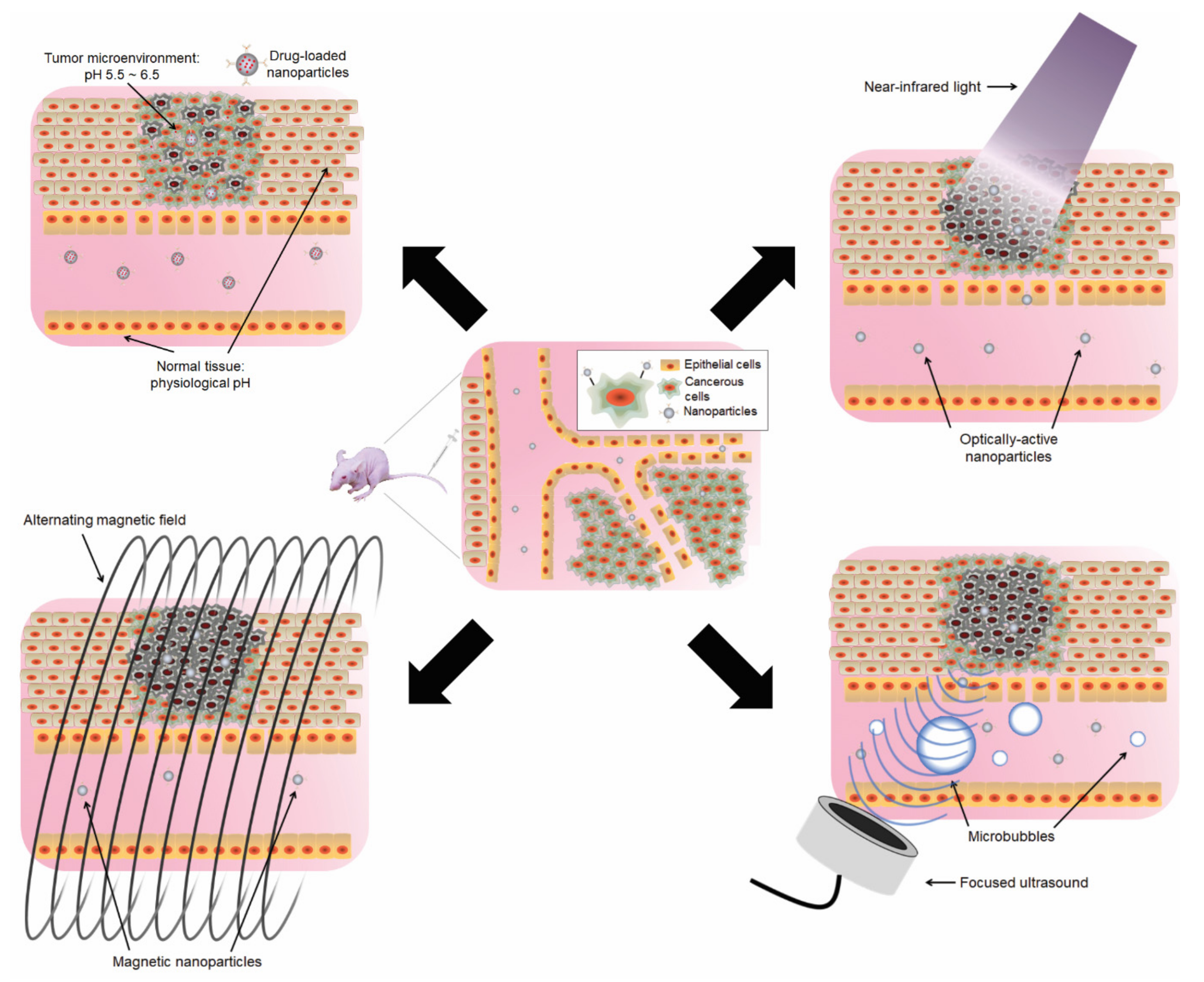
2. General Overview—Nanoparticles and Theranostic Applications
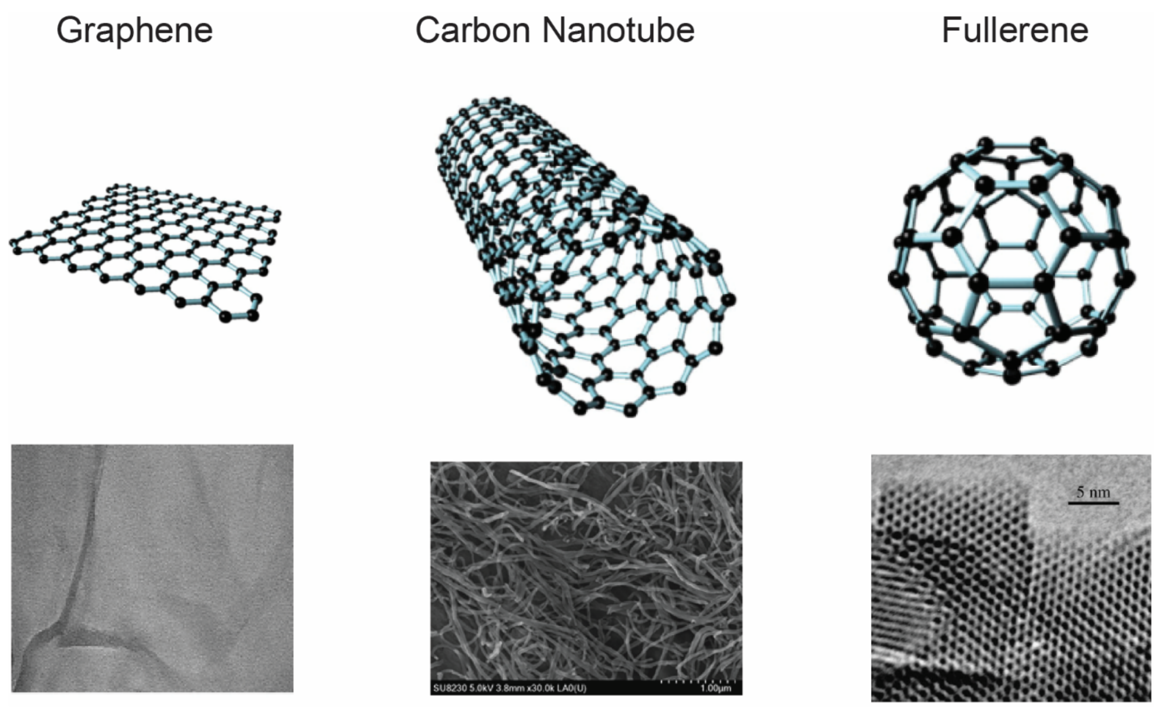
3. CNPs as Theranostic Tools for Cancer
3.1. Carbon Nanotubes (CNTs)
3.2. Graphene
3.3. Fullerene
4. Potential Translation of Nanoparticle Theranostics into the Clinic
- (1)
- The combination of intravenous administration of a targeted CNT formulation with non-invasive focal photothermal therapy will result in tumor ablation with minimal tissue damage. This is a result of the accumulation of a critical mass of nanomaterial, and the amount of heat generated upon light stimulation will confer total tumor ablation even if not every cell has been targeted. This is a positive consequence of treatment, as the ablation has a “near-field” effect on tumor cells and the adjacent surrounding cells. As these adjacent cells are influenced by the tumorigenic properties of the nearby neoplasia, ensuring expansion of the ablation field is important as they have the potential to undergo neoplastic transformation.
- (2)
- The ability to target CNTs to all tumor cells independent of their genetic profiles, the CNT/photothermal effects will be limited to cells expressing the tumor-specific antigen. However, should tumors be proven to be resistant to the targeting moiety, there is the possibility of personalizing and adjusting/adding the targeting moiety against different surface antigens. The adaptability of tumor target agent selection will come from new emerging information that is garnered from biomarker evaluations and unique mutation profiles defined from patient tumor sequencing.
- (3)
- Complete and rapid tumor ablation should not afford the selection of resistant tumors, as is commonly observed with prolonged biological therapeutics. The photoablation of the tumor would be total after the first and single treatment, with the likelihood of tumor recurrence significantly reduced, unlike recurrent chemotherapy or radiation therapy. The procedural safety inferred from using targeted CNT treatment is substantive, considering the consequential side effects resulting from existing therapies [181].
- (4)
- The ability to accurately diagnose and monitor disease onset, recurrence, and progression is critical. Targeted CNTs can provide tumor diagnostic and surveillance tools, as it comes from the ability of dual labeling with an agent that is compatible with existing imaging platforms, i.e., MRI, PET, or even ultrasound.
- (5)
- The overall costs of cancer therapeutic drugs and other treatment options are typically expensive. Although CNTs are much cheaper to manufacture than most nanomaterials, the process of functionalizing CNTs with both the targeting and imaging moieties brings added costs to the final formulation. However, therapeutic CNT platforms should not contribute to additional hospital costs and stay times in the same capacity as surgery and radiation interventions. Altogether, functionalized CNTs as a photothermal treatment protocol can become a standard of care for patients with early-stage and localized disease, with the new treatment being safer and demonstrating a clear improvement in overall survival benefit.
5. Conclusion Remarks
- The FDA will consider the current framework for safety assessment sufficiently robust and flexible to be appropriate for a variety of materials, including nanomaterials, and maintain a product-focused, science-based regulatory policy.
- Technical assessments will be product-specific, and this will consider the effects of nanomaterials in the biological and mode of action (e.g., photothermal) context of each product and its intended use.
- As such, the policies for each product area, both substantive and procedural, will vary according to the statutory authorities and relevant regulatory frameworks. This regulatory policy allows for tailored approaches that adhere to applicable legal frameworks and reflect the characteristics of specific products or product classes and evolving technology and scientific understanding.
- Moreover, the industry remains responsible for ensuring that its products meet all applicable legal requirements, including standards for safety, regardless of the emerging nature of the technology involved in the manufacturing of a product. The FDA also encourages the industry to consult with the Agency early in the product development process to address any questions related to the safety, effectiveness, or other attributes of products that contain nanomaterials or about the regulatory status of such products. Early consultations with the FDA will facilitate a mutual understanding of the specific scientific and regulatory issues for nanotechnology products.
Author Contributions
Funding
Conflicts of Interest
Abbreviations
| ALL | acute lymphocytic leukemia |
| AuNP | gold nanoparticles |
| CD22 | cluster of differentiation-22 |
| CD25 | Interleukin-2 receptor alpha chain |
| CNP | carbon nanoparticles |
| CNT | carbon nanotubes |
| CRPC | castrate resistant prostate cancer |
| EGFR | epidermal growth factor receptor |
| EPR | enhanced permeability and retention |
| FDA | Food and Drug Administration |
| GO | graphene oxide |
| HER2 | human epidermal growth factor receptor 2 |
| HIFU | high-intensity focused ultrasound |
| HNSCC | head and neck squamous cell carcinoma |
| IGFR | insulin-like growth factor receptor |
| IONP | iron oxide nanoparticle |
| MRI | magnetic resonance imaging |
| MWCNT | multi-walled carbon nanotube |
| NCI | National Cancer Institute |
| NIR | near infrared |
| PEG | polyethylene glycol |
| PET | Positron emission tomography |
| PSMA | prostate specific membrane antigen |
| SSC | squamous cell carcinoma |
| SWCNT | single-walled carbon nanotube |
| TSHR | thyroid stimulating hormone receptor |
References
- De Jong, W.H.; Borm, P.J. Drug delivery and nanoparticles: Applications and hazards. Int. J. Nanomed. 2008, 3, 133–149. [Google Scholar] [CrossRef] [Green Version]
- Green, M.R.; Manikhas, G.M.; Orlov, S.; Afanasyev, B.; Makhson, A.M.; Bhar, P.; Hawkins, M.J. Abraxane, a novel Cremophor-free, albumin-bound particle form of paclitaxel for the treatment of advanced non-small-cell lung cancer. Ann Oncol. 2006, 17, 1263–1268. [Google Scholar] [CrossRef] [PubMed]
- Miele, E.; Spinelli, G.P.; Miele, E.; Tomao, F.; Tomao, S. Albumin-bound formulation of paclitaxel (Abraxane ABI-007) in the treatment of breast cancer. Int. J. Nanomed. 2009, 4, 99–105. [Google Scholar] [CrossRef] [Green Version]
- Wu, D.; Si, M.; Xue, H.Y.; Wong, H.L. Nanomedicine applications in the treatment of breast cancer: Current state of the art. Int. J. Nanomed. 2017, 12, 5879–5892. [Google Scholar] [CrossRef] [Green Version]
- Anselmo, A.C.; Mitragotri, S. Nanoparticles in the clinic. Bioeng. Transl. Med. 2016, 1, 10–29. [Google Scholar] [CrossRef] [PubMed]
- Marusyk, A.; Polyak, K. Tumor heterogeneity: Causes and consequences. Biochim. Biophys. Acta 2010, 1805, 105–117. [Google Scholar] [CrossRef] [Green Version]
- Swanton, C. Intratumor heterogeneity: Evolution through space and time. Cancer Res. 2012, 72, 4875–4882. [Google Scholar] [CrossRef] [PubMed] [Green Version]
- Burrell, R.A.; McGranahan, N.; Bartek, J.; Swanton, C. The causes and consequences of genetic heterogeneity in cancer evolution. Nature 2013, 501, 338–345. [Google Scholar] [CrossRef]
- Marusyk, A.; Almendro, V.; Polyak, K. Intra-tumour heterogeneity: A looking glass for cancer? Nat. Rev. Cancer 2012, 12, 323–334. [Google Scholar] [CrossRef]
- Caro, C.; Gamez, F.; Quaresma, P.; Paez-Munoz, J.M.; Dominguez, A.; Pearson, J.R.; Pernia Leal, M.; Beltran, A.M.; Fernandez-Afonso, Y.; De la Fuente, J.M.; et al. Fe(3)O(4)-Au Core-Shell Nanoparticles as a Multimodal Platform for In Vivo Imaging and Focused Photothermal Therapy. Pharmaceutics 2021, 13, 416. [Google Scholar] [CrossRef]
- Beola, L.; Grazu, V.; Fernandez-Afonso, Y.; Fratila, R.M.; de Las Heras, M.; de la Fuente, J.M.; Gutierrez, L.; Asin, L. Critical Parameters to Improve Pancreatic Cancer Treatment Using Magnetic Hyperthermia: Field Conditions, Immune Response, and Particle Biodistribution. ACS Appl. Mater Interfaces 2021, 13, 12982–12996. [Google Scholar] [CrossRef]
- Hashida, Y.; Tanaka, H.; Zhou, S.; Kawakami, S.; Yamashita, F.; Murakami, T.; Umeyama, T.; Imahori, H.; Hashida, M. Photothermal ablation of tumor cells using a single-walled carbon nanotube-peptide composite. J. Control. Release 2014, 173, 59–66. [Google Scholar] [CrossRef] [PubMed] [Green Version]
- Panchapakesan, B.; Lu, S.; Sivakumar, K.; Teker, K.; Cesarone, G.; Wickstrom, E. Single-Wall Carbon Nanotube Nanobomb Agents for Killing Breast Cancer Cells. NanoBiothchnology 2005, 1, 7. [Google Scholar] [CrossRef]
- Burke, A.; Ding, X.; Singh, R.; Kraft, R.A.; Levi-Polyachenko, N.; Rylander, M.N.; Szot, C.; Buchanan, C.; Whitney, J.; Fisher, J.; et al. Long-term survival following a single treatment of kidney tumors with multiwalled carbon nanotubes and near-infrared radiation. Proc. Natl Acad. Sci. USA 2009, 106, 12897–12902. [Google Scholar] [CrossRef] [PubMed] [Green Version]
- Burlaka, A.; Lukin, S.; Prylutska, S.; Remeniak, O.; Prylutskyy, Y.; Shuba, M.; Maksimenko, S.; Ritter, U.; Scharff, P. Hyperthermic effect of multi-walled carbon nanotubes stimulated with near infrared irradiation for anticancer therapy: In vitro studies. Exp. Oncol. 2010, 32, 48–50. [Google Scholar]
- Mendoza-Nava, H.; Ferro-Flores, G.; Ocampo-Garcia, B.; Serment-Guerrero, J.; Santos-Cuevas, C.; Jimenez-Mancilla, N.; Luna-Gutierrez, M.; Camacho-Lopez, M.A. Laser heating of gold nanospheres functionalized with octreotide: In vitro effect on HeLa cell viability. Photomed. Laser Surg. 2013, 31, 17–22. [Google Scholar] [CrossRef]
- Melero, I.; Castanon, E.; Alvarez, M.; Champiat, S.; Marabelle, A. Intratumoural administration and tumour tissue targeting of cancer immunotherapies. Nat. Rev. Clin. Oncol. 2021, 18, 558–576. [Google Scholar] [CrossRef]
- Melancon, M.P.; Lu, W.; Yang, Z.; Zhang, R.; Cheng, Z.; Elliot, A.M.; Stafford, J.; Olson, T.; Zhang, J.Z.; Li, C. In vitro and in vivo targeting of hollow gold nanoshells directed at epidermal growth factor receptor for photothermal ablation therapy. Mol. Cancer Ther. 2008, 7, 1730–1739. [Google Scholar] [CrossRef] [Green Version]
- Boca, S.C.; Potara, M.; Gabudean, A.M.; Juhem, A.; Baldeck, P.L.; Astilean, S. Chitosan-coated triangular silver nanoparticles as a novel class of biocompatible, highly effective photothermal transducers for in vitro cancer cell therapy. Cancer Lett. 2011, 311, 131–140. [Google Scholar] [CrossRef]
- Loo, C.; Lowery, A.; Halas, N.; West, J.; Drezek, R. Immunotargeted nanoshells for integrated cancer imaging and therapy. Nano Lett. 2005, 5, 709–711. [Google Scholar] [CrossRef]
- Panwar, N.; Soehartono, A.M.; Chan, K.K.; Zeng, S.; Xu, G.; Qu, J.; Coquet, P.; Yong, K.T.; Chen, X. Nanocarbons for Biology and Medicine: Sensing, Imaging, and Drug Delivery. Chem. Rev. 2019, 119, 9559–9656. [Google Scholar] [CrossRef]
- Simon, J.; Flahaut, E.; Golzio, M. Overview of Carbon Nanotubes for Biomedical Applications. Materials 2019, 12, 624. [Google Scholar] [CrossRef] [PubMed] [Green Version]
- Raphey, V.R.; Henna, T.K.; Nivitha, K.P.; Mufeedha, P.; Sabu, C.; Pramod, K. Advanced biomedical applications of carbon nanotube. Mater. Sci. Eng. C Mater. Biol. Appl. 2019, 100, 616–630. [Google Scholar] [CrossRef]
- Deng, H.-y.; Wang, L.; Tang, D.; Zhang, Y.; Zhang, L. Review on the laser-induced performance of photothermal materials for ignition application. Energetic Mater. Front. 2021, 2, 201–217. [Google Scholar] [CrossRef]
- Letfullin, R.R.; George, T.F.; Duree, G.C.; Bollinger, B.M. Ultrashort Laser Pulse Heating of Nanoparticles: Comparison of Theoretical Approaches. Adv. Opt. Technol. 2008, 2008, 251718. [Google Scholar] [CrossRef] [Green Version]
- Hu, Q.; Huang, Z.; Duan, Y.; Fu, Z.; Bin, L. Reprogramming Tumor Microenvironment with Photothermal Therapy. Bioconjugate Chem. 2020, 31, 1268–1278. [Google Scholar] [CrossRef]
- Dai, X.; Li, X.; Liu, Y.; Yan, F. Recent advances in nanoparticles-based photothermal therapy synergizing with immune checkpoint blockade therapy. Mater. Des. 2022, 217, 110656. [Google Scholar] [CrossRef]
- Galluzzi, L.; Buqué, A.; Kepp, O.; Zitvogel, L.; Kroemer, G. Immunogenic cell death in cancer and infectious disease. Nat. Rev. Immunol. 2017, 17, 97–111. [Google Scholar] [CrossRef]
- Guo, L.; Yan, D.D.; Yang, D.; Li, Y.; Wang, X.; Zalewski, O.; Yan, B.; Lu, W. Combinatorial photothermal and immuno cancer therapy using chitosan-coated hollow copper sulfide nanoparticles. ACS Nano 2014, 8, 5670–5681. [Google Scholar] [CrossRef]
- Li, W.; Yang, J.; Luo, L.; Jiang, M.; Qin, B.; Yin, H.; Zhu, C.; Yuan, X.; Zhang, J.; Luo, Z.; et al. Targeting photodynamic and photothermal therapy to the endoplasmic reticulum enhances immunogenic cancer cell death. Nat. Commun. 2019, 10, 3349. [Google Scholar] [CrossRef] [PubMed] [Green Version]
- Mroz, P.; Hashmi, J.T.; Huang, Y.Y.; Lange, N.; Hamblin, M.R. Stimulation of anti-tumor immunity by photodynamic therapy. Expert Rev. Clin. Immunol. 2011, 7, 75–91. [Google Scholar] [CrossRef]
- Ng, C.W.; Li, J.; Pu, K. Recent Progresses in Phototherapy-Synergized Cancer Immunotherapy. Adv. Funct. Mater. 2018, 28, 1804688. [Google Scholar] [CrossRef]
- Doane, T.; Burda, C. Nanoparticle mediated non-covalent drug delivery. Adv. Drug Deliv. Rev. 2013, 65, 607–621. [Google Scholar] [CrossRef] [PubMed] [Green Version]
- Kim, S.W.; Lee, Y.K.; Kim, S.H.; Park, J.Y.; Lee, D.U.; Choi, J.; Hong, J.H.; Kim, S.; Khang, D. Covalent, Non-Covalent, Encapsulated Nanodrug Regulate the Fate of Intra- and Extracellular Trafficking: Impact on Cancer and Normal Cells. Sci. Rep. 2017, 7, 6454. [Google Scholar] [CrossRef] [PubMed]
- Werengowska-Ciecwierz, K.; Wisniewski, M.; Terzyk, A.P.; Furmaniak, S. The Chemistry of Bioconjugation in Nanoparticles-Based Drug Delivery System. Adv. Cond. Matter Phys. 2015. [CrossRef] [Green Version]
- Gao, H.; Yang, Z.; Zhang, S.; Cao, S.; Shen, S.; Pang, Z.; Jiang, X. Ligand modified nanoparticles increases cell uptake, alters endocytosis and elevates glioma distribution and internalization. Sci. Rep. 2013, 3, 2534. [Google Scholar] [CrossRef] [Green Version]
- Akinc, A.; Battaglia, G. Exploiting endocytosis for nanomedicines. Cold Spring Harb. Perspect. Biol. 2013, 5, a016980. [Google Scholar] [CrossRef] [Green Version]
- Xin, Y.; Yin, M.; Zhao, L.; Meng, F.; Luo, L. Recent progress on nanoparticle-based drug delivery systems for cancer therapy. Cancer Biol. Med. 2017, 14, 228–241. [Google Scholar] [CrossRef] [Green Version]
- Boedtkjer, E.; Pedersen, S.F. The Acidic Tumor Microenvironment as a Driver of Cancer. Annu. Rev. Physiol. 2020, 82, 103–126. [Google Scholar] [CrossRef] [Green Version]
- Justus, C.R.; Dong, L.; Yang, L.V. Acidic tumor microenvironment and pH-sensing G protein-coupled receptors. Front. Physiol. 2013, 4, 354. [Google Scholar] [CrossRef] [Green Version]
- Lin, B.; Chen, H.; Liang, D.; Lin, W.; Qi, X.; Liu, H.; Deng, X. Acidic pH and High-H(2)O(2) Dual Tumor Microenvironment-Responsive Nanocatalytic Graphene Oxide for Cancer Selective Therapy and Recognition. ACS Appl. Mater. Interfaces 2019, 11, 11157–11166. [Google Scholar] [CrossRef]
- Martinez, G.V.; Zhang, X.; Garcia-Martin, M.L.; Morse, D.L.; Woods, M.; Sherry, A.D.; Gillies, R.J. Imaging the extracellular pH of tumors by MRI after injection of a single cocktail of T1 and T2 contrast agents. NMR Biomed. 2011, 24, 1380–1391. [Google Scholar] [CrossRef] [Green Version]
- Fortuni, B.; Inose, T.; Ricci, M.; Fujita, Y.; Van Zundert, I.; Masuhara, A.; Fron, E.; Mizuno, H.; Latterini, L.; Rocha, S.; et al. Polymeric Engineering of Nanoparticles for Highly Efficient Multifunctional Drug Delivery Systems. Sci. Rep. 2019, 9, 2666. [Google Scholar] [CrossRef]
- Battogtokh, G.; Ko, Y.T. Self-assembling micelle-like nanoparticles with detachable envelopes for enhanced delivery of nucleic acid therapeutics. Mol. Pharm. 2014, 11, 904–912. [Google Scholar] [CrossRef] [PubMed]
- Wang, T.D.; Van Dam, J. Optical biopsy: A new frontier in endoscopic detection and diagnosis. Clin. Gastroenterol. Hepatol. 2004, 2, 744–753. [Google Scholar] [CrossRef] [PubMed] [Green Version]
- Herold, F.; Wiegel, P.; Scholkmann, F.; Muller, N.G. Applications of Functional Near-Infrared Spectroscopy (fNIRS) Neuroimaging in Exercise(-)Cognition Science: A Systematic, Methodology-Focused Review. J. Clin. Med. 2018, 7, 466. [Google Scholar] [CrossRef] [PubMed] [Green Version]
- Gravas, S.; Bachmann, A.; Reich, O.; Roehrborn, C.G.; Gilling, P.J.; De La Rosette, J. Critical review of lasers in benign prostatic hyperplasia (BPH). BJU Int. 2011, 107, 1030–1043. [Google Scholar] [CrossRef]
- Gregorie, H.B., Jr.; Horger, E.O.; Ward, J.L.; Green, J.F.; Richards, T.; Robertson, H.C., Jr.; Stevenson, T.B. Hematoporphyrin-derivative fluorescence in malignant neoplasms. Ann. Surg. 1968, 167, 820–828. [Google Scholar] [CrossRef]
- Kessel, D. Photodynamic Therapy: A Brief History. J. Clin. Med. 2019, 8, 1581. [Google Scholar] [CrossRef] [Green Version]
- Correia, J.H.; Rodrigues, J.A.; Pimenta, S.; Dong, T.; Yang, Z. Photodynamic Therapy Review: Principles, Photosensitizers, Applications, and Future Directions. Pharmaceutics 2021, 13, 1332. [Google Scholar] [CrossRef]
- Gunaydin, G.; Gedik, M.E.; Ayan, S. Photodynamic Therapy for the Treatment and Diagnosis of Cancer-A Review of the Current Clinical Status. Front. Chem. 2021, 9, 686303. [Google Scholar] [CrossRef]
- Costa, D.F.; Mendes, L.P.; Torchilin, V.P. The effect of low- and high-penetration light on localized cancer therapy. Adv. Drug Deliv. Rev. 2019, 138, 105–116. [Google Scholar] [CrossRef]
- Henderson, T.A. Multi-watt near-infrared light therapy as a neuroregenerative treatment for traumatic brain injury. Neural Regen. Res. 2016, 11, 563–565. [Google Scholar] [CrossRef] [PubMed]
- Yi, G.; Hong, S.H.; Son, J.; Yoo, J.; Park, C.; Choi, Y.; Koo, H. Recent advances in nanoparticle carriers for photodynamic therapy. Quant Imaging Med. Surg. 2018, 8, 433–443. [Google Scholar] [CrossRef] [PubMed]
- Liao, J.; Li, W.; Peng, J.; Yang, Q.; Li, H.; Wei, Y.; Zhang, X.; Qian, Z. Combined cancer photothermal-chemotherapy based on doxorubicin/gold nanorod-loaded polymersomes. Theranostics 2015, 5, 345–356. [Google Scholar] [CrossRef] [PubMed] [Green Version]
- Wang, Y.; Black, K.C.L.; Luehmann, H.; Li, W.; Zhang, Y.; Cai, X.; Wan, D.; Liu, S.-Y.; Li, M.; Kim, P.; et al. Comparison Study of Gold Nanohexapods, Nanorods, and Nanocages for Photothermal Cancer Treatment. ACS Nano 2013, 7, 2068–2077. [Google Scholar] [CrossRef] [PubMed]
- Hudson, D.E.; Hudson, D.O.; Wininger, J.M.; Richardson, B.D. Penetration of laser light at 808 and 980 nm in bovine tissue samples. Photomed. Laser Surg. 2013, 31, 163–168. [Google Scholar] [CrossRef] [Green Version]
- Pissuwan, D.; Valenzuela, S.M.; Cortie, M.B. Therapeutic possibilities of plasmonically heated gold nanoparticles. Trends Biotechnol. 2006, 24, 62–67. [Google Scholar] [CrossRef] [PubMed] [Green Version]
- Gul, S.; Khan, S.B.; Rehman, I.U.; Khan, M.A.; Khan, M.I. A Comprehensive Review of Magnetic Nanomaterials Modern Day Theranostics. Front. Mater. 2019, 6, 179. [Google Scholar] [CrossRef] [Green Version]
- Gupta, A.K.; Gupta, M. Synthesis and surface engineering of iron oxide nanoparticles for biomedical applications. Biomaterials 2005, 26, 3995–4021. [Google Scholar] [CrossRef]
- Li, L.; Jiang, W.; Luo, K.; Song, H.; Lan, F.; Wu, Y.; Gu, Z. Superparamagnetic iron oxide nanoparticles as MRI contrast agents for non-invasive stem cell labeling and tracking. Theranostics 2013, 3, 595–615. [Google Scholar] [CrossRef] [PubMed]
- Wang, Y.X. Superparamagnetic iron oxide based MRI contrast agents: Current status of clinical application. Quant Imaging Med. Surg. 2011, 1, 35–40. [Google Scholar] [CrossRef] [PubMed]
- Wang, Y.X.; Hussain, S.M.; Krestin, G.P. Superparamagnetic iron oxide contrast agents: Physicochemical characteristics and applications in MR imaging. Eur. Radiol. 2001, 11, 2319–2331. [Google Scholar] [CrossRef] [Green Version]
- Major, J.L.; Meade, T.J. Bioresponsive, cell-penetrating, and multimeric MR contrast agents. Acc. Chem. Res. 2009, 42, 893–903. [Google Scholar] [CrossRef] [Green Version]
- Bulte, J.W.; Kraitchman, D.L. Iron oxide MR contrast agents for molecular and cellular imaging. NMR Biomed. 2004, 17, 484–499. [Google Scholar] [CrossRef] [PubMed]
- Gao, J.; Gu, H.; Xu, B. Multifunctional magnetic nanoparticles: Design, synthesis, and biomedical applications. Acc. Chem. Res. 2009, 42, 1097–1107. [Google Scholar] [CrossRef]
- Fu, Q.; Li, Z.; Ye, J.; Li, Z.; Fu, F.; Lin, S.L.; Chang, C.A.; Yang, H.; Song, J. Magnetic targeted near-infrared II PA/MR imaging guided photothermal therapy to trigger cancer immunotherapy. Theranostics 2020, 10, 4997–5010. [Google Scholar] [CrossRef]
- Yen, S.K.; Padmanabhan, P.; Selvan, S.T. Multifunctional iron oxide nanoparticles for diagnostics, therapy and macromolecule delivery. Theranostics 2013, 3, 986–1003. [Google Scholar] [CrossRef] [PubMed] [Green Version]
- Cherukuri, P.; Glazer, E.S.; Curley, S.A. Targeted hyperthermia using metal nanoparticles. Adv. Drug Deliv. Rev. 2010, 62, 339–345. [Google Scholar] [CrossRef] [PubMed] [Green Version]
- Hilger, I.; Kaiser, W.A. Iron oxide-based nanostructures for MRI and magnetic hyperthermia. Nanomedicine 2012, 7, 1443–1459. [Google Scholar] [CrossRef]
- Giustini, A.J.; Petryk, A.A.; Cassim, S.M.; Tate, J.A.; Baker, I.; Hoopes, P.J. Magnetic Nanoparticle Hyperthermia in Cancer Treatment. Nano Life 2010, 1. [Google Scholar] [CrossRef] [PubMed]
- Zhou, Y.F. High intensity focused ultrasound in clinical tumor ablation. World J. Clin. Oncol. 2011, 2, 8–27. [Google Scholar] [CrossRef] [PubMed]
- Fisher, D.G.; Price, R.J. Recent Advances in the Use of Focused Ultrasound for Magnetic Resonance Image-Guided Therapeutic Nanoparticle Delivery to the Central Nervous System. Front Pharm. 2019, 10, 1348. [Google Scholar] [CrossRef]
- Vlaisavljevich, E.; Durmaz, Y.Y.; Maxwell, A.; Elsayed, M.; Xu, Z. Nanodroplet-mediated histotripsy for image-guided targeted ultrasound cell ablation. Theranostics 2013, 3, 851–864. [Google Scholar] [CrossRef] [Green Version]
- Wang, M.; Zhang, Y.; Cai, C.; Tu, J.; Guo, X.; Zhang, D. Sonoporation-induced cell membrane permeabilization and cytoskeleton disassembly at varied acoustic and microbubble-cell parameters. Sci. Rep. 2018, 8, 3885. [Google Scholar] [CrossRef] [Green Version]
- Sadeghi-Goughari, M.; Jeon, S.; Kwon, H.J. Enhancing Thermal Effect of Focused Ultrasound Therapy Using Gold Nanoparticles. IEEE Trans. Nanobioscience 2019, 18, 661–668. [Google Scholar] [CrossRef]
- Zhang, N.; Wang, R.; Hao, J.; Yang, Y.; Zou, H.; Wang, Z. Mesoporous composite nanoparticles for dual-modality ultrasound/magnetic resonance imaging and synergistic chemo-/thermotherapy against deep tumors. Int. J. Nanomed. 2017, 12, 7273–7289. [Google Scholar] [CrossRef] [PubMed] [Green Version]
- Canavese, G.; Ancona, A.; Racca, L.; Canta, M.; Dumontel, B.; Barbaresco, F.; Limongi, T.; Cauda, V. Nanoparticle-assisted ultrasound: A special focus on sonodynamic therapy against cancer. Chem. Eng. J. 2018, 340, 155–172. [Google Scholar] [CrossRef]
- Maiti, D.; Tong, X.; Mou, X.; Yang, K. Carbon-Based Nanomaterials for Biomedical Applications: A Recent Study. Front. Pharm. 2018, 9, 1401. [Google Scholar] [CrossRef] [PubMed] [Green Version]
- Mohajeri, M.; Behnam, B.; Sahebkar, A. Biomedical applications of carbon nanomaterials: Drug and gene delivery potentials. J Cell Physiol. 2018, 234, 298–319. [Google Scholar] [CrossRef] [Green Version]
- Dubey, R.; Dutta, D.; Sarkar, A.; Chattopadhyay, P. Functionalized carbon nanotubes: Synthesis, properties and applications in water purification, drug delivery, and material and biomedical sciences. Nanoscale Adv. 2021, 3, 5722–5744. [Google Scholar] [CrossRef]
- Liu, W.; Speranza, G. Functionalization of Carbon Nanomaterials for Biomedical Applications. C 2019, 5, 72. [Google Scholar] [CrossRef] [Green Version]
- Saravanan, S.; Sareen, N.; Abu-El-Rub, E.; Ashour, H.; Sequiera, G.L.; Ammar, H.I.; Gopinath, V.; Shamaa, A.A.; Sayed, S.S.E.; Moudgil, M.; et al. Graphene Oxide-Gold Nanosheets Containing Chitosan Scaffold Improves Ventricular Contractility and Function After Implantation into Infarcted Heart. Sci. Rep. 2018, 8, 15069. [Google Scholar] [CrossRef] [PubMed] [Green Version]
- Dotan, I.; Roche, P.J.; Paliouras, M.; Mitmaker, E.J.; Trifiro, M.A. Engineering Multi-Walled Carbon Nanotube Therapeutic Bionanofluids to Selectively Target Papillary Thyroid Cancer Cells. PLoS ONE 2016, 11, e0149723. [Google Scholar] [CrossRef]
- Goel, A.; Howard, J.B.; Vander Sande, J.B. Size analysis of single fullerene molecules by electron microscopy. Carbon 2004, 42, 1907–1915. [Google Scholar] [CrossRef]
- Markovic, Z.M.; Harhaji-Trajkovic, L.M.; Todorovic-Markovic, B.M.; Kepic, D.P.; Arsikin, K.M.; Jovanovic, S.P.; Pantovic, A.C.; Dramicanin, M.D.; Trajkovic, V.S. In vitro comparison of the photothermal anticancer activity of graphene nanoparticles and carbon nanotubes. Biomaterials 2011, 32, 1121–1129. [Google Scholar] [CrossRef] [PubMed]
- Madani, S.Y.; Naderi, N.; Dissanayake, O.; Tan, A.; Seifalian, A.M. A new era of cancer treatment: Carbon nanotubes as drug delivery tools. Int. J. Nanomed. 2011, 6, 2963–2979. [Google Scholar] [CrossRef] [Green Version]
- Iijima, S. Helical microtubules of graphitic carbon. Nature 1991, 354, 56–58. [Google Scholar] [CrossRef]
- Son, K.H.; Hong, J.H.; Lee, J.W. Carbon nanotubes as cancer therapeutic carriers and mediators. Int. J. Nanomed. 2016, 11, 5163–5185. [Google Scholar] [CrossRef] [Green Version]
- Bekyarova, E.; Ni, Y.; Malarkey, E.B.; Montana, V.; McWilliams, J.L.; Haddon, R.C.; Parpura, V. Applications of Carbon Nanotubes in Biotechnology and Biomedicine. J. Biomed. Nanotechnol. 2005, 1, 3–17. [Google Scholar] [CrossRef] [Green Version]
- Zhang, W.; Zhang, Z.; Zhang, Y. The application of carbon nanotubes in target drug delivery systems for cancer therapies. Nanoscale Res. Lett. 2011, 6, 555. [Google Scholar] [CrossRef]
- Peigney, A.; Laurent, C.; Flahaut, E.; Bacsa, R.R.; Rousset, A. Specific surface area of carbon nanotubes and bundles of carbon nanotubes. Carbon 2001, 39, 507–514. [Google Scholar] [CrossRef] [Green Version]
- Wu, L.; Man, C.; Wang, H.; Lu, X.; Ma, Q.; Cai, Y.; Ma, W. PEGylated multi-walled carbon nanotubes for encapsulation and sustained release of oxaliplatin. Pharm. Res. 2013, 30, 412–423. [Google Scholar] [CrossRef] [PubMed]
- Heister, E.; Brunner, E.W.; Dieckmann, G.R.; Jurewicz, I.; Dalton, A.B. Are Carbon Nanotubes a Natural Solution? Applications in Biology and Medicine. ACS Appl. Mater. Interfaces 2013, 5, 1870–1891. [Google Scholar] [CrossRef]
- Shaffer, M.S.P.; Fan, X.; Windle, A.H. Dispersion and packing of carbon nanotubes. Carbon 1998, 36, 1603–1612. [Google Scholar] [CrossRef]
- Singh, R.; Pantarotto, D.; Lacerda, L.; Pastorin, G.; Klumpp, C.; Prato, M.; Bianco, A.; Kostarelos, K. Tissue biodistribution and blood clearance rates of intravenously administered carbon nanotube radiotracers. Proc. Natl. Acad. Sci. USA 2006, 103, 3357–3362. [Google Scholar] [CrossRef] [Green Version]
- Liu, X.; Hurt, R.H.; Kane, A.B. Biodurability of Single-Walled Carbon Nanotubes Depends on Surface Functionalization. Carbon N Y 2010, 48, 1961–1969. [Google Scholar] [CrossRef] [PubMed] [Green Version]
- Liu, Z.; Cai, W.; He, L.; Nakayama, N.; Chen, K.; Sun, X.; Chen, X.; Dai, H. In vivo biodistribution and highly efficient tumour targeting of carbon nanotubes in mice. Nat. Nanotechnol. 2007, 2, 47–52. [Google Scholar] [CrossRef]
- Wong, B.S.; Yoong, S.L.; Jagusiak, A.; Panczyk, T.; Ho, H.K.; Ang, W.H.; Pastorin, G. Carbon nanotubes for delivery of small molecule drugs. Adv. Drug Deliv. Rev. 2013, 65, 1964–2015. [Google Scholar] [CrossRef]
- Wu, W.; Li, R.; Bian, X.; Zhu, Z.; Ding, D.; Li, X.; Jia, Z.; Jiang, X.; Hu, Y. Covalently combining carbon nanotubes with anticancer agent: Preparation and antitumor activity. ACS Nano 2009, 3, 2740–2750. [Google Scholar] [CrossRef]
- Ji, J.; Liu, M.; Meng, Y.; Liu, R.; Yan, Y.; Dong, J.; Guo, Z.; Ye, C. Experimental Study of Magnetic Multi-Walled Carbon Nanotube-Doxorubicin Conjugate in a Lymph Node Metastatic Model of Breast Cancer. Med. Sci. Monit. 2016, 22, 2363–2373. [Google Scholar] [CrossRef]
- Decatris, M.P.; Sundar, S.; O’Byrne, K.J. Platinum-based chemotherapy in metastatic breast cancer: Current status. Cancer Treat Rev. 2004, 30, 53–81. [Google Scholar] [CrossRef]
- Prato, M.; Kostarelos, K.; Bianco, A. Functionalized carbon nanotubes in drug design and discovery. Acc. Chem. Res. 2008, 41, 60–68. [Google Scholar] [CrossRef] [PubMed]
- Arya, N.; Arora, A.; Vasu, K.S.; Sood, A.K.; Katti, D.S. Combination of single walled carbon nanotubes/graphene oxide with paclitaxel: A reactive oxygen species mediated synergism for treatment of lung cancer. Nanoscale 2013, 5, 2818–2829. [Google Scholar] [CrossRef]
- Kam, N.W.; Dai, H. Carbon nanotubes as intracellular protein transporters: Generality and biological functionality. J. Am. Chem. Soc. 2005, 127, 6021–6026. [Google Scholar] [CrossRef] [Green Version]
- Pantarotto, D.; Briand, J.P.; Prato, M.; Bianco, A. Translocation of bioactive peptides across cell membranes by carbon nanotubes. Chem. Commun. 2004, 16–17. [Google Scholar] [CrossRef]
- Iancu, C.; Mocan, L. Advances in cancer therapy through the use of carbon nanotube-mediated targeted hyperthermia. Int. J. Nanomed. 2011, 6, 1675–1684. [Google Scholar] [CrossRef] [PubMed] [Green Version]
- Raffa, V.; Ciofani, G.; Vittorio, O.; Riggio, C.; Cuschieri, A. Physicochemical properties affecting cellular uptake of carbon nanotubes. Nanomedicine 2010, 5, 89–97. [Google Scholar] [CrossRef] [PubMed] [Green Version]
- Li, J.L.; Bao, H.C.; Hou, X.L.; Sun, L.; Wang, X.G.; Gu, M. Graphene oxide nanoparticles as a nonbleaching optical probe for two-photon luminescence imaging and cell therapy. Angew. Chem. 2012, 51, 1830–1834. [Google Scholar] [CrossRef]
- Kim, J.H.; Cho, M.H.; Kim, K.J.; Kim, S.H. Laser ignition and controlled explosion of nanoenergetic materials: The role of multi-walled carbon nanotubes. Carbon 2017, 118, 268–277. [Google Scholar] [CrossRef]
- Robinson, J.T.; Welsher, K.; Tabakman, S.M.; Sherlock, S.P.; Wang, H.; Luong, R.; Dai, H. High Performance In Vivo Near-IR (>1 mum) Imaging and Photothermal Cancer Therapy with Carbon Nanotubes. Nano Res. 2010, 3, 779–793. [Google Scholar] [CrossRef]
- Weissleder, R. A clearer vision for in vivo imaging. Nat. Biotechnol. 2001, 19, 316–317. [Google Scholar] [CrossRef] [PubMed]
- Siregar, S.; Oktamuliani, S.; Saijo, Y. A Theoretical Model of Laser Heating Carbon Nanotubes. Nanomaterials 2018, 8, 580. [Google Scholar] [CrossRef] [Green Version]
- Torti, S.V.; Byrne, F.; Whelan, O.; Levi, N.; Ucer, B.; Schmid, M.; Torti, F.M.; Akman, S.; Liu, J.; Ajayan, P.M.; et al. Thermal ablation therapeutics based on CN(x) multi-walled nanotubes. Int. J. Nanomed. 2007, 2, 707–714. [Google Scholar]
- Kam, N.W.; O’Connell, M.; Wisdom, J.A.; Dai, H. Carbon nanotubes as multifunctional biological transporters and near-infrared agents for selective cancer cell destruction. Proc. Natl. Acad. Sci. USA 2005, 102, 11600–11605. [Google Scholar] [CrossRef] [Green Version]
- Lee, S.S.; Roche, P.J.; Giannopoulos, P.N.; Mitmaker, E.J.; Tamilia, M.; Paliouras, M.; Trifiro, M.A. Prostate-specific membrane antigen-directed nanoparticle targeting for extreme nearfield ablation of prostate cancer cells. Tumour Biol. 2017, 39, 1010428317695943. [Google Scholar] [CrossRef] [Green Version]
- Lu, G.H.; Shang, W.T.; Deng, H.; Han, Z.Y.; Hu, M.; Liang, X.Y.; Fang, C.H.; Zhu, X.H.; Fan, Y.F.; Tian, J. Targeting carbon nanotubes based on IGF-1R for photothermal therapy of orthotopic pancreatic cancer guided by optical imaging. Biomaterials 2019, 195, 13–22. [Google Scholar] [CrossRef] [PubMed]
- Shao, N.; Lu, S.; Wickstrom, E.; Panchapakesan, B. Integrated molecular targeting of IGF1R and HER2 surface receptors and destruction of breast cancer cells using single wall carbon nanotubes. Nanotechnology 2007, 18, 315101. [Google Scholar] [CrossRef] [Green Version]
- Chakravarty, P.; Marches, R.; Zimmerman, N.S.; Swafford, A.D.; Bajaj, P.; Musselman, I.H.; Pantano, P.; Draper, R.K.; Vitetta, E.S. Thermal ablation of tumor cells with antibody-functionalized single-walled carbon nanotubes. Proc. Natl. Acad. Sci. USA 2008, 105, 8697–8702. [Google Scholar] [CrossRef] [Green Version]
- Khan, S.A.; Kanchanapally, R.; Fan, Z.; Beqa, L.; Singh, A.K.; Senapati, D.; Ray, P.C. A gold nanocage-CNT hybrid for targeted imaging and photothermal destruction of cancer cells. Chem. Commun. 2012, 48, 6711–6713. [Google Scholar] [CrossRef] [PubMed]
- Krishna, V.; Singh, A.; Sharma, P.; Iwakuma, N.; Wang, Q.; Zhang, Q.; Knapik, J.; Jiang, H.; Grobmyer, S.R.; Koopman, B.; et al. Polyhydroxy fullerenes for non-invasive cancer imaging and therapy. Small 2010, 6, 2236–2241. [Google Scholar] [CrossRef] [PubMed]
- Li, J.L.; Hou, X.L.; Bao, H.C.; Sun, L.; Tang, B.; Wang, J.F.; Wang, X.G.; Gu, M. Graphene oxide nanoparticles for enhanced photothermal cancer cell therapy under the irradiation of a femtosecond laser beam. J. Biomed. Mater. Res. A 2014, 102, 2181–2188. [Google Scholar] [CrossRef] [PubMed]
- Robinson, J.T.; Tabakman, S.M.; Liang, Y.; Wang, H.; Casalongue, H.S.; Vinh, D.; Dai, H. Ultrasmall reduced graphene oxide with high near-infrared absorbance for photothermal therapy. J. Am. Chem. Soc. 2011, 133, 6825–6831. [Google Scholar] [CrossRef] [PubMed]
- Yang, K.; Zhang, S.; Zhang, G.; Sun, X.; Lee, S.T.; Liu, Z. Graphene in mice: Ultrahigh in vivo tumor uptake and efficient photothermal therapy. Nano Lett. 2010, 10, 3318–3323. [Google Scholar] [CrossRef]
- Yang, K.; Wan, J.; Zhang, S.; Tian, B.; Zhang, Y.; Liu, Z. The influence of surface chemistry and size of nanoscale graphene oxide on photothermal therapy of cancer using ultra-low laser power. Biomaterials 2012, 33, 2206–2214. [Google Scholar] [CrossRef] [PubMed]
- Chen, J.; Wang, D.; Xi, J.; Au, L.; Siekkinen, A.; Warsen, A.; Li, Z.-Y.; Zhang, H.; Xia, Y.; Li, X. Immuno Gold Nanocages with Tailored Optical Properties for Targeted Photothermal Destruction of Cancer Cells. Nano Lett. 2007, 7, 1318–1322. [Google Scholar] [CrossRef] [Green Version]
- Gormley, A.J.; Greish, K.; Ray, A.; Robinson, R.; Gustafson, J.A.; Ghandehari, H. Gold nanorod mediated plasmonic photothermal therapy: A tool to enhance macromolecular delivery. Int. J. Pharm. 2011, 415, 315–318. [Google Scholar] [CrossRef] [Green Version]
- Hao, Y.; Zhang, B.; Zheng, C.; Ji, R.; Ren, X.; Guo, F.; Sun, S.; Shi, J.; Zhang, H.; Zhang, Z.; et al. The tumor-targeting core-shell structured DTX-loaded PLGA@Au nanoparticles for chemo-photothermal therapy and X-ray imaging. J. Control. Release 2015, 220, 545–555. [Google Scholar] [CrossRef]
- Hirsch, L.R.; Stafford, R.J.; Bankson, J.A.; Sershen, S.R.; Rivera, B.; Price, R.E.; Hazle, J.D.; Halas, N.J.; West, J.L. Nanoshell-mediated near-infrared thermal therapy of tumors under magnetic resonance guidance. Proc. Natl. Acad. Sci. USA 2003, 100, 13549–13554. [Google Scholar] [CrossRef] [Green Version]
- Huang, X.; El-Sayed, I.H.; Qian, W.; El-Sayed, M.A. Cancer Cell Imaging and Photothermal Therapy in the Near-Infrared Region by Using Gold Nanorods. J. Am. Chem. Soc. 2006, 128, 2115–2120. [Google Scholar] [CrossRef]
- Piao, J.G.; Liu, D.; Hu, K.; Wang, L.; Gao, F.; Xiong, Y.; Yang, L. Cooperative Nanoparticle System for Photothermal Tumor Treatment without Skin Damage. ACS Appl. Mater. Interfaces 2016, 8, 2847–2856. [Google Scholar] [CrossRef]
- Sun, T.; Wang, Y.; Wang, Y.; Xu, J.; Zhao, X.; Vangveravong, S.; Mach, R.H.; Xia, Y. Using SV119-gold nanocage conjugates to eradicate cancer stem cells through a combination of photothermal and chemo therapies. Adv. Healthc. Mater. 2014, 3, 1283–1291. [Google Scholar] [CrossRef] [PubMed]
- Topete, A.; Alatorre-Meda, M.; Villar-Alvarez, E.M.; Carregal-Romero, S.; Barbosa, S.; Parak, W.J.; Taboada, P.; Mosquera, V. Polymeric-gold nanohybrids for combined imaging and cancer therapy. Adv. Healthc. Mater. 2014, 3, 1309–1325. [Google Scholar] [CrossRef] [PubMed]
- Topete, A.; Alatorre-Meda, M.; Iglesias, P.; Villar-Alvarez, E.M.; Barbosa, S.; Costoya, J.A.; Taboada, P.; Mosquera, V. Fluorescent drug-loaded, polymeric-based, branched gold nanoshells for localized multimodal therapy and imaging of tumoral cells. ACS Nano 2014, 8, 2725–2738. [Google Scholar] [CrossRef]
- Chu, M.; Shao, Y.; Peng, J.; Dai, X.; Li, H.; Wu, Q.; Shi, D. Near-infrared laser light mediated cancer therapy by photothermal effect of Fe3O4 magnetic nanoparticles. Biomaterials 2013, 34, 4078–4088. [Google Scholar] [CrossRef] [PubMed]
- Espinosa, A.; Di Corato, R.; Kolosnjaj-Tabi, J.; Flaud, P.; Pellegrino, T.; Wilhelm, C. Duality of Iron Oxide Nanoparticles in Cancer Therapy: Amplification of Heating Efficiency by Magnetic Hyperthermia and Photothermal Bimodal Treatment. ACS Nano 2016, 10, 2436–2446. [Google Scholar] [CrossRef]
- Shen, S.; Kong, F.; Guo, X.; Wu, L.; Shen, H.; Xie, M.; Wang, X.; Jin, Y.; Ge, Y. CMCTS stabilized Fe3O4 particles with extremely low toxicity as highly efficient near-infrared photothermal agents for in vivo tumor ablation. Nanoscale 2013, 5, 8056–8066. [Google Scholar] [CrossRef]
- Novoselov, K.S.; Geim, A.K.; Morozov, S.V.; Jiang, D.; Zhang, Y.; Dubonos, S.V.; Grigorieva, I.V.; Firsov, A.A. Electric field effect in atomically thin carbon films. Science 2004, 306, 666–669. [Google Scholar] [CrossRef] [PubMed] [Green Version]
- Novoselov, K.S.; Jiang, D.; Schedin, F.; Booth, T.J.; Khotkevich, V.V.; Morozov, S.V.; Geim, A.K. Two-dimensional atomic crystals. Proc. Natl. Acad. Sci. USA 2005, 102, 10451–10453. [Google Scholar] [CrossRef] [Green Version]
- Yang, K.; Feng, L.; Shi, X.; Liu, Z. Nano-graphene in biomedicine: Theranostic applications. Chem. Soc. Rev. 2013, 42, 530–547. [Google Scholar] [CrossRef]
- Geim, A.K.; Novoselov, K.S. The rise of graphene. Nat. Mater. 2007, 6, 183–191. [Google Scholar] [CrossRef]
- Wang, C.; Li, J.; Amatore, C.; Chen, Y.; Jiang, H.; Wang, X.M. Gold nanoclusters and graphene nanocomposites for drug delivery and imaging of cancer cells. Angew. Chem. 2011, 50, 11644–11648. [Google Scholar] [CrossRef]
- Shi, S.; Chen, F.; Ehlerding, E.B.; Cai, W. Surface engineering of graphene-based nanomaterials for biomedical applications. Bioconjugate Chem. 2014, 25, 1609–1619. [Google Scholar] [CrossRef] [Green Version]
- Zhang, Y.; Ali, S.F.; Dervishi, E.; Xu, Y.; Li, Z.; Casciano, D.; Biris, A.S. Cytotoxicity effects of graphene and single-wall carbon nanotubes in neural phaeochromocytoma-derived PC12 cells. ACS Nano 2010, 4, 3181–3186. [Google Scholar] [CrossRef]
- Pan, Y.; Sahoo, N.G.; Li, L. The application of graphene oxide in drug delivery. Expert Opin. Drug Deliv. 2012, 9, 1365–1376. [Google Scholar] [CrossRef]
- Yang, X.; Zhang, X.; Liu, Z.; Ma, Y.; Huang, Y.; Chen, Y. High-Efficiency Loading and Controlled Release of Doxorubicin Hydrochloride on Graphene Oxide. J. Phys. Chem. C 2008, 112, 17554–17558. [Google Scholar] [CrossRef]
- Depan, D.; Shah, J.; Misra, R.D.K. Controlled release of drug from folate-decorated and graphene mediated drug delivery system: Synthesis, loading efficiency, and drug release response. Mater. Sci. Eng. C 2011, 31, 1305–1312. [Google Scholar] [CrossRef]
- Zheng, X.T.; Ma, X.Q.; Li, C.M. Highly efficient nuclear delivery of anti-cancer drugs using a bio-functionalized reduced graphene oxide. J. Colloid Interface Sci. 2016, 467, 35–42. [Google Scholar] [CrossRef] [Green Version]
- Sahoo, N.G.; Bao, H.; Pan, Y.; Pal, M.; Kakran, M.; Cheng, H.K.; Li, L.; Tan, L.P. Functionalized carbon nanomaterials as nanocarriers for loading and delivery of a poorly water-soluble anticancer drug: A comparative study. Chem. Commun. 2011, 47, 5235–5237. [Google Scholar] [CrossRef]
- Slanina, Z. ISSPIC-5 in konstanz in 1990: Announcements of the C60 preparation and its structure confirmation. NTM Int. J. Hist. Ethics Nat. Sci. Technol. Med. 2001, 9, 41–46. [Google Scholar] [CrossRef]
- Schultz, H.P. Topological Organic Chemistry. Polyhedranes and Prismanes. J. Org. Chem. 1965, 30, 1361–1364. [Google Scholar] [CrossRef]
- Kroto, H.W.; Heath, J.R.; O’Brien, S.C.; Curl, R.F.; Smalley, R.E. C60: Buckminsterfullerene. Nature 1985, 318, 162–163. [Google Scholar] [CrossRef]
- Goodarzi, S.; Da Ros, T.; Conde, J.; Sefat, F.; Mozafari, M. Fullerene: Biomedical engineers get to revisit an old friend. Mater. Today 2017, 20, 460–480. [Google Scholar] [CrossRef] [Green Version]
- Kumar, M.; Raza, K. C60-fullerenes as Drug Delivery Carriers for Anticancer Agents: Promises and Hurdles. Pharm. Nanotechnol. 2017, 5, 169–179. [Google Scholar] [CrossRef]
- Choi, H.S.; Liu, W.; Misra, P.; Tanaka, E.; Zimmer, J.P.; Itty Ipe, B.; Bawendi, M.G.; Frangioni, J.V. Renal clearance of quantum dots. Nat. Biotechnol. 2007, 25, 1165–1170. [Google Scholar] [CrossRef] [Green Version]
- Bakry, R.; Vallant, R.M.; Najam-ul-Haq, M.; Rainer, M.; Szabo, Z.; Huck, C.W.; Bonn, G.K. Medicinal applications of fullerenes. Int. J. Nanomed. 2007, 2, 639–649. [Google Scholar]
- Dellinger, A.; Zhou, Z.; Connor, J.; Madhankumar, A.B.; Pamujula, S.; Sayes, C.M.; Kepley, C.L. Application of fullerenes in nanomedicine: An update. Nanomedicine 2013, 8, 1191–1208. [Google Scholar] [CrossRef] [Green Version]
- Ghiassi, K.B.; Olmstead, M.M.; Balch, A.L. Gadolinium-containing endohedral fullerenes: Structures and function as magnetic resonance imaging (MRI) agents. Dalton Trans. 2014, 43, 7346–7358. [Google Scholar] [CrossRef]
- Braun, K.; Dunsch, L.; Pipkorn, R.; Bock, M.; Baeuerle, T.; Yang, S.; Waldeck, W.; Wiessler, M. Gain of a 500-fold sensitivity on an intravital MR contrast agent based on an endohedral gadolinium-cluster-fullerene-conjugate: A new chance in cancer diagnostics. Int. J. Med. Sci. 2010, 7, 136–146. [Google Scholar] [CrossRef] [Green Version]
- Wang, L.; Zhu, X.; Tang, X.; Wu, C.; Zhou, Z.; Sun, C.; Deng, S.L.; Ai, H.; Gao, J. A multiple gadolinium complex decorated fullerene as a highly sensitive T(1) contrast agent. Chem. Commun. 2015, 51, 4390–4393. [Google Scholar] [CrossRef]
- Shultz, M.D.; Duchamp, J.C.; Wilson, J.D.; Shu, C.-Y.; Ge, J.; Zhang, J.; Gibson, H.W.; Fillmore, H.L.; Hirsch, J.I.; Dorn, H.C.; et al. Encapsulation of a Radiolabeled Cluster Inside a Fullerene Cage, 177LuxLu(3−x)N@C80: An Interleukin-13-Conjugated Radiolabeled Metallofullerene Platform. J. Am. Chem. Soc. 2010, 132, 4980–4981. [Google Scholar] [CrossRef] [Green Version]
- Yamago, S.; Tokuyama, H.; Nakamura, E.; Kikuchi, K.; Kananishi, S.; Sueki, K.; Nakahara, H.; Enomoto, S.; Ambe, F. In vivo biological behavior of a water-miscible fullerene: 14C labeling, absorption, distribution, excretion and acute toxicity. Chem. Biol. 1995, 2, 385–389. [Google Scholar] [CrossRef] [PubMed] [Green Version]
- Lu, F.; Haque, S.A.; Yang, S.T.; Luo, P.G.; Gu, L.; Kitaygorodskiy, A.; Li, H.; Lacher, S.; Sun, Y.P. Aqueous Compatible Fullerene-Doxorubicin Conjugates. J. Phys. Chem. C Nanomater. Interfaces 2009, 113, 17768. [Google Scholar] [CrossRef]
- Zakharian, T.Y.; Seryshev, A.; Sitharaman, B.; Gilbert, B.E.; Knight, V.; Wilson, L.J. A fullerene-paclitaxel chemotherapeutic: Synthesis, characterization, and study of biological activity in tissue culture. J. Am. Chem. Soc. 2005, 127, 12508–12509. [Google Scholar] [CrossRef] [PubMed]
- Prylutskyy, Y.I.; Cherepanov, V.V.; Evstigneev, M.P.; Kyzyma, O.A.; Petrenko, V.I.; Styopkin, V.I.; Bulavin, L.A.; Davidenko, N.A.; Wyrzykowski, D.; Woziwodzka, A.; et al. Structural self-organization of C60 and cisplatin in physiological solution. Phys. Chem. Chem. Phys. PCCP 2015, 17, 26084–26092. [Google Scholar] [CrossRef]
- Sitharaman, B.; Zakharian, T.Y.; Saraf, A.; Misra, P.; Ashcroft, J.; Pan, S.; Pham, Q.P.; Mikos, A.G.; Wilson, L.J.; Engler, D.A. Water-soluble fullerene (C60) derivatives as nonviral gene-delivery vectors. Mol. Pharm. 2008, 5, 567–578. [Google Scholar] [CrossRef]
- Grebinyk, A.; Grebinyk, S.; Prylutska, S.; Ritter, U.; Matyshevska, O.; Dandekar, T.; Frohme, M. C60 fullerene accumulation in human leukemic cells and perspectives of LED-mediated photodynamic therapy. Free Radic. Biol. Med. 2018, 124, 319–327. [Google Scholar] [CrossRef]
- Guan, M.; Ge, J.; Wu, J.; Zhang, G.; Chen, D.; Zhang, W.; Zhang, Y.; Zou, T.; Zhen, M.; Wang, C.; et al. Fullerene/photosensitizer nanovesicles as highly efficient and clearable phototheranostics with enhanced tumor accumulation for cancer therapy. Biomaterials 2016, 103, 75–85. [Google Scholar] [CrossRef] [Green Version]
- Guan, M.; Dong, H.; Ge, J.; Chen, D.; Sun, S.; Wang, C.; Yan, C.; Wang, P.; Shu, C. Multifunctional upconversion–nanoparticles–trismethylpyridylporphyrin–fullerene nanocomposite: A near-infrared light-triggered theranostic platform for imaging-guided photodynamic therapy. Npg Asia Mater. 2015, 7, e205. [Google Scholar] [CrossRef] [Green Version]
- Hamblin, M.R. Fullerenes as photosensitizers in photodynamic therapy: Pros and cons. Photochem. Photobiol. Sci. 2018, 17, 1515–1533. [Google Scholar] [CrossRef] [PubMed]
- Fidler, I.J.; Hart, I.R. Biological diversity in metastatic neoplasms: Origins and implications. Science 1982, 217, 998–1003. [Google Scholar] [CrossRef]
- Pleasance, E.D.; Cheetham, R.K.; Stephens, P.J.; McBride, D.J.; Humphray, S.J.; Greenman, C.D.; Varela, I.; Lin, M.L.; Ordonez, G.R.; Bignell, G.R.; et al. A comprehensive catalogue of somatic mutations from a human cancer genome. Nature 2010, 463, 191–196. [Google Scholar] [CrossRef] [PubMed] [Green Version]
- Fiala, C.; Diamandis, E.P. Mutations in normal tissues-some diagnostic and clinical implications. BMC Med. 2020, 18, 283. [Google Scholar] [CrossRef]
- Salgia, R.; Kulkarni, P. The Genetic/Non-genetic Duality of Drug ‘Resistance’ in Cancer. Trends Cancer 2018, 4, 110–118. [Google Scholar] [CrossRef]
- Watson, P.A.; Arora, V.K.; Sawyers, C.L. Emerging mechanisms of resistance to androgen receptor inhibitors in prostate cancer. Nat. Rev. Cancer 2015, 15, 701–711. [Google Scholar] [CrossRef] [Green Version]
- Chism, D.D.; De Silva, D.; Whang, Y.E. Mechanisms of acquired resistance to androgen receptor targeting drugs in castration-resistant prostate cancer. Expert Rev. Anticancer Ther. 2014, 14, 1369–1378. [Google Scholar] [CrossRef] [PubMed] [Green Version]
- Boudadi, K.; Antonarakis, E.S. Resistance to Novel Antiandrogen Therapies in Metastatic Castration-Resistant Prostate Cancer. Clin. Med. Insights. Oncol. 2016, 10, 1–9. [Google Scholar] [CrossRef] [Green Version]
- Hobisch, A.; Hoffmann, J.; Lambrinidis, L.; Eder, I.E.; Bartsch, G.; Klocker, H.; Culig, Z. Antagonist/agonist balance of the nonsteroidal antiandrogen bicalutamide (Casodex) in a new prostate cancer model. Urol. Int. 2000, 65, 73–79. [Google Scholar] [CrossRef] [PubMed]
- Culig, Z.; Hoffmann, J.; Erdel, M.; Eder, I.E.; Hobisch, A.; Hittmair, A.; Bartsch, G.; Utermann, G.; Schneider, M.R.; Parczyk, K.; et al. Switch from antagonist to agonist of the androgen receptor bicalutamide is associated with prostate tumour progression in a new model system. Br. J. Cancer 1999, 81, 242–251. [Google Scholar] [CrossRef]
- Wadosky, K.M.; Koochekpour, S. Androgen receptor splice variants and prostate cancer: From bench to bedside. Oncotarget 2017, 8, 18550–18576. [Google Scholar] [CrossRef] [Green Version]
- Steentjes, L.; Siesling, S.; Drummond, F.J.; van Manen, J.G.; Sharp, L.; Gavin, A. Factors associated with current and severe physical side-effects after prostate cancer treatment: What men report. Eur. J. Cancer Care 2018, 27. [Google Scholar] [CrossRef] [Green Version]
- Kiessling, F.; Mertens, M.E.; Grimm, J.; Lammers, T. Nanoparticles for imaging: Top or flop? Radiology 2014, 273, 10–28. [Google Scholar] [CrossRef] [Green Version]
- Blair, H.A.; Deeks, E.D. Albumin-Bound Paclitaxel: A Review in Non-Small Cell Lung Cancer. Drugs 2015, 75, 2017–2024. [Google Scholar] [CrossRef]
- Cecco, S.; Aliberti, M.; Baldo, P.; Giacomin, E.; Leone, R. Safety and efficacy evaluation of albumin-bound paclitaxel. Expert Opin. Drug Saf. 2014, 13, 511–520. [Google Scholar] [CrossRef] [PubMed]
- Gradishar, W.J.; Tjulandin, S.; Davidson, N.; Shaw, H.; Desai, N.; Bhar, P.; Hawkins, M.; O’Shaughnessy, J. Phase III trial of nanoparticle albumin-bound paclitaxel compared with polyethylated castor oil-based paclitaxel in women with breast cancer. J. Clin. Oncol. 2005, 23, 7794–7803. [Google Scholar] [CrossRef] [PubMed]
- Socinski, M.A.; Manikhas, G.M.; Stroyakovsky, D.L.; Makhson, A.N.; Cheporov, S.V.; Orlov, S.V.; Yablonsky, P.K.; Bhar, P.; Iglesias, J. A dose finding study of weekly and every-3-week nab-Paclitaxel followed by carboplatin as first-line therapy in patients with advanced non-small cell lung cancer. J. Thorac. Oncol.: Off. Publ. Int. Assoc. Study Lung Cancer 2010, 5, 852–861. [Google Scholar] [CrossRef] [Green Version]
- Keefe, S.M.; Hoffman-Censits, J.; Cohen, R.B.; Mamtani, R.; Heitjan, D.; Eliasof, S.; Nixon, A.; Turnbull, B.; Garmey, E.G.; Gunnarsson, O.; et al. Efficacy of the nanoparticle-drug conjugate CRLX101 in combination with bevacizumab in metastatic renal cell carcinoma: Results of an investigator-initiated phase I-IIa clinical trial. Ann. Oncol. 2016, 27, 1579–1585. [Google Scholar] [CrossRef] [PubMed]
- Von Hoff, D.D.; Mita, M.M.; Ramanathan, R.K.; Weiss, G.J.; Mita, A.C.; LoRusso, P.M.; Burris, H.A., III; Hart, L.L.; Low, S.C.; Parsons, D.M.; et al. Phase I Study of PSMA-Targeted Docetaxel-Containing Nanoparticle BIND-014 in Patients with Advanced Solid Tumors. Clin. Cancer Res. 2016, 22, 3157–3163. [Google Scholar] [CrossRef] [Green Version]
- Autio, K.A.; Dreicer, R.; Anderson, J.; Garcia, J.A.; Alva, A.; Hart, L.L.; Milowsky, M.I.; Posadas, E.M.; Ryan, C.J.; Graf, R.P.; et al. Safety and Efficacy of BIND-014, a Docetaxel Nanoparticle Targeting Prostate-Specific Membrane Antigen for Patients With Metastatic Castration-Resistant Prostate Cancer: A Phase 2 Clinical Trial. JAMA Oncol. 2018, 4, 1344–1351. [Google Scholar] [CrossRef] [Green Version]
- Rastinehad, A.R.; Anastos, H.; Wajswol, E.; Winoker, J.S.; Sfakianos, J.P.; Doppalapudi, S.K.; Carrick, M.R.; Knauer, C.J.; Taouli, B.; Lewis, S.C.; et al. Gold nanoshell-localized photothermal ablation of prostate tumors in a clinical pilot device study. Proc. Natl. Acad. Sci. USA 2019, 116, 18590–18596. [Google Scholar] [CrossRef] [Green Version]
- Schultz, J. Nanospectra Biosciences Announces Enrollment Completion of Pivotal Study of AuroLase Therapy for Ablation of Prostate Tissue. GlobeNewswire, 2 November 2021. [Google Scholar]
- Pottier, A.; Borghi, E.; Levy, L. New use of metals as nanosized radioenhancers. Anticancer Res. 2014, 34, 443–453. [Google Scholar]
- Bonvalot, S.; Rutkowski, P.L.; Thariat, J.; Carrere, S.; Ducassou, A.; Sunyach, M.P.; Agoston, P.; Hong, A.; Mervoyer, A.; Rastrelli, M.; et al. NBTXR3, a first-in-class radioenhancer hafnium oxide nanoparticle, plus radiotherapy versus radiotherapy alone in patients with locally advanced soft-tissue sarcoma (Act.In.Sarc): A multicentre, phase 2-3, randomised, controlled trial. Lancet Oncol. 2019, 20, 1148–1159. [Google Scholar] [CrossRef] [PubMed]
- Hoffmann, C.; Calugaru, V.; Borcoman, E.; Moreno, V.; Calvo, E.; Liem, X.; Salas, S.; Doger, B.; Jouffroy, T.; Mirabel, X.; et al. Phase I dose-escalation study of NBTXR3 activated by intensity-modulated radiation therapy in elderly patients with locally advanced squamous cell carcinoma of the oral cavity or oropharynx. Eur. J. Cancer 2021, 146, 135–144. [Google Scholar] [CrossRef]
- Scher, N.; Bonvalot, S.; Le Tourneau, C.; Chajon, E.; Verry, C.; Thariat, J.; Calugaru, V. Review of clinical applications of radiation-enhancing nanoparticles. Biotechnol. Rep. 2020, 28, e00548. [Google Scholar] [CrossRef] [PubMed]
- Hu, Y.; Paris, S.; Barsoumian, H.; Abana, C.O.; He, K.; Wasley, M.; Younes, A.I.; Masrorpour, F.; Chen, D.; Yang, L.; et al. Radiation Therapy Enhanced by NBTXR3 Nanoparticles Overcomes Anti-PD1 Resistance and Evokes Abscopal Effects. Int. J. Radiat. Oncol. Biol. Phys. 2021, 111, 647–657. [Google Scholar] [CrossRef]
- Chandrasekar, T.; Yang, J.C.; Gao, A.C.; Evans, C.P. Mechanisms of resistance in castration-resistant prostate cancer (CRPC). Transl. Androl. Urol. 2015, 4, 365–380. [Google Scholar] [CrossRef]


| Study | Type of Nanoparticles | Indication | Target | Amount of Nanoparticles Used | Laser Wavelength (nm) | Laser Power (W/cm2) | Time | In Vitro/Vivo (Route) 2 | Results and Comments |
|---|---|---|---|---|---|---|---|---|---|
| Carbon-based | |||||||||
| Chakravarty et al. (2008) [119] | SWCNT | Daudi (Burkitt’s lymphoma) | CD22, CD25 | 40 μL, 0.09 mg/mL | 808 | 5 | 7 min | in vitro | Specific in vitro photothermal cell ablation by antibody-directed methods. Antibody-bound CNTs remained with cells in serum solution despite washing with PBS. |
| Dotan et al. (2015) [84] | MWCNT | BCPAP (thyroid) | TSHR | n/a | 532 | 2.7 | 30 s | in vitro | In vitro evaluation of MWCNT-mediated cell ablation. ~73% cell ablation achieved. |
| Kam et al. (2005) [115] | SWCNT | HeLa (cervical) | Folic acid | Up to 5 mg/L | 808 | 1.4 | 2 min | in vitro | In vitro evaluation of SWCNT endocytosis and CNT-induced photothermal ablation. |
| Khan et al. (2012) [120] | Gold nanocage-SWCNT | LNCaP (prostate) | A9 RNA aptamer targeting PSMA | 3 nM | 1064 | 2 | 10 min | in vitro | In vitro photothermal ablation of LNCaP cells using GNC-CNT hybrid resulted in over 95% of cell death. |
| Krishna et al. (2010) [121] | Fullerenes | BT474 (breast) | No | 30 μL of 0.45 mg/mL | 785 | 0.5 | 10 min | in vivo (i.t.) | Histological sections of tumors obtained 20 h post-treatment showed up to 40% necrosis in the nanoparticle-treated mice, compared to 20~50% necrosis in the control. |
| Lee et al. (2017) [116] | MWCNT | LNCaP (prostate) | PSMA | 75 μL of 60 μg/mL | 532 | 2.7 | 30 s | in vitro | >65% in vitro cell ablation. The temperature of the bulk cell-CNT solution remains relatively consistent after laser exposure. |
| Li et al. (2014) [122] | Reduced graphene oxide | AGS (gastric cancer) | Transferrin | 50 μg/mL | 800 | Up to 0.03 | Up to 45 raster scans | in vitro | Femtosecond lasers are used to evaluate in vitro photothermal therapy of graphene oxides. |
| Lu et al. (2019) [117] | SWCNT | BXPC-3 (pancreas) | IGF1-R | 300 μg/mL, 200 μL | 785 | 1 | 5 min | in vitro/in vivo (i.v.) | SWCNT-based, image-guided photothermal therapy on an orthotopic murine model of pancreatic cancer-bearing BXPC-3. |
| Markovic et al. (2011) [86] | Graphene and SWCNT | U251 (glioma) | No | Up to 10 μg/mL | 808 | 2, spot size 6 × 8 mm | Up to 5 min | in vitro | Graphene functionalized by PVP, CNT by DNA. In vitro IC50 of 0.3 ± 0.0 μM and 4.2 ± 0.3 μM for graphene and CNT respectively when irradiated for 5 min. |
| Robinson et al. (2010) [111] | SWCNT | 4T1 (murine breast) | No | 200 μL, 0.35 mg/mL | 808 | 0.6 | 5 min | in vivo (i.v.) | Compared the photothermal efficiency of SWCNT vs gold nanorods in vivo. GNR required higher power (2 W/cm2) to achieve complete tumor remission than CNT (0.6 W/cm2) |
| Robinson et al. (2011) [123] | Reduced graphene oxide | U87MG (glioma) | RGD peptide | 6.6 mg/L | 808 | 15.3 | 8 min | in vitro | Destruction of U87MG cells when incubated with RGD peptide-functionalized reduce graphene oxide in vitro. |
| Shao et al. (2007) [118] | SWCNT | BT474, MCF7 (breast) | IGF1R, HER2 | 4 μL, 0.1 mg/mL | 808 | 0.8 | 3 min | in vitro | Two different functionalization—one with α-IGF1R antibodies, and another with α-HER2 antibodies on different sets of CNTs were prepared, incubated with the cells, and irradiated with laser in vitro. |
| Torti et al. (2007) [114] | Nitrogen-doped MWCNT | CRL1932 (kidney) | No | Up to 0.083 mg/mL | 1064 | 3 | 4 min | in vitro | Evaluated the correlation of the length of MWCNT and their photothermal effects. Longer (1100 nm) CNTs were better in cell ablation (up to 95%) than shorter (300 nm) CNT in vitro. |
| Yang et al. (2010) [124] | Nanographene sheets | 4T1 (murine breast) | No | 200 μL, 20 mg/kg | 808 | 2 | 5 min | in vivo (i.v.) | PEG-functionalized graphene sheets, passive targeting. Complete remission of tumor after laser irradiation in vivo. |
| Yang et al. (2012) [125] | Reduced graphene oxide | 4T1 (murine breast) | No | 200 μL, 20 mg/kg | 808 | 0.15 | 5 min | in vivo (i.v.) | PEG-functionalized graphene sheets, passive targeting. Complete remission of the tumor using reduced graphene oxide and laser irradiation in vivo. Non-reduced graphene oxides did not induce photothermal effects in mouse models. |
| Gold-based | |||||||||
| Chen et al. (2007) [126] | Gold nanocage | SK-BR-3 (breast) | HER2 | Not specified | 810 | 1.8 | 5 min | in vitro | In vitro cytotoxicity was evaluated with calcein AM and EthD-1 assay; a laser power density of 1.5 W/cm2 was sufficient to induce a significant decrease in cell viability. |
| Gormley et al. (2011) [127] | Gold nanorod | S-180 (murine sarcoma) | No | 200 μL, 9.6 mg/kg, OD = 120 | 808 | 1.6 | 10 min | in vivo (i.v.) | In vivo measurement of the internal temperature of the tumor, ∆T of 13.7 ± 2.9 °C after 10 min of exposure. |
| Hao et al. (2015) [128] | Gold nanoshell-deposited PLGA nanoparticle | U87MG (glioma) | Angiopep-2 peptide | Up to 1.2 mg/mL | 808 | Not specified | 5 min | in vitro/in vivo (i.v.) | Loaded with docetaxel; the combination of photothermal effects + DTX inhibited up to 70% of cell growth in vitro, and up to 70% inhibition of tumor growth in vivo. |
| Not specified | 1.5 | 1.5 min | |||||||
| Hirsch et al. (2003) [129] | Gold-silica nanoshell | SK-BR-3 (breast) | No | Not specified | 820 | 35 | 7 min | in vitro/ in vivo (i.s.) | Temperature profiles were monitored with MRI. In vivo temperature increase of 37.4 ± 6.6 °C on 4–6 min NIR exposure. |
| TVT (canine venereal) | 4, 5mm spot diameter | <6 min | |||||||
| Huang et al. (2006) [130] | Gold nanorod | HOC313 clone 8, HSC3 (oral SCC) | EGFR | OD800nm = 0.5 | 800 | 10, 1mm spot diameter | 4 min | in vitro | In vitro cell viability was evaluated by trypan blue staining. At 20 W/cm2 exposure for 4 min, normal untargeted HaCat cells were also injured. |
| Liao et al. (2015) [55] | Gold nanorod-loaded PCL polymersome | C26 (murine colon) | No | 200 μL, 50 mg/kg | 808 | 2.5 | 5 min | in vitro/ in vivo (i.v.) | Loaded with doxorubicin; hyperthermia-triggered release of DOX. Up to 73% in vitro cell ablation with a combination of GNR-DOX and laser, and the same protocol evaluated in vivo caused tumor cell necrosis and the complete removal of the tumor. |
| Loo et al. (2005) [20] | Gold nanoshell | SK-BR-3 (breast) | HER2 | 100 μL of 3 × 10⁹/mL | 820 | 8 × 10⁻⁷ | 7 min | in vitro | In vitro cytotoxicity was evaluated with calcein fluorescence. |
| Melancon et al. (2008) [18] | Hollow gold nanoshell | A431 (skin) | EGFR | 100 μL of 7.3 × 10¹⁰/mL | 808 | 40 | 5 min | in vitro/in vivo (i.v.) | In vitro cell death was confirmed with EthD-1 staining. Enhanced tumor accumulation with antibody targeting compared to non-targeting GNS (not significant), confirmed using 111InCl3 radiolabeling. |
| Piao et al. (2016) [131] | Gold nanocage | HeLa (cervical) | No | 10 μg/mL | 850 | 0.4 | 5 min | in vitro/ in vivo (i.v.) | Loaded with cTL peptide for additional cell membrane disruption. Applying laser to bare GNCs did not cause cell damage in vitro, but the addition of cTL peptide did (cTL mediated cell death,~60%). Compared 0.4 W/cm2 (no skin damage) vs 1.0 W/cm2 (skin damage) in vivo with cTL-GNC. |
| 4T1 (murine breast) | 100 μL, 4 mg/mL | 10 min | |||||||
| Sun et al. (2014) [132] | Gold nanocage | MDA-MB-435 (breast) | SV119 targeting sigma-2 receptor | 0.2 nM | 808 | 0.8 | 20 min | in vitro | Loaded with doxorubicin; 80% reduction of cell growth in vitro with laser-induced photothermal + DOX treatment, while ~25% of cell growth without laser. |
| Topete et al. (2014) [133] | Gold nanoshell | HeLa (cervical) | Folic acid | Up to 13 μM | 808 | 2.5 | 5 min | in vitro | In vitro data; loaded with doxorubicin and SPION; hyperthermia-triggered release of doxorubicin, magnetic guidance to the target using SPION. |
| Topete et al. (2014) [134] | Branched gold nanoshell | MDA-MB-231 (breast) | Folic acid | Not specified | 808 | 2 | 10 min | in vitro | Functionalized with human serum albumin and indocyanine green, loaded with doxorubicin for light-triggered release. In vitro: >90% cell death with DOX loaded, albumin/ICG/folic acid-functionalized gold nanoshell. |
| Magnetic | |||||||||
| Chu et al. (2013) [135] | Iron oxide nanoparticles | Eca-109 (esophagus) | No | 100 μL, Up to 0.5 mg/mL | 808 | 0.25 | 20 min | in vitro/ in vivo (i.t.) | Iron oxide nanoparticles of different shapes (spherical, hexagonal, and wire-like shapes) were evaluated; 50% of cell death post laser irradiation In vitro, and significant tumor growth inhibition with continuous laser treatment after nanoparticle administration in vivo. |
| 70 μL, 8 mg/mL | 20 min/day, 24 days | ||||||||
| Espinosa et al. (2016) [136] | Iron oxide nanocubes | SKOV3 (ovarian), PC3 (prostate), A431 (skin) | No | [Fe] of 0.7 mg/mL, of the nanocage itself not specified | 808 | 0.3, 0.8 | 5 min | in vitro/ in vivo (i.t.) | In vitro results show that for SKOV3 cells, laser irradiation resulted in 64% cell ablation while for dual laser and magnetic hyperthermia combination over 85% of cell death was observed. Similar patterns were observed for in vivo experiments—dual protocols for tumor ablation were most efficient than either laser or magnetic by themselves. |
| A431 | 50 μL, 14 mg/mL | 0.3 | 10 min | ||||||
| Fu et al. (2020) [67] | TiS2 nanosheet anchored iron oxide nanoparticles | 4T1 (murine breast) | No | Up to 100 μg/mL | 808/1064 | 0.3/1.0 | 5 min | in vitro/ in vivo (i.v.) | Used magnets to pull the nanoparticles toward the target. A significant difference between targeting and non-targeting groups in vitro was observed when 50 μg/mL concentration was used (<5% cell viability). Tumor weight was 30% of the control with photothermal ablation in vivo and further enhanced when combined with immune checkpoint inhibitors. |
| 1064 | 1 | ||||||||
| Shen et al. (2013) [137] | Chitosan-modified iron oxide | KB (oral SCC), MCF-7 (breast) | No | 500 μL, Up to 300 μg/mL | 808 | 2 | Up to 3 min | in vitro/ in vivo (i.v.) | Cell viability dropped below 10% in vitro when the nanoparticle concentration of 75 μg/mL was incubated and irradiated with cells for 3 min. Low toxicity associated with the chitosan-modified iron oxide in vivo, complete tumor remission after the combination of nanoparticle administration and laser treatment. |
| S180 (murine ascites) | 200 μL, 10 mg/mL | 1.5 | 5 min | ||||||
| Company | Drug | Nanostructure | Mode of Action | Clinical Trial ID/Study Indication 1/Cancer Type |
|---|---|---|---|---|
| Nanobiotix | NBTXR3 | hafnium oxide (HfO2) nanoparticles | Radioenhancer | NCT04484909/&/Pancreatic cancer |
| NCT04505267/&/Non-small cell lung cancer | ||||
| NCT02721056/**/Liver cancer | ||||
| NCT04862455/&/HNSCC | ||||
| NCT04615013/&/Esophageal cancer | ||||
| NCT03589339/*/Multiple advanced cancer | ||||
| NCT02805894/**/Prostate cancer | ||||
| NCT01946867/&/Locally advanced SSC | ||||
| NCT01433068/*/Soft Tissue Sarcoma | ||||
| NCT02379845/*/Soft Tissue Sarcoma | ||||
| NCT04892173/&*/HNSCC | ||||
| NCT02901483/%/HNSCC | ||||
| NCT02465593/%/Rectal Cancer | ||||
| NCT04834349/&*/HNSCC | ||||
| NCT05039632/&*/Lung and Liver metastasis | ||||
| NewLink Genetics (Lumos Pharma) | CRLX101 | cyclodextrin-based polymer | Drug payload delivery (camptothecin) | NCT02389985/%/Ovarian cancer |
| NCT02648711/%/Solid tumors | ||||
| NCT01612546/**/Esophageal cancer | ||||
| NCT01380769/**/Non-small cell lung cancer | ||||
| NCT02187302/*/Metastatic renal cell carcinoma | ||||
| NCT03531827/%*/CRPC | ||||
| NCT01803269/%*/Solid tumor | ||||
| NCT00333502/*/Solid tumors | ||||
| NCT02010567/%*/Rectal cancer | ||||
| NCT01625936/*/Renal cell carcinoma | ||||
| NCT00753740/W/Ovarian Cancer | ||||
| NCT01652079/*/Ovarian cancer, fallopian tube cancer | ||||
| Ensysce Biosciences | SWCNTs | siRNA and Drug payload delivery (taxol/doxorubicin) | Unknown pre-clinical stage | |
| Bind Therapeutics (Pfizer) | BIND-014 | Accurin polymer | Drug payload delivery (Docetaxel) | NCT01812746/*/CRPC |
| NCT01792479/*/Non-small cell lung cancer | ||||
| NCT01300533/*/Metastatic cancers, solid tumors | ||||
| NCT02283320/*/Squamous cell non-small lung cancer | ||||
| NCT02479178/%/multiple cancers | ||||
| Nanospectra Biosciences Inc. | AuroLase | Gold nanoshells | Laser assisted ablation | NCT01679470/%/Primary or metastatic lung tumors |
| NCT00848042/**/Head and neck cancer | ||||
| NCT04240639/&*/Prostate cancer | ||||
| NCT02680535/*/Prostate cancer |
Disclaimer/Publisher’s Note: The statements, opinions and data contained in all publications are solely those of the individual author(s) and contributor(s) and not of MDPI and/or the editor(s). MDPI and/or the editor(s) disclaim responsibility for any injury to people or property resulting from any ideas, methods, instructions or products referred to in the content. |
© 2023 by the authors. Licensee MDPI, Basel, Switzerland. This article is an open access article distributed under the terms and conditions of the Creative Commons Attribution (CC BY) license (https://creativecommons.org/licenses/by/4.0/).
Share and Cite
Lee, S.S.; Paliouras, M.; Trifiro, M.A. Functionalized Carbon Nanoparticles as Theranostic Agents and Their Future Clinical Utility in Oncology. Bioengineering 2023, 10, 108. https://doi.org/10.3390/bioengineering10010108
Lee SS, Paliouras M, Trifiro MA. Functionalized Carbon Nanoparticles as Theranostic Agents and Their Future Clinical Utility in Oncology. Bioengineering. 2023; 10(1):108. https://doi.org/10.3390/bioengineering10010108
Chicago/Turabian StyleLee, Seung S., Miltiadis Paliouras, and Mark A. Trifiro. 2023. "Functionalized Carbon Nanoparticles as Theranostic Agents and Their Future Clinical Utility in Oncology" Bioengineering 10, no. 1: 108. https://doi.org/10.3390/bioengineering10010108