
Recent Progress in Microencapsulation of Active Peptides—Wall Material, Preparation, and Application: A Review

Abstract
:1. Introduction
2. Wall Materials of Peptide Microcapsules
2.1. Natural Polymer Wall Materials
2.1.1. Polysaccharides
2.1.2. Protein
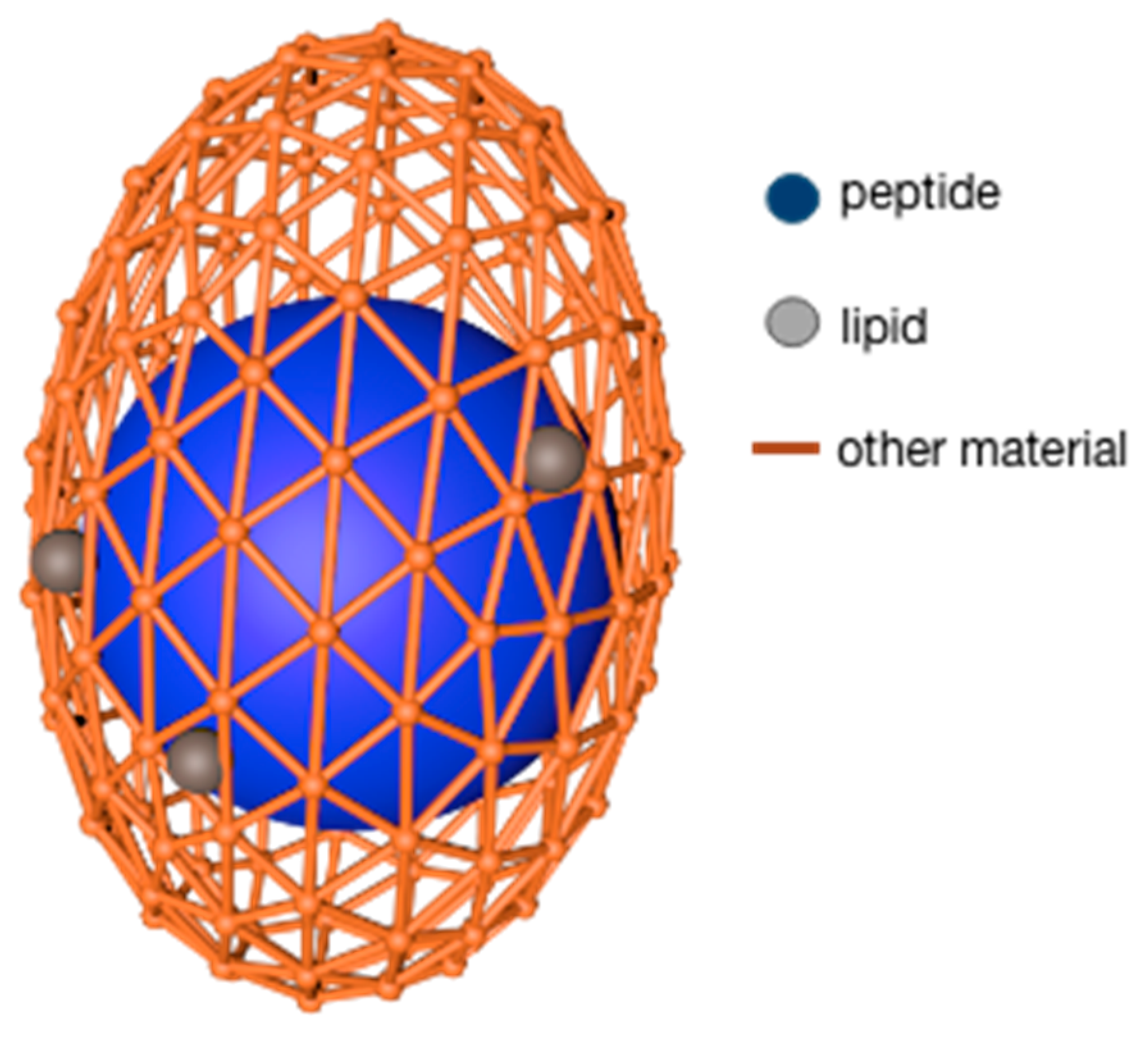
2.1.3. Lipids
2.2. Modified Polymer Wall Materials
2.2.1. Modified Cellulose
2.2.2. Modified Protein
2.3. Synthetic Polymer Wall Materials
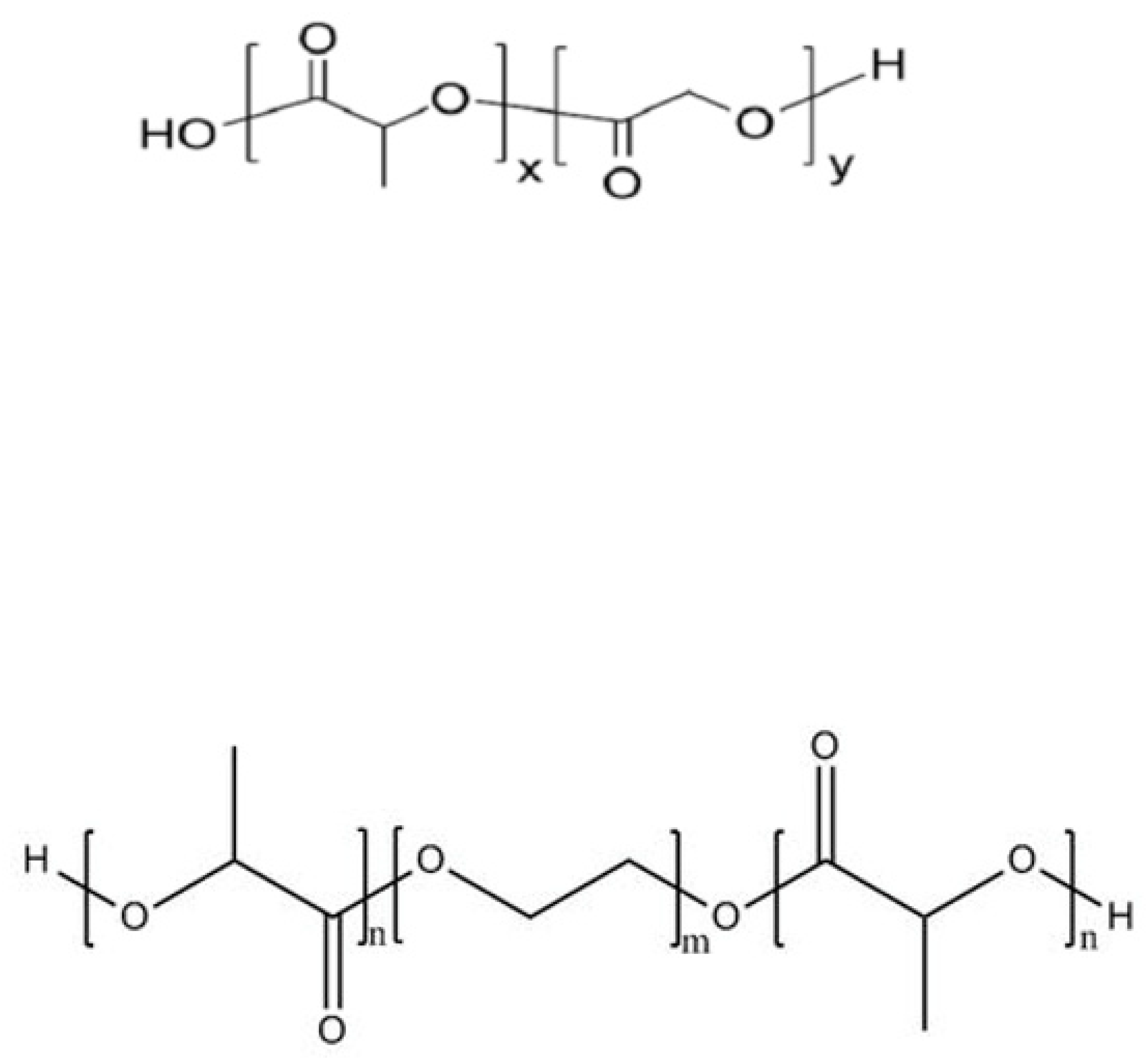
2.3.1. Polylactic Acid
2.3.2. Polydextrose
2.3.3. Polypropylene
2.4. Potential Material
3. Preparation Technology of Peptide Microcapsules
3.1. Micro-Controlled Flow (Microfluidic) Method
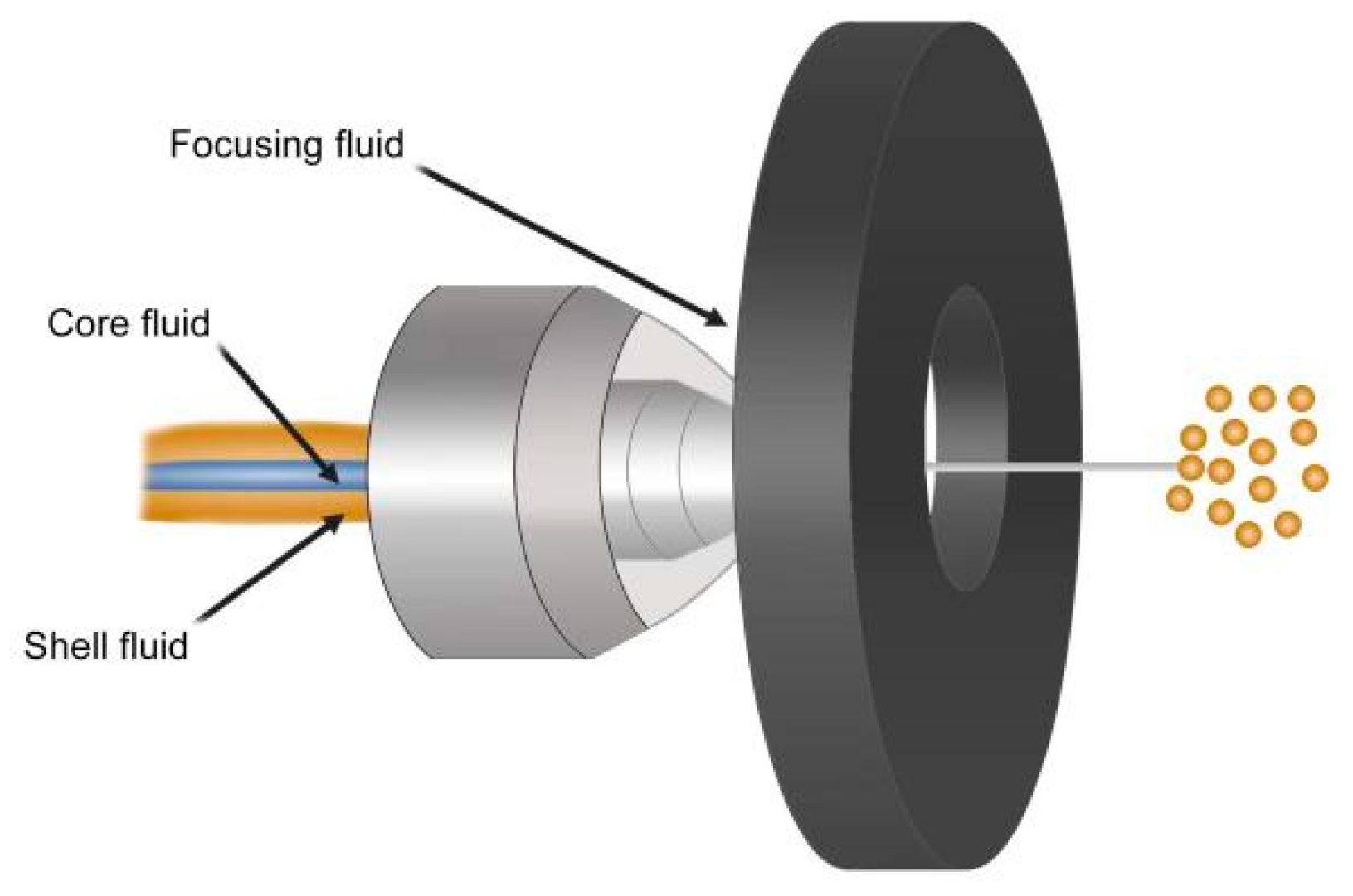
3.2. Microjet Method
3.3. Layer-by-Layer Self-Assembly (LBL) Method
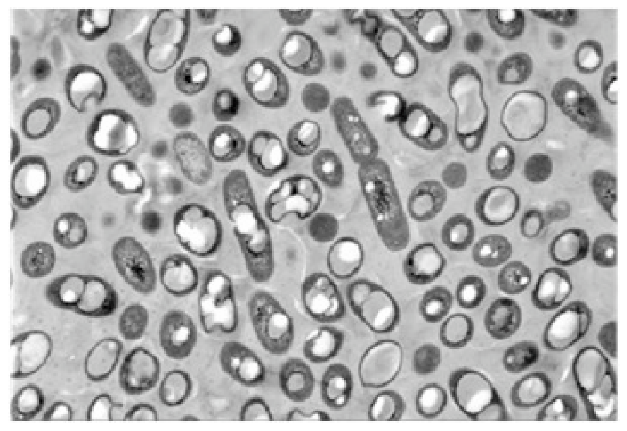
3.4. Yeast Cell Wall Method
4. Application of Peptides Microcapsules
4.1. Application of Peptides Microcapsules in Biopharmaceuticals
4.2. Application of Peptide Microcapsules in Food
4.3. Application of Peptide Microcapsules in Other Fields
5. Summary
Author Contributions
Funding
Data Availability Statement
Conflicts of Interest
References
- Yang, F.J.; Chen, X.; Huang, M.C.; Yang, Q.; Cai, X.X.; Chen, X.; Wang, S.Y. Molecular characteristics and structure–activity relationships of food-derived bioactive peptides. J. Integr. Agric. 2021, 20, 2313–2332. [Google Scholar] [CrossRef]
- Jose, M.L.; Paulo, E.S.; Munekata, B.G.; Francisco, J.B.; Leticia, M.; Cristina, P.S.; Fidel, T. Bioactive peptides as natural antioxidants in food products—A review. Trends Food Sci. Technol. 2018, 79, 136–147. [Google Scholar] [CrossRef]
- Shukla, P.; Chopda, K.; Sakure, A.; Hati, S. Current Trends and Applications of Food Derived Antihypertensive Peptides for the Management of Cardiovascular Disease. Protein Pept. Lett. 2022, 29, 408–428. [Google Scholar] [CrossRef]
- Moscoso, M.G.; Zavaleta, A.I.; Mujica, Á.; Arnao, I.; Moscoso, N.C.; Santos, M.; Sánchez, J. Antimicrobial peptides purified from hydrolysates of kanihua (Chenopodium pallidicaule Aellen) seed protein fractions. Food Chem. 2021, 360, 129951. [Google Scholar] [CrossRef]
- Ding, B.; Xu, Z.H.; Qian, C.D.; Jiang, F.S.; Ding, X.H.; Ruan, Y.P.; Ding, Z.S.; Fan, Y.S. Antiplatelet Aggregation and Antithrombosis Efficiency of Peptides in the Snake Venom of Deinagkistrodon acutus: Isolation, Identification, and Evaluation. Evid.-Based Complement. Altern. Med. 2015, 2015, 412841. [Google Scholar] [CrossRef] [PubMed] [Green Version]
- Huang, T.H.; Liu, P.Y.; Lin, Y.L.; Tai, J.S. Hypoglycemic peptide-enriched hydrolysates of Corbicula fluminea and Chlorella sorokiniana possess synergistic hypoglycemic activity through inhibiting pseudo-glucosidase and dipeptidyl peptidase-4 activity. J. Sci. Food Agric. 2021, 102, 716–723. [Google Scholar] [CrossRef]
- Chen, M.W.; Zhang, F.; Su, Y.J.; Chang, C.H.; Li, J.H.; Gu, L.P.; Yang, Y.J. Immunomodulatory effects of egg white peptides on immunosuppressed mice and sequence identification of immunomodulatory peptides. Food Biosci. 2020, 49, 101873. [Google Scholar] [CrossRef]
- Zhao, L.; Huang, S.L.; Cai, X.X.; Hong, J.; Wang, S.Y. A specific peptide with calcium chelating capacity isolated from whey protein hydrolysate. J. Funct. Foods 2014, 10, 46–53. [Google Scholar] [CrossRef]
- Luan, X.; Wu, Y.; Shen, Y.W.; Zhang, H.; Zhou, Y.D.; Chen, H.Z.; Nagle, D.G.; Zhang, W.D. Cytotoxic and antitumor peptides as novel chemotherapeutics. Nat. Prod. Rep. 2021, 38, 7–17. [Google Scholar] [CrossRef]
- Wu, Y.H.; Farrag, H.N.; Kato, T.; Li, H.; Ikeno, S. Design and Synthesis of Novel Peptides to Protect Ferulic Acid against Ultraviolet Radiation Based on Domain Site IIA of Bovine Serum Albumin. Biomolecules 2021, 11, 1285. [Google Scholar] [CrossRef]
- Zhong, H.; Shi, J.Y.; Zhang, J.H.; Wang, Q.Q.; Zhang, Y.P.; Yu, P.; Guan, R.; Feng, F.Q. Soft-Shelled Turtle Peptide Supplementation Modifies Energy Metabolism and Oxidative Stress, Enhances Exercise Endurance, and Decreases Physical Fatigue in Mice. Foods 2022, 11, 600. [Google Scholar] [CrossRef] [PubMed]
- Zhang, H.R.; Zhao, L.Y.; Shen, Q.S.; Qi, L.W.; Jiang, S.; Guo, Y.J.; Zhang, C.H.; Riche, A. Preparation of cattle bone collagen peptides-calcium chelate and its structural characterization and stability. LWT-Food Sci. Technol. 2021, 144, 111264. [Google Scholar] [CrossRef]
- Mofieed, A.; Amit, K.V.; Rajan, P. Collagen extraction and recent biological activities of collagen peptides derived from sea-food waste: A review. Sustain. Chem. Pharm. 2020, 18, 100315. [Google Scholar] [CrossRef]
- Miguel, M.; Vassallo, D.V.; Wiggers, G.A. Bioactive peptides and hydrolysates from egg proteins as a new tool for protection against cardiovascular problems. Curr. Pharm. Des. 2020, 26, 3676–3683. [Google Scholar] [CrossRef]
- Cynthia, L.; Emilie, M.; Joachim, V.L.; Lina, V.; Jozef, V.B. Peptides in insect oogenesis. Curr. Opin. Insect Sci. 2019, 31, 58–64. [Google Scholar] [CrossRef]
- Xu, H.J.; Chen, Y.L.; Li, J.W.; Luo, J.Y.; Wang, Y.M.; Ma, W.M. A novel unique terminal ampullae-expressed insulin-like peptide in male white shrimp. Penaeus Vannamei. Aquac. Rep. 2022, 23, 101011. [Google Scholar] [CrossRef]
- Zhang, C.; Zhang, Y.X.; Liu, G.R.; Li, W.H.; Xia, S.Q.; Li, H.; Liu, X.Q. Effects of soybean protein isolates and peptides on the growth and metabolism of Lactobacillus rhamnosus. J. Funct. Foods 2021, 77, 104335. [Google Scholar] [CrossRef]
- Wang, L.Y.; Lei, L.; Wan, K.; Fu, Y.; Hu, H.W. Physicochemical Properties and Biological Activity of Active Films Based on Corn Peptide Incorporated Carboxymethyl Chitosan. Coatings 2021, 11, 604. [Google Scholar] [CrossRef]
- Sun, S.L.; Zhang, G.W.; Mu, H.Y.; Zhang, H.; Chen, Y. The mixture of corn and wheat peptide prevent diabetes in NOD mice. J. Funct. Foods 2019, 56, 163–170. [Google Scholar] [CrossRef]
- Gupta, N.; Bhagyawant, S.S. Bioactive peptide of Cicer arietinum L. induces apoptosis in human endometrial cancer via DNA fragmentation and cell cycle arrest. 3 Biotech 2021, 11, 63. [Google Scholar] [CrossRef]
- Guo, H.K.; Guo, S.Y.; Liu, H.M. Antioxidant activity and inhibition of ultraviolet radiation-induced skin damage of Selenium-rich peptide fraction from selenium-rich yeast protein hydrolysate. Bioorganic Chem. 2020, 105, 104431. [Google Scholar] [CrossRef] [PubMed]
- Kenichiro, N.; Nobuhiro, K.; Noriko, S.; Chisato, Y.; Hiroshi, T. Synthesis and antimycobacterial activity of calpinactam derivatives. Bioorganic Med. Chem. Lett. 2012, 22, 7739–7741. [Google Scholar] [CrossRef]
- Zhou, J.J.; Chen, M.F.; Wu, S.J.; Liao, X.Y.; Wang, J.; Wu, Q.P.; Zhuang, M.Z.; Ding, Y. A review on mushroom-derived bioactive peptides: Preparation and biological activities. Food Res. Int. 2020, 134, 109230. [Google Scholar] [CrossRef] [PubMed]
- Chen, Y.H.; Wang, F.; Zhou, J.W.; Niu, T.T.; Xuan, R.R.; Chen, H.M.; Wu, W. In Vivo Antifatigue Activity of Spirulina Peptides Achieved by Their Antioxidant Activity and by Acting on Fat Metabolism Pathway in Mice. Nat. Prod. Commun. 2020, 15, 1934578X20946233. [Google Scholar] [CrossRef]
- Mendis, E.; Rajapakse, N.; Kim, S.K. Antioxidant properties of a radical-scavenging peptide purified from enzymatically prepared fish skin gelatin hydrolysate. J. Agric. Food Chem. 2005, 53, 581–587. [Google Scholar] [CrossRef]
- Gulay, O.; Paola, F.; Iolanda, D.M.; Jianbo, X.; Esra, C. A review of microencapsulation methods for food antioxidants: Principles, advantages, drawbacks and applications. Food Chem. 2019, 272, 494–506. [Google Scholar] [CrossRef]
- Diamante, M.; Annachiara, D.P.; Antonietta, L.S.; Teresa, C.; Francesco, E.; Gianluigi, M. Microencapsulation of nisin in alginate beads by vibrating technology: Preliminary investigation. LWT-Food Sci. Technol. 2016, 66, 436–443. [Google Scholar] [CrossRef]
- Wang, Y.F.; Qi, W.; Huang, R.L.; Su, R.X.; He, Z.M. Counterion-Directed Assembly: Counterion-Directed, Structurally Tunable Assembly of Hydrogels, Membranes, and Sacs at Aqueous Liquid-Liquid Interfaces (Adv. Mater. Interfaces 5/2016). Adv. Mater. Interfaces 2016, 3, 1500327. [Google Scholar] [CrossRef]
- Santana, A.A.; Cano, H.D.M.; Oliveira, R.A.; Telis, V.R.N. Influence of different combinations of wall materials on the microencapsulation of jussara pulp (Euterpe edulis) by spray drying. Food Chem. 2016, 212, 1–9. [Google Scholar] [CrossRef]
- Javier, D.H.L.; Luis, A.B.P.; Alvarez, R.J.; Hugo, S.G. Microencapsulation using starch as wall material: A review. Food Rev. Int. 2017, 34, 148–161. [Google Scholar] [CrossRef]
- Yang, M.Y.; Liang, Z.; Wang, L.; Qi, M.; Luo, Z.S.; Li, L. Microencapsulation Delivery System in Food Industry-Challenge and the Way Forward. Adv. Polym. Technol. 2020, 13, 7531810. [Google Scholar] [CrossRef]
- Jéssica, S.R.; Cristiane, M.V. Microencapsulation of natural dyes with biopolymers for application in food: A review. Food Hydrocoll. 2021, 112, 106374. [Google Scholar] [CrossRef]
- Ramprakash, B.; Incharoensakdi, A. Alginate encapsulated nanobio-hybrid system enables improvement of photocatalytic biohydrogen production in the presence of oxygen. Int. J. Hydrog. Energy 2022, 47, 11492–11499. [Google Scholar] [CrossRef]
- Abdel, A.M.S.; Salama, H.E. Developing multifunctional edible coatings based on alginate for active food packaging. Int. J. Biol. Macromol. 2021, 190, 837–844. [Google Scholar] [CrossRef] [PubMed]
- Pratiksha, S.; Pankaj, B.; Omprakash, S.Y. Synthesis, characterization and application of crosslinked alginate as green packaging material. Heliyon 2020, 6, e03026. [Google Scholar] [CrossRef] [Green Version]
- Sikorski, P.; Mo, F.; Skjak, B.G.; Stokke, B.T. Evidence for egg-box-compatible interactions in calcium-alginate gels from fiber X-ray diffraction. Biomacromolecules 2007, 8, 2098–2103. [Google Scholar] [CrossRef]
- Kumar, A.; Belhaj, M.; DiPette, D.J.; Potts, J.D. A Novel Alginate-Based Delivery System for the Prevention and Treatment of Pressure-Overload Induced Heart Failure. Front. Pharmacol. 2021, 11, 602952. [Google Scholar] [CrossRef] [PubMed]
- Oki, Y.; Kirita, K.; Ohta, S.; Ohba, S.; Horiguchi, I.; Sakai, Y.; Ito, T. Switching of Cell Proliferation/ Differentiation in Thiol-Maleimide Clickable Microcapsules Triggered by in Situ Conjugation of Biomimetic Peptides. Biomacromolecules 2019, 20, 2350–2359. [Google Scholar] [CrossRef] [PubMed]
- Ambaye, T.G.; Vaccari, M.; Prasad, S.; van Hullebusch, E.D.; Rtimi, S. Preparation and applications of chitosan and cellulose composite materials. J. Environ. Manag. 2022, 301, 113850. [Google Scholar] [CrossRef]
- Zhao, M.G.; He, H.; Guo, D.J.; Zhang, X.; Jia, L.; Hou, T.; Ma, A.M. Chitosan oligosaccharides-tripolyphosphate microcapsules as efficient vehicles for desalted duck egg white peptides-calcium: Fabrication, entrapment mechanism and in vivo calcium absorption studies. LWT-Food Sci. Technol. 2022, 154, 112869. [Google Scholar] [CrossRef]
- Li, Z.L.; Chen, P.; Xu, X.Z.; Ye, X.; Wang, J. Preparation of chitosan-sodium alginate microcapsules containing ZnS nanoparticles and its effect on the drug release. Mater. Sci. Eng. C 2009, 29, 2250–2253. [Google Scholar] [CrossRef]
- Salvatore, D.G.; Chasper, P.; Lipps, G. Stable and selective permeable hydrogel microcapsules for high-throughput cell cultivation and enzymatic analysis. Microb. Cell Factories 2020, 19, 170. [Google Scholar] [CrossRef]
- Ansari, Z.; Goomer, S. Natural Gums and Carbohydrate-Based Polymers: Potential Encapsulants. Indo Glob. J. Pharm. Sci. 2022, 12, 1–20. [Google Scholar] [CrossRef]
- Alicia, H.; Fabra, M.J.; Frédéric, D.; Cécile, D.B.; Andrée, V. Interface and aroma barrier properties of iota-carrageenan emulsion–based films used for encapsulation of active food compounds. J. Food Eng. 2009, 93, 80–88. [Google Scholar] [CrossRef]
- Joanna, T.; Ewelina, J.; Ewa, P.; Barbara, B.; Joanna, K.D. Furcellaran-Coated Microcapsules as Carriers of Cyprinus carpio Skin-Derived Antioxidant Hydrolysate: An In Vitro and In Vivo Study. Nutrients 2019, 11, 2502. [Google Scholar] [CrossRef] [Green Version]
- Raú, l.E.C.; Pablo, R.S.; Adriana, N.M.; Silvina, R.D. Pyropia columbina phycocolloids as microencapsulating material improve bioaccessibility of brewers’ spent grain peptides with ACE-I inhibitory activity. Int. J. Food Sci. Technol. 2020, 55, 1311–1317. [Google Scholar] [CrossRef]
- Nazia, T.; Suhani, D.K. Synthesis, characterization and applications of copolymer of β-cyclodextrin: A review. J. Polym. Res. 2020, 27, 1–30. [Google Scholar] [CrossRef]
- Crini, G.; Fourmentin, S.; Fenyvesi, É.; Torri, G.; Fourmentin, M.; Morin, C.N. Cyclodextrins, from molecules to applications. Environ. Chem. Lett. 2018, 16, 1361–1375. [Google Scholar] [CrossRef]
- Pawar, S.; Shende, P. A Comprehensive Patent Review on β-cyclodextrin Cross-linked Nanosponges for Multiple Applications. Recent Pat. Nanotechnol. 2020, 14, 75–89. [Google Scholar] [CrossRef]
- Desai, D.; Shende, P. Monodispersed cyclodextrin-based nanocomplex of neuropeptide Y for targeting MCF-7 cells using a central composite design. J. Drug Deliv. Sci. Technol. 2021, 65, 102692. [Google Scholar] [CrossRef]
- Chen, L.Y.; Gabriel, E.R.; Muriel, S. Food protein-based materials as nutraceutical delivery systems. Trends Food Sci. Technol. 2006, 17, 272–283. [Google Scholar] [CrossRef]
- Dave, J.; Ye, X.; Jethro, M.; Xiao, H. Protein-Based Drug-Delivery Materials. Materials 2017, 5, 517. [Google Scholar] [CrossRef] [Green Version]
- Fan, Q.Q.; Ma, J.Z.; Xu, Q.; Zhang, J.; Demetra, S.; Gaidău, C.; Guo, C. Animal-derived natural products review: Focus on novel modifications and applications. Colloids Surf. B Biointerfaces 2015, 128, 181–190. [Google Scholar] [CrossRef]
- Kantrol, K.S.; Monika, K.; Ravi, S.P. Chylomicron mimicking solid lipid nanoemulsions encapsulated enteric minicapsules targeted to colon for immunization against hepatitis B. Int. Immunopharmacol. 2019, 66, 317–329. [Google Scholar] [CrossRef]
- Ahmady, A.; Hayati, A.S.N. A review: Gelatine as a bioadhesive material for medical and pharmaceutical applications. Int. J. Pharm. 2021, 608, 121037. [Google Scholar] [CrossRef] [PubMed]
- Favaro, C.S.; Santana, A.S.; Monterrey, E.S.; Trindade, M.A.; Netto, F.M. The use of spray drying technology to reduce bitter taste of casein hydrolysate. Food Hydrocoll. 2009, 24, 336–340. [Google Scholar] [CrossRef]
- Niu, H.X.; Chang, J.; Jia, Y.D. Microencapsulation of crystalline-methionine enclosed with gelatine and sodium alginate by spray-drying. Mater. Res. Innov. 2015, 19, 257–262. [Google Scholar] [CrossRef]
- Ashaolu, T.J. Applications of soy protein hydrolysates in the emerging functional foods: A review. Int. J. Food Sci. Technol. 2020, 55, 421–428. [Google Scholar] [CrossRef]
- Gao, X.Q.; Xiong, G.Y.; Fu, L.; Liu, S.L. Water distribution of raw and heat-induced gelation of minced pork paste prepared by soy protein isolates and carrageenan: Ingredients modify the gelation of minced pork. J. Food Process. Preserv. 2019, 43, e14221. [Google Scholar] [CrossRef]
- Wei, C.L. Construction of soybean peptide-curcumin nanoparticles and their microencapsulation. South China Univ. Technol. 2019, 24, 1–86. [Google Scholar] [CrossRef]
- Zhao, C.H.; Chen, N.; Ashaolu, T.J. Whey proteins and peptides in health-promoting functions—A review. Int. Dairy J. 2022, 126, 105269. [Google Scholar] [CrossRef]
- Farizano, J.V.; Díaz, V.L.I.; Masias, E.; Baillo, A.A.; Torino, M.I.; Fadda, S.; Vanden, B.N.L.; Montenegro, M.A.; Saavedra, L.; Minahk, C. Biotechnological use of dairy by-products for the production and microencapsulation of the food preservative enterocin CRL35. FEMS Microbiol. Lett. 2022, 369, fnac033. [Google Scholar] [CrossRef] [PubMed]
- Zubair, M.; Pradhan, R.A.; Arshad, M.; Ullah, A. Recent Advances in Lipid Derived Bio-Based Materials for Food Packaging Applications. Macromol. Mater. Eng. 2021, 306, 1–35. [Google Scholar] [CrossRef]
- Blanco-Pascual, N.; Koldeweij, R.B.J.; Stevens, R.S.A.; Montero, M.P.; Gómez-Guillén, M.C.; Cate, A.T. Peptide Microencapsulation by Core-Shell Printing Technology for Edible Film Application. Food Bioprocess Technol. 2014, 7, 2472–2483. [Google Scholar] [CrossRef] [Green Version]
- Jiang, Z.L.; To, N. Recent advances in chemically modified cellulose and its derivatives for food packaging applications: A review. Polymers 2022, 14, 1533. [Google Scholar] [CrossRef] [PubMed]
- Aomatsu, Y.; Nakajima, Y.; Ohyama, T.; Kin, T.; Kanehiro, H.; Hisanaga, M.; Ko, S.; Nagao, M.; Tatekawa, Y.; Sho, M.; et al. Efficacy of agarose/polystyrene sulfonic acid microencapsulation for islet xenotransplantation. Transplant. Proc. 2000, 32, 1071–1072. [Google Scholar] [CrossRef]
- Nishimura, M.; Iizuka, N.; Fujita, Y.; Sawamoto, O.; Matsumoto, S. Effects of encapsulated porcine islets on glucose and C-peptide concentrations in diabetic nude mice 6 months after intraperitoneal transplantation. Xenotransplantation 2017, 24, e12313. [Google Scholar] [CrossRef]
- Cesar, A.R.B.; Larissa, P.P.; Elisabete, A.L.G.; Nilce, M.S.; Priscilla, A.B.M.L.; Douglas, D.A.S.; Andreia, B.M.; Marlus, C.; Eduardo, F.V. HPMCP-coated microcapsules containing the ctx (Ile21)-ha antimicrobial peptide reduce the mortality rate caused by resistant salmonella enteritidis in laying hens. Antibiotics 2021, 10, 616. [Google Scholar] [CrossRef]
- Jenny, K.R.; Luz, S.; Mary, A.A. Stabilization of oils by microencapsulation with heated protein-glucose syrup mixtures. J. Am. Oil Chem. Soc. 2006, 83, 965–972. [Google Scholar] [CrossRef]
- Pavel, S.; Vladimir, M. Protein interaction with charged macromolecules: From model polymers to unfolded proteins and post- translational modifications. Int. J. Mol. Sci. 2019, 20, 1252. [Google Scholar] [CrossRef] [Green Version]
- Swati, K.; Aasima, R.; Savita, S. Protein engineering and its applications in food industry. Taylor Fr. 2017, 57, 2321–2329. [Google Scholar] [CrossRef]
- Wang, Z.G.; Ju, X.R.; He, R.; Yuan, J.; Wang, L.F. Effect of rapeseed protein structural modification on microstructural properties of peptide microcapsules. Food Bioprocess Technol. 2015, 8, 1305–1318. [Google Scholar] [CrossRef]
- Deborah, M.S.; Joachim, K. A synthetic polymer matrix for the delayed or pulsatile release of water-soluble peptides. J. Control. Release 2002, 78, 143–153. [Google Scholar] [CrossRef]
- Li, X.M.; Xu, Y.L.; Chen, G.G.; Wei, P.; Ping, Q.N. PLGA nanoparticles for the oral delivery of 5-Fluorouracil using high pressure homogenization-emulsification as the preparation method and in vitro/in vivo studies. Drug Dev. Ind. Pharm. 2008, 34, 107–115. [Google Scholar] [CrossRef] [PubMed]
- Justin, K.Y.H.; Steven, P.S. Characterization of octreotide-PLGA binding by isothermal titration calorimetry. Biomacromolecules 2020, 21, 4087–4093. [Google Scholar] [CrossRef]
- Lim, S.M.; Eom, H.N.; Jiang, H.H.; Sohn, M.J.; Lee, K.C. Evaluation of PEGylated exendin-4 released from poly (lactic-co-glycolic acid) microspheres for antidiabetic therapy. J. Pharm. Sci. 2015, 104, 72–80. [Google Scholar] [CrossRef]
- Zhang, Y.; Wu, X.H.; Han, Y.R.; Mo, F.; Duan, Y.R.; Li, S.M. Novel thymopentin release systems prepared from bioresorbable PLA-PEG-PLA hydrogels. Int. J. Pharm. 2010, 386, 15–22. [Google Scholar] [CrossRef]
- Burdock, G.A.; Flamm, W.G. A review of the studies of the safety of polydextrose in food. Food Chem. Toxicol. 1999, 37, 233–264. [Google Scholar] [CrossRef]
- Marília, P.F.; Bruna, G.; Maria, E.C.S.; Izabela, D.A.; Maria, T.B.P. Microencapsulation performance of Fe-peptide complexes and stability monitoring. Food Res. Int. 2019, 125, 108505. [Google Scholar] [CrossRef]
- Günay, K.A.; Berthier, D.L.; Jerri, H.A.; Benczédi, D.; Klok, H.-A.; Herrmann, A. Selective Peptide-Mediated Enhanced Deposition of Polymer Fragrance Delivery Systems on Human Hair. ACS Appl. Mater. Interfaces 2017, 9, 24238–24249. [Google Scholar] [CrossRef]
- Shashi, K.B.; Ranjit, G.; Choi, T.R.; Jung, H.R.; Yang, S.Y.; Song, H.S.; Jeon, J.M.; Kim, J.S.; Lee, Y.K.; Yang, Y.H. Poly(3-hydroxybutyrate-co-3-hydroxyhexanoate) production from engineered Ralstonia eutropha using synthetic and anaerobically digested food waste derived volatile fatty acids. Int. J. Biol. Macromol. 2019, 133, 1–10. [Google Scholar] [CrossRef]
- Sharma, V.; Sehgal, R.; Gupta, R. Polyhydroxyalkanoate (PHA): Properties and Modifications. Polymer 2020, 212, 123161. [Google Scholar] [CrossRef]
- Cao, L.D.; Liu, Y.J.; Xu, C.L.; Zhou, Z.L.; Zhao, P.Y.; Niu, S.J.; Huang, Q.L. Biodegradable poly(3-hydroxybutyrate-co-4-hydroxybutyrate) microcapsules for controlled release of trifluralin with improved photostability and herbicidal activity. Mater. Sci. Eng. C 2019, 102, 134–141. [Google Scholar] [CrossRef] [PubMed]
- Svetlana, U.; Cruz, M.J.; Cabeza, L.F.; Grágeda, M. Preparation and Characterization of Inorganic PCM Microcapsules by Fluidized Bed Method. Materials 2016, 9, 24. [Google Scholar] [CrossRef] [Green Version]
- Jin, Y.; Zhou, Q.; Li, Z.H.; Yang, Z.H.; Fan, H.J. Calcium-cross linked polysaccharide microcapsules for controlled release and antimicrobial applications. Colloids Surf. A Physicochem. Eng. Asp. 2020, 600, 125025. [Google Scholar] [CrossRef]
- Chatterjee, S.; Judeh, Z.M.A. Impact of encapsulation on the physicochemical properties and gastrointestinal stability of fish oil. LWT-Food Sci. Technol. 2016, 65, 206–213. [Google Scholar] [CrossRef]
- Sahar, A.; Elham, A.; Iman, K.; Seid, M.J. Lipid nano scale cargos for the protection and delivery of food bioactive ingredients and nutraceuticals. Trends Food Sci. Technol. 2018, 74, 132–146. [Google Scholar] [CrossRef]
- Beghetto, V.; Sole, R.; Buranello, C.; AlAbkal, M.; Facchin, M. Recent advancements in plastic packaging recycling: A mini-review. Materials 2021, 14, 4782. [Google Scholar] [CrossRef]
- Gou, M.L.; Wei, X.W.; Men, K.; Wang, B.L.; Luo, F.; Zhao, X.; Wei, Y.Q.; Qian, Z.Y. PCL/PEG copolymeric nanoparticles: Potential nanoplatforms for anticancer agent delivery. Curr. Drug Targets 2011, 12, 1131–1150. [Google Scholar] [CrossRef]
- Kim, M.R.; Feng, T.; Zhang, Q.; Chan, H.Y.E.; Chau, Y. Co-Encapsulation and Co-Delivery of Peptide Drugs via Polymeric Nanoparticles. Polymers 2019, 11, 288. [Google Scholar] [CrossRef] [Green Version]
- Zhang, D.M.; Zhang, Q.; Lu, Y.L.; Yao, Y.; Li, S.; Jiang, J.; Liu, G.L.; Liu, Q.J. Peptide Functionalized Nanoplasmonic Sensor for Explosive Detection. Nano-Micro Lett. 2016, 8, 36–43. [Google Scholar] [CrossRef] [PubMed] [Green Version]
- Song, J.Y.; CortezJugo, C.; Shirbin, S.J.; Lin, Z.X.; Pan, S.J.; Qiao, G.G.; Caruso, F. Immobilization and Intracellular Delivery of Structurally Nanoengineered Antimicrobial Peptide Polymers Using Polyphenol-Based Capsules. Adv. Funct. Mater. 2022, 32, 2107341. [Google Scholar] [CrossRef]
- Miléna, L.; Nikolett, K.S.; Vince, A.; András, J.L.; István, A. Microparticles, Microspheres, and Microcapsules for Advanced Drug Delivery. Sci. Pharm. 2019, 87, 20. [Google Scholar] [CrossRef] [Green Version]
- Ghiman, R.; Pop, R.; Rugina, D.; Focsan, M. Recent progress in preparation of microcapsules with tailored structures for bio-medical applications. J. Mol. Struct. 2022, 1248, 131366. [Google Scholar] [CrossRef]
- Jaganathan, M.; Madhumitha, D.; Dhathathreyan, A. Protein microcapsules: Preparation and applications. Adv. Colloid Interface Sci. 2014, 209, 1–7. [Google Scholar] [CrossRef]
- Alexander, G.; Martin, W.; Andreas, B.S. Oral peptide delivery: In-vitro evaluation of thiolated alginate/poly(acrylic acid) microparticles. J. Pharm. Pharmacol. 2010, 59, 1191–1198. [Google Scholar] [CrossRef]
- Tang, Y.T.; Arbaugh, B.; Park, H.; Scher, H.B.; Bai, L.; Mao, L.; Jeoh, T. Targeting enteric release of therapeutic peptides by encapsulation in complex coacervated matrix microparticles by spray drying. J. Drug Deliv. Sci. Technol. 2022, 79, 104063. [Google Scholar] [CrossRef]
- Raúl, E.C.; Andrea, C.S.; Luis, C.G.; Silvina, R.D.; David, B.A. Bioactive Phaseolus lunatus peptides release from maltodextrin/gum arabic microcapsules obtained by spray drying after simulated gastrointestinal digestion. Int. J. Food Sci. Technol. 2019, 54, 2002–2009. [Google Scholar] [CrossRef] [Green Version]
- Situ, W.B.; Li, X.X.; Liu, J.; Chen, L. Preparation and characterization of glycoprotein-resistant starch complex as a coating material for oral bioadhesive microparticles for colon-targeted polypeptide delivery. J. Agric. Food Chem. 2015, 63, 4138–4147. [Google Scholar] [CrossRef]
- Situ, W.B.; Chen, L.; Wang, X.Y.; Li, X.X. Resistant starch film-coated microparticles for an oral colon-specific polypeptide delivery system and its release behaviors. J. Agric. Food Chem. 2014, 62, 3599–3609. [Google Scholar] [CrossRef] [PubMed]
- Agrawal, H.; Joshi, R.; Gupta, M. Optimization of pearl millet-derived bioactive peptide microspheres with double emulsion solvent evaporation technique and its release characterization. Food Struct. 2021, 29, 100200. [Google Scholar] [CrossRef]
- Wu, Z.M.; Zhou, L.Y.; Guo, X.D.; Jiang, W.; Ling, L.; Qian, Y.; Luo, K.Q.; Zhang, L.J. HP55-coated capsule containing PLGA/RS nanoparticles for oral delivery of insulin. Int. J. Pharm. 2012, 425, 1–8. [Google Scholar] [CrossRef] [PubMed]
- Kondiah, P.P.D.; Choonara, Y.E.; Tomar, L.K.; Tyagi, C.; Kumar, P.; Toit, L.C.; Marimuthu, T.; Modi, G.; Pillay, V. Development of a Gastric Absorptive, Immediate Responsive, Oral Protein-Loaded Versatile Polymeric Delivery System. AAPS PharmSciTech 2017, 18, 2479–2493. [Google Scholar] [CrossRef]
- Sun, X.T.; Liu, M.; Xu, Z.R. Microfluidic fabrication of multifunctional particles and their analytical applications. Talanta 2014, 121, 163–177. [Google Scholar] [CrossRef]
- Shimanovich, U.; Levin, A.; Eliaz, D.; Michaels, T.; Toprakcioglu, Z.; Frohm, B.; De, G.E.; Linse, S.; ÅkerfeldT, K.S.; Knowles, T.P.J. pH-Responsive capsules with a fibril scaffold shell assembled from an amyloidogenic peptide. Small 2021, 17, 2007188. [Google Scholar] [CrossRef] [PubMed]
- Han, F.Y.; Xu, W.Z.; Kumar, V.; Cui, C.S.; Li, X.R.; Jiang, X.Y.; Woodruff, T.M.; Whittaker, A.K.; Smith, M.T. Optimisation of a Microfluidic Method for the Delivery of a Small Peptide. Pharmaceutics 2021, 13, 1505. [Google Scholar] [CrossRef] [PubMed]
- Calva, E.S.J.; Lugo, C.E.; Jiménez, F.M. Microencapsulation of cocoa liquor nanoemulsion with whey protein using spray drying to protection of volatile compounds and antioxidant capacity. J. Microencapsul. 2019, 36, 447–458. [Google Scholar] [CrossRef]
- Zhang, C.; Siew, L.A.K.; Chen, X.D.; Siew, Y.Q. Microencapsulation of fermented noni juice via micro-fluidic-jet spray drying: Evaluation of powder properties and functionalities. Powder Technol. 2020, 361, 995–1005. [Google Scholar] [CrossRef]
- Miriam, G.V.; Hugo, E.A.; Guadalupe, M.G.; Hugo, E.S.; Enrique, A.G. Oxidative stability of green coffee oil (Coffea arabica) microencapsulated by spray drying. Processes 2019, 7, 734. [Google Scholar] [CrossRef] [Green Version]
- Alfonso, G.G.; Elena, C.H.; María, F.M.; Lucía, M.B. Massive, generic, and controlled microencapsulation by flow focusing: Some physicochemical aspects and new applications. J. Flow Chem. 2015, 5, 48–54. [Google Scholar] [CrossRef]
- Laura, A.; Graça, F.; Claúdia, M.; Joana, V.; Isabel, C. Gouveia. Bioactive microsphere-based coating for biomedical-textiles with encapsulated antimicrobial peptides (AMPs). Ciência Tecnol. Dos Mater. 2014, 26, 118–125. [Google Scholar] [CrossRef]
- Jaime, M.G.; Teresa, G.M.; Juan, C.M.; Juan, M. Yeast Immobilization Systems for Alcoholic Wine Fermentations: Actual Trends and Future Perspectives. Front. Microbiol. 2018, 9, 241. [Google Scholar] [CrossRef] [Green Version]
- Huizar, C.C.; Ji, N.; Reddick, R.; Ostroff, G.R.; Forsthuber, T.G. Glucan particles as a novel adjuvant for the induction of experimental autoimmune encephalomyelitis. Cell. Immunol. 2021, 366, 104383. [Google Scholar] [CrossRef] [PubMed]
- Mochizuki, Y.; Kogawa, R.; Takegami, R.; Nakamura, K.; Wakabayashi, A.; Ito, T.; Yoshioka, Y. Co-microencapsulation of islets and msc cellsaics, mosaic-like aggregates of mscs and recombinant peptide pieces, and therapeutic effects of their subcutaneous transplantation on diabetes. Biomedicines 2020, 8, 318. [Google Scholar] [CrossRef]
- Jiang, T.; Singh, B.J.; Sushila, M.; Li, H.S.; Kang, S.K.; Bok, J.D.; Cho, C.G.; Choi, Y.J. Oral delivery of probiotic expressing M cell homing peptide conjugated BmpB vaccine encapsulated into alginate/chitosan/alginate microcapsules. Eur. J. Pharm. Biopharm. 2014, 88, 768–777. [Google Scholar] [CrossRef]
- Wang, Y.; Hou, L.L.; Su, D.; Wang, Z.X.; Jia, Y.M. Optimization of Brevilaterin Microencapsulation and Analysis of Slow-release Characteristics. Food Sci. 2021, 42, 38–43. [Google Scholar] [CrossRef]
- Hu, Y.Q.; Chen, Y.; Lin, L.J.; Zhang, J.H.; Lan, R.G.; Wu, B.L. Studies on antimicrobial peptide-loaded nanomaterial for root caries restorations to inhibit periodontitis related pathogens in periodontitis care. J. Microencapsul. 2021, 38, 89–99. [Google Scholar] [CrossRef]
- Cesar, R.A.; Saraiva, M.M.; Daniel, F.M. Alginate-based microparticles coated with HPMCP/AS cellulose-derivatives enable the Ctx(Ile 21)-Ha antimicrobial peptide application as a feed additive. Int. J. Biol. Macromol. 2022, 8, 472–481. [Google Scholar] [CrossRef]


| Classification | Source | Common Types | Advantages | Disadvantages | References |
|---|---|---|---|---|---|
Natural polymer wall materials | Extracted from natural substances | Carbohydrates. protein, lipids | Biocompatibility, environmentally friendly and wide range of sources at low cost | Low mechanical strength, low loading capacity | [33,34,35,36,39], [49,50,51,52], [87] |
| Modified polymer wall materials | Modification of natural material | Modified protein, modified cellulose | Good stability and Does not age easily | Difficult to prepare, low preparation efficiency | [59], [61,62,63], [65] |
Synthetic polymer wall materials | Artificial synthesis | Biodegradable, non-biodegradable | High drug loading capacity, high mechanical strength | Expensive | [63,65] [69] [88,89,90] |
| Metallic material | - | Au, Fe3+ | Good stability | Low drug loading capacity | [91,92] |
| Method | Equipment | Advantages | Disadvantages | References |
|---|---|---|---|---|
| Ionotropic gelation | - | Simple operation | Time-consuming | [40,96] |
| Spray drying | Spray dryer | Easy operation, continuous production | Particle size heterogeneity | [97,98] |
| Extrusion–spheronisation | Constant flow pump | Low temperature | Low production rate | [99,100] |
| Molecular embedding | Ultrasonic | Easy operation | Core material limited | [49] |
| Solvent evaporation | Vacuum freeze dryer | Low temperature | Residual solvents | [101,102] |
| Free radical polymerization | - | Simple operation, low cost | Low production rate | [103] |
| Applications | Peptides | Objective | Function | Reference |
|---|---|---|---|---|
| Biopharmaceuticals | Antimicrobial peptide | Antipneumonia drug | Preventing peptide degradation, reducing clearance, enhancing intracellular delivery | [92] |
| Recombinant peptides and rat islets | Subcutaneous Islet drug | Improving blood sugar normalization | [114] | |
| M-cell homing peptide | Oral vaccine | Improving the vitality and survival rate of peptides | [115] | |
| Food | Duck egg white peptides-calcium | Calcium supplement | promoting calcium absorption, avoiding stomach digestion | [40] |
| Nisin | Nisin storage | Improving survival rate of nisin | [27] | |
| Antibacterial peptide | Bread preservative | Increasing preservation time of bread | [116] | |
| Others | Antibacterial peptide | Anti-cavity drug | Increasing bacteriostasis rate | [117] |
| Phage identification peptides | Fragrance retaining agent | Increasing the deposition and release of aromatic substances on the hair | [80] | |
| Ctx(Ile21)-Ha antimicrobial peptide | Feed additive | Reducing systemic infection of Streptococcus enteritidis in chickens | [118] |
Disclaimer/Publisher’s Note: The statements, opinions and data contained in all publications are solely those of the individual author(s) and contributor(s) and not of MDPI and/or the editor(s). MDPI and/or the editor(s) disclaim responsibility for any injury to people or property resulting from any ideas, methods, instructions or products referred to in the content. |
© 2023 by the authors. Licensee MDPI, Basel, Switzerland. This article is an open access article distributed under the terms and conditions of the Creative Commons Attribution (CC BY) license (https://creativecommons.org/licenses/by/4.0/).
Share and Cite
Li, M.; Guo, Q.; Lin, Y.; Bao, H.; Miao, S. Recent Progress in Microencapsulation of Active Peptides—Wall Material, Preparation, and Application: A Review. Foods 2023, 12, 896. https://doi.org/10.3390/foods12040896
Li M, Guo Q, Lin Y, Bao H, Miao S. Recent Progress in Microencapsulation of Active Peptides—Wall Material, Preparation, and Application: A Review. Foods. 2023; 12(4):896. https://doi.org/10.3390/foods12040896
Chicago/Turabian StyleLi, Mengjie, Quanyou Guo, Yichen Lin, Hairong Bao, and Song Miao. 2023. "Recent Progress in Microencapsulation of Active Peptides—Wall Material, Preparation, and Application: A Review" Foods 12, no. 4: 896. https://doi.org/10.3390/foods12040896