Article

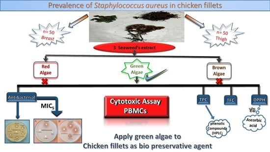
Evaluation of the Prevalence of Staphylococcus aureus in Chicken Fillets and Its Bio-Control Using Different Seaweed Extracts

1
Department of Food Technology, Arid Lands Cultivation Research Institute, City of Scientific Research and Technological Applications, New Borg El-Arab 21934, Egypt
2
Department of Food Hygiene and Control, Faculty of Veterinary Medicine, Alexandria University, Alexandria 21544, Egypt
3
Department of Food Control, Faculty of Veterinary Medicine, Kafrelsheikh University, Kafrelsheikh 33516, Egypt
4
Department of Botany and Microbiology, Faculty of Science, Damietta University, Damietta 34511, Egypt
5
Department of Food Hygiene and Control, Faculty of Veterinary Medicine, Benha University, Qaluobia 13736, Egypt
6
Department of Microbiology, Faculty of Veterinary Medicine, Matrouh University, Matrouh 51511, Egypt
7
Department of Food Development and Food Quality, Institute of Food Science and Human Nutrition, Gottfried Wilhelm Leibniz University Hannover, Am Kleinen Felde 30, 30167 Hannover, Germany
8
Department of Food Science, Faculty of Agriculture (Saba Basha), Alexandria University, Alexandria 21531, Egypt
9
Department of Functional Materials and Electronics, Center for Physical Sciences and Technology, Saulėtekio al. 3, 10257 Vilnius, Lithuania
*
Authors to whom correspondence should be addressed.
Foods 2023, 12(1), 20; https://doi.org/10.3390/foods12010020
Submission received: 22 November 2022 / Revised: 12 December 2022 / Accepted: 16 December 2022 / Published: 21 December 2022

Abstract

:
This study aims to assess the occurrence of Staphylococcus aureus in chicken fillets and to control its growth using various lyophilized seaweed extracts (i.e., Halimeda opuntia (HO), Actinotrichia fragilis, and Turbinaria turbinata) by an agar disk diffusion assay in vitro. Results showed that prevalence of S. aureus in breast and thigh samples reached of 92% and 84%, respectively. Lyophilized HO extract was the only seaweed that showed the antibacterial activity against S aureus with a significant difference at p < 0.05. The minimum inhibitory concentration (MIC) of HO extract was 1.5%, with an inhibition zone of 8.16 ± 0.73 mm. Regarding 1,1-diphenyl-2-picrylhydrazyl (DPPH) scavenging activity, IC50 was recorded at 55.36 μg/mL, whereas cytotoxic IC50 of the lyophilized HO extract on peripheral blood mononuclear cells (PBMCs) was 33.7 µg/mL; a higher IC50 of HO extracts permits their use as a safe food additive in meat products. Moreover, total phenolic compounds and total flavonoids compounds recorded 20.36 ± 0.092 and 16.59 ± 0.029 mg/mL, respectively. HPLC analyses of phenolic compounds profiles exhibited many bioactive substances and the higher ratio was daidzein with 10.84 ± 0.005 µg/mL and followed by gallic acid with a value of 4.06 ± 0.006 µg/mL. In a challenge study, chicken fillet (CHF) experimentally inoculated with S. aureus (ST) and treated with the lyophilized HO algal extract at 4% and 6% (CHF/ST/HO) showed a complete reduction of S. aureus count on the 6th and 4th days in chicken fillet stored at 4 °C, respectively. Moreover, CHF/ST/HO at 4% and 6% of HO extract enhanced the sensory attributes of grilled un-inoculated chicken fillet. Thus, lyophilized HO extracts are promising antibacterial and antioxidant candidates in the chicken meat industry.

Graphical Abstract

1. Introduction

Food safety is a top priority for both public health and the economy. Approximately 1 out of 10 yearly suffers from food poisoning from contaminated food consumption [1]. Providing high-quality, safe, and nutritious food will become increasingly difficult in the next decades [2], as both nutrition and food safety are interlinked for health outcomes from food systems [3].
Chicken meat products are commonly recognized as an important source of protein worldwide. In 2018, world poultry production reached 123 million tons in 1 year, with a prediction to increase [4]. However, chicken meat products commonly harbor S. aureus, a food poisoning bacterium [5] and a pathogen that contaminates food during handling and processing [6]. It is a Gram-positive, facultative anaerobe, and toxic bacterium [7]. S. aureus is a major concern in chicken meat and shows resistance to various antibiotic types, even methicillin [8]. Recent research has focused on using natural antimicrobials in meat products [9]. A novel natural antimicrobial that has antibacterial impacts against S. aureus is seaweed (macroalgae). Marine algae are consumed as food worldwide and are used to extract polysaccharides and gelatinous substances [10]. Moreover, it is considered a functional diet rich in antioxidants, phenolic acids, flavonoids, pigments, protein, vitamins, essential amino acids, minerals, fats, polysaccharides, and proteins. Hence, the antioxidative characteristics of several algae have been analyzed in recent studies by different in vitro and in vivo assays [11,12]. Moreover, algae enhance the antioxidant properties of chicken meat [13]. However, there is a need for further information and the application of seaweeds as food [14], in particular their cytotoxity.
This study aimed to assess the following topics: (I) prevalence of S. aureus infection in the breast and thigh of chicken meat samples; (II) in vitro anti-S. aureus effect of three lyophilized seaweed extracts (i.e., Halimeda opuntia, Actinotrichia fragilis, and Turbinaria turbinata); (III) MIC, DPPH scavenging activity, cytotoxicity, TFC, TPC, HPLC phenolic profile of lyophilized HO extract; and (IV) antibacterial capability of lyophilized HO extract in chicken fillet experimentally inoculated with S. aureus concerning sensory attributes of grilled un-inoculated chicken fillet.

2. Materials and Methods

2.1. Collection of Chicken Fillet and Determination of S. aureus

A total of 100 chicken breast and thigh samples were collected from several local markets in Alexandria Governorate, Egypt. The chicken meat was collected randomly from the local retails which sold in pieces, at refrigerated temperature, and packaged in polyethylene bags. These samples were transferred to the laboratory in an ice box to be bacteriologically examined without delay. Isolation of S. aureus was applied in Baird Parker selective media in duplicate at 37 °C after 24 h [15].

2.2. Bacterial Strain

Pathogenic S. aureus EMCC 1351 was obtained from Microbiological Resources Center (MERCIN), Faculty of Agriculture, Ain Shams University, Cairo, Egypt. Bacterial strain was prepared and adjusted at a bacterial density of 1 × 10⁷ CFU/mL according to Eldin et al. [16].

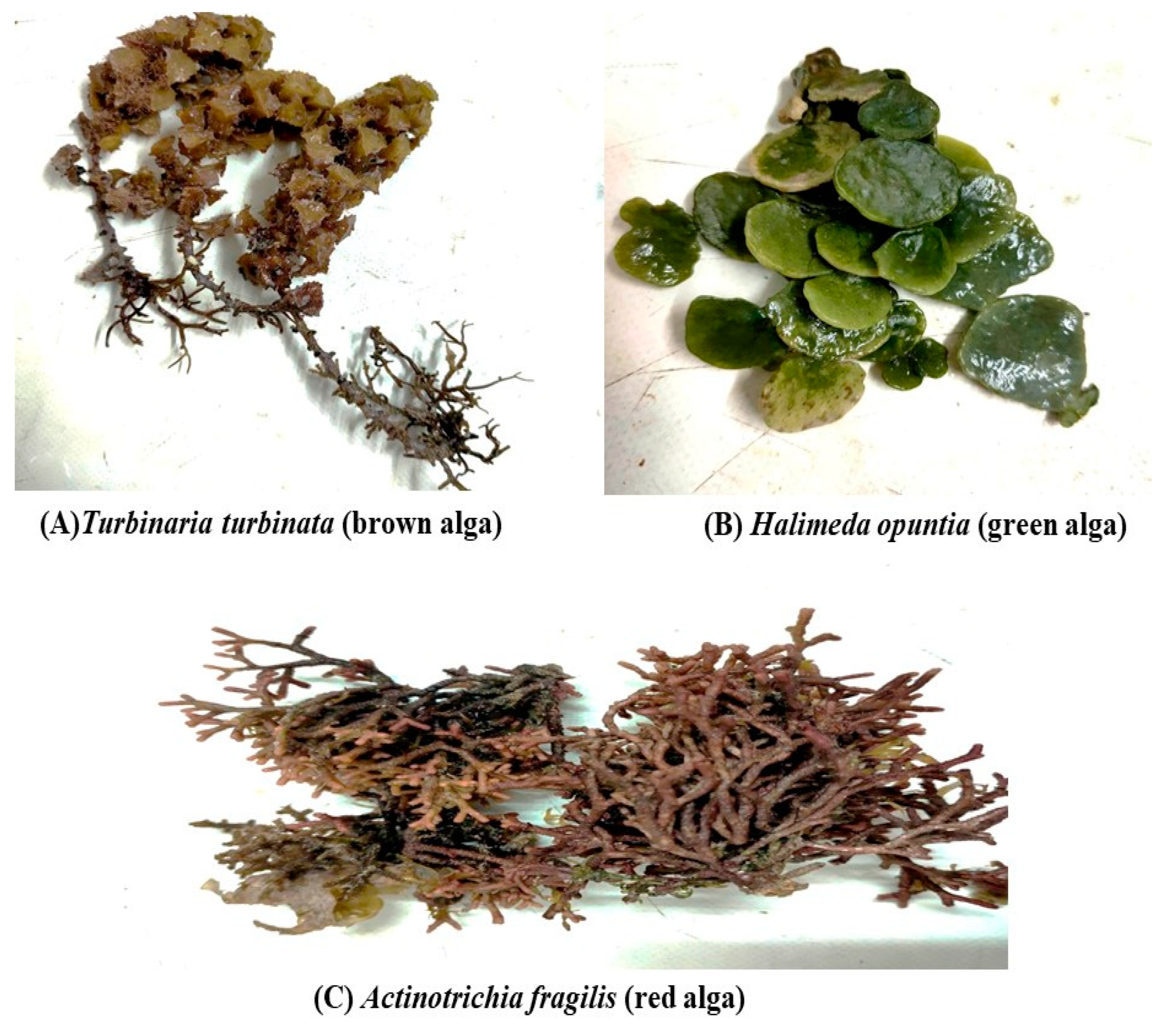
2.3. Algal Materials and Extraction

Three seaweeds were collected from Hurghada city, Red Sea Governorate, Egypt i.e., Halimeda opuntia (HO) (green algae), Actinotrichia fragilis (AF) (red algae), and Turbinaria turbinata (TT) (brown algae) (see Figure 1). Algal species were carefully cleaned from the epiphytes and then dried and powdered. Each algal powder was prepared as a lyophilized ethanolic extract (70% ethanol: deionized water v/v). The identification of the seaweed species was carried out according to Salem et al. [17] and Yang et al. [18].

2.4. Antibacterial Activity

2.4.1. Assessment of the Antibacterial Activity of Lyophilized Seaweed Extracts

The ability of lyophilized seaweed extracts as an antibacterial against S. aureus reference strain EMCC1351 (prepared in Microbiological Resources Center (MERCIN), Faculty of Agriculture, Ain Shams University, Cairo, Egypt) was evaluated using agar disk diffusion assay [19,20]. Overnight culture of S. aureus was enriched on Mueller Hinton Medium (MHM) broth (Oxoid, Cheshire, UK) at 37 °C/48 h and then spread on MHM plates. After dryness, the lyophilized seaweed extracts were loaded onto each separate disk (20 µL), and the plates were maintained at 4 °C/30 min and then incubated at 37 °C/24 h. The clear inhibitory zones obtained were recorded in mm, considering the anti-S. aureus activity of various lyophilized seaweed extracts. In addition, the results of the inhibitory zones were compared with those of three antibiotic disks, tetracycline, chloramphenicol, and sulfamethoxazole.

2.4.2. Evaluation of the Minimum Inhibitory Concentrations (MICs) of Lyophilized HO Extract

On the basis of the antibacterial activity results of the three seaweed extracts, we evaluated MIC for the only one that has antibacterial power against S. aureus, thus, HO algal extracts minimum inhibitory concentrations against S. aureus were evaluated according to Kadaikunnan et al. [21] using descending concentrations. It was performed using different concentrations, i.e., 100, 50, 25, 12.5, 6.25, 3.12, 1.56, and 0.78 mg/mL lyophilized HO algal extract. S. aureus suspension of grown cultures was prepared and adjusted to a density of 106 colony forming unit (CFU)/mL [16].

2.5. Phytochemical Analysis of the Lyophilized HO Algal Extract

2.5.1. Assessment of the Radical Scavenging Capacity by the DPPH Assay

The ability of the lyophilized HO algal extract to scavenge DPPH free radicals was assessed according to Catarino et al. [22] and Hamad et al. [23] with few modifications. Ascorbic acid was used as a positive control. Values were expressed as IC50 (the lyophilized HO extract’s concentration inhibited 50% DPPH). IC50 values were compared with a concentration plot using a nonlinear regression algorithm. Inhibition % was calculated according to Equation (1).
Inhibition   ( % ) = A   of   control     A   of   the   sample A   of   control   × 100  
where: A = absorbance.

2.5.2. Total Phenolic and Total Flavonoid Contents of Lyophilized HO Algal Extract

Total phenolic content (TPC) of lyophilized HO algal extract (green algae) was evaluated by Folin-Ciocalteu technique at 765 nm using a UV/Vis spectrophotometer (PG Instrument Ltd. Lutterworth, UK) as follow; 1 mL of an 70% ethanolic HO extract (1%) was added to 0.1 mL of Folin-Ciocalteu reagent. The mixture was left for 15 min at room temperature. Then, 3 mL of 2% Na2CO3 was added. The prepared mixture was left for 30 min at room temperature before the measurement. The TPC was calculated using standard calibration curve of gallic acid, and TPC results was expressed in mg of gallic acid equivalents (GAE) per mL of HO extract (mg GAE/mL) [24]; hence, ethanol was used as blank sample.
On the other hand, the total flavonoid content (TFC) of lyophilized HO algal extract (green algae) were analyzed by a UV/Vis spectrophotometer at 510 nm. One milliliter of an 70% ethanolic HO extract (1%) was added to 4 mL of distilled H2O and mixed vigorously. After approximately 5 min, 0.3 mL of NaNO2 (5%) was added to the mixture and 0.3 mL of AlCl3 (10%) was added. Further, 2 mL of NaOH (1 M) after an extra 6 min was added and the volume of the prepared mixture was increased up to 10 mL of distilled H2O. TFC were expressed as mg of quercetin equivalent (QE) per mL of the HO extract (mg QE/mL) [25].

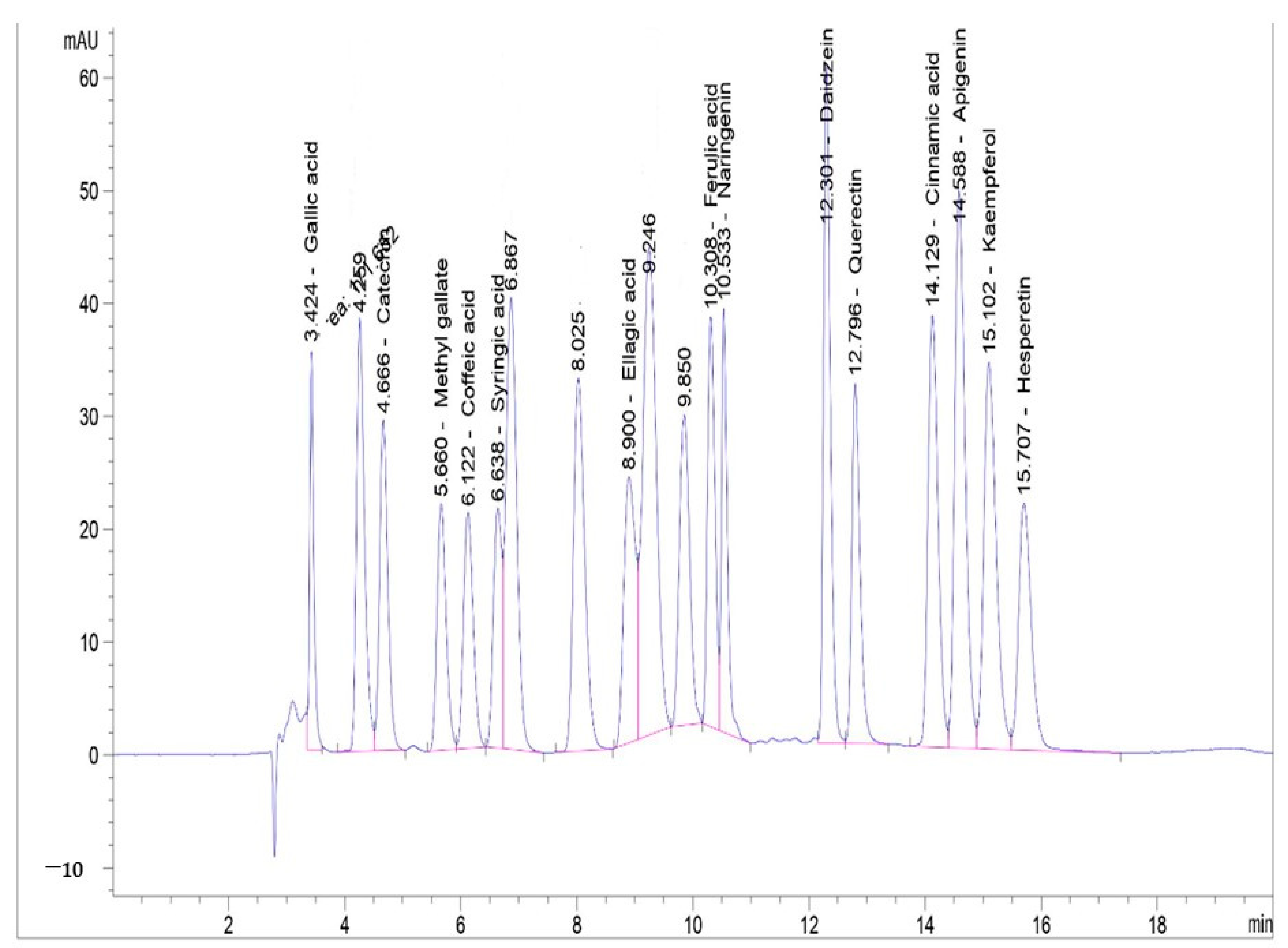
2.6. HPLC Evaluation of Phenolic Compounds Profiles of the Lyophilized HO Algal Extract

HPLC (Agilent 1260 infinity HPLC Series, Santa Clara, CA, USA) was used to screen lyophilized HO algal extract’s phenolic profile. Phenolic compounds were separated using an Eclipse C18 column (4.6 mm × 250 mm i.d., 5 μm) at 40 °C. The separation was achieved using a ternary linear elution gradient with (A): HPLC-grade water- 0.2% H3PO4 (v/v), obtained from Sigma-Aldrich, St. Louis, MO, USA; (B) methanol (Thermo Fisher Scientific, Waltham, MA, USA); and (C) acetonitrile (Thermo Fisher Scientific, Waltham, MA, USA). The mobile phase was applied at a 0.9 mL/min flow rate, and the multi-wavelength detector was adjusted to 280 nm. Approximately 5 μL injection volume was used. HPLC phenolic profile screening was conducted according to Hamad et al. [26].

2.7. Safety and Cytotoxicity Assay of Lyophilized HO Algal Extract

Lyophilized HO algal extract was evaluated for its effect on the viability of peripheral blood mononuclear cells (PBMCs). Cell viability was investigated using PBMCs maintained in Roswell Park Memorial Institute RPMI medium. To isolate PBMCs, whole blood, firstly diluted with PBS, was then gently layered over an equal volume of Ficoll in a Falcon and finally centrifuged for 30 min at 500 rpm without brake. Blank wells (150 µL PBS), control wells (150 µL PBMCs), and tested wells (150 µL PBMCs) were allocated on a 96-well microtiter plate. Lyophilized HO algal extracts at different concentrations were inoculated to test wells and then incubated for 24 h, according to Popiołkiewicz et al. [27]. Neutral red (150 µL) was added and then incubated at 37 °C for 2 h. After washing the cells, the plates were cleaned with a de-staining solution (1% acetic acid: 49% deionized water: 50% ethanol, 150 µL/well. At 540 nm, absorbance was monitored using a T80 UV/VIS spectrophotometer [28]. Lyophilized HO algal extract inhibition% was calculated using Equation (2) and IC50 values were calculated online: www.aatbio.com/tools/IC50-calculator, (accessed on 27 July 2022).
Lyophilized   HO   algal   extract   inhibition % = 100 O . D   Control     O . D   Treatment O . D   Control
where O.D. = optical density; control = 150 µL PBMCs, treatment = 150 µL HO extract.

2.8. Assessment of the Antibacterial Effect of Lyophilized HO Algal Extract against S. aureus Experimentally Inoculated into Chicken Fillet

Raw chicken breast fillets (boneless) were sliced into 5 cm × 5 cm pieces using a sterile knife. Before the experiment, chicken fillets were sterilized with ultraviolet light (UV) for 15 min/side to control background micro-flora, according to Morsy et al. [29]. Prepared chicken fillet samples were divided into six groups as follows: Group 1, chicken fillet without any treatments (CHF); Group 2, chicken fillet treated with direct addition of lyophilized HO algal extract 4% (CHF/HO 4%); Group 3, chicken fillet treated with lyophilized HO algal extract 6% (CHF/HO 6%); Group 4, chicken fillet experimentally inoculated with 107 CFU/mL S. aureus (CHF/ST); Group 5, chicken fillet experimentally inoculated with S. aureus and treated with lyophilized HO algal extract 4% (CHF/ST/HO 4%); and Group 6, chicken fillet experimentally inoculated with S. aureus and treated with lyophilized HO algal extract 6% (CHF/ST/HO 6%).
Samples were maintained at room temperature for 15 min to allow cell attachment after inoculation and were then chilled at 4 °C and examined bacteriologically every 2 days for S. aureus till the cells completely loss their viability. This experiment was repeated in triplicate to obtain the mean values for statistical analysis (n = 3).
Samples were bacteriologically examined at 0, 2, 4, 6, 8, and 10th days of storage for S. aureus count according to FDA [30].

2.9. Assessment of the Acceptability of Chicken Fillet Fortified with the Lyophilized HO Algal Extract

A total of 10 experienced panelists applied the evaluation at the Food Technology Department, City of Scientific Research and Technological Applications, New Borg El Arab, Egypt. Sensory evaluation was applied on a grilled un-inoculated chicken fillet fortified with lyophilized HO algal extract to evaluate its acceptability as a food additive. The first three groups of experiments [(CHF), (CHF/HO 4%), and (CHF/HO 6%)] were evaluated after grilling for sensory attributes.
The samples were maintained at room temperature 25 °C/10 min before evaluation. Panelists evaluated the chicken fillet for the degree of acceptability depending on the following criteria: color, odor, taste, texture, and overall acceptance (10 points/each item), with a scale ranging from 1 to 10, where 10 is more accepted as described by Hamad et al. [31]. In addition, the average sensory attribute data with its standard deviations were evaluated.

2.10. Statistical Analysis

All calculations were implemented based on SPSS, version 23 (IBM SPSS Statistics for Windows, IBM Corp., Armonk, NY, USA). The means ± standard error (SE) was used for the data analyses. One-way analysis of variance (ANOVA) using the Duncan test was used, where the probability was considered statistically significant when p < 0.01 or p < 0.05.

3. Results and Discussion

3.1. Prevalence of S. aureus in Chicken Fillet

One of the main demands that will be increased by 2050 is protein. Chicken meat constitutes a major protein part depending on the diet [32,33]. Unfortunately, contamination of chicken meat with food poisoning bacteria occurs during any step of processing [34]. S. aureus is a common food poisoning hazard in chicken meat that secretes a heat resistance toxin that affects human health.
In the current study, chicken meat samples were evaluated bacteriologically for the presence of S. aureus. Results in Table S1 revealed the occurrence of S. aureus in breast and thigh chicken meat samples, which was 92% and 84%, respectively. There was no significant difference (p > 0.05) between the prevalence of S. aureus in the breast and thigh of the chicken. These results were higher than those by Momtaz et al. [35], who isolated S. aureus from 22.77% of fresh raw chicken meats. Qian et al. [5] confirm S. aureus contamination of chicken meat in all processing plant steps. Meat contamination with S. aureus negatively impacts human health and causes serious diseases [36] because it secretes enterotoxins in food. These toxins are thermo-stable and resistant to gastrointestinal proteases [37].
In this study, chicken meat was sampled as an equivalent criterion to the buying of chicken for consumers from suppliers with a low hygienic level for the tools utilized to cut the chicken, poor sanitation levels, and frequent direct contact between the meat and market visitors. The high prevalence of S. aureus in the detected chicken samples in the present research is due to contamination of the tested samples with the pathogenic microorganisms anywhere in the supply chain from farm to market. Similarly, contamination can also occur during contact with the facility’s equipment, such as belts, grinders, and saws, or by contact with chicken meat handlers like knives and hand contact. Furthermore, due to abundance of proteins, carbohydrates, fats, vitamins, in chicken and its high-water holding capacity (WHC) allow the formation of a suitable conditions and environment for S. aureus contamination and growth.

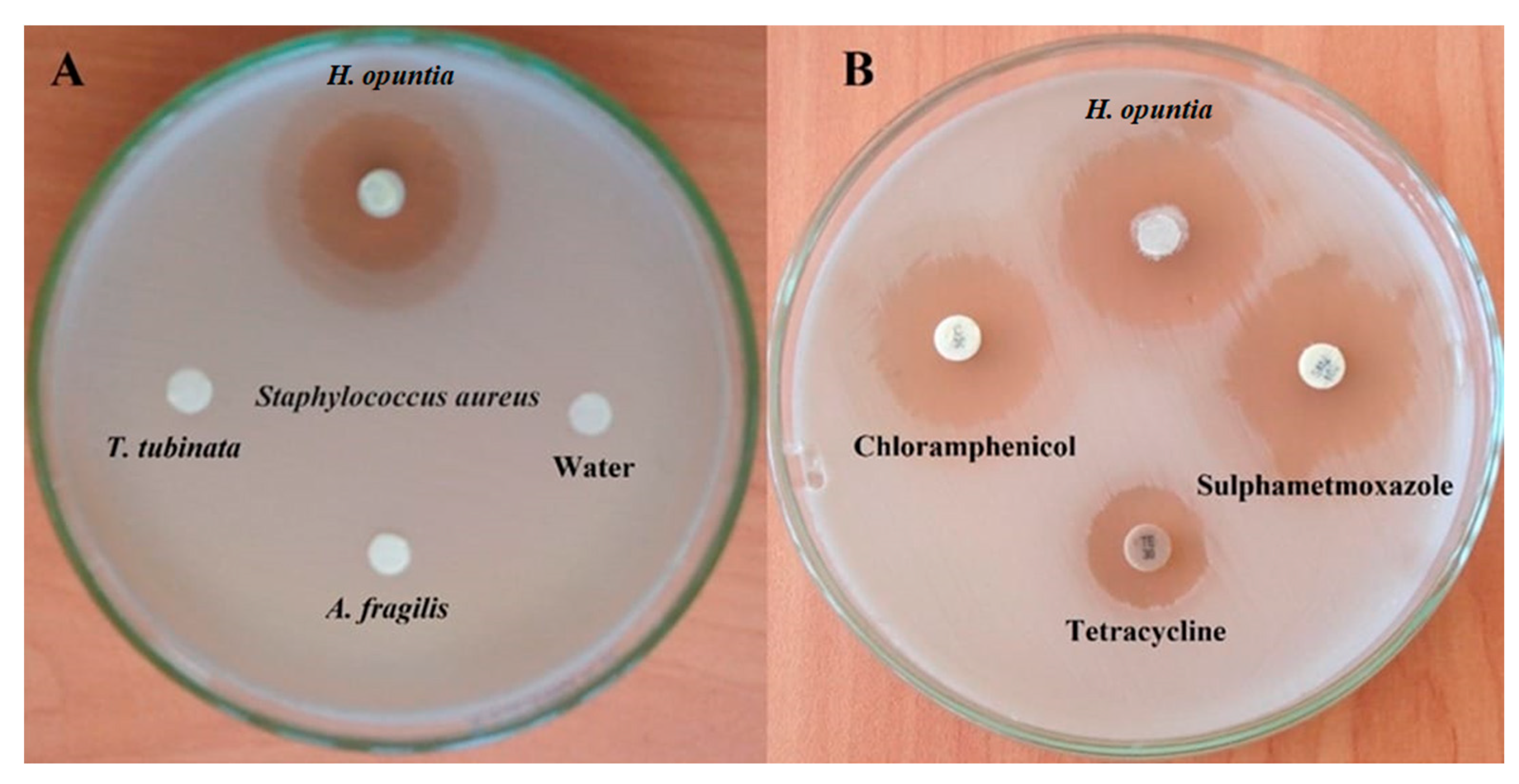
3.2. Antibacterial Activity of Lyophilized Seaweed Extracts

Producing food free from foodborne diseases based on natural antimicrobials has become a great interest in food safety sector. Marine algae consider novel food additives to produce natural and functional products that fulfill consumer demand because of their secondary bioactive metabolites as phenolic compounds [38]. Moreover, it has antimicrobial effects on different forms of bacteria [39].
This study evaluated the antibacterial effect of three lyophilized seaweed extracts on S. aureus using an agar disk diffusion assay. Results in Table 1, Figure 2A,B reveal a comparative study of in vitro antibacterial effects of lyophilized HO (green algae), lyophilized AF (red algae), and lyophilized TT (brown algae), as well as three types of antibiotics, were used as references against S. aureus. Furthermore, the lyophilized HO extract was the only algae that exhibited an anti-S. aureus effect with an inhibition zone of 43.16 ± 0.44 mm (Figure 2A) and even higher than those of chloramphenicol, tetracycline, and sulfamethoxazole antibiotics with a significant difference at p < 0.05 (Figure 2B). This result agrees with that of Ely et al. [40] and Manivannan et al. [41], who confirmed the algal extracts have in vitro antibacterial activity against Gram-positive and Gram-negative.

3.3. Minimum Inhibitory Concentrations (MICs) of Lyophilized HO Extract

MIC of the lyophilized HO extract against S. aureus in vitro and the antibacterial effect of different concentrations was evaluated. Results in Table 1 and Figure 3 showed that lyophilized HO extracts at a minimum concentration of 3.12 mg/mL exhibited an anti-S. aureus with inhibition zone of 8.16 ± 0.73 mm. Furthermore, the anti-S. aureus activity increased relatively by the gradually increment of the extract concentration.

3.4. DPPH Radical Scavenging Capacity

The DPPH assay is an accurate, reliable, and cost-effective way to assess antioxidant radical scavenging activity. The antioxidant capacity of the lyophilized HO extract was evaluated in Table 2 depending on the DPPH radical scavenging capacity. Results compared with ascorbic acid as a standard antioxidant. It was found that the IC50 of ascorbic acid was 26.36 µg/mL while that of the lyophilized HO extract was 55.3 µg/mL. The highest DPPH scavenging activity of the lyophilized HO extract was 95.34% at a concentration of 100 µg/mL. These results were higher than those by Nazarudin et al. [42], who found that the HO highest DPPH reduction is 63.61% at 1000 mg/mL concentration. This seaweed radical scavenging ability enhances its antioxidant effect on carcinogenesis [43].

3.5. TPC and TFC of Lyophilized HO Extract

From the current findings, it was found that TPC was 20.36 ± 0.092 mg/mL, while TFC was 16.59 ± 0.029 mg/mL. These results were lower than those of Nazarudin et al. [40], who found that TPC and TFC of HO extracts are 55.04 ± 0.98 mg/g and 40.02 ± 0.02 mg/g, respectively. Many factors affect the variation in phenolic content: location, ecological classification, season, temperature, pH, light incidence, water salinity, and water nutrient composition [44]. The higher antioxidant potential (Table 2) is mainly due to the high level of TPC and TFC.

3.6. Phenolic Profile of Lyophilized HO Algal Extract by HPLC

HPLC evaluates the phenolic profile content in marine microalgae [45]. Therefore, a detailed profile of the phenolic content of the lyophilized HO extract was illustrated by HPLC in Table S2 and Figure 4. It was found that lyophilized HO extract exhibited many phenolic bioactive compounds that could be explain its antioxidant and antibacterial activity. The highest bioactive compound was daidzein with 10.84 ± 0.005 µg/mL, followed by gallic acid with 4.06 ± 0.006 µg/mL. Indeed, daidzein has antioxidant and anticancer properties [46]. Gallic acid is used in the food industry as an antioxidant and preservative owing to its strong radical scavenging and antioxidant activities [47]. Moreover, it is a potent antimicrobial, gastroprotective, anticancer, antioxidant, promotes many health benefits to humans, and protects both the heart and kidney [48].
The determined phenolics could be attributed to the lyophilized HO extract’s antimicrobial and antioxidant roles proved in this study. Kurhekar, [49] refers to the antimicrobial effect of marine algae for its content of various bioactive compounds, e.g., ascorbic acid, phenolic acids, lutein, α -tocopherol, α -carotene, β-carotene, and flavonoids.

3.7. Safety and Cytotoxicity Assay of Lyophilized HO Algal Extract

The PBMCs cytotoxicity approach, utilizes cells isolated from multiple individuals, provides a high throughput evaluation of the cytotoxicity in vitro of candidate drugs. Additionally, PBMCs assay provide a primary reflection into how immune cell from different donors respond to the candidate compounds in development. Indeed, the safety of any new antimicrobials added to food is of great concern [42,50,51,52], therefore, the safety and/or cytotoxicity of the lyophilized HO extract were assessed. The cytotoxic effect of the lyophilized HO extract on the viability of PBMCs (Table S3) revealed that the cytotoxicity of PBMCs showed a positive correlation with the lyophilized HO extract concentration. The concentrations of lyophilized HO extract ranged from a maximum of 250 µg/mL to a minimum of 7.8 µg/mL, which showed inhibition of the viability of PBMCs with 69.27%, respectively. Moreover, the amount of lyophilized HO extract required to cause 50% inhibition of PBMC cells (IC50) was 33.7 µg/mL. A higher IC50 permits the use of lyophilized HO extract as a safe food additive in meat products.

3.8. Chicken Fillets Challenge Study

Regarding the rising chicken meat consumption global concern [53], the safety of chicken meat is considered a common consumer demand. In a challenge study, lyophilized HO algal extract was applied in chicken fillets experimentally inoculated with S. aureus to evaluate its antibacterial effect. Results in Table 3 revealed that lyophilized HO algal extract showed an anti-S. aureus effect in chicken fillets stored at 4 °C. CHF/ST/HO 4% and 6% caused a complete reduction of S. aureus to count on the 6th and 4th days of storage, while the count of S. aureus in the CHF/ST group gradually increased. Furthermore, there was a significant difference (p < 0.05) between treated and untreated groups with lyophilized HO across the entire storage period.
Chicken fillet without any treatments (CHF); chicken fillet treated with lyophilized HO algal extract 4% (CHF/HO 4%); chicken fillet treated with lyophilized HO algal extract 6% (CHF/HO 6%); chicken fillet experimentally inoculated with 107 CFU/mL S. aureus (CHF/ST); chicken fillet experimentally inoculated with S. aureus and treated with lyophilized HO algal extract 4% (CHF/ST/HO 4%); and chicken fillet experimentally inoculated with S. aureus and treated with lyophilized HO algal extract 6% (CHF/ST/HO 6%).
The challenge study results go with the previous evaluation of the antioxidant and in vitro anti-S. aureus effect of lyophilized HO algal extract. The anti-S. aureus effects of lyophilized HO algal extract in chicken fillet stored at 4 °C may be attributed to the fact that marine algae contain phenolic compounds that compete against invading bacteria [54,55], it considers a broad-spectrum antiviral and antibacterial [56] those from the Arabian Gulf and the Saudi Arabia Red Sea inhibit methicillin-resistant S. aureus [45]. HO was not evaluated before against S. aureus. However, it activates the antibacterial effect of zinc oxide nanoparticles on pathogenic Vibrio harveyi [57]. Moreover, the lyophilized algal extract contains higher bioactive compounds and hence more antimicrobial activity [58]. Thus, it is considered a sustainable food for humans.

3.9. Acceptability of Chicken Fillet Fortified with Lyophilized HO Algal Extract

The sensory attributes of grilled un-inoculated chicken fillet fortified with lyophilized HO algal extract were evaluated. Results in Table 4 revealed that CHF/HO 4% and CHF/HO 6% enhanced the color, odor, taste, texture, and overall acceptability of grilled chicken fillet. Moreover, there was a significant difference (p < 0.05) between all treatments. Further, HO algae showed acceptability to consumers. The recent studies confirmed that, HO algae had pleasant organoleptic attributes, and even a protein alternative in meat products [59,60].

4. Conclusions

The current finding revealed that, the occurrence of S. aureus in tested breast and thigh chicken meat samples was recorded at 92% and 84%, respectively. There was no significant difference (p > 0.05) between the prevalence of S. aureus in the breast and thigh of the chicken. The high occurrence of S. aureus in the chicken samples detected in the present research is due to the contamination occurred by the pathogenic microorganisms anywhere in the supply chain, from farm to market.
This study evaluated the antibacterial effect of three lyophilized seaweed extracts on S. aureus. Amongst these three seaweeds, results demonstrated that HO extract was the only algae that exhibited an antibacterial activity against S. aureus due to the high content of total phenolic compounds, total flavonoids compounds, as well as several phenolic compounds illustrated by HPLC profile. In addition, HO extract exhibited antioxidant effect owing to the various bioactive molecules, which have the ability to scavenge free radicals (DPPH). A higher IC50 of HO extracts permits their use as a safe food additive in meat products. In a challenging study, the lyophilized HO extract displayed an anti-S. aureus effect in chicken fillets stored at 4 °C. HO extract also enhanced the sensory attributes of grilled un-inoculated chicken fillet. In sum, lyophilized HO extracts are promising food supplements in the chicken meat industry with eminent antibacterial and antioxidant properties.

Supplementary Materials

The following supporting information can be downloaded at: https://www.mdpi.com/article/10.3390/foods12010020/s1, Table S1. Prevalence of S. aureus in chicken fillet collected from different local markets (n = 100); Table S2. HPLC evaluation for phenolic profile of lyophilized HO algal extract; Table S3. Evaluation of safety and cytotoxicity assay to lyophilized HO algal extract on the viability of PBMCs cells.

Author Contributions

Conceptualization, G.H.; methodology, G.H.; software, T.M., G.H., A.A., G.K., A.Z., R.A.E., R.E., A.T. and E.M.E.; validation, T.M., G.H., T.E., A.T., A.Z., A.A., G.K., R.A.E., R.E. and E.M.E.; formal analysis, T.M., T.E., A.A., G.K., A.Z., R.A.E., R.E. and E.M.E.; investigation, T.M.; resources, G.H.; data curation, T.M.; writing—original draft preparation, G.H., and T.M.; writing—review and editing, T.M., G.H. and T.E.; visualization, T.E., A.T., A.Z., A.A., G.K., R.A.E., R.E., E.M.E. and T.M.; supervision, G.H.; project administration, G.H., T.E. and T.M.; funding acquisition, T.E., A.T., A.Z. and T.M. All authors have read and agreed to the published version of the manuscript.

Funding

The publication of this article was supported by the Open Access Fund of Leibniz Universität Hannover. This study did not receive external fundings.

Institutional Review Board Statement

Not applicable.

Informed Consent Statement

Not applicable.

Data Availability Statement

The data presented in this study are available on request from the corresponding author.

Acknowledgments

This study was conducted in the Food Technology Department of Arid Lands Cultivation Research Institute, City of Scientific Research and Technological Applications, Alexandria, Egypt. The authors would thank the reviewers and editors for their helpful recommendations.

Conflicts of Interest

The authors declare no conflict of interest.

References

  1. Lee, H.; Yoon, Y. Etiological agents Implicated in foodborne illness worldwide. Food Sci. Anim. Resour. 2021, 41, 1–7. [Google Scholar] [CrossRef] [PubMed]
  2. European Commission. Farm to Fork Strategy for a Fair, Healthy and Environmentally Friendly Food System. 2020. Available online: https//ec.europa.eu/food/horizontal-topics/farm-fork-strategyEn (accessed on 14 June 2022).
  3. WHO. Draft WHO Global Strategy for Food Safety 2022–2030, towards Stronger Food Safety Systems and Global Cooperation, Department of Nutrition and Food Safety Prepared by WHO Secretariat. 2021. Available online: https://www.who.int/publications/m/item/draft-who-global-strategy-for-food-safety-2022-2030 (accessed on 5 August 2022).
  4. PIANZ. Industry Information. 2021. Available online: http://www.pianz.org.nz/industry-information/industry-statistics/meat-consumption/chicken-remains-new-zealands-favourite-meat (accessed on 21 July 2022).
  5. Qian, C.; Castaneda-Gulla, K.; Sattlegger, E.; Mutukumira, A.N. Enterotoxigenicity and genetic relatedness of Staphylococcus aureus in a commercial poultry plant and poultry farm. Int. J. Food Microbiol. 2022, 363, 109454. [Google Scholar] [CrossRef] [PubMed]
  6. Davies, A.R.; Board, R.G. The Microbiology of Meat and Poultry, 1st ed.; Thomson Science: Exmouth, UK, 1998. [Google Scholar]
  7. Massawe, H.F.; Mdegela, R.H.; Kurwijila, L.R. Antibiotic resistance of Staphylococcus aureus isolates from milk produced by smallholder dairy farmers in Mbeya Region, Tanzania. Int. J. One Health 2019, 5, 31–37. [Google Scholar] [CrossRef] [Green Version]
  8. Dehkordi, F.S.; Gandomi, H.; Basti, A.A.; Misaghi, A.; Rahimi, E. Phenotypic and genotypic characterization of antibiotic resistance of methicillin-resistant Staphylococcus aureus isolated from hospital food. Antimicrob. Resist. Infect. Control 2017, 6, 104. [Google Scholar] [CrossRef]
  9. Alizadeh Sani, M.; Ehsani, A.; Hashemi, M. Whey protein isolate/cellulose nanofiber/TiO2 nanoparticle/rosemary essential oil nanocomposite film: Its effect on microbial and sensory quality of lamb meat and growth of common foodborne pathogenic bacteria during refrigeration. Int. J. Food Microbiol. 2017, 251, 8–14. [Google Scholar] [CrossRef]
  10. García-Vaquero, M.; Rajauria, G.; O’doherty, J.; Sweeney, T. Polysaccharides from macroalgae: Recent advances, innovative technologies and challenges in extraction and purification. Food Res. Int. 2017, 99, 1011–1020. [Google Scholar] [CrossRef] [Green Version]
  11. Bhuvana, P.; Sangeetha, P.; Anuradha, V.; Syed Ali, M. Spectral characterization of bioactive compounds from microalgae. N. oculata C. vulgaris. Biocatal. Agric. Biotechnol. 2019, 19, 101094. [Google Scholar] [CrossRef]
  12. Santhakumaran, P.; Ayyappan, S.M.; Ray, J.G. Nutraceutical applications of twenty-five species of rapid-growing green microalgae as indicated by their antibacterial, antioxidant and mineral content. Algal Res. 2020, 47, 101878. [Google Scholar] [CrossRef]
  13. Pestana, J.M.; Puerta, B.; Santos, H. Impact of dietary incorporation of Spirulina (Arthrospira platensis) and exogenous enzymes on broiler performance, carcass traits, and meat quality. Poult. Sci. 2020, 99, 2519–2532. [Google Scholar] [CrossRef]
  14. Banach, J.; Burg, S.; Fels-Klerx, H. Food safety during seaweed cultivation at offshore wind farms: An exploratory study in the North Sea. Mar. Policy. 2020, 120, 104082. [Google Scholar] [CrossRef]
  15. El-Khawas, K.M.; Hendy, B.A.S. Assessment and improvement of hygienic status of chicken fillet from slaughterhouses using organic acids from natural sources. Assiut Vet. Med. J. 2015, 61, 8–17. [Google Scholar]
  16. Eldin, R.M.B.; Talaat, D.; Elbaba, A.H.; Ibrahim, M.S. Antibacterial activity y of some plant extracts on different bacteria in chicken fillet. Eur. J. Pharm. Med. Res. 2020, 7, 84–95. [Google Scholar]
  17. Salem, W.M.; Galal, H.; Nasr El-deen, F. Screening for antibacterial activities in some marine algae from the red sea (Hurghada, Egypt). Afr. J. Microbiol. Res. 2011, 5, 2160–2167. [Google Scholar] [CrossRef] [Green Version]
  18. Yang, Y.; Zhang, M.; Alalawy, A.I.; Almutairi, F.M.; Al-Duais, M.A.; Wang, J.; Salama, E.S. Identification and characterization of marine seaweeds for biocompounds production. Environ. Technol. Innov. 2021, 24, 101848. [Google Scholar] [CrossRef]
  19. Weese, J.S.; Anderson, M.E.C.; Lowe, A.; Penno, R.; Da Costa, T.M.; Button, L.; Goth, K.C. Screening of the equine intestinal microflora for potential probiotic organisms. Equine Vet. J. 2004, 36, 351–355. [Google Scholar] [CrossRef] [PubMed]
  20. Hamad, G.M.; Abdelmotilib, N.M.; Darwish, A.M.G.; Zeitoun, A.M. Commercial probiotic cell-free supernatants for inhibition of Clostridium perfringens poultry meat infection in Egypt. Anaerobe 2020, 62, 102181. [Google Scholar] [CrossRef] [PubMed]
  21. Kadaikunnan, S.; Rejiniemon, T.; Khaled, J.M.; Alharbi, N.S.; Mothana, R. In-vitro antibacterial, antifungal, antioxidant and functional properties of Bacillus amyloliquefaciens Ann. Clin. Microbiol. Antimicrob. 2015, 14, 9. [Google Scholar] [CrossRef] [Green Version]
  22. Catarino, M.D.; Silva, A.M.S.; Saraiva, S.C.; Sobral, A.J.F.N.; Cardoso, S.M. Characterization of phenolic constituents and evaluation of antioxidant properties of leaves and stems of Eriocephalus africanus. Arab. J. Chem. 2018, 11, 62–69. [Google Scholar] [CrossRef] [Green Version]
  23. Hamad, G.M.; Mohdaly, A.A.A.; El-Nogoumy, B.A.; Ramadan, M.F.; Hassan, S.A.; Zeitoun, A.M. Detoxification of Aflatoxin B1 and Ochratoxin A Using Salvia farinacea and Azadirachta indica Water Extract and Application in Meat Products. Appl. Biochem. Biotechnol. 2021, 193, 3098–3120. [Google Scholar] [CrossRef]
  24. Hamad, G.; Ombarak, R.A.; Eskander, M.; Mehany, T.; Anees, F.R.; Elfayoumy, R.A.; Omar, S.A.; Lorenzo, J.M.; Abou-Alella, S.A.-E. Detection and inhibition of Clostridium botulinum in some Egyptian fish products by probiotics cell-free supernatants as bio-preservation agents. LWT 2022, 163, 113603. [Google Scholar] [CrossRef]
  25. El Sohaimy, A.A.S.; El-Sheikh, H.M.; Refaay, M.T.; Zaytoun, A.M.M. Effect of harvesting in different ripening stages on olive (Olea europea) oil quality. Am. J. Food Technol. 2016, 11, 1–11. [Google Scholar] [CrossRef]
  26. Hamad, G.M.; Abu-serie, M.M.; Ali, S.H.; Hafez, E.E. Combination Probiotic Supernatants Reduce Growth and Aflatoxin Production by Aspergillus spp in Food Contamination. Am. J. Food Sci. Technol. 2018, 6, 57–67. [Google Scholar]
  27. Popiołkiewicz, J.; Polkowski, K.; Skierski, J.S.; Mazurek, A.P. In vitro toxicity evaluation in the development of new anticancer drugs-Genistein glycosides. Cancer Lett. 2005, 229, 67–75. [Google Scholar] [CrossRef] [PubMed]
  28. Ryan, R.M.; Deci, E.L. Self-Determination Theory: Basic Psychological Needs in Motivation, Development, and Wellness; The Guilford Press: New York, USA, 2017. [Google Scholar]
  29. Morsy, M.K.; Elsabagh, R.; Trinetta, V. Evaluation of novel synergistic antimicrobial activity of nisin, lysozyme, EDTA nanoparticles, and/or ZnO nanoparticles to control foodborne pathogens on minced beef. Food Control 2018, 92, 249–254. [Google Scholar] [CrossRef]
  30. FDA (Food and Drug Administration Center for Food Safety & Applied Nutrition). Staphylococcus aureus toxin formation in hydrated batter mixes. In Fish and Fisheries Products Hazards and Controls Guidance; Food and Drug Administration Center for Food Safety & Applied Nutrition: Washington, DC, USA, 2001; pp. 201–208. [Google Scholar]
  31. Hamad, G.M.; Omar, S.A.; Mostafa, A.G.M.; Cacciotti, I.; Saleh, S.M.; Allam, M.G.; El-Nogoumy, B.; Abou-Alella, S.A.-E.; Mehany, T. Binding and removal of polycyclic aromatic hydrocarbons in cold smoked sausage and beef using probiotic strains. Food Res. Int. 2022, 161, 111793. [Google Scholar] [CrossRef] [PubMed]
  32. Chia, S.Y.; Tanga, C.M.; Loon, J.J.A.; Dicke, M. Insects for sustainable animal feed: Inclusive business models involving smallholder. Curr. Opin. Environ. Sustain. 2019, 41, 23–30. [Google Scholar] [CrossRef]
  33. Elahi, U.; Ma, Y.B.; Wu, S.G.; Wang, J.; Zhang, H.J.; Qi, G.H. Growth performance, carcass characteristics, meat quality and serum profile of broiler chicks fed on housefly maggot meal as a replacement of soybean meal. J. Anim. Physiol. Anim. Nutr. 2020, 104, 1075–1084. [Google Scholar] [CrossRef]
  34. Incili, G.K.; Çalıcıoglu, M. Change in scalding fluids by time in poultry slaughterhouse and its effect on microbiological quality of carcasses. J. Food Saf. 2015, 38, 12485. [Google Scholar] [CrossRef]
  35. Momtaz, H.; Dehkordi, F.S.; Rahimi, E.; Asgarifar, A.; Momeni, M. Virulence genes and antimicrobial resistance profiles of Staphylococcus aureus isolated from chicken meat in Isfahan province, Iran. J. Appl. Poult. Res. 2012, 22, 913–992. [Google Scholar] [CrossRef]
  36. Taylor, T.A.; Unakal, C.G. Staphylococcus aureus. In StatPearls [Internet]; StatPearls Publishing: Treasure Island, FL, USA, 2020. Available online: https://www.ncbi.nlm.nih.gov/books/NBK441868/ (accessed on 11 June 2022).
  37. Alves, V.F.; Nino-Arias, F.C.; Pitondo-Silva, A.; Frazilio, D.D.; Oliveira Gonçalves, L.D.; Toubas, L.C.; Sapateiro Torres, I.M.; Oxaran, V.; Dittmann, K.K.; De Martinis, E.C.P. Molecular characterization of Staphylococcus aureus from some artisanal Brazilian dairies. Int. Dairy J. 2018, 85, 247–253. [Google Scholar] [CrossRef] [Green Version]
  38. Jimenez-Lopez, C.; Pereira, A.G.; Lourenço-Lopes, C.; Garcia-Oliveira, P.; Cassani, L.; Fraga-Corral, M.; Prieto, M.A.; Simal-Gandara, J. Main bioactive phenolic compounds in marine algae and their mechanisms of action supporting potential health benefits. Food Chem. 2021, 341, 128262. [Google Scholar] [CrossRef] [PubMed]
  39. Surendhiran, D.; Cui, H.; Lin, L. Encapsulation of Phlorotannin in Alginate/PEO blended nanofibers to preserve chicken meat from Salmonella contaminations. Food Packag. Shelf Life. 2019, 21, 100346. [Google Scholar] [CrossRef]
  40. Ely, R.; Supriya, T.; Naik, C.G. Antimicrobial activity of marine organisms collected off the coast of South East India. J. Exp. Biol. Ecol. 2004, 309, 121–127. [Google Scholar] [CrossRef]
  41. Manivannan, K.; Karthikaidevi, G.; Anantharaman, P.; Balasubramanian, T. Antimicrobial potential of selected brown seaweeds from Vedalai coastal waters. Gulf Mannar. Asian Pac. Trop. Biomed. 2011, 1, 114–120. [Google Scholar] [CrossRef] [PubMed] [Green Version]
  42. Nazarudin, M.F.; Yasin, I.S.M.; Mazli, N.A.I.N.; Saadi, A.R.; Azizee, M.H.S.; Nooraini, M.A.; Saad, N.; Ferdous, U.T.; Fakhrulddin, I.M. Preliminary screening of antioxidant and cytotoxic potential of green seaweed, Halimeda opuntia (Linnaeus) Lamouroux. Saudi J. Biol. Sci. 2022, 29, 2698–2705. [Google Scholar] [CrossRef]
  43. Ferdous, U.T.; Yusof, Z.N.B. Medicinal prospects of antioxidants from algal sources in cancer therapy. Front. Pharmacol. 2021, 12, 593116. [Google Scholar] [CrossRef] [PubMed]
  44. Lomartire, S.; Cotas, J.; Pacheco, D.; Marques, J.C.; Pereira, L.; Gonçalves, A.M.M. Environmental impact on seaweed phenolic production and activity: An important step for compound exploitation. Mar. Drugs 2021, 19, 245. [Google Scholar] [CrossRef]
  45. Moubayed, N.M.S.; Al Houri, H.J.; Al Khulaifi, M.M.; Al Farraj, D.A. Antimicrobial, antioxidant properties and chemical composition of seaweeds collected from Saudi Arabia (Red Sea and Arabian Gulf). Saudi J. Biol. Sci. 2017, 24, 162–169. [Google Scholar] [CrossRef] [Green Version]
  46. Sattarinezhad, E.; Fani, N.; Bordbar, A.; Hatami, P.; Abbasi Kajani, A.; Taki, M. Probing the physico-chemical, antioxidant and anticancer influence of β-lactoglobulin on dietary flavonoid daidzein. Inform. Med. Unlocked 2021, 25, 100643. [Google Scholar] [CrossRef]
  47. Kahkeshani, N.; Farzaei, F.; Fotouhi, M.; Shaghayegh Alavi, S.; Bahramsoltani, R.; Naseri, R.; Momtaz, S.; Abbasabadi, Z.; Rahimi, R.; Hosein Farzaei, M.; et al. Pharmacological effects of gallic acid in health and diseases: A mechanistic review. Iran J. Basic Med. Sci. 2019, 22, 25–237. [Google Scholar]
  48. AL Zahrani, N.A.; El-Shishtawy, R.M.; Asiri, A.M. Recent developments of gallic acid derivatives and their hybrids in medicinal chemistry: A review. Eur. J. Med. Chem. 2019, 204, 112609. [Google Scholar] [CrossRef] [PubMed]
  49. Kurhekar, J.V. Antimicrobial lead compounds from marine plants. In Phytochemicals as Lead Compounds for New Drug Discovery; Elsevier: Amsterdam, The Netherlands, 2020; pp. 257–274. [Google Scholar]
  50. Surendhiran, D.; Li, C.; Cui, H.; Lin, L. Fabrication of high stability active nanofibers encapsulated with pomegranate peel extract using chitosan/PEO for meat preservation. Food Packag. Shelf Life. 2020, 23, 100439. [Google Scholar] [CrossRef]
  51. Kurt, O.; Özdal-Kurt, F.; Tuğlu, M.I.; Akçora, C.M. The cytotoxic, neurotoxic, apoptotic and antiproliferative activities of extracts of some marine algae on the MCF-7 cell line. Biotech. Histochem. 2014, 89, 568–576. [Google Scholar] [CrossRef] [PubMed]
  52. Nazarudin, M.F.; Isha, A.; Mastuki, S.N.; Ain, N.M.; Mohd Ikhsan, N.F.; Abidin, A.Z.; Aliyu-Paiko, M.; Vilas-Boas, M. Chemical composition and evaluation of the a-glucosidase inhibitory and cytotoxic properties of marine algae Ulva intestinalis, Halimeda macroloba, and Sargassum ilicifolium. Evid. Based Complement. Altern. Med. 2020, 2020, 2753945. [Google Scholar] [CrossRef]
  53. Daghir, N.; Diab-El-Harake, M.; Kharroubi, S. Poultry production and its effects on food security in the mena region. J. Appl. Poult. Res. 2020, 30, 100110. [Google Scholar] [CrossRef]
  54. Klepacka, J.; Gujska, E.; Michalak, J. Phenolic compounds as cultivar- and variety-distinguishing factors in some plant products. Plant Foods Hum. Nutr. 2011, 66, 64–69. [Google Scholar] [CrossRef] [Green Version]
  55. Shukla, A.; Mongal, D.; Mukherjee, G.; Sil, A.K. Edible Marine Algae: A Wellspring of Bioactive Agents Towards Sustainable Management of Human Welfare. In Reference Module in Food Science; Elsevier: Amsterdam, The Netherlands, 2023. [Google Scholar]
  56. Chakraborthy, K.; Lipton, A.P.; Paulraj, R.; Vijayan, K.K. Antibacterial diterpernoids of Ulva fasciata Delile from South-western coast of Indian Peninsula. Food Chem. 2010, 119, 1399–1408. [Google Scholar] [CrossRef]
  57. Anand, K.V.; Mahalakshmi, D.; Muthamil Selvan, S.; Ravi, M.; Kannan, M.; Govindaraju, K.; Shalan, A.E. Biomass extract of green macroalga Halimeda opuntia assisted ZnO nanoparticles: Preparation, physico-chemical characterization, and antibacterial activity against Vibrio harveyi. Biomass Convers. Biorefin. 2022, 1–9. [Google Scholar] [CrossRef]
  58. Salvador, N.; Go´mez Garreta, A.; Lavelli, L.; Ribera, M.A. Antimicrobial activity of Iberian macroalgae. Sci. Mar. 2007, 71, 101–113. [Google Scholar] [CrossRef] [Green Version]
  59. Grahl, S.; Palanisamy, M.; Strack, M.; Meier-Dinkel, L.; Toepfl, S.; Morlein, D. Towards more sustainable meat alternatives: How technical parameters affect the sensory properties of extrusion products derived from soy and algae. J. Clean. Prod. 2018, 198, 962–971. [Google Scholar] [CrossRef]
  60. Weinrich, R.; Gassler, B. Beyond classical van Westendorp: Assessing price sensitivity for variants of algae-based meat substitutes. J. Retail. Consum. Serv. 2021, 63, 102719. [Google Scholar] [CrossRef]
Figure 1. Pictures of the seaweeds investigated in the study: (A) Turbinaria turbinata (TT) (brown alga); (B) Halimeda opuntia (HO) (green alga), and (C) Actinotrichia fragilis (AF) (red alga).
Foods 12 00020 g001
Figure 2. Antibacterial activity of lyophilized seaweed extracts (Halimeda opuntia, Actinotrichia fragilis, and Turbinaria turbinata) against S. aureus using agar disk diffusion assay vs. chloramphenicol, tetracycline, and sulfamethoxazole antibiotics. Inhibitions zones are measured in mm. (A) Antibacterial effect of three lyophilized seaweed extracts, (B) Antibacterial effect of antibiotics compared with H. opuntia.
Foods 12 00020 g002
Figure 3. Determination of the minimum inhibitory concentration (MIC) of HO extract against S. aureus EMCC1351.
Foods 12 00020 g003
Figure 4. HPLC chromatogram of phenolic compounds profile of lyophilized HO algal extract. (1) Gallic acid, (3) Catechin, (4) Methyl gallate, (5) Caffeic acid, (6) Syringic acid, (9) Ellagic acid, (12) Ferulic acid, (13) Naringenin, (14) Daidzein, (15) Quercetin, (16) Cinnamic acid, (17) Apigenin, (18) Kaempferol, and (19) Hesperetin.
Foods 12 00020 g004
Table 1. Antibacterial activity and minimum inhibitory concentrations (MICs) of lyophilized seaweeds’ extracts against S. aureus using agar disk diffusion assay.
Extract/MaterialConcentration/VolumeInhibition Zone (mm) Against S. aureus
lyophilized HO extract (green algae)100 mg/mL43.16 ± 0.44 a
lyophilized TT extract (brown algae)100 mg/mLNZ
lyophilized AF extract (red algae)100 mg/mLNZ
Water20 µLNZ
Tetracycline30 mg/mL15.26 ± 0.34 d
Chloramphenicol30 mg/mL28.17 ± 0.42 b
Sulphametmoxazole100 mg/mL23.33 ± 0.60 c
Minimum inhibitory concentrations (MICs)
Strainlyophilized HO extract against S. aureus (mg/mL)
S. aureusConc. (mg/mL)Inhibition zone (mm)
10042.0 ± 0.28
5031.17 ± 0.43
2520.33 ± 0.72
12.516.17 ± 0.44
6.2513.0 ± 0.26
3.1210.23 ± 0.57
1.56ND
NZ: No Zone; MIC: Minimum Inhibition Concentration; ND: Not detected; HO: Halimeda opuntia, AF: Actinotrichia fragilis; TT: Turbinaria turbinata. a,b,c,d Data in the same column followed by different superscript letters differ significantly (p < 0.05).
Table 2. DPPH radical scavenging capacity of lyophilized HO extract comparing with ascorbic acid as standard.
Conc. (µg/mL)Ascorbic AcidLyophilized HO Extract (Green Algae)
Inhibition (%)IC50 (µg/mL)Inhibition (%)IC50 (µg/mL)
105.12 ± 0.005 b26.369.62 ± 0.006 a55.36
2035.19 ± 0.006 a18.51 ± 0.008 b
3056.89 ± 0.007 a27.64 ± 0.007 b
4080.03 ± 0.035 a35.53 ± 0.003 b
5089.61 ± 0.003 a43.75 ± 0.006 b
6094.72 ± 0.004 a54.19 ± 0.005 b
7097.20 ± 0.005 a67.51 ± 0.004 b
8098.68 ± 0.003 a80.23 ± 0.007 b
9099.34 ± 0.004 a89.63 ± 0.006 b
10099.67 ± 0.002 a95.34 ± 0.011 b
a,b Data in the same row between different antioxidant activity (%) followed by different superscript letters differ significantly (p < 0.01).
Table 3. Antibacterial effect of different concentration from lyophilized HO algal extract against S. aureus experimentally inoculated in chicken fillet stored at 4 °C (mean ± SE).
Storage (Days)CHFCHF/HO 4%CHF/HO 6%CHF/STCHF/ST/HO 4%CHF/ST/HO 6%
00.000.000.007.04 ± 0.022 Aa7.04 ± 0.022 Aa7.04 ± 0.022 Aa
2nd0.000.000.007.18 ± 0.016 Ba6.71 ± 0.008 Bab5.78 ± 0.004 Bb
4th0.000.000.007.32 ± 0.020 Ca4.38 ± 0.00 Cb2.61 ± 0.008 Cc
6th0.000.000.007.40 ± 0.021 Da3.49 ± 0.014 Db0.00 Dc
8th0.000.000.007.50 ± 0.007 Ea0.00 Eb0.00 Db
10th0.000.000.007.72 ± 0.005 Fa0.00 Eb0.00 Db
CHF: chicken fillet without any treatments, CHF/HO 4%: chicken fillet treated with HO algal extract 4%, CHF/HO 6%: chicken fillet treated with lyophilized HO algal extract 6%, CHF/ST: chicken fillet experimentally inoculated with 107 CFU/mL S. aureus CHF/ST/HO 4%: chicken fillet experimentally inoculated with S. aureus and treated with lyophilized HO algal extract 4%, and CHF/ST/HO 6%: chicken fillet experimentally inoculated with S. aureus and treated with lyophilized HO algal extract 6%. S. aureus counts are in (Log10 CFU/g). A,B,C,D,E,F Data in the same column between same treatment at different storage periods followed by different superscript letters differ significantly (p < 0.05). a,b,c Data in the same row between different treatments at same storage periods followed by different superscript letters differ significantly (p < 0.05).
Table 4. Acceptability of grilled un-inoculated chicken fillet fortified with lyophilized HO algal extract depending on sensory attributes.
SamplesColorOdorTasteTextureOverall Acceptance
CHF8.00 ± 0.13 B7.85 ± 0.29 C8.20 ± 0.25 B8.00 ± 0.27 B8.30 ± 0.15 B
CHF/HO 4%8.50 ± 0.15 A8.05 ± 0.16 B8.65 ± 0.15 A8.45 ± 0.17 A8.44 ± 0.14 A
CHF/HO 6%8.35 ± 0.19 A8.50 ± 0.13 A8.70 ± 0.11 A8.50 ± 0.14 A8.65 ± 0.11 A
CHF: chicken fillet without any treatments; CHF/HO 4%: chicken fillet treated with lyophilized HO algal extract 4%; CHF/HO 6%: chicken fillet treated with lyophilized HO algal extract 6%. A,B,C Data in the same column between different treatment followed by different superscript letters differ significantly (p < 0.05).
Disclaimer/Publisher’s Note: The statements, opinions and data contained in all publications are solely those of the individual author(s) and contributor(s) and not of MDPI and/or the editor(s). MDPI and/or the editor(s) disclaim responsibility for any injury to people or property resulting from any ideas, methods, instructions or products referred to in the content.

