
Cardioprotective Peptides from Milk Processing and Dairy Products: From Bioactivity to Final Products including Commercialization and Legislation


Abstract
:
1. Introduction
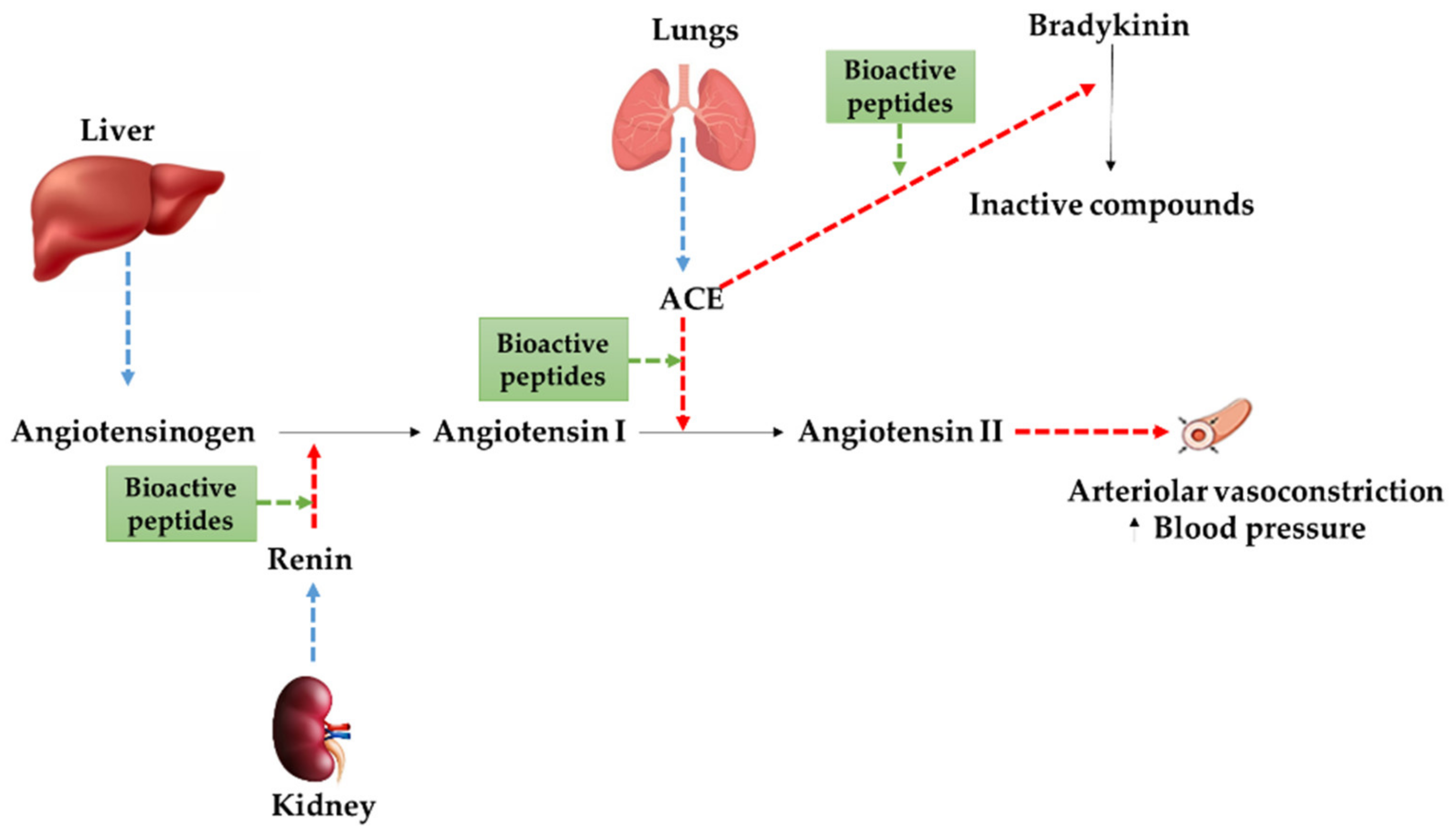
2. Bioactive Peptides Derived from Milk

| Original Proteins | Microbial Cultures | Peptide Sequences | References |
|---|---|---|---|
| β-casein and k-casein | Lactobacillus helveticus and Saccharomyces cerevisiae | VPP and IPP | [48] |
| β-casein and k-casein | Lactobacillus helveticus LBK16H | VPP and IPP | [49] |
| β-casein and αS1-casein | Lactobacillus GG and enzymes + pepsin and trypsin | YPFPAVPYPQRTTMPLW | [50] |
| Whey proteins | Lactobacillus helveticus CPN 4 | YP | [51] |
| β-casein | Lactobacillus rhamnosus + digestion with pepsin and Corolase PP | DKIHPFYQEPVLVKEAMAPK | [52] |
| β-casein | Lactobacillus delbrueckii ssp. bulgaricus | SKVYPFPGPI | [53] |
| β-casein | Streptococcus salivarius ssp. thermophiles + Lactococcus lactis biovar. diacetylactis | SKVYP | [53] |
| β-lactoglobulin | Kluyveromyces marxianus var. marxianus | YLLF | [54] |
| β-casein | Enterococcus faecalis CECT 5727 | LHLPLP and LVYPFPGPIPNSLPQNIPP | [55] |
| Whole milk | Enterococcus faecalis TH563 and Lactobacillus delbrueckii subsp. bulgaricus LA2 | Not identified | [56] |
| Whole milk | Bifidobacterium bifidum MF 20/5 and Lactobacillus casei YIT 9029 | Not identified | [57] |
| β-casein | Enterococcus faecalis BCS27 | VVVPPF and ENLLRF | [58] |
| β-casein | Bifidobacterium bifidum MF 20/5 | LVYPFP | [59] |
| κ-casein and αs2-casein | Kluyveromyces marxianus Z17 | VLSRYP and LSFF | [60] |
| Whole goat milk | Wild Lactobacillus plantarum 69 | Not identified | [61] |
| β-casein derived from camel milk | Leuconostoc lactis PTCC1899 | MVPYPQR | [62] |
3. Bioactive Peptides from Yogurt
4. Bioactive Peptides from Cheese
5. Bioactive Peptides from Kefir
6. In vivo and Ex vivo Studies
7. Structure-Function Relationships of Bioactive Peptides
8. Effect of GI Digestion on Bioactive Peptides
9. Allergenicity and Toxicity of Bioactive Peptides
10. Legislation and Regulatory Requirements for Bioactive Peptides
11. Future Perspective and Conclusions
Author Contributions
Funding
Institutional Review Board Statement
Informed Consent Statement
Data Availability Statement
Acknowledgments
Conflicts of Interest
References
- Aluko, R.E. Antihypertensive peptides from food proteins. Annu. Rev. Food Sci. Technol. 2015, 6, 235–262. [Google Scholar] [CrossRef]
- Cicero, A.F.; Fogacci, F.; Colletti, A. Potential role of bioactive peptides in prevention and treatment of chronic diseases: A narrative review. Br. J. Pharmacol. 2017, 174, 1378–1394. [Google Scholar] [CrossRef]
- FitzGerald, R.J.; Cermeño, M.; Khalesi, M.; Kleekayai, T.; Amigo-Benavent, M. Application of in silico approaches for the generation of milk protein-derived bioactive peptides. J. Funct. Foods 2020, 64, 103636. [Google Scholar] [CrossRef]
- Marcone, S.; Belton, O.; Fitzgerald, D.J. Milk-derived bioactive peptides and their health promoting effects: A potential role in atherosclerosis. Br. J. Clin. Pharmacol. 2017, 83, 152–162. [Google Scholar] [CrossRef] [Green Version]
- Gobbetti, M.; Stepaniak, L.; De Angelis, M.; Corsetti, A.; Di Cagno, R. Latent bioactive peptides in milk proteins: Proteolytic activation and significance in dairy processing. Crit. Rev. Food Sci. Nutr. 2002, 42, 223–239. [Google Scholar] [CrossRef]
- Mohanty, D.; Mohapatra, S.; Misra, S.; Sahu, P. Milk derived bioactive peptides and their impact on human health—A review. Saudi J. Biol. Sci. 2016, 23, 577–583. [Google Scholar] [CrossRef] [PubMed] [Green Version]
- Oliveira, D.; Fox, P.; O’Mahony, J.A. Byproducts from Dairy Processing. In Byproducts from Agriculture and Fisheries: Adding Value for Food, Feed, Pharma, and Fuels; Wiley: West Sussex, UK, 2019; pp. 57–106. [Google Scholar]
- Semb, A.G.; Ikdahl, E.; Wibetoe, G.; Crowson, C.; Rollefstad, S. Atherosclerotic cardiovascular disease prevention in rheumatoid arthritis. Nat. Rev. Rheumatol. 2020, 16, 361–379. [Google Scholar] [CrossRef] [PubMed]
- Balakumar, P.; Maung, U.K.; Jagadeesh, G. Prevalence and prevention of cardiovascular disease and diabetes mellitus. Pharmacol. Res. 2016, 113, 600–609. [Google Scholar] [CrossRef] [PubMed]
- FitzGerald, R.J.; Murray, B.A.; Walsh, D.J. Hypotensive peptides from milk proteins. J. Nutr. 2004, 134, 980S–988S. [Google Scholar] [CrossRef] [Green Version]
- Aluko, R.E. Food protein-derived renin-inhibitory peptides: In vitro and in vivo properties. J. Food Biochem. 2019, 43, e12648. [Google Scholar] [CrossRef] [Green Version]
- Baptista, D.P.; Gigante, M.L. Bioactive peptides in ripened cheeses: Release during technological processes and resistance to the gastrointestinal tract. J. Sci. Food Agric. 2021, 101, 4010–4017. [Google Scholar] [CrossRef]
- Chaudhary, A.; Bhalla, S.; Patiyal, S.; Raghava, G.P.; Sahni, G. FermFooDb: A database of bioactive peptides derived from fermented foods. Heliyon 2021, 7, e06668. [Google Scholar] [CrossRef]
- Kleekayai, T.; Cermeño, M.; FitzGerald, R.J. The Production of Bioactive Peptides from Milk Proteins. In Agents of Change; Springer: Cham, Switzerland, 2021; pp. 447–497. [Google Scholar]
- Choi, J.; Sabikhi, L.; Hassan, A.; Anand, S. Bioactive peptides in dairy products. Int. J. Dairy Technol. 2012, 65, 1–12. [Google Scholar] [CrossRef]
- Capriotti, A.L.; Cavaliere, C.; Piovesana, S.; Samperi, R.; Laganà, A. Recent trends in the analysis of bioactive peptides in milk and dairy products. Anal. Bioanal. Chem. 2016, 408, 2677–2685. [Google Scholar] [CrossRef]
- Tidona, F.; Criscione, A.; Guastella, A.M.; Zuccaro, A.; Bordonaro, S.; Marletta, D. Bioactive peptides in dairy products. Ital. J. Anim. Sci. 2009, 8, 315–340. [Google Scholar] [CrossRef]
- Hernández-Ledesma, B.; del Mar Contreras, M.; Recio, I. Antihypertensive peptides: Production, bioavailability and incorporation into foods. Adv. Colloid Interface Sci. 2011, 165, 23–35. [Google Scholar] [CrossRef] [Green Version]
- Korhonen, H.; Pihlanto, A. Technological options for the production of health-promoting proteins and peptides derived from milk and colostrum. Curr. Pharm. Des. 2007, 13, 829–843. [Google Scholar] [CrossRef]
- Shahbandeh, M. Global Cow Milk Production 2015–2021. Available online: https://www.statista.com/statistics/263952/production-of-milk-worldwide/ (accessed on 21 April 2022).
- Punia, H.; Tokas, J.; Malik, A.; Sangwan, S.; Baloda, S.; Singh, N.; Singh, S.; Bhuker, A.; Singh, P.; Yashveer, S. Identification and detection of bioactive peptides in milk and dairy products: Remarks about agro-foods. Molecules 2020, 25, 3328. [Google Scholar] [CrossRef]
- Danquah, M.K.; Agyei, D. Pharmaceutical applications of bioactive peptides. OA Biotechnol. 2012, 1, 1–7. [Google Scholar] [CrossRef] [Green Version]
- Guo, Y.; Jiang, X.; Xiong, B.; Zhang, T.; Zeng, X.; Wu, Z.; Sun, Y.; Pan, D. Production and transepithelial transportation of angiotensin-I-converting enzyme (ACE)-inhibitory peptides from whey protein hydrolyzed by immobilized Lactobacillus helveticus proteinase. J. Dairy Sci. 2019, 102, 961–975. [Google Scholar] [CrossRef] [Green Version]
- Huang, S.; Gong, Y.; Li, Y.; Ruan, S.; Azam, S.R.; Duan, Y.; Ye, X.; Ma, H. Preparation of ACE-inhibitory peptides from milk protein in continuous enzyme membrane reactor with gradient dilution feeding substrate. Process Biochem. 2020, 92, 130–137. [Google Scholar] [CrossRef]
- Shu, G.; Huang, J.; Bao, C.; Meng, J.; Chen, H.; Cao, J. Effect of different proteases on the degree of hydrolysis and angiotensin I-converting enzyme-inhibitory activity in goat and cow milk. Biomolecules 2018, 8, 101. [Google Scholar] [CrossRef] [Green Version]
- Villadóniga, C.; Cantera, A.M.B. New ACE-inhibitory peptides derived from α-lactalbumin produced by hydrolysis with Bromelia antiacantha peptidases. Biocatal. Agric. Biotechnol. 2019, 20, 101258. [Google Scholar] [CrossRef]
- Wu, Q.; Zhang, X.; Jia, J.; Kuang, C.; Yang, H. Effect of ultrasonic pretreatment on whey protein hydrolysis by alcalase: Thermodynamic parameters, physicochemical properties and bioactivities. Process Biochem. 2018, 67, 46–54. [Google Scholar] [CrossRef]
- Guo, Y.; Pan, D.; Tanokura, M. Optimisation of hydrolysis conditions for the production of the angiotensin-I converting enzyme (ACE) inhibitory peptides from whey protein using response surface methodology. Food Chem. 2009, 114, 328–333. [Google Scholar] [CrossRef]
- Mirzapour-Kouhdasht, A.; Moosavi-Nasab, M.; Lee, C.W.; Yun, H.; Eun, J.-B. Structure–function engineering of novel fish gelatin-derived multifunctional peptides using high-resolution peptidomics and bioinformatics. Sci. Rep. 2021, 11, 7401. [Google Scholar] [CrossRef]
- Noman, A.; Xu, Y.; AL-Bukhaiti, W.Q.; Abed, S.M.; Ali, A.H.; Ramadhan, A.H.; Xia, W. Influence of enzymatic hydrolysis conditions on the degree of hydrolysis and functional properties of protein hydrolysate obtained from Chinese sturgeon (Acipenser sinensis) by using papain enzyme. Process Biochem. 2018, 67, 19–28. [Google Scholar] [CrossRef]
- Islam, M.S.; Hongxin, W.; Admassu, H.; Noman, A.; Ma, C.; An Wei, F. Degree of hydrolysis, functional and antioxidant properties of protein hydrolysates from Grass Turtle (Chinemys reevesii) as influenced by enzymatic hydrolysis conditions. Food Sci. Nutr. 2021, 9, 4031–4047. [Google Scholar] [CrossRef] [PubMed]
- Mazorra-Manzano, M.A.; Mora-Cortes, W.G.; Leandro-Roldan, M.M.; González-Velázquez, D.A.; Torres-Llanez, M.J.; Ramírez-Suarez, J.C.; González-Córdova, A.F.; Vallejo-Córdoba, B. Production of whey protein hydrolysates with angiotensin-converting enzyme-inhibitory activity using three new sources of plant proteases. Biocatal. Agric. Biotechnol. 2020, 28, 101724. [Google Scholar] [CrossRef]
- Lin, K.; Zhang, L.-W.; Han, X.; Cheng, D.-Y. Novel angiotensin I-converting enzyme inhibitory peptides from protease hydrolysates of Qula casein: Quantitative structure-activity relationship modeling and molecular docking study. J. Funct. Foods 2017, 32, 266–277. [Google Scholar] [CrossRef]
- Lin, K.; Zhang, L.; Han, X.; Meng, Z.; Zhang, J.; Wu, Y.; Cheng, D. Quantitative structure–activity relationship modeling coupled with molecular docking analysis in screening of angiotensin I-converting enzyme inhibitory peptides from Qula Casein hydrolysates obtained by two-enzyme combination hydrolysis. J. Agric. Food Chem. 2018, 66, 3221–3228. [Google Scholar] [CrossRef]
- Corrons, M.A.; Liggieri, C.S.; Trejo, S.A.; Bruno, M.A. ACE-inhibitory peptides from bovine caseins released with peptidases from Maclura pomifera latex. Food Res. Int. 2017, 93, 8–15. [Google Scholar] [CrossRef]
- Tagliazucchi, D.; Shamsia, S.; Helal, A.; Conte, A. Angiotensin-converting enzyme inhibitory peptides from goats’ milk released by in vitro gastro-intestinal digestion. Int. Dairy J. 2017, 71, 6–16. [Google Scholar] [CrossRef]
- Abdel-Hamid, M.; Otte, J.; De Gobba, C.; Osman, A.; Hamad, E. Angiotensin I-converting enzyme inhibitory activity and antioxidant capacity of bioactive peptides derived from enzymatic hydrolysis of buffalo milk proteins. Int. Dairy J. 2017, 66, 91–98. [Google Scholar] [CrossRef]
- Salami, M.; Moosavi-Movahedi, A.A.; Moosavi-Movahedi, F.; Ehsani, M.R.; Yousefi, R.; Farhadi, M.; Niasari-Naslaji, A.; Saboury, A.A.; Chobert, J.M.; Haertlé, T. Biological activity of camel milk casein following enzymatic digestion. J. Dairy Res. 2011, 78, 471–478. [Google Scholar] [CrossRef]
- Tagliazucchi, D.; Shamsia, S.; Conte, A. Release of angiotensin converting enzyme-inhibitory peptides during in vitro gastro-intestinal digestion of camel milk. Int. Dairy J. 2016, 56, 119–128. [Google Scholar] [CrossRef]
- Baba, W.N.; Baby, B.; Mudgil, P.; Gan, C.-Y.; Vijayan, R.; Maqsood, S. Pepsin generated camel whey protein hydrolysates with potential antihypertensive properties: Identification and molecular docking of antihypertensive peptides. LWT 2021, 143, 111135. [Google Scholar] [CrossRef]
- Koirala, S.; Prathumpai, W.; Anal, A.K. Effect of ultrasonication pretreatment followed by enzymatic hydrolysis of caprine milk proteins and on antioxidant and angiotensin converting enzyme (ACE) inhibitory activity of peptides thus produced. Int. Dairy J. 2021, 118, 105026. [Google Scholar] [CrossRef]
- Lin, K.; Ma, Z.; Ramachandran, M.; De Souza, C.; Han, X.; Zhang, L.-W. ACE inhibitory peptide KYIPIQ derived from yak milk casein induces nitric oxide production in HUVECs and diffuses via a transcellular mechanism in Caco-2 monolayers. Process Biochem. 2020, 99, 103–111. [Google Scholar] [CrossRef]
- Cui, Q.; Sun, Y.; Cheng, J.; Guo, M. Effect of two-step enzymatic hydrolysis on the antioxidant properties and proteomics of hydrolysates of milk protein concentrate. Food Chem. 2022, 366, 130711. [Google Scholar] [CrossRef]
- Bao, Q.; Liu, W.; Yu, J.; Wang, W.; Qing, M.; Chen, X.; Wang, F.; Zhang, J.; Zhang, W.; Qiao, J. Isolation and identification of cultivable lactic acid bacteria in traditional yak milk products of Gansu Province in China. J. Gen. Appl. Microbiol. 2012, 58, 95–105. [Google Scholar] [CrossRef] [PubMed] [Green Version]
- Chen, H.; Ji, Z.; Shu, G.W.; Xing, H.N. Effect of Probiotic Lactobacillus Strains on Angiotensin I Converting Enzyme Inhibitory Activity from Fermented Goat Milk. In Advanced Materials Research; Trans Tech Publications Ltd.: Bäch, Switzerland, 2012; pp. 442–445. [Google Scholar]
- Shu, G.; Yang, H.; Chen, H.; Zhang, Q.; Tian, Y. Effect of incubation time, inoculum size, temperature, pasteurization time, goat milk powder and whey powder on ACE inhibitory activity in fermented milk by L. plantarum LP69. Acta Sci. Pol. Technol. Aliment. 2015, 14, 107–116. [Google Scholar] [CrossRef] [PubMed]
- Li, S.; Tang, S.; He, Q.; Hu, J.; Zheng, J. In vitro antioxidant and angiotensin-converting enzyme inhibitory activity of fermented milk with different culture combinations. J. Dairy Sci. 2020, 103, 1120–1130. [Google Scholar] [CrossRef] [PubMed]
- Takano, D.T. Anti-hypertensive activity of fermented dairy products containing biogenic peptides. Antonie Van Leeuwenhoek 2002, 82, 333–340. [Google Scholar] [CrossRef]
- Sipola, M.; Finckenberg, P.; Korpela, R.; Vapaatalo, H.; Nurminen, M.L. Effect of long-term intake of milk products on blood pressure in hypertensive rats. J. Dairy Res. 2002, 69, 103–111. [Google Scholar] [CrossRef]
- Wan, J.; Mawson, R.; Ashokkumar, M.; Ronacher, K.; Coventry, M.J.; Roginski, H.; Versteeg, C. Emerging processing technologies for functional foods. Aust. J. Dairy Technol. 2005, 60, 167–169. [Google Scholar]
- Yamamoto, N.; Maeno, M.; Takano, T. Purification and characterization of an antihypertensive peptide from a yogurt-like product fermented by Lactobacillus helveticus CPN4. J. Dairy Sci. 1999, 82, 1388–1393. [Google Scholar] [CrossRef]
- Korhonen, H.; Pihlanto, A. Bioactive peptides: New challenges and opportunities for the dairy industry. Aust. J. Dairy Technol. 2003, 58, 129. [Google Scholar]
- Ashar, M.N.; Chand, R. Antihypertensive peptides purified from milks fermented with Lactobacillus delbrueckii ssp. bulgaricus. Milchwissenschaft 2004, 59, 14–17. [Google Scholar]
- Barrett, E.; Hayes, M.; Fitzgerald, G.F.; Hill, C.; Stanton, C.; Rose, R.P. Fermentation, cell factories and bioactive peptides: Food grade bacteria for production of biogenic compounds. Aust. J. Dairy Technol. 2005, 60, 157–162. [Google Scholar]
- Quirós, A.; Ramos, M.; Muguerza, B.; Delgado, M.A.; Miguel, M.; Aleixandre, A.; Recio, I. Identification of novel antihypertensive peptides in milk fermented with Enterococcus faecalis. Int. Dairy J. 2007, 17, 33–41. [Google Scholar] [CrossRef]
- Regazzo, D.; Da Dalt, L.; Lombardi, A.; Andrighetto, C.; Negro, A.; Gabai, G. Fermented milks from Enterococcus faecalis TH563 and Lactobacillus delbrueckii subsp. bulgaricus LA2 manifest different degrees of ACE-inhibitory and immunomodulatory activities. Dairy Sci. Technol. 2010, 90, 469–476. [Google Scholar] [CrossRef]
- Gonzalez-Gonzalez, C.R.; Tuohy, K.M.; Jauregi, P. Production of angiotensin-I-converting enzyme (ACE) inhibitory activity in milk fermented with probiotic strains: Effects of calcium, pH and peptides on the ACE-inhibitory activity. Int. Dairy J. 2011, 21, 615–622. [Google Scholar] [CrossRef]
- Gútiez, L.; Gómez-Sala, B.; Recio, I.; del Campo, R.; Cintas, L.M.; Herranz, C.; Hernández, P.E. Enterococcus faecalis strains from food, environmental, and clinical origin produce ACE-inhibitory peptides and other bioactive peptides during growth in bovine skim milk. Int. J. Food Microbiol. 2013, 166, 93–101. [Google Scholar] [CrossRef]
- Gonzalez-Gonzalez, C.; Gibson, T.; Jauregi, P. Novel probiotic-fermented milk with angiotensin I-converting enzyme inhibitory peptides produced by Bifidobacterium bifidum MF 20/5. Int. J. Food Microbiol. 2013, 167, 131–137. [Google Scholar] [CrossRef]
- Li, Y.; Sadiq, F.A.; Liu, T.; Chen, J.; He, G. Purification and identification of novel peptides with inhibitory effect against angiotensin I-converting enzyme and optimization of process conditions in milk fermented with the yeast Kluyveromyces marxianus. J. Funct. Foods 2015, 16, 278–288. [Google Scholar] [CrossRef]
- Chen, L.; Zhang, Q.; Ji, Z.; Shu, G.; Chen, H. Production and fermentation characteristics of angiotensin-I-converting enzyme inhibitory peptides of goat milk fermented by a novel wild Lactobacillus plantarum 69. LWT 2018, 91, 532–540. [Google Scholar] [CrossRef]
- Soleymanzadeh, N.; Mirdamadi, S.; Mirzaei, M.; Kianirad, M. Novel β-casein derived antioxidant and ACE-inhibitory active peptide from camel milk fermented by Leuconostoc lactis PTCC1899: Identification and molecular docking. Int. Dairy J. 2019, 97, 201–208. [Google Scholar] [CrossRef]
- Solanki, D.; Hati, S. Considering the potential of Lactobacillus rhamnosus for producing Angiotensin I-Converting Enzyme (ACE) inhibitory peptides in fermented camel milk (Indian breed). Food Biosci. 2018, 23, 16–22. [Google Scholar] [CrossRef]
- Rani, S.; Pooja, K.; Pal, G.K. Exploration of potential angiotensin converting enzyme inhibitory peptides generated from enzymatic hydrolysis of goat milk proteins. Biocatal. Agric. Biotechnol. 2017, 11, 83–88. [Google Scholar] [CrossRef]
- El Hatmi, H.; Jrad, Z.; Khorchani, T.; Jardin, J.; Poirson, C.; Perrin, C.; Cakir-Kiefer, C.; Girardet, J.-M. Identification of bioactive peptides derived from caseins, glycosylation-dependent cell adhesion molecule-1 (GlyCAM-1), and peptidoglycan recognition protein-1 (PGRP-1) in fermented camel milk. Int. Dairy J. 2016, 56, 159–168. [Google Scholar] [CrossRef]
- Mann, B.; Athira, S.; Sharma, R.; Bajaj, R. Bioactive Peptides in Yogurt. In Yogurt in Health and Disease Prevention; Elsevier: London, UK, 2017; pp. 411–426. [Google Scholar]
- Buendia, J.R.; Li, Y.; Hu, F.B.; Cabral, H.J.; Bradlee, M.L.; Quatromoni, P.A.; Singer, M.R.; Curhan, G.C.; Moore, L.L. Regular Yogurt Intake and Risk of Cardiovascular Disease Among Hypertensive Adults. Am. J. Hypertens. 2018, 31, 557–565. [Google Scholar] [CrossRef] [PubMed]
- Chabance, B.; Marteau, P.; Rambaud, J.C.; Migliore-Samour, D.; Boynard, M.; Perrotin, P.; Guillet, R.; Jollès, P.; Fiat, A.M. Casein peptide release and passage to the blood in humans during digestion of milk or yogurt. Biochimie 1998, 80, 155–165. [Google Scholar] [CrossRef]
- Fitzgerald, R.J.; Murray, B.A. Bioactive peptides and lactic fermentations. Int. J. Dairy Technol. 2006, 59, 118–125. [Google Scholar] [CrossRef]
- Nguyen, H.T.H.; Gathercole, J.L.; Day, L.; Dalziel, J.E. Differences in peptide generation following in vitro gastrointestinal digestion of yogurt and milk from cow, sheep and goat. Food Chem. 2020, 317, 126419. [Google Scholar] [CrossRef] [PubMed]
- Maes, W.; Van Camp, J.; Vermeirssen, V.; Hemeryck, M.; Ketelslegers, J.M.; Schrezenmeir, J.; Van Oostveldt, P.; Huyghebaert, A. Influence of the lactokinin Ala-Leu-Pro-Met-His-Ile-Arg (ALPMHIR) on the release of endothelin-1 by endothelial cells. Regul. Pept. 2004, 118, 105–109. [Google Scholar] [CrossRef] [PubMed]
- Perpetuo, E.A.; Juliano, L.; Lebrun, I. Biochemical and Pharmacological Aspects of Two Bradykinin-Potentiating Peptides Obtained from Tryptic Hydrolysis of Casein. J. Protein Chem. 2003, 22, 601–606. [Google Scholar] [CrossRef]
- Nurminen, M.L.; Sipola, M.; Kaarto, H.; Pihlanto-Leppälä, A.; Piilola, K.; Korpela, R.; Tossavainen, O.; Korhonen, H.; Vapaatalo, H. Alpha-lactorphin lowers blood pressure measured by radiotelemetry in normotensive and spontaneously hypertensive rats. Life Sci. 2000, 66, 1535–1543. [Google Scholar] [CrossRef]
- Hayes, M.; Stanton, C.; Fitzgerald, G.F.; Ross, R.P. Putting microbes to work: Dairy fermentation, cell factories and bioactive peptides. Part II: Bioactive peptide functions. Biotechnol. J. 2007, 2, 435–449. [Google Scholar] [CrossRef]
- Giacometti Cavalheiro, F.; Parra Baptista, D.; Domingues Galli, B.; Negrão, F.; Nogueira Eberlin, M.; Lúcia Gigante, M. High protein yogurt with addition of Lactobacillus helveticus: Peptide profile and angiotensin-converting enzyme ACE-inhibitory activity. Food Chem. 2020, 333, 127482. [Google Scholar] [CrossRef]
- Papadimitriou, C.G.; Vafopoulou-Mastrojiannaki, A.; Silva, S.V.; Gomes, A.-M.; Malcata, F.X.; Alichanidis, E. Identification of peptides in traditional and probiotic sheep milk yoghurt with angiotensin I-converting enzyme (ACE)-inhibitory activity. Food Chem. 2007, 105, 647–656. [Google Scholar] [CrossRef]
- Pina, A.S.; Roque, A.C. Studies on the molecular recognition between bioactive peptides and angiotensin-converting enzyme. J. Mol. Recognit. 2009, 22, 162–168. [Google Scholar] [CrossRef]
- Lebrun, I.; Lebrun, F.L.; Henriques, O.B.; Carmona, A.K.; Juliano, L.; Camargo, A.C. Isolation and characterization of a new bradykinin potentiating octapeptide from gamma-casein. Can. J. Physiol. Pharmacol. 1995, 73, 85–91. [Google Scholar] [CrossRef] [PubMed]
- Robinson, R.C.; Nielsen, S.D.; Dallas, D.C.; Barile, D. Can cheese mites, maggots and molds enhance bioactivity? Peptidomic investigation of functional peptides in four traditional cheeses. Food Funct. 2021, 12, 633–645. [Google Scholar] [CrossRef]
- Sánchez-Rivera, L.; Martínez-Maqueda, D.; Cruz-Huerta, E.; Miralles, B.; Recio, I. Peptidomics for discovery, bioavailability and monitoring of dairy bioactive peptides. Food Res. Int. 2014, 63, 170–181. [Google Scholar] [CrossRef]
- Gómez-Ruiz, J.Á.; Taborda, G.; Amigo, L.; Recio, I.; Ramos, M. Identification of ACE-inhibitory peptides in different Spanish cheeses by tandem mass spectrometry. Eur. Food Res. Technol. 2006, 223, 595–601. [Google Scholar] [CrossRef]
- Gupta, A.; Mann, B.; Kumar, R.; Sangwan, R. Antioxidant activity of Cheddar cheeses at different stages of ripening. Int. J. Dairy Technol. 2009, 62, 339–347. [Google Scholar] [CrossRef]
- Iwaniak, A.; Mogut, D. Metabolic Syndrome-Preventive Peptides Derived from Milk Proteins and Their Presence in Cheeses: A Review. Appl. Sci. 2020, 10, 2772. [Google Scholar] [CrossRef]
- Summer, A.; Formaggioni, P.; Franceschi, P.; Di Frangia, F.; Righi, F.; Malacarne, M. Cheese as Functional Food: The Example of Parmigiano Reggiano and Grana Padano. Food Technol. Biotechnol. 2017, 55, 277–289. [Google Scholar] [CrossRef]
- Bütikofer, U.; Meyer, J.; Sieber, R.; Wechsler, D. Quantification of the angiotensin-converting enzyme-inhibiting tripeptides Val-Pro-Pro and Ile-Pro-Pro in hard, semi-hard and soft cheeses. Int. Dairy J. 2007, 17, 968–975. [Google Scholar] [CrossRef]
- Pihlanto, A. Lactic Fermentation and Bioactive Peptides. In Lactic Acid Bacteria–R & D for Food, Health and Livestock Purposes; IntechOpen: London, UK, 2013; pp. 310–331. [Google Scholar]
- McSweeney, P.L. Biochemistry of cheese ripening: Introduction and overview. In Cheese; Elsevier: London, UK, 2017; pp. 379–387. [Google Scholar]
- Ardö, Y.; McSweeney, P.L.H.; Magboul, A.A.A.; Upadhyay, V.K.; Fox, P.F. Chapter 18–Biochemistry of Cheese Ripening: Proteolysis. In Cheese, 4th ed.; McSweeney, P.L.H., Fox, P.F., Cotter, P.D., Everett, D.W., Eds.; Academic Press: London, UK, 2017; pp. 445–482. [Google Scholar] [CrossRef]
- Ong, L.; Shah, N.P. Release and identification of angiotensin-converting enzyme-inhibitory peptides as influenced by ripening temperatures and probiotic adjuncts in Cheddar cheeses. LWT–Food Sci. Technol. 2008, 41, 1555–1566. [Google Scholar] [CrossRef]
- Sahingil, D.; Hayaloglu, A.A.; Kirmaci, H.A.; Özer, B.; Simsek, O. Changes of proteolysis and angiotensin-I converting enzyme-inhibitory activity in white-brined cheese as affected by adjunct culture and ripening temperature. J. Dairy Res. 2014, 81, 394–402. [Google Scholar] [CrossRef] [PubMed]
- Sánchez-Rivera, L.; Diezhandino, I.; Gómez-Ruiz, J.Á.; Fresno, J.M.; Miralles, B.; Recio, I. Peptidomic study of Spanish blue cheese (Valdeón) and changes after simulated gastrointestinal digestion. Electrophoresis 2014, 35, 1627–1636. [Google Scholar] [CrossRef]
- Stuknytė, M.; Cattaneo, S.; Masotti, F.; De Noni, I. Occurrence and fate of ACE-inhibitor peptides in cheeses and in their digestates following in vitro static gastrointestinal digestion. Food Chem. 2015, 168, 27–33. [Google Scholar] [CrossRef] [PubMed]
- Basiricò, L.; Catalani, E.; Morera, P.; Cattaneo, S.; Stuknytė, M.; Bernabucci, U.; De Noni, I.; Nardone, A. Release of angiotensin converting enzyme-inhibitor peptides during in vitro gastrointestinal digestion of Parmigiano Reggiano PDO cheese and their absorption through an in vitro model of intestinal epithelium. J. Dairy Sci. 2015, 98, 7595–7601. [Google Scholar] [CrossRef] [PubMed] [Green Version]
- Baptista, D.P.; Salgaço, M.K.; Sivieri, K.; Gigante, M.L. Use of static and dynamic in vitro models to simulate Prato cheese gastrointestinal digestion: Effect of Lactobacillus helveticus LH-B02 addition on peptides bioaccessibility. LWT 2020, 134, 110229. [Google Scholar] [CrossRef]
- Atanasova, J.; Dalgalarrondo, M.; Iliev, I.; Moncheva, P.; Todorov, S.D.; Ivanova, I.V. Formation of Free Amino Acids and Bioactive Peptides During the Ripening of Bulgarian White Brined Cheeses. Probiotics Antimicrob. Proteins 2021, 13, 261–272. [Google Scholar] [CrossRef] [PubMed]
- Iwaniak, A.; Mogut, D.; Minkiewicz, P.; Żulewska, J.; Darewicz, M. Gouda Cheese with Modified Content of β-Casein as a Source of Peptides with ACE- and DPP-IV-Inhibiting Bioactivity: A Study Based on In Silico and In Vitro Protocol. Int. J. Mol. Sci. 2021, 22, 2949. [Google Scholar] [CrossRef]
- Guzel-Seydim, Z.B.; Kok-Tas, T.; Greene, A.K.; Seydim, A.C. Review: Functional properties of kefir. Crit. Rev. Food Sci. Nutr. 2011, 51, 261–268. [Google Scholar] [CrossRef]
- Muir, D.; Tamime, A.; Wszolek, M. Comparison of the sensory profiles of kefir, buttermilk and yogurt. Int. J. Dairy Technol. 1999, 52, 129–134. [Google Scholar] [CrossRef]
- Simova, E.; Beshkova, D.; Angelov, A.; Hristozova, T.; Frengova, G.; Spasov, Z. Lactic acid bacteria and yeasts in kefir grains and kefir made from them. J. Ind. Microbiol. Biotechnol. 2002, 28, 1–6. [Google Scholar] [CrossRef]
- Şanli, T.; Akal, H.C.; Yetişemiyen, A.; Hayaloglu, A.A. Influence of adjunct cultures on angiotensin-converting enzyme (ACE)-inhibitory activity, organic acid content and peptide profile of kefir. Int. J. Dairy Technol. 2018, 71, 131–139. [Google Scholar] [CrossRef]
- López-Fandiño, R.; Otte, J.; van Camp, J. Physiological, chemical and technological aspects of milk-protein-derived peptides with antihypertensive and ACE-inhibitory activity. Int. Dairy J. 2006, 16, 1277–1293. [Google Scholar] [CrossRef]
- Nielsen, M.S.; Martinussen, T.; Flambard, B.; Sørensen, K.I.; Otte, J. Peptide profiles and angiotensin-I-converting enzyme inhibitory activity of fermented milk products: Effect of bacterial strain, fermentation pH, and storage time. Int. Dairy J. 2009, 19, 155–165. [Google Scholar] [CrossRef]
- Shu, G.; Ma, L.; Chen, L.; Guo, M.; Guo, Y.; Chen, H. Goat milk Kefir with ACE inhibitory activity: Preparation and storage stability evaluation. J. Food Process. Preserv. 2020, 44, e14417. [Google Scholar] [CrossRef]
- Amorim, F.G.; Coitinho, L.B.; Dias, A.T.; Friques, A.G.F.; Monteiro, B.L.; Rezende, L.C.D.; Pereira, T.M.C.; Campagnaro, B.P.; De Pauw, E.; Vasquez, E.C.; et al. Identification of new bioactive peptides from Kefir milk through proteopeptidomics: Bioprospection of antihypertensive molecules. Food Chem. 2019, 282, 109–119. [Google Scholar] [CrossRef]
- Zarei, M.; Forghani, B.; Ebrahimpour, A.; Abdul Hamid, A.; Anwar, F.; Saari, N. In vitro and in vivo antihypertensive activity of palm kernel cake protein hydrolysates: Sequencing and characterization of potent bioactive peptides. Ind. Crop. Prod. 2015, 76, 112–120. [Google Scholar] [CrossRef]
- Zou, P.; Wang, J.L.; He, G.Q.; Wu, J. Purification, identification, and in vivo activity of angiotensin I-converting enzyme inhibitory peptide, from ribbonfish (Trichiurus haumela) backbone. J. Food Sci. 2014, 79, C1–C7. [Google Scholar] [CrossRef]
- Gleeson, J.P.; Brayden, D.J.; Ryan, S.M. Evaluation of PepT1 transport of food-derived antihypertensive peptides, Ile-Pro-Pro and Leu-Lys-Pro using in vitro, ex vivo and in vivo transport models. Eur. J. Pharm. Biopharm. 2017, 115, 276–284. [Google Scholar] [CrossRef] [Green Version]
- Koyama, D.; Sasai, M.; Matsumura, S.; Inoue, K.; Ohinata, K. A milk-derived pentapeptide reduces blood pressure in advanced hypertension in a CCK system-dependent manner. Food Funct. 2020, 11, 9489–9494. [Google Scholar] [CrossRef]
- Lee, N.-Y.; Cheng, J.-T.; Enomoto, T.; Nakamura, I. The antihypertensive activity of angiotensin-converting enzyme inhibitory peptide containing in bovine lactoferrin. Chin. J. Physiol. 2006, 49, 67–73. [Google Scholar] [PubMed]
- Centeno, J.M.; Burguete, M.C.; Castelló-Ruiz, M.; Enrique, M.; Vallés, S.; Salom, J.B.; Torregrosa, G.; Marcos, J.F.; Alborch, E.; Manzanares, P. Lactoferricin-Related Peptides with Inhibitory Effects on ACE-Dependent Vasoconstriction. J. Agric. Food Chem. 2006, 54, 5323–5329. [Google Scholar] [CrossRef]
- Ruiz-Giménez, P.; Burguete, M.C.; Castelló-Ruiz, M.; Marcos, J.F.; Salom, J.B.; Vallés, S.; Torregrosa, G.; Alborch, E.; Manzanares, P. Bovine lactoferrin pepsin hydrolysate exerts inhibitory effect on angiotensin I-converting enzyme-dependent vasoconstriction. Int. Dairy J. 2007, 17, 1212–1215. [Google Scholar] [CrossRef]
- Ruiz-Giménez, P.; Ibáñez, A.; Salom, J.B.; Marcos, J.F.; López-Díez, J.J.; Vallés, S.; Torregrosa, G.; Alborch, E.; Manzanares, P. Antihypertensive Properties of Lactoferricin B-Derived Peptides. J. Agric. Food Chem. 2010, 58, 6721–6727. [Google Scholar] [CrossRef] [PubMed]
- Sánchez-Rivera, L.; Santos, P.F.; Miralles, B.; Carrón, R.; José Montero, M.; Recio, I. Peptide fragments from β-casein f(134–138), HLPLP, generated by the action of rat blood plasma peptidases show potent antihypertensive activity. Food Res. Int. 2016, 88, 348–353. [Google Scholar] [CrossRef] [Green Version]
- Khedr, S.; Deussen, A.; Kopaliani, I.; Zatschler, B.; Martin, M. Effects of tryptophan-containing peptides on angiotensin-converting enzyme activity and vessel tone ex vivo and in vivo. Eur. J. Nutr. 2018, 57, 907–915. [Google Scholar] [CrossRef] [PubMed]
- Xue, L.; Wang, X.; Hu, Z.; Wu, Z.; Wang, L.; Wang, H.; Yang, M. Identification and characterization of an angiotensin-converting enzyme inhibitory peptide derived from bovine casein. Peptides 2018, 99, 161–168. [Google Scholar] [CrossRef] [PubMed]
- Ruiz-Giménez, P.; Salom, J.B.; Marcos, J.F.; Vallés, S.; Martínez-Maqueda, D.; Recio, I.; Torregrosa, G.; Alborch, E.; Manzanares, P. Antihypertensive effect of a bovine lactoferrin pepsin hydrolysate: Identification of novel active peptides. Food Chem. 2012, 131, 266–273. [Google Scholar] [CrossRef]
- Geerlings, A.; Villar, I.C.; Hidalgo Zarco, F.; Sánchez, M.; Vera, R.; Zafra Gomez, A.; Boza, J.; Duarte, J. Identification and Characterization of Novel Angiotensin-Converting Enzyme Inhibitors Obtained from Goat Milk. J. Dairy Sci. 2006, 89, 3326–3335. [Google Scholar] [CrossRef]
- Clare, D.A.; Swaisgood, H.E. Bioactive Milk Peptides: A Prospectus1. J. Dairy Sci. 2000, 83, 1187–1195. [Google Scholar] [CrossRef]
- Rodríguez-Figueroa, J.C.; González-Córdova, A.F.; Torres-Llanez, M.J.; Garcia, H.S.; Vallejo-Cordoba, B. Novel angiotensin I-converting enzyme inhibitory peptides produced in fermented milk by specific wild Lactococcus lactis strains. J. Dairy Sci. 2012, 95, 5536–5543. [Google Scholar] [CrossRef] [Green Version]
- Cushman, D.W.; Cheung, H.S.; Sabo, E.F.; Ondetti, M.A. Design of potent competitive inhibitors of angiotensin-converting enzyme. Carboxyalkanoyl and mercaptoalkanoyl amino acids. Biochemistry 1977, 16, 5484–5491. [Google Scholar] [CrossRef]
- Rohrbach, M.S.; Williams, E.B.; Rolstad, R.A. Purification and substrate specificity of bovine angiotensin-converting enzyme. J. Biol. Chem. 1981, 256, 225–230. [Google Scholar] [CrossRef]
- Gómez-Ruiz, J.Á.; Ramos, M.; Recio, I. Angiotensin-converting enzyme-inhibitory peptides in Manchego cheeses manufactured with different starter cultures. Int. Dairy J. 2002, 12, 697–706. [Google Scholar] [CrossRef]
- Pan, D.; Guo, H.; Zhao, B.; Cao, J. The molecular mechanisms of interactions between bioactive peptides and angiotensin-converting enzyme. Bioorg. Med. Chem. Lett. 2011, 21, 3898–3904. [Google Scholar] [CrossRef]
- Abdelhedi, O.; Nasri, R.; Jridi, M.; Mora, L.; Oseguera-Toledo, M.E.; Aristoy, M.-C.; Amara, I.B.; Toldrá, F.; Nasri, M. In silico analysis and antihypertensive effect of ACE-inhibitory peptides from smooth-hound viscera protein hydrolysate: Enzyme-peptide interaction study using molecular docking simulation. Process Biochem. 2017, 58, 145–159. [Google Scholar] [CrossRef]
- Nie, X.; Zhao, L.; Deng, S.; Su, W.; Zhang, Y. A review of molecular simulation applied in vapor-liquid equilibria (VLE) estimation of thermodynamic cycles. J. Mol. Liq. 2018, 264, 652–674. [Google Scholar] [CrossRef]
- Tao, X.; Huang, Y.; Wang, C.; Chen, F.; Yang, L.; Ling, L.; Che, Z.; Chen, X. Recent developments in molecular docking technology applied in food science: A review. Int. J. Food Sci. Technol. 2020, 55, 33–45. [Google Scholar] [CrossRef]
- Fu, Y.; Young, J.F.; Løkke, M.M.; Lametsch, R.; Aluko, R.E.; Therkildsen, M. Revalorisation of bovine collagen as a potential precursor of angiotensin I-converting enzyme (ACE) inhibitory peptides based on in silico and in vitro protein digestions. J. Funct. Foods 2016, 24, 196–206. [Google Scholar] [CrossRef]
- Nongonierma, A.B.; FitzGerald, R.J. Learnings from quantitative structure–activity relationship (QSAR) studies with respect to food protein-derived bioactive peptides: A review. RSC Adv. 2016, 6, 75400–75413. [Google Scholar] [CrossRef]
- Sandberg, M.; Eriksson, L.; Jonsson, J.; Sjöström, M.; Wold, S. New Chemical Descriptors Relevant for the Design of Biologically Active Peptides. A Multivariate Characterization of 87 Amino Acids. J. Med. Chem. 1998, 41, 2481–2491. [Google Scholar] [CrossRef] [PubMed]
- Wu, J.; Aluko, R.E.; Nakai, S. Structural requirements of Angiotensin I-converting enzyme inhibitory peptides: Quantitative structure-activity relationship study of di- and tripeptides. J. Agric. Food Chem. 2006, 54, 732–738. [Google Scholar] [CrossRef] [PubMed]
- Jing, P.; Qian, B.; He, Y.; Zhao, X.; Zhang, J.; Zhao, D.; Lv, Y.; Deng, Y. Screening milk-derived antihypertensive peptides using quantitative structure activity relationship (QSAR) modelling and in vitro/in vivo studies on their bioactivity. Int. Dairy J. 2014, 35, 95–101. [Google Scholar] [CrossRef]
- Lin, K.; Zhang, L.W.; Han, X.; Xin, L.; Meng, Z.X.; Gong, P.M.; Cheng, D.Y. Yak milk casein as potential precursor of angiotensin I-converting enzyme inhibitory peptides based on in silico proteolysis. Food Chem. 2018, 254, 340–347. [Google Scholar] [CrossRef]
- Majumder, K.; Wu, J. A new approach for identification of novel antihypertensive peptides from egg proteins by QSAR and bioinformatics. Food Res. Int. 2010, 43, 1371–1378. [Google Scholar] [CrossRef]
- Pripp, A.H.; Isaksson, T.; Stepaniak, L.; Sørhaug, T. Quantitative structure-activity relationship modelling of ACE-inhibitory peptides derived from milk proteins. Eur. Food Res. Technol. 2004, 219, 579–583. [Google Scholar] [CrossRef]
- Sagardia, I.; Roa-Ureta, R.H.; Bald, C. A new QSAR model, for angiotensin I-converting enzyme inhibitory oligopeptides. Food Chem. 2013, 136, 1370–1376. [Google Scholar] [CrossRef]
- Tripaldi, P.; Pérez-González, A.; Rojas, C.; Radax, J.; Ballabio, D.; Todeschini, R. Classification-based QSAR Models for the Prediction of the Bioactivity of ACE-inhibitor Peptides. Protein Pept. Lett. 2018, 25, 1015–1023. [Google Scholar] [CrossRef]
- Vukic, V.R.; Vukic, D.V.; Milanovic, S.D.; Ilicic, M.D.; Kanuric, K.G.; Johnson, M.S. In silico identification of milk antihypertensive di- and tripeptides involved in angiotensin I-converting enzyme inhibitory activity. Nutr. Res. 2017, 46, 22–30. [Google Scholar] [CrossRef]
- Wu, J.; Aluko, R.E.; Nakai, S. Structural Requirements of Angiotensin I-Converting Enzyme Inhibitory Peptides: Quantitative Structure-Activity Relationship Modeling of Peptides Containing 4–10 Amino Acid Residues. QSAR Comb. Sci. 2006, 25, 873–880. [Google Scholar] [CrossRef]
- Wu, J.; Liao, W.; Udenigwe, C.C. Revisiting the mechanisms of ACE inhibitory peptides from food proteins. Trends Food Sci. Technol. 2017, 69, 214–219. [Google Scholar] [CrossRef]
- Udenigwe, C.C.; Li, H.; Aluko, R.E. Quantitative structure-activity relationship modeling of renin-inhibiting dipeptides. Amino Acids 2012, 42, 1379–1386. [Google Scholar] [CrossRef]
- Wada, Y.; Lönnerdal, B. Bioactive peptides derived from human milk proteins–mechanisms of action. J. Nutr. Biochem. 2014, 25, 503–514. [Google Scholar] [CrossRef]
- Li, M.; Xia, S.; Zhang, Y.; Li, X. Optimization of ACE inhibitory peptides from black soybean by microwave-assisted enzymatic method and study on its stability. LWT 2018, 98, 358–365. [Google Scholar] [CrossRef]
- Mirzapour-Kouhdasht, A.; Moosavi-Nasab, M.; Kim, Y.M.; Eun, J.B. Antioxidant mechanism, antibacterial activity, and functional characterization of peptide fractions obtained from barred mackerel gelatin with a focus on application in carbonated beverages. Food Chem. 2021, 342, 128339. [Google Scholar] [CrossRef]
- Wang, B.; Xie, N.; Li, B. Influence of peptide characteristics on their stability, intestinal transport, and in vitro bioavailability: A review. J. Food Biochem. 2019, 43, e12571. [Google Scholar] [CrossRef] [Green Version]
- Mirzaei, M.; Mirdamadi, S.; Safavi, M.; Soleymanzadeh, N. The stability of antioxidant and ACE-inhibitory peptides as influenced by peptide sequences. LWT 2020, 130, 109710. [Google Scholar] [CrossRef]
- Xie, N.; Wang, B.; Jiang, L.; Liu, C.; Li, B. Hydrophobicity exerts different effects on bioavailability and stability of antioxidant peptide fractions from casein during simulated gastrointestinal digestion and Caco-2 cell absorption. Food Res. Int. 2015, 76, 518–526. [Google Scholar] [CrossRef]
- Picariello, G.; Ferranti, P.; Fierro, O.; Mamone, G.; Caira, S.; Di Luccia, A.; Monica, S.; Addeo, F. Peptides surviving the simulated gastrointestinal digestion of milk proteins: Biological and toxicological implications. J. Chromatogr. B Anal. Technol. Biomed. Life Sci. 2010, 878, 295–308. [Google Scholar] [CrossRef]
- Mohan, A.; Rajendran, S.R.C.K.; He, Q.S.; Bazinet, L.; Udenigwe, C.C. Encapsulation of food protein hydrolysates and peptides: A review. RSC Adv. 2015, 5, 79270–79278. [Google Scholar] [CrossRef]
- Perry, S.L.; McClements, D.J. Recent Advances in Encapsulation, Protection, and Oral Delivery of Bioactive Proteins and Peptides using Colloidal Systems. Molecules 2020, 25, 1161. [Google Scholar] [CrossRef] [PubMed] [Green Version]
- Zhang, T.; Su, M.; Jiang, X.; Xue, Y.; Zhang, J.; Zeng, X.; Wu, Z.; Guo, Y.; Pan, D. Transepithelial Transport Route and Liposome Encapsulation of Milk-Derived ACE-Inhibitory Peptide Arg-Leu-Ser-Phe-Asn-Pro. J. Agric. Food Chem. 2019, 67, 5544–5551. [Google Scholar] [CrossRef] [PubMed]
- Tordesillas, L.; Berin, M.C.; Sampson, H.A. Immunology of Food Allergy. Immunity 2017, 47, 32–50. [Google Scholar] [CrossRef] [Green Version]
- Berni Canani, R.; Di Costanzo, M.; Bedogni, G.; Amoroso, A.; Cosenza, L.; Di Scala, C.; Granata, V.; Nocerino, R. Extensively hydrolyzed casein formula containing Lactobacillus rhamnosus GG reduces the occurrence of other allergic manifestations in children with cow’s milk allergy: 3-year randomized controlled trial. J. Allergy Clin. Immunol. 2017, 139, 1906–1913.e1904. [Google Scholar] [CrossRef] [Green Version]
- Cuadrado, C.; Cheng, H.; Sanchiz, A.; Ballesteros, I.; Easson, M.; Grimm, C.C.; Dieguez, M.C.; Linacero, R.; Burbano, C.; Maleki, S.J. Influence of enzymatic hydrolysis on the allergenic reactivity of processed cashew and pistachio. Food Chem. 2018, 241, 372–379. [Google Scholar] [CrossRef] [PubMed]
- Jiménez-Saiz, R.; Benedé, S.; Molina, E.; López-Expósito, I. Effect of processing technologies on the allergenicity of food products. Crit. Rev. Food Sci. Nutr. 2015, 55, 1902–1917. [Google Scholar] [CrossRef] [Green Version]
- Verhoeckx, K.C.M.; Vissers, Y.M.; Baumert, J.L.; Faludi, R.; Feys, M.; Flanagan, S.; Herouet-Guicheney, C.; Holzhauser, T.; Shimojo, R.; van der Bolt, N.; et al. Food processing and allergenicity. Food Chem. Toxicol. Int. J. Publ. Br. Ind. Biol. Res. Assoc. 2015, 80, 223–240. [Google Scholar] [CrossRef]
- Yang, A.; Long, C.; Xia, J.; Tong, P.; Cheng, Y.; Wang, Y.; Chen, H. Enzymatic characterisation of the immobilised Alcalase to hydrolyse egg white protein for potential allergenicity reduction. J. Sci. Food Agric 2017, 97, 199–206. [Google Scholar] [CrossRef]
- Golkar, A.; Milani, J.M.; Vasiljevic, T. Altering allergenicity of cow’s milk by food processing for applications in infant formula. Crit. Rev. Food Sci. Nutr. 2019, 59, 159–172. [Google Scholar] [CrossRef]
- Hill, D.J.; Cameron, D.J.; Francis, D.E.; Gonzalez-Andaya, A.M.; Hosking, C.S. Challenge confirmation of late-onset reactions to extensively hydrolyzed formulas in infants with multiple food protein intolerance. J. Allergy Clin. Immunol. 1995, 96, 386–394. [Google Scholar] [CrossRef]
- Saylor, J.D.; Bahna, S.L. Anaphylaxis to casein hydrolysate formula. J. Pediatrics 1991, 118, 71–74. [Google Scholar] [CrossRef]
- Dziuba, M.; Minkiewicz, P.; Dąbek, M. Peptides, specific proteolysis products, as molecular markers of allergenic proteins–in silico studies. Acta Sci. Pol. Technol. Aliment. 2013, 12, 101–112. [Google Scholar]
- Reddi, S.; Kapila, R.; Dang, A.; Kapila, S. Evaluation of allergenic response of milk bioactive peptides using mouse mast cell. Milchwissenschaft 2012, 67, 2. [Google Scholar]
- Brouwer, C.P.; Rahman, M.; Welling, M.M. Discovery and development of a synthetic peptide derived from lactoferrin for clinical use. Peptides 2011, 32, 1953–1963. [Google Scholar] [CrossRef]
- Maeno, M.; Nakamura, Y.; Mennear, J.H.; Bernard, B.K. Studies of the Toxicological Potential of Tripeptides (L-Valyl-L-prolyl-L-proline and L-lsoleucyl-L-prolyl-L-proline): III. Single- and/or Repeated-Dose Toxicity of Tripeptides-Containing Lactobacillus helveticus-Fermented Milk Powder and Casein Hydrolysate in Rats. Int. J. Toxicol. 2005, 24 (Suppl. S4), 13–23. [Google Scholar] [CrossRef]
- Ponstein-Simarro Doorten, A.Y.; Vd Wiel, J.A.; Jonker, D. Safety evaluation of an IPP tripeptide-containing milk protein hydrolysate. Food Chem. Toxicol. Int. J. Publ. Br. Ind. Biol. Res. Assoc. 2009, 47, 55–61. [Google Scholar] [CrossRef]
- Van der Velden, W.J.F.M.; van Iersel, T.M.P.; Blijlevens, N.M.A.; Donnelly, J.P. Safety and tolerability of the antimicrobial peptide human lactoferrin 1-11 (hLF1-11). BMC Med. 2009, 7, 44. [Google Scholar] [CrossRef] [Green Version]
- Hartmann, R.; Wal, J.M.; Bernard, H.; Pentzien, A.K. Cytotoxic and allergenic potential of bioactive proteins and peptides. Curr. Pharm. Des. 2007, 13, 897–920. [Google Scholar] [CrossRef]
- Gleeson, J.P.; Heade, J.; Ryan, S.M.; Brayden, D.J. Stability, toxicity and intestinal permeation enhancement of two food-derived antihypertensive tripeptides, Ile-Pro-Pro and Leu-Lys-Pro. Peptides 2015, 71, 1–7. [Google Scholar] [CrossRef]
- Gatehouse, D. Bacterial mutagenicity assays: Test methods. Methods Mol. Biol. 2012, 817, 21–34. [Google Scholar] [CrossRef]
- Bernard, B.K.; Nakamura, Y.; Aihara, K.; Mennear, J.H. Studies of the toxicological potential of tripeptides (L-valyl-L-prolyl-L-proline and L-isoleucyl-L-prolyl-L-proline): IX. Evaluation of the mutagenic potential of synthesized L-valyl-L-prolyl-L-proline in the Salmonella-Escherichia coli/microsome, incorporation assay. Int. J. Toxicol. 2005, 24 (Suppl. S4), 107–113. [Google Scholar] [CrossRef]
- Aguilar-Toalá, J.E.; Santiago-López, L.; Peres, C.M.; Peres, C.; Garcia, H.S.; Vallejo-Cordoba, B.; González-Córdova, A.F.; Hernández-Mendoza, A. Assessment of multifunctional activity of bioactive peptides derived from fermented milk by specific Lactobacillus plantarum strains. J. Dairy Sci. 2017, 100, 65–75. [Google Scholar] [CrossRef]
- Halavach, T.M.; Dudchik, N.V.; Tarun, E.I.; Zhygankov, V.G.; Kurchenko, V.P.; Romanovich, R.V.; Khartitonov, V.D.; Asafov, V.A. Biologically Active Properties of Hydrolysed and Fermented Milk Proteins. 2020. Available online: https://elib.belstu.by/handle/123456789/40195 (accessed on 21 April 2022).
- Hussein, F.A.; Chay, S.Y.; Ghanisma, S.B.M.; Zarei, M.; Auwal, S.M.; Hamid, A.A.; Ibadullah, W.Z.W.; Saari, N. Toxicity study and blood pressure–lowering efficacy of whey protein concentrate hydrolysate in rat models, plus peptide characterization. J. Dairy Sci. 2020, 103, 2053–2064. [Google Scholar] [CrossRef]
- Nakamura, Y.; Bando, I.; Mennear, J.H.; Bernard, B.K. Studies of the toxicological potential of tripeptides (L-valyl-L-prolyl-L-proline and L-isoleucyl-L-prolyl-L-proline): IV. Assessment of the repeated-dose toxicological potential of synthesized L-valyl-L-prolyl-L-proline in male and female rats and dogs. Int. J. Toxicol. 2005, 24 (Suppl. S4), 25–39. [Google Scholar] [CrossRef]
- European Union. Regulation (EC) No 258/97 of the European Parliament and of the Council of 27 January 1997 concerning novel foods and novel food ingredients. Off. J. Eur. Union 1997, 43, 1–6. [Google Scholar]
- Chalamaiah, M.; Keskin Ulug, S.; Hong, H.; Wu, J. Regulatory requirements of bioactive peptides (protein hydrolysates) from food proteins. J. Funct. Foods 2019, 58, 123–129. [Google Scholar] [CrossRef]
- Vargas-Bello-Pérez, E.; Márquez-Hernández, R.I.; Hernández-Castellano, L.E. Bioactive peptides from milk: Animal determinants and their implications in human health. J. Dairy Res. 2019, 86, 136–144. [Google Scholar] [CrossRef] [Green Version]
- Lammi, C.; Bollati, C.; Aiello, G.; Arnoldi, A. Chapter 22–Application in Nutrition: Cholesterol-Lowering Activity. In Biologically Active Peptides; Toldrá, F., Wu, J., Eds.; Academic Press: London, UK, 2021; pp. 551–568. [Google Scholar] [CrossRef]
- Sirtori, C.R. From hypertension to hyperlipidaemia: The nutraceutical properties of egg proteins. Int. J. Nutraceuticals Funct. Foods Nov. Foods 2021, 15, 85–91. [Google Scholar] [CrossRef]
- Chasanah, E.; Susilowati, R.; Yuwono, P.; Zilda, D.S.; Fawzya, Y.N. Amino acid profile of biologically processed fish protein hydrolysate (FPH) using local enzyme to combat stunting. IOP Conf. Ser. Earth Environ. Sci. 2019, 278, 012013. [Google Scholar] [CrossRef]
- European Union. Regulation (EC) No 1924/2006 of the European Parliament and of the Council of 20 December 2006 on nutrition and health claims made on foods. Off. J. Eur. Union 2007, 12, 3–18. [Google Scholar]
- EFSA Panel on Dietetic Products, Nutrition and Allergies. General scientific guidance for stakeholders on health claim applications. EFSA J. 2016, 14, 4367. [Google Scholar] [CrossRef]
- EFSA Panel on Dietetic Products, Nutrition and Allergies. Scientific Opinion on the substantiation of health claims related to bonito protein peptide and maintenance of normal blood pressure (ID 1716) pursuant to Article 13(1) of Regulation (EC) No 1924/2006. EFSA J. 2010, 8, 1730. [Google Scholar] [CrossRef]
- FDA. Available online: https://www.fda.gov/food/food-labeling-nutrition (accessed on 21 April 2022).
- Hartmann, R.; Meisel, H. Food-derived peptides with biological activity: From research to food applications. Curr. Opin. Biotechnol. 2007, 18, 163–169. [Google Scholar] [CrossRef]
- Available online: https://www.mhlw.go.jp/english/topics/foodsafety/fhc/02.html (accessed on 22 April 2022).
- Sánchez, A.; Vázquez, A. Bioactive peptides: A review. Food Qual. Saf. 2017, 1, 29–46. [Google Scholar] [CrossRef]


| Original Proteins | Proteolytic Procedures | Peptide Sequences | Cardioprotective Mechanisms | References |
|---|---|---|---|---|
| Bovine casein | Peptidase from Maclura pomifera latex | YQEPVLGPVRGPFPIIV and RFFVAPFPE | ACE inhibitory | [35] |
| Goat milk | Simulated gastro-intestinal digestion | AEK, AI, IPP, AY, andVP | ACE inhibitory | [36] |
| Buffalo skimmed milk | Papain, pepsin, or trypsin | FPGPIPK, IPPK, IVPN, and QPPQ | ACE inhibitory | [37] |
| Camel milk casein | Pepsin, trypsin, and chymotrypsin | Not identified | ACE inhibitory | [38] |
| Camel milk | Simulated gastro-intestinal digestion | IPP | ACE inhibitory | [39] |
| Defatted milk powder | Continuous enzyme membrane reactor (EMR) | Not identified | ACE inhibitory | [24] |
| Camel milk whey hydrolysates | Pepsin | PVAAAPVM and LRPFL | Renin inhibitory and ACE inhibitory | [40] |
| Caprine milk | Neutral protease and pepsin | Not identified | ACE inhibitory | [41] |
| Yak milk casein | Screened using quantitative structure-activity relationship (QSAR) models | KYIPIQ | Production of nitric oxide by ACE inhibition | [42] |
| Milk protein concentrate | Alcalase, protamex, flavourzyme, proteAXH, and protease A2SD | QEPVLGPVRGPFP and YPFPGPIPN | ACE inhibitory | [43] |
| Cheese Type | Protein | Culture | Peptide Sequence | References |
|---|---|---|---|---|
| Cheddar | κ-CN | Lactococcus lactis subsp. lactis and L. lactis subsp. cremoris with adjunct culture of Lactobacillus acidophilus LAFTI® L10 | ARHPHPH | [89] |
| αs1-CN | RPKHPIKHQ | |||
| αs1-CN | RPKHPIK | |||
| αs1-CN | RPKHPI | |||
| αs1-CN | FVAPFPEVF | |||
| β-CN | YQEPVLGPVRGPFPIIV | |||
| White brined-cheese | Casein | L. lactis subsp. lactis and L. lactis subsp. cremoris with adjunct culture of Lactobacillus helveticus | Not identified | [90] |
| Valdeón | αs1-CN β-CN | Penicillium roqueforti | DAYPSGAW DKIHPF | [91] |
| Grana Padano | β-CN | L. helveticus is added to raw milk prior to renneting | HLPLP | [92] |
| Parmigiano Reggiano | β-CN β-CN β-CN | Natural whey starter prior to renneting | VPP IPP LHLPLP HLPLP | [93] |
| Prato | αs1-CN | L. lactis subsp. lactis and L. lactis subsp. cremoris | FVAPFPEVF | [94] |
| Peptides Sequences | Original Proteins | Methods-Models | Cardioprotective Results | References |
|---|---|---|---|---|
| LRPVAA | Whey protein (Lactoferrin) | In vivo—SHR * | 1 nmol/kg body weight of peptides decreased systolic pressure of SHR | [109] |
| FKCRRWQWRMKKLGA | Whey protein (lactoferricin B17-31) | Ex vivo—RCAS ** | 20 µM of peptides inhibited ACE-dependent angiotensin I-induced contraction | [110] |
| NI | Whey protein (Lactoferrin) | Ex vivo—RCAS *** | 4.49 mg/mL of peptides inhibited ACE-dependent angiotensin I-induced contraction | [111] |
| RRWQWR and WQ | Whey protein (lactoferricin B20-25) | In vivo—SHR | 10 mg/kg body weight of peptides decreased systolic pressure of SHR | [112] |
| FKCRRWQWRMKKLGAP RRWQWR CRRWQWR KCRRWQWR FKCRRWQWR FKCRRWQW FKCRRW | Whey protein (lactoferricin B17-32) (lactoferricin B20-25) (lactoferricin B19-25) (lactoferricin B18-25) (lactoferricin B17-25) (lactoferricin B17-24) (lactoferricin B17-22) | Ex vivo—RCAS | 20 µM of peptides inhibited ACE-dependent angiotensin I-induced contraction | [112] |
| HLPLP, HLPL, LPLP and HLP | Milk (β-casein) | In vivo—SHR | 7 mg/kg body weight of peptides decreased systolic pressure of SHR | [113] |
| IPP and LKP | Milk | In vivo—Isoflurane-anaesthetised rats intra-jejunal Ex vivo—isolated jejunal tissue from Isoflurane-anaesthetised rats | 24 µM of peptides showed transepithelial permeability 3 µM of peptides showed transepithelial permeability | [107] |
| IW IW, EW and WL | Milk | In vivo—SHR Ex vivo—isolated arota of male Wistar rats | 19 mg/kg body weight of peptides decreased systolic pressure of SHR 10 µM of peptides decreased the angiotensin I/angiotensin II ratio | [114] |
| YQK | Milk (casein) | In vivo—SHR | 1–9 19 mg/kg body weight of peptides decreased systolic pressure of SHR | [115] |
| Item Names | Type | Peptides Sequences | Manufacturers | References |
|---|---|---|---|---|
| Calpis | Sour milk | IPP and VPP | Calpis Co., Ltd., Tokyo, Japan | Patent EP0323283 |
| Evolus | Fermented milk | IPP and VPP | Valio, Ltd., Helsinki, Finland | Patent US6972282 |
| Amealbp | Tablets | IPP and VPP | Calpis Co., Ltd., Tokyo, Japan | |
| Ameal Peptide | Ingredient | IPP and VPP | Calpis Co., Ltd., Tokyo, Japan | |
| Casein DP | Soft drink | FFVAPFPEVFGK | Kanebo Co., Ltd., Tokyo, Japan | Patent JP62270533 |
| C12 Peptide | Ingredient | FFVAPFPEVFGK | DMV International, Veghel, Netherlands | |
| Lowpept | Ingredient | RYLGY and AYFYPEL | Innaves S.A., Pontevedra, Spain | Patent WO012355 |
| BioZate 1 | Ingredient | β-lactoglobulin fragments | Davisco Foods, Minnesota, USA | Patent US6998259 |
Publisher’s Note: MDPI stays neutral with regard to jurisdictional claims in published maps and institutional affiliations. |
© 2022 by the authors. Licensee MDPI, Basel, Switzerland. This article is an open access article distributed under the terms and conditions of the Creative Commons Attribution (CC BY) license (https://creativecommons.org/licenses/by/4.0/).
Share and Cite
Mirzapour-Kouhdasht, A.; Garcia-Vaquero, M. Cardioprotective Peptides from Milk Processing and Dairy Products: From Bioactivity to Final Products including Commercialization and Legislation. Foods 2022, 11, 1270. https://doi.org/10.3390/foods11091270
Mirzapour-Kouhdasht A, Garcia-Vaquero M. Cardioprotective Peptides from Milk Processing and Dairy Products: From Bioactivity to Final Products including Commercialization and Legislation. Foods. 2022; 11(9):1270. https://doi.org/10.3390/foods11091270
Chicago/Turabian StyleMirzapour-Kouhdasht, Armin, and Marco Garcia-Vaquero. 2022. "Cardioprotective Peptides from Milk Processing and Dairy Products: From Bioactivity to Final Products including Commercialization and Legislation" Foods 11, no. 9: 1270. https://doi.org/10.3390/foods11091270