Review

A Comprehensive Review of Moringa oleifera Bioactive Compounds—Cytotoxicity Evaluation and Their Encapsulation

by
Oana Lelia Pop
1,2,*,†,
Andreea Diana Kerezsi
1,3,† and
Călina Ciont (Nagy)
1,2,*
1
Department of Food Science, University of Agricultural Sciences and Veterinary Medicine, 400372 Cluj-Napoca, Romania
2
Molecular Nutrition and Proteomics Lab, CDS3, Life Science Institute, University of Agricultural Science and Veterinary Medicine, 400372 Cluj-Napoca, Romania
3
Gembloux Agro-Bio Tech, Department of Food Science and Formulation, University of Liège, B-5030 Gembloux, Belgium
*
Authors to whom correspondence should be addressed.
These authors contributed equally to this work.
Foods 2022, 11(23), 3787; https://doi.org/10.3390/foods11233787
Submission received: 28 October 2022 / Revised: 15 November 2022 / Accepted: 21 November 2022 / Published: 24 November 2022
(This article belongs to the Special Issue Plant Extracts: From Extract Technology to Health Benefits)

Abstract

:
Moringa oleifera Lam. has gained a lot of attention due to its potential use as a functional food not only for human health but also for animal health. Its bioactive molecules include carbohydrates, phenolic compounds, carotenoids, fatty acids, essential amino acids, and functional peptides. Despite significant efforts to isolate and characterize bioactive metabolites with health functions, few effective metabolites are accessible. The current review aims to describe the main processes for extracting and encapsulating bioactive compounds from Moringa oleifera for potential impact on food science and public health. Researchers have shown that different extraction techniques significantly impact the Moringa polysaccharides’ molecular structure and biological activity. Encapsulation has been proposed to reduce oxidative stability and entrap active agents within a carrier material to deliver bioactive molecules into foods. Currently, polysaccharides and proteins, followed by lipids, are used for material encapsulation. Recent techniques include spray drying, cross-linking gelation, freeze-drying, nanoencapsulation, electrospinning, and electrospraying. Moreover, these encapsulations can overlap concerns regarding the Moringa oleifera compounds’ cytotoxicity. Future studies should prioritize the effect of new encapsulation materials on Moringa extract and develop new techniques that consider both encapsulation cost and efficiency.

1. Introduction

Moringa oleifera Lam. is gaining worldwide appreciation, especially for its extensive nutritional profile and for combating the malnutrition problem. The plant is rich in phytochemicals (flavonoids, isothiocyanates, phenolic acids, tannins) and has many nutrients, such as proteins, fibers, minerals, and a balanced composition of amino acids. It includes 13 species of shrubs and trees native to India and Africa, and it is distributed in many other tropical and arid countries [1,2].
According to different studies, the bioactive components of Moringa plants are developed in novel functional foods or they have other applications in the cosmetic and pharmaceutical fields. M. oleifera leaf powder may act as a dietary supplement when it is added in different types of food, such as sauces or soups. Furthermore, scientists show that the nutritional and quality value of cereals, cookies, bread, yogurts, cakes, or cheese was improved after adding the Moringa leaf powder. Moringa is also rich in ben oil, considered a substitute for olive oil due to its similar characteristics, containing oleic acid, sterols, and tocopherols. The plant can be utilized in the food industry as a natural additive as well; thus, it might replace synthetic ones in the future [3,4,5,6,7].
Moreover, almost every part of the plant (seeds, leaves, flowers, bark, roots, fruit, and immature pods) can be exploited because it offers a multitude of human health benefits due to its antibacterial, antidiabetic, anti-inflammatory, antioxidant, and hepatoprotective properties [8]. Aside from the diversity of therapeutic properties, Moringa is also known for its anticancer activity. One of the most common diseases, cancer, affects many people, and one in seven dies because of inadequate medication. This is the reason why many medicinal plants are being investigated to help treat this problem, including M. oleifera. The leaves that contain benzyl isothiocyanate, glucosinolates, and niazimicin are believed to be responsible for its anticancer activity [9].
The encapsulation technique is applied in order to have better access to the bioactive compounds found in Moringa oleifera. This technique protects the compounds, and it can be an excellent physical barrier against different conditions such as temperature, water activity, pH, oxygen, and light conditions. Moreover, the stability is improved by encapsulation [10]. Prior to encapsulation, the bioactive compounds need to be extracted. Several techniques are used (i.e., supercritical fluid extraction, microwave-assisted extraction, and cold pressing) [11].
Different factors, such as solvent type, extraction temperature, time, pH, extraction methods, or solvent concentration, can impact the efficacy of bioactive compounds release. For example, 12 bioactive compounds in Moringa oleifera seeds were identified as having the optimum conditions for the extraction: ethanol extraction (49.8%) at 80 °C, 45 min extraction time, and 0.62 mm particle size [12]. Through the encapsulation method, several carriers or coatings are used to protect the bioactive compounds, such as biopolymers, plant-based or animal-based proteins, polysaccharides, and gums [13]. Cytotoxicity of the carrier materials has become an interesting topic for researchers. In a study conducted in 2020, the lack of toxicity of tragacanth gum used in spray drying of Moringa was confirmed by a scientific group. However, more studies in this direction must be addressed [14].
The research of the studies meant to be included in this review was performed in Web of Science, Google Scholar, and Pubmed using the following keywords: Moringa oleifera, alkaloids, antimicrobial, antioxidant, bioactive compounds, carotenoids, cytotoxicity, diabetes, emulsion, encapsulation, extraction, fatty acids, folates, glucosinolates, hepatoprotective, isothiocyanates, metabolic syndrome, obesity, oil, phenolic compounds, polyphenols, polysaccharides, proteins, peptides, saponins, spray drying and tannins. We included all the studies that brought relevant information and were not redundant.
In this way, our paper provides an overview of the main processes for extracting and encapsulating the bioactive compounds of Moringa oleifera and their cytotoxicity. Thus, several extraction techniques (solvent, hot water, solid-state fermentation, ultrasound, microwave) are described to perform the encapsulation of the bioactive profile of Moringa (microencapsulation, nanoencapsulation, electrospinning, and electrospraying). To our knowledge, this is the first article that unites two essential aspects, the cytotoxicity of the bioactive compounds and their encapsulation, and has an impact on food science and public health.

2. Bioactive Components of Moringa oleifera

It is well known that, due to the rapidly growing drug resistance, scientists are shifting their attention to finding alternatives for bioactive compounds, especially those found in medicinal plants [15]. Because of Moringa’s high resistance to arid conditions and the fact that almost every part of the tree is valuable, its application varies from medicine usage and functional food preparation to biodiesel production and water purification [16]. Among the most important bioactive compounds of M. oleifera are carotenoids, phenolic compounds, alkaloids, glucosinolates, isothiocyanates, folates, tannins, saponins, and fatty acids [1].
Flavonoids (apigenin, quercetin, luteolin, myricetin, kaempferol), lignans (secoisolariciresinol, isolariciresinol, medioresinol, epipinoresinol glycosides), and phenolcarboxylic acids and their derivatives (coumaroylquinic, caffeoylquinic, feruloylquinic acids) are the main phenolic compounds found in Moringa leaves. The hydroxyl groups are responsible for the antioxidant activity [1,17,18].
Carotenoids are natural pigments in plants and foods, and they are responsible for protecting against cellular damage by acting as antioxidants. In research conducted by Saini (2014), six main carotenoids were observed, which include 13-Z-lutein, all-E-lutein, all-E-luteoxanthin, all-E-zeaxanthin, all-E-β-carotene, and 15-Z-β-carotene [19].
Different secondary metabolites are found in Moringa oleifera leaves, such as alkaloids, glucosinolates, and isothiocyanates. Myrosinase, an enzyme found in the idioblasts of the cell plant, is activated when the plant is damaged, processed, harvested, or chewed and produced β-D-glucose hydrolysis when the pH was neutral, resulting in isothiocyanates (I.T.C.s), thiocyanates, sulfates, and nitriles. The last-mentioned compounds give plants a pungent taste and smell and have biological properties, such as antifungal and antibacterial [20,21].
In addition, proteins and peptide fractions, which have a high nutritional profile, have been investigated as attractive molecules in Moringa. These compounds exhibit many biological activities, namely anticancer, antibacterial, antioxidant, hepatoprotective, and antidiabetic. For example, the aromatic and hydrophobic amino acids of the peptides are responsible for the antioxidant activity. Moreover, in Moringa oleifera seeds, researchers reported 7 essential and 10 nonessential amino acids, with glutamic acid having the highest value (22.71 g/100 g protein), followed by arginine (15.78 g/100 g protein) [22,23,24].
An underestimated by-product, Moringa oleifera seed cake, is obtained after the extraction of the oil, and it has gained attention due to its chemical profile. A higher concentration of Ca and K were observed, and 24 bioactive compounds were identified in the cake residue, such as oleic acid, 3-Hydroxy-2-p-tolyl-2-butenenitril, erucic acid, and eicosanoic acid. Another promising usage of this by-product is to improve the fertility of the soil because of its mineral profile (magnesium, phosphorus, nitrogen copper, calcium, manganese, nickel, zinc, and iron) and the higher quantity of the protein contained (60%) [11,25,26].
Due to the complexity of their chemical composition, all these mentioned bioactive compounds positively impact human health.

2.1. Impact on the Human Health

Aside from the high nutritional profile, M. oleifera offers many advantages for our health. Different parts of the plant (leaves, seeds, bark, roots, flowers, fruit, and immature pods) have different roles. They can act as an antibacterial, antiasthmatic, diuretic, antidiabetic, antipyretic, anticancer, anti-inflammatory, hepatoprotective, cardiac, and circulatory stimulant [17,27,28,29,30]. A few studies are presented to highlight the benefits of this plant.
Before evaluating the antimicrobial activity against Pseudomonas aeruginosa and Erwinia carotovora, various parts of Moringa (leaves, flowers, roots, seeds, and bark) were extracted in different solvents, such as methanol, water, acetone, and ethyl acetate. It was found that leaves extracted in methanol, ethyl acetate, and ethanol presented antibacterial action for both mentioned strains [31].
Moreover, Moringa leaves are reported to impact decreasing glucose levels in the blood. This was proved in a study by Momoh (2013), where tablets based on M. oleifera leaf powder were orally administered to Wistar albino rats. It was shown that after 8 h, glucose levels were reduced with 54.4%. The tablets also presented good physicochemical properties, and therefore, Moringa leaves can be used as a supplement [32].
In addition, releasing γ-amino butyric acid (GABA) from ethanol extract of M. oleifera leaves can treat diseases such as epilepsy by causing anticonvulsing action [33].
Other studied bioactive compounds concerning human health are alkaloids, which are responsible for cardiac stimulation and stabilizing blood pressure. They can also decrease fat and cholesterol by preventing hyperlipidemia [8]. This was proved by Aekthammarat (2019), who studied the effect of an aqueous extract of Moringa leaves on hypertensive Wistar rats. Moringa extract was administered to the Wistar rats at doses of 30 and 60 mg/kg/day, along with N-nitro-L-arginine-methyl ester at a three-week course of 50 mg/kg/day, which increased the animals’ blood pressure and heart rates. After the administration of the extract, blood pressure dropped, and tachycardia occurred [34].
The presence of quercetin released from the alcoholic and aqueous extracts of M. oleifera might be responsible for the hepatoprotective activity of the plant [35]. Quercetin, together with other compounds (kaempferol, gallic acid, chlorogenic acid, rosmarinic acid, vicenin-2, and rutin), seem to have another attribution, namely, to heal wounds [36].
Furthermore, it was proven that Moringa oleifera had the potential to treat nervous system problems when the Moringa extract, in combination with fluoxetine, was administrated to Swiss albino mice [37]. In addition, the bioactive compounds from the alcoholic fraction of Moringa leaf juice may have antimigraine activity [38].
Another therapeutic potential of Moringa oleifera described by the researchers is related to the thyroid. It was shown that a lower dose of Moringa aqueous leaf extract (175 mg kg−1 body wt.) is safer and more useful than using a higher dose (350 mg kg−1 body wt.) for treating hyperthyroidism [39].
Generally, leaves can treat asthma, hyperglycemia, syphilis, headaches, skin problems, pneumonia, ear infections, and flu. Crohn’s disease, rheumatism, cramps, gout, epilepsy, arthritis, and sexually transmitted diseases might be treated with the Moringa seeds. A remedy for urinary problems is associated with the flowers of Moringa. Moreover, the root bark is used as a cardiac stimulant, and the pods can help with liver, diarrhea, or spleen problems [8,40,41].
The anticancer properties of M. oleifera will be described in the following subchapter. It might be considered the most essential activity of the plant because cancer is a prominent cause of death worldwide. Furthermore, some significant anticancer compounds cannot be synthesized chemically due to their complex structures, so they are extracted from different plants [42].

2.2. Cytotoxicity of Moringa oleifera

The definitions of cytotoxicity may vary depending on the purpose of the study and whether or not cells are killed or have their metabolism changed. The objective of testing the potential cytotoxicity of the compounds is either because these compounds are believed to be anticancer agents and cytotoxicity may be vital to their effect or because the compounds are used in the pharmaceutical or beauty industry and any harmful side effects must be eliminated [43].
Several bioactive compounds, including alkaloids, flavonoids, and phenolics, have been described in M. oleifera as responsible for anticancer and antioxidant activity. It seems that antioxidants inhibit the proliferation of cancer cells. Cell proliferation decreases due to the cytostatic effect, and cell survival decreases due to the cytotoxic effect [44].
To illustrate the cytotoxicity of M. oleifera on different cells, some relevant studies are discussed in the following paragraphs.
Starting from 1999, bioactive compounds of M. oleifera have been explored. 4 (alpha-L-rhamnosyloxy)-benzyl isothiocyanate, glycerol-1-(9-octadecanoate), 3-O-(6′-O-oleoyl-beta-D-glucopyranosyl)-beta-sitosterol, beta-sitosterol-3-O-beta-D-glucopyranoside, beta-sitosterol, niazimicin, and niazirin, were studied against Epstein–Barr Virus-Early Antigen (EBV-EA). From all the abovementioned, niazimicin seems to be considered a potent antitumor promoter. 4 (alpha-L-rhamnosyloxy)-benzyl isothiocyanate and beta-sitosterol-3-O-beta-D-glucopyranoside also showed significant activity [45].
The impact of M. oleifera Lam. extract against human multiple myeloma (U266 B1) cell lines was investigated by Parvathy and Umamaheshwari. The neutral red dye uptake method was used to test the cytotoxicity of the extract in different solvents, such as methanol, ethanol, ethyl acetate, and chloroform. The results showed that methanolic extract had the most cytotoxic activity in the cells. Some compounds from Moringa leaves similar to vinblastine and vincristine might be used as a herbal treatment for people who suffer from myeloma (i.e., cancer of the plasma cells) [46]. Figure 1 shows an overview of the steps for detecting the cytotoxicity of M. oleifera.
The Moringa leaves were encapsulated by spray drying with tragacanth gum as a carrier to increase the bioactivity and bioaccessibility of the compounds. The total polyphenolic content, antioxidant activity, and cytotoxicity (on colon cancer cells—Caco-2) were evaluated on the microencapsulated Moringa. After the in vitro digestion, the total polyphenolic content increased gradually from the mouth (9.7%) to the stomach environment (35.2%) and intestinal one (57.6%). The highest antioxidant activity was associated with the intestinal phase. Regarding cytotoxicity, the Caco-2 cells were tested on microencapsulated and nonencapsulated M. oleifera extract. After 48 h of incubation, at the highest concentration (0.125 mg mL−1), cell viability was 82% for nonencapsulated and 87% for the encapsulated extract [14].
Moringa leaves extracted in cold water were evaluated for their anticancer activity against human hepatocellular carcinoma cells (HepG2) and human non-small cell lung cancer (A549). When Moringa extracts were orally administered, a significant reduction in proliferation (44–52%) was observed for HepG2 and A549 cells. After these results, the Moringa cold extract might be a treatment for people with liver and lung cancers [47].
Moreover, different extraction methods (cold water, hot water, ethanol 80%) of M. oleifera were performed against acute lymphoblastic leukemia (ALL), acute myeloid leukemia (A.M.L.), and HepG2 cells by using the M.T.T. (3-(4, 5-dimethylthiazol-2-yl)-2, 5- diphenyl tetrazolium bromide) assay. After 24 h of incubation, very high activity was observed for the ethanolic extract, with 82% of the A.M.L. cells destructed and 86% of the ALL cells. In contrast, 75% of A.M.L. cells were destroyed by the hot water extract, compared to 84% of ALL cells. Additionally, in the case of HepG2 cells, hot water extract presented the strongest anticancer activity (81% of cells destructed) [42].
Another study was conducted on the human cervix carcinoma cell line (Hela). The M. oleifera leaf and callus were extracted in ethanol-water (70–30%), and the test against Hela was carried out using the M.T.T. method. Hela cell viability was considerably reduced by M. oleifera callus and leaf extracts; the number of cells survival decreased gradually with increasing concentration. However, M. oleifera leaf extract was stronger than the callus extract because of the higher phenolic content. This means that the phenolic compounds are involved in cytotoxicity, which other scientists have also observed [44,48,49].
As we can see from these studies, the bioactive compounds of M. oleifera could be used as a natural treatment against different types of cancer due to the prominent source of antioxidants and chemical profile. The cytotoxicity of Moringa seems to be related to its phenolic compounds. Moreover, different extraction methods in cold water, hot water, and solvents might impact the release of the plant’s active ingredients.

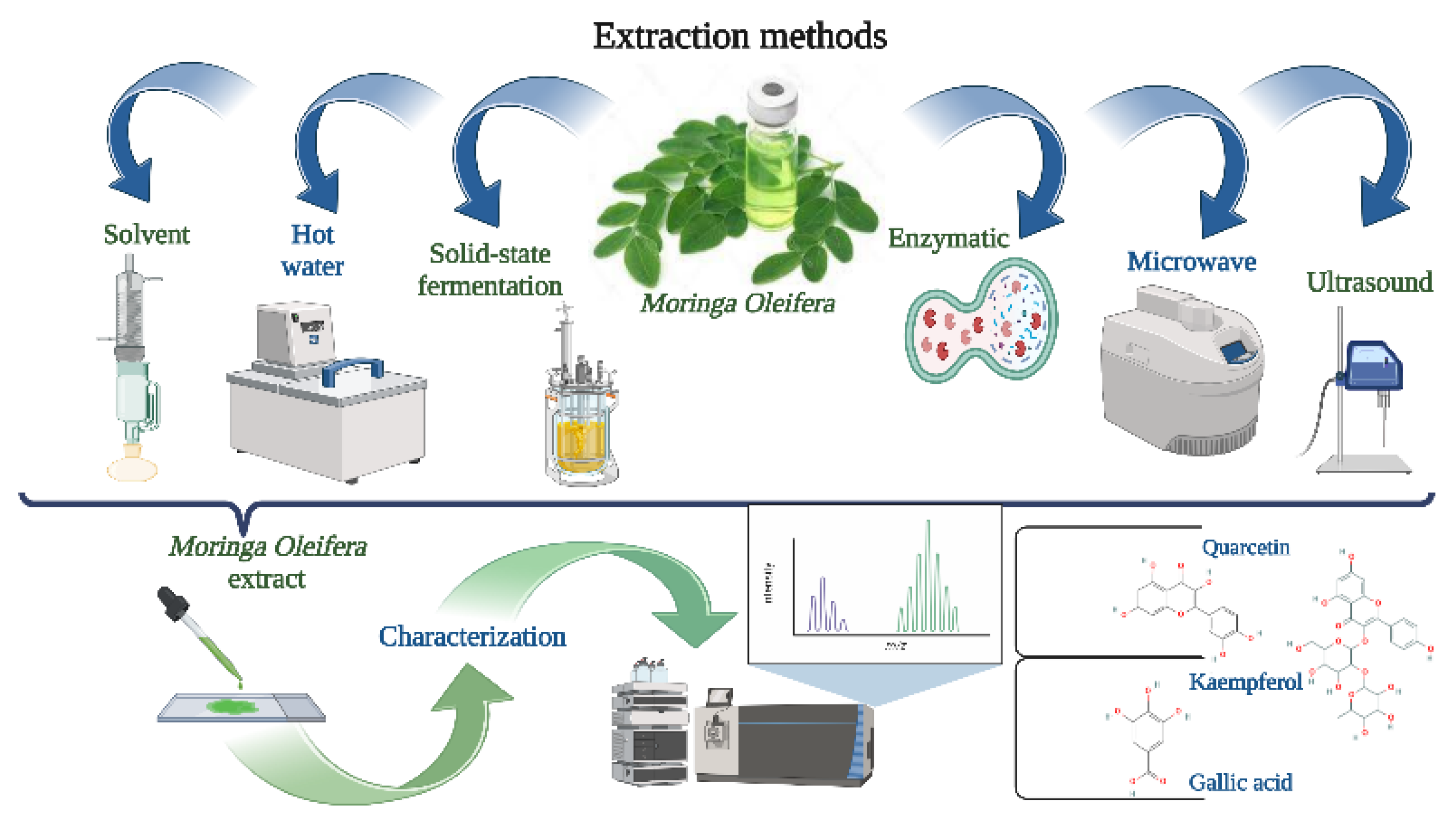
3. Extraction Techniques Prior to Encapsulation

Over the years, several extraction techniques have been developed (Figure 2), including traditional extraction (solvent, hot water, solid state fermentation) and nontraditional techniques (ultrasound, microwave, enzymatic) [50,51,52,53,54,55,56,57]. Whereas conventional extraction methods include longer extraction durations, toxicity, volatility, flammability, and lower extraction efficiency [58,59,60], nontraditional are considered to be better for the environment since they use fewer synthetic chemicals and produce a better yield and quality of extract [14,61,62,63,64].

3.1. Traditional Extraction Techniques

3.1.1. Solvent Extraction

The process of solvent extraction is one of the earliest methods of biological compound extraction. Solvent extraction (solid–liquid or liquid–liquid) requires solvents and heat or stirring. For example, operational factors such as temperature (80–110 °C), solvent ratio (chloroform: methanol from 1:1, 2:1, 3:1, 4:1), and solvent type on the oil extraction yield from Moringa have been examined. Maximum yield (41%) was achieved with increasing temperature (100 °C) and at a solvent ratio of 3:1; however, oil degradation began between 425 and 450 °C [65]. Nobossé et al. investigated how the extraction solvent (methanol, ethanol, and water) and age (30, 45, and 60 days) of fresh Moringa leaves influenced the extraction of the bioactive compounds. Maximum total polyphenols (4.57 g GAE/100 g) and tocopherol content (106 mg/100 g) were all found in leaves aged 60 days by methanol extraction, whereas flavonoids were extracted by ethanol (1.8 g CE/100 g). In addition, total antioxidant capacity was higher in an aqueous solvent. Leaves that were 30 days old had the most significant levels of vitamin C, chlorophyll, carotenoids, and antiperoxide activity (p < 0.05) [66].
However, in order to use the extract in food applications, it is still necessary to identify the optimum extraction solvents and to determine if extraction generates cytotoxicity [67,68]. It has been shown that methanolic solvents used to extract biological compounds from M. oleifera leaves had 17 times higher antioxidant activity than aqueous solvents (Trolox equivalents of 160.18 nmol/mL). The aqueous extract was less cytotoxic than the methanolic, as cells that resulted were more susceptible to concentrations ranging from 0.05 to 5 mg/L. [68]. In addition, the ethanolic extract of M. oleifera seeds at 1000 mg/kg had a significant decrease in erythrocytes in Wistar rats [67]. Despite the drawbacks (toxicity, volatility, flammability), traditional solvent techniques are still commonly used since the extraction units are easily accessible and less expensive than nontraditional methods.

3.1.2. Hot Water

Since the selective extraction process, the route from Moringa to bioactive recovery is challenging [51,52,54,58]. It has been reported that temperature is the primary factor that impacts the selectivity and recovery of phenolic compounds [51,52,58]. To fully extract the phenolic and flavonoid components, several researchers have also suggested that the maximum temperature for hot water extraction should be lower than 200 °C [51,54,58]. For example, the effect of temperature (50–200 °C) and duration (5–60 min) on the extractability pattern of macronutrient (Ca, K, and Mg) and micronutrient components (Al, Co, Cr, Cu, Fe, Ni, and Zn) from M. oleifera leaves during hot water extraction was evaluated. The proper temperature and extraction time were discovered to be 90 °C and 60 min, respectively. Micronutrient extraction improves with temperatures over 90 °C, which may increase the risk of recovering toxic elements from the extract [51].
In another study, at different extraction temperature (25–200 °C), the behavior of three flavonols’ (myricetin, quercetin, and kaempferol) content of Moringa leaves powder was characterized. Myricetin (2699 mg/kg) and kaempferol (3440 mg/kg) concentrations were found to be at their highest at 100 °C, but they began to decrease (1496 mg/kg, and 1730 mg/kg, respectively) at 150 °C. However, the concentration of quercetin (1429–1480 mg/kg) remained constant as the extraction temperature increased [58].
Comparing pressurized hot water extractions and carbon dioxide-expanded ethanol, the maximum yield of total phenolics from Moringa leaves was obtained for pressurized hot water extractions (62.4 vs. 20.3 mg GAE/g leaves) [54].

3.1.3. Solid-State Fermentation

Today’s world continuously develops, and creativity is the fundamental impetus for technical progress and innovation. For this reason, researchers are trying to find more sustainable methods, such as solid-state fermentation (S.S.F.). It is a fermentation method with a solid substrate/support and flow-free water. To overcome the low organoleptic properties and digestibility of Moringa leaf, extracts from leaf flour were investigated following S.S.F. with Aspergillus niger. The fermentation process was performed at 30 °C for 168 h using spore suspension (1.0–4.0 × 10⁷/g of solid) and different concentrations of extraction solvents (40% ethanol (v/v), 40% acetone (v/v), 80% ethanol (v/v), and 80% acetone (v/v)). The highest levels of total phenolics (136.4%) and flavonoids (783.1%) were identified using extraction solvents (80% ethanol and 80% acetone, respectively). Moreover, in vitro antioxidant activity in ferric reduction (277.2 mol Trolox/g M.L.F.) was observed with the 80% acetone solvent [59]. Zhang et al. used four bacterial strains (Trichoderma reesei CICC 2626, Bacillus pumilus CICC 10440, Bacillus subtilis subsp. CICC 21934, and Bacillus sp. CICC 21942) for S.S.F. The most soluble protein from Moringa leaf powder (285 mg/g) was generated utilizing Bacillus pumilus CICC 10440 in a 1:60 ratio over 24 h [60]. S.S.F. is a dynamic replacement for renowned traditional methods of metabolite processing that plays a significant role in producing food or biofuels.

3.2. Nontraditional Extraction Techniques

3.2.1. Ultrasound

Ultrasound extraction (U.E.) is based on cavitation, which disintegrates cell walls and releases active compounds from their natural matrices [69]. The cavitation that contains the active compounds will be compressed during sonication, providing a high-temperature and pressure microenvironment, thus speeding up the extraction process [56]. One study used response surface methodology to determine the optimum conditions for performing U.E. for M. oleifera leaves with methanol extraction solvent. Most active compounds (quercetin 3-glucoside, quercetin malonyl glucoside) were released at 11 °C, pH 7.7, time of 15 min, and a methanol concentration of 74.5% [56]. Pollini et al. highlight the impact of solvent composition in the U.E. on the biological properties of M. Oleifera leaves. The highest concentrations (216.4 µg/g quercetin 3-O-rhamnoside and 293.9 µg/g quercetin 3-O-(6″-O-malonyl)-β-D-glucoside) were found under the following U.E. factors: 50% water, 60:1 liquid/solid ratio, and 60 °C for 60 min. [53]. In addition, the effectiveness of the U.E. in increasing the extraction efficiency of oil from M. oleifera seeds is also studied by several researchers [50,57,70]. There were no significant differences between U.E. and conventional extraction in oil yield, but the time for oil extraction, U.E. (20 min), and conventional extraction (50 min) was significantly lower (p < 0.05) [57].

3.2.2. Microwave

Due to the polar solvents’ dipolar rotation, microwave radiation can cross the material structure, causing molecular friction that speeds up the mass transfer of the molecules of interest [14]. As reported by Chen et al. and Da Porto et al., microwave extraction (M.E.) may be used to preheat seeds before oil extraction [14,62]. The extracted oil using the Soxhlet method was more successful when M.E. pretreatments were performed (40.0%) at 100 W for 30, 60, and 90 s compared to the extracted oil using untreated seeds (+9.9%) [62].
Based on their research, Rodriguez et al. suggest the following M.E. optimal parameters for Moringa leaves: 20 min of extraction time, 42% ethanol, and 158 °C. The extraction yields on 26 ± 2%, 15 ± 2 g/mL DPPH, Trolox equivalent antioxidant capacity 16 ± 1 Trolox/100 g dry leaf [55]. Moreover, for the protein extraction, ME conditions (solvent–solid ratio 91:1 mL/g, extraction time 148 s, extraction temperature 41°C, microwave power 81 W) showed significantly high extraction efficacy (82.07 mg/g) compared to conventional solvent extraction method (68.99 mg/g) [61].

3.2.3. Enzymatic

Enzyme extraction (E.E.) seems to be a promising advanced technique for selecting biological compounds compared to the traditional solvent process [63,64]. For example, Latif et al. [63] use a commercial enzyme (protease; 580 000 DU/g) to extract oil and protein from the Moringa seed. The weight percentage of oil extracted by E.E. (22.5%) was significantly higher than that of aqueous extraction (7.8%) but significantly lower (p < 0.05) than hexane extraction (32.4%). Regarding protein content in the aqueous and creamy phase, E.E. shows the highest range (25.8%) compared to aqueous extraction (15.0%). Moreover, it was suggested that by using E.E. (protease and cellulase), the maximum oil recovery (70%) was achieved with a pH of 4.5, a moisture/kernel ratio of 8:1 (w/w), and a shaking rate of 300 strokes/min at 40 °C for a 1 h incubation period [64]. For the E.E. leaf protein, pH and the enzyme/substrate ratio had no significant impact, whereas the essential factors, including temperature 30 °C, multienzyme complex concentration 60 fungal beta-glucanase units (arabinose, cellulase, ß-glucanase, hemicellulose, and xylanase enzyme), and incubation time of 0.3 h have a substantial influence [71].
Enzymatic treatment can potentially improve polyphenol extraction yield and reduce energy consumption with lower process temperatures compared to current methods.

4. Encapsulation Methods of Bioactive Compounds from Moringa oleifera

Since the bioactive molecules extracted from different parts of M. oleifera, in most cases, cannot be used in food applications as they are, encapsulation is proposed. For example, M. oleifera leaf extracts have low bioavailability, limited water solubility, and are very unstable and oxidation-prone [13]. M. oleifera bioactive compounds react to several elements used in food processing procedures (pH, water activity, light conditions, oxygen, and temperature). Therefore, it is important to stop them from deteriorating and increase their stability in those circumstances. Encapsulation technology can offer a strong physical barrier against the abovementioned elements [14].
To create microcapsules that have various desirable qualities, the essential bioactive chemicals are protected by encapsulation in either a homogeneous or heterogeneous matrix. By using this method, unstable liquid compounds can be converted into free-flowing stable powder. Core bioactive components have been encapsulated using a variety of techniques, including cross-linking gelation, freeze-drying, microemulsion, spray drying, and different nanoformulations (nanoliposomes) (Figure 3) [72,73,74].
Microencapsulation makes the bioactive compounds easier to handle while protecting them from light, air, and other elements. In the encapsulation process, biopolymers (alginate, chitosan, etc.) and polysaccharides (maltodextrin, starch, etc.) are frequently used as carriers or coatings along with animal-based (whey) or plant-based proteins (soy, pea proteins) and gums (arabic, acacia gum) [13,64,65,66,67]. The optimal encapsulation material should have excellent water solubility, good emulsification, and film-forming properties, quick drying rate, and low viscosity, even in a highly concentrated solution [75]. Of the encapsulating techniques, spray drying is the least expensive and most widely used for turning emulsions into stable powder [64] with increased bioavailability, reduced oxidation rate, controlled release, and the unpleasant flavor masked.
Encapsulation parameters, such as nozzle size, inlet temperature, flow rate, outlet temperature, rotations per minute, surfactant, polymer solution feed rate, and cross-linking agent concentration directly influence the yield of the process, the size and the shape of the obtained particles, and the amount of bioactive compound [13]. For example, Airouyuwa (2019) observed that the yield of the spray drying was higher at the highest temperature (140, 160, and 180 °C), explained by the fact that when using greater inner temperature, the capacity of the heat and mass transfer process are increased and the evaporation rate is higher [13]. In obtaining microemulsions, the time, temperature, and rotations influence the emulsion and the bioactive compound stability. In this process, the type of oil phase used and the amount of bioactive material impact the microemulsion size and appearance (for a clear, transparent, or translucent microemulsion, the droplet sizes must be under 100 nm) [76,77]. The distance between the pipette nozzle and the surface of the hardening bath during the cross-linking gelation is a factor that directly affects the size and shape of the beads.
Additionally, it has been shown that phenolic compounds can reduce the surface tension of solutions, which might account for the observed reduction in particle size [78]. Interestingly, in the electrospraying process, surface tension repulsion outweighs viscoelastic forces at relatively low concentrations of gelatin (7%), causing beads to form. However, if core concentration rises and a mixed bead-fiber structure develops, this equilibrium may be harmed [79]. Among the other already-stated parameters, the polymer concentration, molecular weight, and ratio between polymers (when a mixture) can modify the shape and size of the obtained particles [78,80]. As a result of the improvement in surface-to-volume ratio, encapsulated materials with smaller particle sizes are more advantageous for food applications. Overall, these results suggest that well-designed and tailored encapsulation protocols can maximize the use of M. oleifera bioactive compounds.

4.1. Microencapsulation of Moringa oleifera and Their Food Applications

Due to the nature of its components, M. oleifera has many applications. The seed powder is utilized as a natural coagulant for water treatment, removing many suspended particles from rivers and murky streams [81]. The oil from the seeds is used for cosmetics, biodiesel production, and even as a fertilizer. Zeatin may be made from M. oleifera extracts, which promote plant growth and raises crop yields [82]. In addition to these uses, M. oleifera utilization in different food matrixes is becoming more widespread. For instance, in Mexico, fishmeal was replaced with M. oleifera due to the plant’s high protein and carbohydrate content in tilapia feed [83]. Oyenyinca (2018) and his collaborators describe how different bioactive compounds belonging to M. oleifera are used in various foods as fortificants, mostly in an encapsulated form.
However, they conclude that it might not always be considered fortification or enrichment when M. oleifera is used to enhance the nutritional content of staple foods [84]. It would seem from the results presented by Dollah (2014) that mixing M. oleifera oil with other vegetable oils would make it possible to modify or alter the original properties of the oils and provide functional and nutritional attributes for use in various food applications, expanding the opportunities for the commercial use of these oils [85].
To extend the shelf life of chicken burgers, Alqurashi et al. investigated the antibacterial properties of M. oleifera leaves extract (0.5–2%). The results showed that chicken burgers with 1–2% extract had a significant impact (p < 0.05) in reducing the total number of plates up to 6 days of refrigeration storage at 4 °C [86]. Moreover, through cross-linking gelation method, the addition of alginate encapsulation of Lactobacillus plantarum (9.32–9.58 Log10 CFU/g) and Pediococcus acidilactici (9.82–9.81 Log10 CFU/g) with Moringa oleifera extract (0.05–0.1%) in fruit juice and drinkable yogurt was evaluated. After 28 days of storage, the plant extract increased bacterial stability compared with the control in both products [87]. Table 1 shows the most-used encapsulation techniques for different bioactive compounds from M. oleifera and their possible applications. Nevertheless, despite their health advantages, additional human/clinical trials and evaluations are required to further rule out any health concerns [5,11].

4.2. Nanoencapsulation

Recently, nanoencapsulation has been used as a strategy for overcoming the drawbacks of traditional drug delivery methods, such as low bioavailability, adverse reactions, and unpleasant organoleptic qualities [79,91,92,93,94].
The effectiveness of nanoencapsulation ethanolic M. oleifera leaf extract in a fish-sourced gelatin matrix was assessed. M. oleifera extract encapsulation at 1%, 3%, and 5% produced bead-free and uniform nanofibers. According to scanning electron microscopy (S.E.M.) and transmission electron microscopy (T.E.M.) analysis, pure gelatin and encapsulated nanofiber have internal dimensions of 115 nm and 39 nm, respectively. Encapsulation efficiency was 80–85%, and toxicological results suggest that the extract was safe for consumption [79]. Moreover, a symbiotic containing Saccharomyces cerevisiae yeast (11 × 1012 CFU) and M. oleifera leaf extract was developed for growing rabbits using the nanoencapsulation technique. Alginate (1% w/v) and CaCl2 (0.5% w/v) successfully encapsulated the supplement (51.38 nm) with encapsulation efficiencies of 57.55% [91].
For the biocontrol of Listeria monocytogenes and Staphylococcus aureus (103 CFU/g) on cheese, an active food packaging material with Moringa oil (20 mg/mL)-loaded chitosan (3.0 mg/mL) nanoparticles (MO@CNPs) and MO@CNP (9.0 mg/mL)-embedded gelatin nanofibers has been developed. When applied to cheese, MO@CNPs nanofibers showed solid antibacterial activity (78.63% and 98.67%, respectively) at 4 °C for 10 days, extending the shelf life with no negative Impact on the flavor or texture of the cheese [92]. Moreover, alginate nanoparticles (0.1%) with Moringa oleifera extract (0.1%) showed antimicrobial activity against Staphylococcus aureus, Escherichia coli, Proteus vulgaris, Citrobacter diversus, and Salmonella enterica [95].
Nanoencapsulation is still one of the most promising technologies because of its potential to encapsulate bioactive components.

4.3. Electrospinning Method

Electrospinning has been used to make active food packaging with incorporated antioxidants, antiripening, and antimicrobials to maintain fresh and processed foods safe and increase their shelf life [96,97]. Particularly, nanofiber products made by electrospinning have received a lot of attention due to their better filtration efficiency and breathability. For example, different polymer concentrations (30–45%) and an injection flow rate (0.7 mL/h) of zein nanofiber with M. oleifera kernel oil were evaluated. Hydrophobic electrospun nanofibers averaged 450 ± 24 nm and encapsulated the oil efficiently, thus prolonging the oil shelf life [97]. Narayan et al. [93] show the beneficial effect (∼88%) of 7% polyacrylonitrile-supported biosorbent (M. oleifera 20 mg/mL) for the absorption of Congo red dye from an aqueous solution.
More research into electrospinning technology to generate biological macromolecule materials for the encapsulation and sustained release of food-active compounds is required.

4.4. Electrospraying Method

Electrospraying has been proposed to produce biological microcapsules; however, its usage in food encapsulation is currently unexplored [94,98]. For the encapsulation of the bioactive extract from M. oleifera (1–5%, w/w) through the electrospraying technique, a gelatin matrix (7.5%) was used to produce nanocapsules (140–179 nm). Electrospraying parameters were 20 kV voltage, 0.5 mL/h flow rate, and 10 cm emitter/collector distance; thus, extract-loaded nanocapsules had an encapsulation effectiveness of 83.0 ± 4.0% [79]. Additionally, the physical and antioxidant characteristics of chocolate alginate beads with Moringa oleifera leaf extract (0–6%) were assessed. Extract concentration (6%) made the beads smaller (4.3 mm) and more spherical, whereas hardening time just influenced circularity. For beads prepared with 2% extract and allowed 20 min in CaCl2, a minimum total phenolic concentration of 0.84 mg GAE/g beads was reported. However, the radical scavenging activity was concentration-related and required a level of 6% MLE to obtain inhibitory impact [78].
According to these studies, electrospraying is a more feasible and efficient alternative to encapsulate biological compounds than spray drying, which uses high temperatures [94,98].

5. Conclusions and Future Directions

A variety of therapeutic properties (anticancer, antibacterial, antiproliferative, antihypertensive, and anti-inflammatory) of bioactive compounds belonging to M. oleifera leaves, roots, seeds, and oils are sustained by in vitro and in vivo studies.
Including M. oleifera Lam. bioactive molecules in the humans diet are crucial for improving food nutrient value, human health sustainment, and environmental and sustainability reasons. The impact of these bioactive molecules on their applications is dependent primarily on two characteristics: on the one hand, their activity and, on the other hand, stability. These traits are closely related to their structural elements and ability to incorporate and maintain their bioactive features in the food matrix.
A combination of conventional and cutting-edge methodologies, such as thermal, chemical, and biotechnological methods, can be used to increase their use in food and beverages. Encapsulation is one of the advanced methods that has recently gained interest in the food sector. The bioactive compounds of M. oleifera can be encapsulated for optimal delivery within the human stomach or intestinal tract to produce pleasant, safe, and affordable foods. Therefore, compared to everyday food items, highly nutritious foods rich in M. oleifera bioactive compounds will need to be customized more precisely. In addition, using this valuable technique must ensure cost efficiency and sustainability. After analyzing the literature, we can conclude that spray drying is a cost-effective method, and it is very versatile. As we have seen, encapsulation efficiency can be maximized by using specific biopolymers, especially proteins or protein derivatives. The entrapped compounds’ stability also supports the use of this technique over time. Studies related to the sustainability of this method should be started, considering that obtaining the encapsulation matrices must also be part of this objective.
However, these encapsulations can overlap concerns regarding the cytotoxicity of the M. oleifera compounds. In addition to studying the cytotoxicity of Moringa bioactive compounds, the cytotoxicity of the carrier materials has also become an interesting topic for researchers. However, more studies in this direction have to be explored. Furthermore, we believe that implementing sustainable extraction methods and well-designed encapsulation techniques can maximize the utilization of M. oleifera bioactive compounds in the food industry, cosmetics, pharmaceutics, and water cleaning. More precisely, using green extraction methods, biopolymers, adequate concentrations, and suitable encapsulation methods will increase the benefits derived from the use of M. oleifera bioactive compounds. Because new encapsulation techniques involve pricy devices, we concluded that spray drying is the most suitable technique for designing and implementing the bioactive compound’s application in human health, with good yield, a broad palette of applicability, and high versatility.

Author Contributions

Conceptualization, O.L.P., A.D.K. and C.C.; initial draft preparation, A.D.K. and C.C.; writing—review and editing, O.L.P., A.D.K. and C.C.; reviewing the paper, O.L.P., A.D.K. and C.C.; funding acquisition, O.L.P. All authors have read and agreed to the published version of the manuscript.

Funding

This research was funded by a grant of the Romanian Ministry of Education and Research, CCCDI-UEFISCDI, project number PN-III-P4-ID-PCE-2020-2126, within PNCDI III.

Institutional Review Board Statement

Not applicable.

Informed Consent Statement

Not applicable.

Data Availability Statement

Not applicable.

Acknowledgments

The authors would like to thank Simon Elisabeta-Brighita for helping to improve the English level of the article.

Conflicts of Interest

The authors declare no competing financial interest.

References

  1. Kashyap, P.; Kumar, S.; Riar, C.S.; Jindal, N.; Baniwal, P.; Guine, R.P.F.; Correia, P.M.R.; Mehra, R.; Kumar, H. Recent Advances in Drumstick (Moringa oleifera) Leaves Bioactive Compounds: Composition, Health Benefits, Bioaccessibility, and Dietary Applications. Antioxidants 2022, 11, 402. [Google Scholar] [CrossRef]
  2. Saucedo-Pompa, S.; Torres-Castillo, J.A.; Castro-Lopez, C.; Rojas, R.; Sanchez-Alejo, E.J.; Ngangyo-Heya, M.; Martinez-Avila, G.C.G. Moringa plants: Bioactive compounds and promising applications in food products. Food Res. Int. 2018, 111, 438–450. [Google Scholar] [CrossRef] [PubMed]
  3. Alam, M.A. Development of Fiber Enriched Herbal Biscuits: A Preliminary Study on Sensory Evaluation and chemical Composition. Int. J. Nutr. Food Sci. 2014, 3, 246. [Google Scholar] [CrossRef] [Green Version]
  4. D’Auria, G.; Nitride, C.; Ferranti, P. Moringa oleifera Lam. Proteins: Properties and Food Applications. In Reference Module in Food Science; Elsevier: Amsterdam, The Netherlands, 2023. [Google Scholar] [CrossRef]
  5. Giuberti, G.; Rocchetti, G.; Montesano, D.; Lucini, L. The potential of Moringa oleifera in food formulation: A promising source of functional compounds with health-promoting properties. Curr. Opin. Food Sci. 2021, 42, 257–269. [Google Scholar] [CrossRef]
  6. Liu, Y.; Wang, X.-y.; Wei, X.-m.; Gao, Z.-t.; Han, J.-p. Values, properties and utility of different parts of Moringa oleifera: An overview. Chin. Herb. Med. 2018, 10, 371–378. [Google Scholar] [CrossRef]
  7. Nur, R.; Demak, I.P.K.; Radhiah, S.; Rusydi, M.; Mantao, E.; Larasati, R.D. The effect of moringa leaf extracton increasing hemoglobin and bodyweight in post-disaster pregnant women. Enfermería Clínica 2020, 30, 79–82. [Google Scholar] [CrossRef]
  8. Islam, Z.; Islam, S.M.R.; Hossen, F.; Mahtab-Ul-Islam, K.; Hasan, M.R.; Karim, R. Moringa oleifera is a Prominent Source of Nutrients with Potential Health Benefits. Int. J. Food Sci. 2021, 2021, 6627265. [Google Scholar] [CrossRef]
  9. Gopalakrishnan, L.; Doriya, K.; Kumar, D.S. Moringa oleifera: A review on nutritive importance and its medicinal application. Food Sci. Hum. Wellness 2016, 5, 49–56. [Google Scholar] [CrossRef] [Green Version]
  10. Sun-Waterhouse, D.; Wadhwa, S.S.; Waterhouse, G.I.N. Spray-Drying Microencapsulation of Polyphenol Bioactives: A Comparative Study Using Different Natural Fibre Polymers as Encapsulants. Food Bioprocess Technol. 2012, 6, 2376–2388. [Google Scholar] [CrossRef]
  11. Dzuvor, C.K.O.; Pan, S.; Amanze, C.; Amuzu, P.; Asakiya, C.; Kubi, F. Bioactive components from Moringa oleifera seeds: Production, functionalities and applications—A critical review. Crit. Rev. Biotechnol. 2022, 42, 271–293. [Google Scholar] [CrossRef]
  12. Premi, M.; Sharma, H.K. Effect of extraction conditions on the bioactive compounds from Moringa oleifera (PKM 1) seeds and their identification using LC–MS. J. Food Meas. Charact. 2017, 11, 213–225. [Google Scholar] [CrossRef]
  13. Osamede Airouyuwa, J.; Kaewmanee, T. Microencapsulation of Moringa oleifera leaf extracts with vegetable protein as wall materials. Food Sci. Technol. Int. 2019, 25, 533–543. [Google Scholar] [CrossRef] [PubMed]
  14. Chen, C.; Zhang, B.; Huang, Q.; Fu, X.; Liu, R.H. Microwave-assisted extraction of polysaccharides from Moringa oleifera Lam. leaves: Characterization and hypoglycemic activity. Ind. Crops Prod. 2017, 100, 1–11. [Google Scholar] [CrossRef]
  15. Mulat, M.; Khan, F.; Muluneh, G.; Pandita, A. Phytochemical Profile and Antimicrobial Effects of Different Medicinal Plant: Current Knowledge and Future Perspectives. Curr. Tradit. Med. 2020, 6, 24–42. [Google Scholar] [CrossRef]
  16. Saini, R.K.; Sivanesan, I.; Keum, Y.S. Phytochemicals of Moringa oleifera: A review of their nutritional, therapeutic and industrial significance. 3 Biotech 2016, 6, 203. [Google Scholar] [CrossRef] [PubMed] [Green Version]
  17. Nouman, W.; Anwar, F.; Gull, T.; Newton, A.; Rosa, E.; Domínguez-Perles, R. Profiling of polyphenolics, nutrients and antioxidant potential of germplasm’s leaves from seven cultivars of Moringa oleifera Lam. Ind. Crops Prod. 2016, 83, 166–176. [Google Scholar] [CrossRef]
  18. Zhao, B.; Deng, J.; Li, H.; He, Y.; Lan, T.; Wu, D.; Gong, H.; Zhang, Y.; Chen, Z. Optimization of Phenolic Compound Extraction from Chinese Moringa oleifera Leaves and Antioxidant Activities. J. Food Qual. 2019, 2019, 5346279. [Google Scholar] [CrossRef] [Green Version]
  19. Saini, R.K.; Shetty, N.P.; Giridhar, P. Carotenoid content in vegetative and reproductive parts of commercially grown Moringa oleifera Lam. cultivars from India by LC–APCI–MS. Eur. Food Res. Technol. 2014, 238, 971–978. [Google Scholar] [CrossRef]
  20. Bischoff, K.L. Chapter 40—Glucosinolates. In Nutraceuticals; Gupta, R.C.., Ed.; Academic Press: Boston, MA, USA, 2016. [Google Scholar] [CrossRef]
  21. Ishida, M.; Hara, M.; Fukino, N.; Kakizaki, T.; Morimitsu, Y. Glucosinolate metabolism, functionality and breeding for the improvement of Brassicaceae vegetables. Breed. Sci. 2014, 64, 48–59. [Google Scholar] [CrossRef] [Green Version]
  22. Gu, X.; Yang, Y.; Wang, Z. Nutritional, phytochemical, antioxidant, α-glucosidase and α-amylase inhibitory properties of Moringa oleifera seeds. S. Afr. J. Bot. 2020, 133, 151–160. [Google Scholar] [CrossRef]
  23. Liang, L.-L.; Cai, S.-Y.; Gao, M.; Chu, X.-M.; Pan, X.-Y.; Gong, K.-K.; Xiao, C.-W.; Chen, Y.; Zhao, Y.-Q.; Wang, B.; et al. Purification of antioxidant peptides of Moringa oleifera seeds and their protective effects on H2O2 oxidative damaged Chang liver cells. J. Funct. Foods 2020, 64, 103698. [Google Scholar] [CrossRef]
  24. Kumar, M.; Selvasekaran, P.; Kapoor, S.; Barbhai, M.D.; Lorenzo, J.M.; Saurabh, V.; Potkule, J.; Changan, S.; ElKelish, A.; Selim, S.; et al. Moringa oleifera Lam. seed proteins: Extraction, preparation of protein hydrolysates, bioactivities, functional food properties, and industrial application. Food Hydrocoll. 2022, 131, 107791. [Google Scholar] [CrossRef]
  25. Zaid, A.Q.; Ghazali, S.B. Dataset on physicochemical properties of particle-sized Moringa oleifera seed cake and its application as bio-coagulants in water treatment application. Chem. Data Collect. 2019, 24, 100284. [Google Scholar] [CrossRef]
  26. Emmanuel, S.; Zaku, S. Moringa oleifera seed-cake, alternative biodegradable and biocompatibility organic fertilizer for modern farming. Agric. Biol. J. N. Am. 2011, 2, 1289–1292. [Google Scholar] [CrossRef]
  27. Irfan, H.M.; Asmawi, M.Z.; Khan, N.A.K.; Sadikun, A. Effect of Ethanolic Extract of Moringa oleifera Lam. Leaves on Body Weight and Hyperglycemia of Diabetic Rats. Pak. J. Nutr. 2016, 15, 112–117. [Google Scholar] [CrossRef]
  28. Nandave, M.; Ojha, S.K.; Joshi, S.; Kumari, S.; Arya, D.S. Moringa oleifera leaf extract prevents isoproterenol-induced myocardial damage in rats: Evidence for an antioxidant, antiperoxidative, and cardioprotective intervention. J. Med. Food 2009, 12, 47–55. [Google Scholar] [CrossRef]
  29. Pontual, E.V.; Pires-Neto, D.F.; Fraige, K.; Higino, T.M.M.; Carvalho, B.E.A.; Alves, N.M.P.; Lima, T.A.; Zingali, R.B.; Coelho, L.C.B.B.; Bolzani, V.S.; et al. A trypsin inhibitor from Moringa oleifera flower extract is cytotoxic to Trypanosoma cruzi with high selectivity over mammalian cells. Nat. Prod. Res. 2017, 32, 2940–2944. [Google Scholar] [CrossRef]
  30. Ujah, O.; Ujah, I.; Johnson, J.T.; Ekam, V.; Udenze, E. Hepatoprotective property of ethanolic leaf extract of Moringa oleifera oncarbon tetrachloride (CCl4) induced hepatotoxicity. J. Nat. Prod. Plant Resour. 2013, 3, 15–22. [Google Scholar]
  31. Prabakaran, M.; Kim, S.-H.; Sasireka, A.; Chandrasekaran, M.; Chung, I.-M. Polyphenol composition and antimicrobial activity of various solvent extracts from different plant parts of Moringa oleifera. Food Biosci. 2018, 26, 23–29. [Google Scholar] [CrossRef]
  32. Momoh, M.A.; Chime, S.A.; Kenechukwu, F.C. Novel drug delivery system of plant extract for the management of diabetes: An antidiabetic study. J. Diet. Suppl. 2013, 10, 252–263. [Google Scholar] [CrossRef] [PubMed]
  33. Bakre, A.G.; Aderibigbe, A.O.; Ademowo, O.G. Studies on neuropharmacological profile of ethanol extract of Moringa oleifera leaves in mice. J. Ethnopharmacol. 2013, 149, 783–789. [Google Scholar] [CrossRef]
  34. Aekthammarat, D.; Pannangpetch, P.; Tangsucharit, P. Moringa oleifera leaf extract lowers high blood pressure by alleviating vascular dysfunction and decreasing oxidative stress in L-NAME hypertensive rats. Phytomedicine 2019, 54, 9–16. [Google Scholar] [CrossRef] [PubMed]
  35. Anwar, F.; Latif, S.; Ashraf, M.; Gilani, A.H. Moringa oleifera: A food plant with multiple medicinal uses. Phytother. Res. PTR 2007, 21, 17–25. [Google Scholar] [CrossRef]
  36. Chin, C.Y.; Jalil, J.; Ng, P.Y.; Ng, S.F. Development and formulation of Moringa oleifera standardised leaf extract film dressing for wound healing application. J. Ethnopharmacol. 2018, 212, 188–199. [Google Scholar] [CrossRef]
  37. Kaur, G.; Invally, M.; Sanzagiri, R.; Buttar, H.S. Evaluation of the antidepressant activity of Moringa oleifera alone and in combination with fluoxetine. J. Ayurveda Integr. Med. 2015, 6, 273–279. [Google Scholar] [CrossRef] [PubMed] [Green Version]
  38. Upadhye, K.; Rangari, V.; Mathur, V. Antimigraine activity study of Moringa oleifera leaf juice. Int. J. Green Pharm. 2012, 6, 204. [Google Scholar] [CrossRef]
  39. Tahiliani, P.; Kar, A. Role of Moringa oleifera leaf extract in the regulation of thyroid hormone status in adult male and female rats. Pharm. Res. 2000, 41, 319–323. [Google Scholar] [CrossRef] [PubMed]
  40. Kasolo, J.N.; Bimenya, G.S.; Ojok, L.; Ochieng, J.; Ogwal-Okeng, J.W. Phytochemicals and uses of Moringa oleifera leaves in Ugandan rural communities. J. Med. Plants Res. 2010, 4, 753–757. [Google Scholar]
  41. Mbikay, M. Therapeutic Potential of Moringa oleifera Leaves in Chronic Hyperglycemia and Dyslipidemia: A Review. Front. Pharm. 2012, 3, 24. [Google Scholar] [CrossRef] [PubMed] [Green Version]
  42. Khalafalla, M.M.; Abdellatef, E.; Dafalla, H.M.; Nassrallah, A.A.; Aboul-Enein, K.M.; Lightfoot, D.A.; El-Deeb, F.E.; El-Shemy, H.A. Active principle from Moringa oleifera Lam leaves effective against two leukemias and a hepatocarcinoma. Afr. J. Biotechnol. 2010, 9, 8467–8471. [Google Scholar] [CrossRef]
  43. Freshney, R.I. Cytotoxicity. In Culture of Animal Cells: A Manual of Basic Technique; John Wiley & Sons: Hoboken, NJ, USA, 2005. [Google Scholar] [CrossRef]
  44. Szlachetka, K.; Kut, P.; Stępień, A. Cytotoxic and anticancer activity of Moringa oleifera. Eur. J. Clin. Exp. Med. 2020, 18, 214–220. [Google Scholar] [CrossRef]
  45. Guevara, A.P.; Vargas, C.; Sakurai, H.; Fujiwara, Y.; Hashimoto, K.; Maoka, T.; Kozuka, M.; Ito, Y.; Tokuda, H.; Nishino, H. An antitumor promoter from Moringa oleifera Lam. Mutat. Res. 1999, 440, 181–188. [Google Scholar] [CrossRef]
  46. Parvathy, M.V.S.; Umamaheshwari, A. Cytotoxic Effect of Moringa Oleifera Leaf Extracts On Human Multiple Myeloma Cell Lines. Trends Med. Res. 2007, 2, 44–50. [Google Scholar] [CrossRef] [Green Version]
  47. Jung, I.L.; Lee, J.H.; Kang, S.C. A potential oral anticancer drug candidate, Moringa oleifera leaf extract, induces the apoptosis of human hepatocellular carcinoma cells. Oncol. Lett. 2015, 10, 1597–1604. [Google Scholar] [CrossRef]
  48. Jafarain, A.; Asghari, G.; Ghassami, E. Evaluation of cytotoxicity of Moringa oleifera Lam. callus and leaf extracts on Hela cells. Adv. Biomed. Res. 2014, 3, 194. [Google Scholar] [CrossRef]
  49. Sohaib, M.; Al-Barakah, F.N.I.; Migdadi, H.M.; Husain, F.M. Comparative study among Avicennia marina, Phragmites australis, and Moringa oleifera based ethanolic-extracts for their antimicrobial, antioxidant, and cytotoxic activities. Saudi J. Biol. Sci. 2022, 29, 111–122. [Google Scholar] [CrossRef] [PubMed]
  50. Mohammadpour, H.; Sadrameli, S.M.; Eslami, F.; Asoodeh, A. Optimization of ultrasound-assisted extraction of Moringa peregrina oil with response surface methodology and comparison with Soxhlet method. Ind. Crops Prod. 2019, 131, 106–116. [Google Scholar] [CrossRef]
  51. Nuapia, Y.; Al-Hamimi, S.; Matshediso, P.G.; Cukrowska, E.; Tutu, H.; Turner, C.; Chimuka, L. Selective pressurized hot water extraction of nutritious macro-nutrients vs. micro-nutrients in Moringa oleifera leaves-a chemometric approach. Anal. Bioanal. Chem. 2020, 412, 2495–2503. [Google Scholar] [CrossRef]
  52. Nuapia, Y.; Cukrowska, E.; Tutu, H.; Chimuka, L. Statistical comparison of two modeling methods on pressurized hot water extraction of vitamin C and phenolic compounds from Moringa oleifera leaves. S. Afr. J. Bot. 2020, 129, 9–16. [Google Scholar] [CrossRef]
  53. Pollini, L.; Tringaniello, C.; Ianni, F.; Blasi, F.; Manes, J.; Cossignani, L. Impact of Ultrasound Extraction Parameters on the Antioxidant Properties of Moringa Oleifera Leaves. Antioxidants 2020, 9, 277. [Google Scholar] [CrossRef] [PubMed]
  54. Rodríguez-Pérez, C.; Mendiola, J.A.; Quirantes-Piné, R.; Ibáñez, E.; Segura-Carretero, A. Green downstream processing using supercritical carbon dioxide, CO2-expanded ethanol and pressurized hot water extractions for recovering bioactive compounds from Moringa oleifera leaves. J. Supercrit. Fluids 2016, 116, 90–100. [Google Scholar] [CrossRef]
  55. Rodriguez-Perez, C.; Gilbert-Lopez, B.; Mendiola, J.A.; Quirantes-Pine, R.; Segura-Carretero, A.; Ibanez, E. Optimization of microwave-assisted extraction and pressurized liquid extraction of phenolic compounds from Moringa oleifera leaves by multiresponse surface methodology. Electrophoresis 2016, 37, 1938–1946. [Google Scholar] [CrossRef]
  56. Yerena-Prieto, B.J.; Gonzalez-Gonzalez, M.; Vázquez-Espinosa, M.; González-de-Peredo, A.V.; García-Alvarado, M.Á.; Palma, M.; Rodríguez-Jimenes, G.d.C.; Barbero, G.F. Optimization of an Ultrasound-Assisted Extraction Method Applied to the Extraction of Flavonoids from Moringa Leaves (Moringa oleífera Lam.). Agronomy 2022, 12, 261. [Google Scholar] [CrossRef]
  57. Zhong, J.; Wang, Y.; Yang, R.; Liu, X.; Yang, Q.; Qin, X. The application of ultrasound and microwave to increase oil extraction from Moringa oleifera seeds. Ind. Crops Prod. 2018, 120, 1–10. [Google Scholar] [CrossRef]
  58. Matshediso, P.G.; Cukrowska, E.; Chimuka, L. Development of pressurised hot water extraction (PHWE) for essential compounds from Moringa oleifera leaf extracts. Food Chem. 2015, 172, 423–427. [Google Scholar] [CrossRef]
  59. Feitosa, P.R.B.; Santos, T.R.J.; Gualberto, N.C.; Narain, N.; de Aquino Santana, L.C.L. Solid-state fermentation with Aspergillus niger for the bio-enrichment of bioactive compounds in Moringa oleifera (moringa) leaves. Biocatal. Agric. Biotechnol. 2020, 27, 101709. [Google Scholar] [CrossRef]
  60. Zhang, M.; Huang, Y.; Zhao, H.; Wang, T.; Xie, C.; Zhang, D.; Wang, X.; Sheng, J. Solid-state fermentation of Moringa oleifera leaf meal using Bacillus pumilus CICC 10440. J. Chem. Technol. Biotechnol. 2017, 92, 2083–2089. [Google Scholar] [CrossRef]
  61. Cheng, F.; Shu, G.; Chen, L.; Dai, C.; Wan, H.; Chen, H.; Dong, X. Ultrasound-microwave assisted extraction of proteins from Moringa oleifera leaves: Comparative optimization study and LC-MS analysis of the protein concentrate. J. Food Process. Preserv. 2021, 45, e15547. [Google Scholar] [CrossRef]
  62. Da Porto, C.; Decorti, D.; Natolino, A. Microwave pretreatment of Moringa oleifera seed: Effect on oil obtained by pilot-scale supercritical carbon dioxide extraction and Soxhlet apparatus. J. Supercrit. Fluids 2016, 107, 38–43. [Google Scholar] [CrossRef]
  63. Latif, S.; Anwar, F.; Hussain, A.I.; Shahid, M. Aqueous enzymatic process for oil and protein extraction from Moringa oleifera seed. Eur. J. Lipid Sci. Technol. 2011, 113, 1012–1018. [Google Scholar] [CrossRef]
  64. Mat Yusoff, M.; Gordon, M.H.; Ezeh, O.; Niranjan, K. Aqueous enzymatic extraction of Moringa oleifera oil. Food Chem. 2016, 211, 400–408. [Google Scholar] [CrossRef]
  65. Bhutada, P.R.; Jadhav, A.J.; Pinjari, D.V.; Nemade, P.R.; Jain, R.D. Solvent assisted extraction of oil from Moringa oleifera Lam. seeds. Ind. Crops Prod. 2016, 82, 74–80. [Google Scholar] [CrossRef]
  66. Nobosse, P.; Fombang, E.N.; Mbofung, C.M.F. Effects of age and extraction solvent on phytochemical content and antioxidant activity of fresh Moringa oleifera L. leaves. Food Sci. Nutr. 2018, 6, 2188–2198. [Google Scholar] [CrossRef]
  67. Chivapat, S.; Sincharoenpokai, P.; Suppajariyawat, P.; Rungsipipat, A.; Phattarapornchaiwat, S.; Chantarateptawan, V. Safety Evaluations of Ethanolic Extract of Moringa oleifera Lam. Seed in Experimental Animals. Thai J. Vet. Med. 2012, 42, 343–352. [Google Scholar]
  68. Landazuri, A.C.; Gualle, A.; Castaneda, V.; Morales, E.; Caicedo, A.; Orejuela-Escobar, L.M. Moringa oleifera Lam. leaf powder antioxidant activity and cytotoxicity in human primary fibroblasts. Nat. Prod. Res. 2021, 35, 6194–6199. [Google Scholar] [CrossRef] [PubMed]
  69. Abu Bakar, S.N.H.; Abu Hasan, H.; Abdullah, S.R.S.; Kasan, N.A.; Muhamad, M.H.; Kurniawan, S.B. A review of the production process of bacteria-based polymeric flocculants. J. Water Process Eng. 2021, 40, 101915. [Google Scholar] [CrossRef]
  70. Kayanan, B.U.R.; Sagum, R.S. Microwave and Ultrasound Pretreatment of Moringa oleifera Lam. Seeds: Effects on Oil Expression, Oil Quality, and Bioactive Component. J. Oleo Sci. 2021, 70, 875–884. [Google Scholar] [CrossRef] [PubMed]
  71. Benhammouche, T.; Melo, A.; Martins, Z.; Faria, M.A.; Pinho, S.C.M.; Ferreira, I.; Zaidi, F. Nutritional quality of protein concentrates from Moringa Oleifera leaves and in vitro digestibility. Food Chem. 2021, 348, 128858. [Google Scholar] [CrossRef]
  72. Wang, Y.; Ouyang, Q.; Chang, X.; Yang, M.; He, J.; Tian, Y.; Sheng, J. Anti-photoaging effects of flexible nanoliposomes encapsulated Moringa oleifera Lam. isothiocyanate in UVB-induced cell damage in HaCaT cells. Drug Deliv. 2022, 29, 871–881. [Google Scholar] [CrossRef]
  73. Militao, I.M.; Roddick, F.; Bergamasco, R.; Fan, L. Rapid adsorption of PFAS: Application of Moringa oleifera seed powder encapsulated in alginate beads. Environ. Technol. Innov. 2022, 28, 102761. [Google Scholar] [CrossRef]
  74. Castro-López, C.; Espinoza-González, C.; Ramos-González, R.; Boone-Villa, V.D.; Aguilar-González, M.A.; Martínez-Ávila, G.C.; Aguilar, C.N.; Ventura-Sobrevilla, J.M. Spray-drying encapsulation of microwave-assisted extracted polyphenols from Moringa oleifera: Influence of tragacanth, locust bean, and carboxymethyl-cellulose formulations. Food Res. Int. 2021, 144, 110291. [Google Scholar] [CrossRef] [PubMed]
  75. Gürbüz, E.; Keresteci, B.; Günneç, C.; Baysal, G. Encapsulation Applications and Production Techniques in the Food Industry. J. Nutr. Health Sci. 2020, 7, 106. [Google Scholar]
  76. Batra, G.; Gortzi, O.; Lalas, S.I.; Galidi, A.; Alibade, A.; Nanos, G.D. Enhanced antioxidant activity of Capsicum annuum L. and Moringa oleifera L. extracts after encapsulation in microemulsions. ChemEngineering 2017, 1, 15. [Google Scholar] [CrossRef] [Green Version]
  77. Kaltsa, O.; Alibade, A.; Batra, G.; Bozinou, E.; Makris, D.P.; Lalas, S.I. Fortification of chocolate using Moringa oleifera extract encapsulated in microemulsions. OCL 2021, 28, 38. [Google Scholar] [CrossRef]
  78. Kaltsa, O.; Alibade, A.; Bozinou, E.; Makris, D.P.; Lalas, S.I. Encapsulation of Moringa oleifera extract in Ca-alginate chocolate beads: Physical and antioxidant properties. J. Food Qual. 2021, 2021, 5549873. [Google Scholar] [CrossRef]
  79. Hani, N.; Azarian, M.H.; Torkamani, A.E.; Kamil Mahmood, W.A. Characterisation of gelatin nanoparticles encapsulated with Moringa oleifera bioactive extract. Int. J. Food Sci. Technol. 2016, 51, 2327–2337. [Google Scholar] [CrossRef]
  80. Pop, O.L.; Vodnar, D.C.; Suharoschi, R.; Mudura, E.; Socaciu, C.L. plantarum ATCC 8014 Entrapment with Prebiotics and Lucerne Green Juice and Their Behavior in Simulated Gastrointestinal Conditions. J. Food Process Eng. 2016, 39, 433–441. [Google Scholar] [CrossRef]
  81. Radhakrishnan, K.; Sethuraman, L.; Panjanathan, R.; Natarajan, A.; Solaiappan, V.; Thilagaraj, W.R. Biosorption of heavy metals from actual electroplating wastewater using encapsulated Moringa oleifera beads in fixed bed column. Desalination Water Treat. 2016, 57, 3572–3587. [Google Scholar] [CrossRef]
  82. Jain, P.; Farooq, B.; Lamba, S.; Koul, B. Foliar spray of Moringa oleifera Lam. leaf extracts (MLE) enhances the stevioside, zeatin and mineral contents in Stevia rebaudiana Betoni. S. Afr. J. Bot. 2020, 132, 249–257. [Google Scholar] [CrossRef]
  83. Milla, P.G.; Penalver, R.; Nieto, G. Health Benefits of Uses and Applications of Moringa oleifera in Bakery Products. Plants 2021, 10, 318. [Google Scholar] [CrossRef] [PubMed]
  84. Oyeyinka, A.T.; Oyeyinka, S.A. Moringa oleifera as a food fortificant: Recent trends and prospects. J. Saudi Soc. Agric. Sci. 2018, 17, 127–136. [Google Scholar] [CrossRef]
  85. Dollah, S.; Abdulkarim, S.M.; Ahmad, S.H.; Khoramnia, A.; Ghazali, H.M. Physicochemical properties and potential food applications of Moringa oleifera seed oil blended with other vegetable oils. J. Oleo Sci. 2014, 63, 811–822. [Google Scholar] [CrossRef] [PubMed] [Green Version]
  86. Alqurashi, R.M.; Aldossary, H.M. In vitro antioxidant and antimicrobial activity of Moringa oleifera leaf as a natural food preservative in chicken burgers. Emir. J. Food Agric. 2021, 33, 450–457. [Google Scholar] [CrossRef]
  87. Shehata, M.G.; Abd-Rabou, H.S.; El-Sohaimy, S.A. Plant Extracts in Probiotic Encapsulation: Evaluation of their Effects on Strains Survivability in Juice and Drinkable Yogurt During Storage and an in-vitro Gastrointestinal Model. J. Pure Appl. Microbiol. 2019, 13, 609–617. [Google Scholar] [CrossRef] [Green Version]
  88. George, T.T.; Oyenihi, A.B.; Rautenbach, F.; Obilana, A.O. Characterization of Moringa oleifera Leaf Powder Extract Encapsulated in Maltodextrin and/or Gum Arabic Coatings. Foods 2021, 10, 3044. [Google Scholar] [CrossRef]
  89. Premi, M.; Sharma, H.K. Effect of different combinations of maltodextrin, gum arabic and whey protein concentrate on the encapsulation behavior and oxidative stability of spray dried drumstick (Moringa oleifera) oil. Int. J. Biol. Macromol. 2017, 105, 1232–1240. [Google Scholar] [CrossRef] [PubMed]
  90. Castro-López, C.; Gonçalves, C.; Ventura-Sobrevilla, J.M.; Pastrana, L.M.; Aguilar-González, C.N.; Martínez-Ávila, G.C. Moringa oleifera—Storage Stability, In Vitro-Simulated Digestion and Cytotoxicity Assessment of Microencapsulated Extract. Processes 2020, 8, 770. [Google Scholar] [CrossRef]
  91. Hashem, N.M.; Hosny, N.S.; El-Desoky, N.I.; Shehata, M.G. Effect of Nanoencapsulated Alginate-Synbiotic on Gut Microflora Balance, Immunity, and Growth Performance of Growing Rabbits. Polymers 2021, 13, 4191. [Google Scholar] [CrossRef]
  92. Lin, L.; Gu, Y.; Cui, H. Moringa oil/chitosan nanoparticles embedded gelatin nanofibers for food packaging against Listeria monocytogenes and Staphylococcus aureus on cheese. Food Packag. Shelf Life 2019, 19, 86–93. [Google Scholar] [CrossRef]
  93. Narayan, M.; Sadasivam, R.; Packirisamy, G.; Pichiah, S. Electrospun polyacrylonitrile-Moringa Olifera based nanofibrous bio-sorbent for remediation of Congo red dye. J. Environ. Manag. 2022, 317, 115294. [Google Scholar] [CrossRef]
  94. Pisoschi, A.M.; Pop, A.; Cimpeanu, C.; Turcus, V.; Predoi, G.; Iordache, F. Nanoencapsulation techniques for compounds and products with antioxidant and antimicrobial activity—A critical view. Eur. J. Med. Chem. 2018, 157, 1326–1345. [Google Scholar] [CrossRef] [PubMed]
  95. El-Houssiny, A.S.; Fouad, E.A.; Hegazi, A.G. A Comparative Antimicrobial Activity Study of Moringa oleifera Extracts Encapsulated within ALg Nanoparticles. Nanosci. Nanotechnol.-Asia 2021, 11, 144–152. [Google Scholar] [CrossRef]
  96. Mele, E. Electrospinning of Essential Oils. Polymers 2020, 12, 908. [Google Scholar] [CrossRef] [PubMed] [Green Version]
  97. Afolabi-owolabi, O.T.; Abidin, S.Z.; Ariffin, F. Electrospun Polymer Nanofiber from Moringa Oleifera Kernel Oil with Coaxial Electrospinning Method. Curr. Nutr. Food Sci. 2020, 16, 90–97. [Google Scholar] [CrossRef]
  98. Tanhaei, A.; Mohammadi, M.; Hamishehkar, H.; Hamblin, M.R. Electrospraying as a novel method of particle engineering for drug delivery vehicles. J. Control. Release 2021, 330, 851–865. [Google Scholar] [CrossRef] [PubMed]
Figure 1. Cytotoxicity bioassay of M. oleifera (created with BioRender.com).
Foods 11 03787 g001
Figure 2. Extraction techniques used before encapsulation of the bioactive compounds from M. oleifera (created with BioRender.com).
Foods 11 03787 g002
Figure 3. Most-studied encapsulation methods for M. oleifera bioactive compounds (created with BioRender.com).
Foods 11 03787 g003
Table 1. Moringa oleifera bioactive compounds encapsulation techniques and features of the encapsulated product.
Encapsulation TechniqueEncapsulated MaterialEncapsulation MatrixEncapsulation FormulationFeaturesApplicationRef.
Cross-linking
gelation
Moringa
oleifera leaf extract
Alginate, whey protein, cacao powder0, 2, 4, and 6%
extract
concentration
beads size 4.30–5.02 mm
phenolic content 0.88–1.38 mg GAE/g of beads
20–50 mL/L dissolved
organic carbon release in the first 30 min
food
ingredient
[78]
Moringa
oleifera seed
powder (MSP)
Sodium alginate
3% Alg, 2% MSP
2% Alg, 1% MSP
blank 2%/3% Alg
↑perfluoroalkyl and polyfluoroalkyl substances absorption
90–95% bead moisture content
pollutants removal[73]
Moringa
oleifera seed powder (MSP)
Sodium alginate
mean diameter 3 mm
Cu2+ 99.3%, Ni2+ 77.98%, Co2+ 70.80%, Zn2+ 60.90%, Mn2+ 49.96% biosorption
pollutants removal[81]
Freeze-dryingMoringa
oleifera leaf powder ethanol extract
Maltodextrin, arabic gumMalto-dextrin, arabic gum
moisture content 1.47–1.77%
hygroscopicity 11.13–15.86%
water solubility index 86.35–98.74%
food
fortification
[88]
Micro-emulsionCarotenoids Moringa
oleifera L. leaves
Lecithin, sunflower oil, absolute ethanol, water
encapsulation efficiency 59.67%
stable 24 h at room temperature
stable after centrifugation
stable under thermal test
antioxidant[76]
Moringa
oleifera leaves extract
Coconut/
soybean oil, tween80, 
span80
4%, 8% Moringa
oleifera leaves extract
microemulsion size 15.1–82 nm
stable after centrifugation
stable under thermal test
food
fortification
[77]
Spray
drying
Drumstick oil (30%)Maltodextrin (MD)
Gum Arabic (GA)
Whey Protein (WP)
MD: GA (25:75, 50:50, 75:25)
MD: WPC (25:75, 50:50, 75:25)
encapsulation efficiency 66.23–91.05%
surface oil content 1.65–6.11%
moisture content 1.44–1.91%
particle size distribution 11.43–28.03 µm
density 1.15–1.34 g/cm3
porosity 0.36–0.58
↑ oil stability
food
fortification
[89]
Polyphenols
(M. oleifera) extracted by microwave-
assisted
extraction
Tragacanth gum (TG), locust bean gum (LBG), and
carboxymethyl-cellulose (CMC)
TG 1%, LBG 1%, CMC 1%, LBG 0.5% + TG 0.5%, TG 0.5% + CMC 0.5%, CMC 0.5% + LBG 0.5%
encapsulation efficiency 24.28–35.74%
total phenolic content 25.17–27.49 (mg GAE g−1 of microparticles)
total flavonoid content 23.20–26.82 (mg QE g−1 of microparticles)
DPPH 5.96–6.95 (mg GAE g−1 of microparticles)
ABTS 5.61–6.18 (mg TE g−1 of microparticles)
antioxidant[74]
Moringa
oleifera leaves extract (MLE)
Soy protein
isolate (SPI),
pea protein
isolate (PPI)
10% plant-based protein
11–22% (w/v) (MLE)
encapsulation yield 65.46–85.22% (SPI)/63.63–88.64% (PPI)
total polyphenols content 6.76–11.96 mg GAE/g powder (SPI formulation) and 4.95–8.81 mg GAE/g (PPI formulation)
antioxidant[13]
Moringa
oleifera leaves extract (MLE)
Tragacanth gum1% tragacanth gum
total polyphenols 42.47–45.28% (from the initial content)
antioxidant activity 39.7–75.32% across 30 days
storage
polyphenols release in simulated gastrointestinal test 9.7% oral conditions, 27.2–35.2% gastric conditions (30–60 min), 44.1, 51.8 and 57.6% intestinal conditions (2, 3 and 4 h)
antioxidant[90]
Publisher’s Note: MDPI stays neutral with regard to jurisdictional claims in published maps and institutional affiliations.

