Article

Acyclic Diterpene Phytol from Hemp Seed Oil (Cannabis sativa L.) Exerts Anti-Inflammatory Activity on Primary Human Monocytes-Macrophages

by
Carmen M. Claro-Cala
1,*,
Elena Grao-Cruces
2,
Rocio Toscano
2,
Maria C. Millan-Linares
2,
Sergio Montserrat-de la Paz
2 and
Maria E. Martin
3
1
Department of Pharmacology, Pediatric and Radiology, Faculty of Medicine, Universidad de Sevilla, Av. Sanchez Pizjuan s/n, 41009 Seville, Spain
2
Department of Medical Biochemistry, Molecular Biology and Immunology, School of Medicine, Universidad de Sevilla, Av. Sanchez Pizjuan s/n, 41009 Seville, Spain
3
Department of Cell Biology, Faculty of Biology, Universidad de Sevilla, Av. Reina Mercedes s/n, 41012 Seville, Spain
*
Author to whom correspondence should be addressed.
Foods 2022, 11(15), 2366; https://doi.org/10.3390/foods11152366
Submission received: 5 July 2022 / Revised: 31 July 2022 / Accepted: 5 August 2022 / Published: 7 August 2022

Abstract

:
Seeds from non-drug varieties of hemp (Cannabis sativa L.) have been used for traditional medicine, food, and fiber production. Our study shows that phytol obtained from hemp seed oil (HSO) exerts anti-inflammatory activity in human monocyte-macrophages. Fresh human monocytes and human macrophages derived from circulating monocytes were used to evaluate both plasticity and anti-inflammatory effects of phytol from HSO at 10–100 mM using FACS analysis, ELISA, and RT-qPCR methods. The quantitative study of the acyclic alcohol fraction isolated from HSO shows that phytol is the most abundant component (167.59 ± 1.81 mg/Kg of HSO). Phytol was able to skew monocyte-macrophage plasticity toward the anti-inflammatory non-classical CD14+CD16++ monocyte phenotype and toward macrophage M2 (CD200Rhigh and MRC-1high), as well as to reduce the production of IL-1β, IL-6, and TNF-α, diminishing the inflammatory competence of mature human macrophages after lipopolysaccharide (LPS) treatment. These findings point out for the first time the reprogramming and anti-inflammatory activity of phytol in human monocyte-macrophages. In addition, our study may help to understand the mechanisms by which phytol from HSO contributes to the constant and progressive plasticity of the human monocyte-macrophage linage.

1. Introduction

Hemp seeds (Cannabis sativa L.) of non-drug varieties have been widely used for many years as a source of medicines, foods, or fibers [1,2,3,4,5,6]. Compared to other vegetable oils, hemp seed oil (HSO) offers many benefits for the food industry, as an ingredient itself, or as an additive to increase the quality of animal-derived food products [2,3]. Additionally, regarding the ratio omega-6/omega-3 (3:1) of linoleic and linolenic acids, two essential polyunsaturated fatty acids (PUFAs) for human nutrition, HSO is considered to be remarkably balanced [7]. In line with our investigations of medicinal and food plants as a source of secondary metabolites with bioactivity, our group has recently evaluated a preliminary analytic screening of HSO [8]. The composition of HSO includes terpenes, phytosterols, and some tocopherols with remarkable anti-oxidative activity to scavenge free radicals that may have a role in certain signaling pathways related to the regulation of the inflammatory response [9,10,11]. Therefore, HSO-related compounds emerge with an anti-inflammatory and immune-modulatory role as a novel added value of HSO [2,3].
Conventional food waxes contain acyclic fatty alcohols, which have been isolated using saponification and solvent extraction from spinach, sugarcane, or beeswax [12]. These alcohols are responsible for health benefits, including reduction of cholesterol levels, platelet aggregation, or endothelial damage [13]. Phytol (3,7,11,15-tetramethylhexadec-2-en-1-ol) is one of those acyclic diterpene alcohols present in the composition of the essential oils of some aromatic plants and in the unsaponifiable fraction of vegetable oils [14]. The most abundant compounds in the unsaponifiable fraction of HSO are β-sitosterol, campesterol, phytol, cycloartenol, and γ-tocopherol [8]. Previous data report many therapeutic activities of phytol against mycobacteria and as an anticonvulsant, antispasmodic, or antitumoral agent [15,16,17]. Although some data point to the promising anti-inflammatory activity of diterpenes [14,18,19,20,21], little is known about the bioactivity of phytol [22]. In addition, although it is a major component of the unsaponifiable fraction of HSO, the biological activity in primary human monocytes of isolated HSO phytol has not been studied to date.
Both monocytes and macrophages appear to be notable therapeutic targets as the main sources of pro-inflammatory and anti-inflammatory cytokines, contributing to oxidative-mediated inflammatory cascades that occur during all stages of inflammatory disorders such as atherosclerosis [23,24,25]. In humans, monocyte cells are classified in classical monocytes (CD14++CD16), intermediate monocytes (CD14++CD16+) and non-classical monocytes (CD14+CD16++) [26,27,28]. The highest proportion (around 85% of the total) of monocytes is classical monocytes. These cells behave like expert phagocytes, greatly express CCR2, and give rise to the M1 macrophage phenotype, which causes reactive oxygen species and cytokines (TNF-α, IL-1β, and IL-6) during inflammation or infection in response to LPS [29,30]. On the other hand, intermediate monocytes secrete pro-inflammatory cytokines and highly express CCR5, CD163, TLR4, and HLA-DR during activation [31]. Finally, non-classical monocytes are smaller and less granular cells, showing lower expression of CCR2 compared to classical or intermediate monocytes [31], but rich in CD16. Non-classical monocytes tend to the anti-inflammatory phenotype of polarizing M2 macrophages after various stimuli, such as IL-4, and play a role in patrolling, tissue repairing, and wound healing [30,32]. The purpose of this paper is to examine the effects of phytol, the acyclic diterpene alcohol isolated from the unsaponifiable fraction of HSO, on the activation of primary monocytes and mature macrophage human cells as indicators of inflammatory disorders.

2. Materials and Methods

2.1. Isolation of Phytol from an Unsaponifiable Fraction of Hemp Seed Oil

Following conventional protocols, from 1 Kg of HSO (Naturgreen, Murcia, Spain), the unsaponifiable fraction was isolated. The composition of this fraction was analyzed as described in Montserrat-de la Paz et al. using the IUPAC method [33]. Ethanol was used for the extraction of the acyclic alcohol fraction three times at room temperature, removing the solvent under vacuum conditions, to obtain a residue (225 mg) that was suspended using water, to continue the extraction with petroleum ether and chloroform. Using silica gel column chromatography, the residue from the chloroform layer was fractioned with eluents CHCl3–Me2CO, followed by purification using exclusion resin (RP C18 Si-gel column) with eluent CH3OH–water (8:2–7:3). The GC-MS study was assessed using a capillary gas chromatograph HP 5890 Series II Plus connected to a mass spectrometer HP 5972 (both models from Hewlett-Packard). Operating conditions and equipment were as follows: a capillary column (30 m by 0.25 mm internal diameter, Hewlett-Packard) coated with HP silicon 5% phenyl methyl, an oven temperature increasing 30 °C min−1 from 60 °C to 130 °C, followed by 4 °C min−1 rising from 130 °C to 300 °C, a carrier gas pressure (He) fixed until the end of the temperature program at 1.05 × 10⁵ Pa, increasing then at 0.04 × 10⁵ Pa min−1 from 1.05 × 10⁵ to 1.5 × 10⁵ Pa, temperature of 50 °C for injector (on column), 70 eV of electron energy, 170 °C of source temperature and 1.5 s of cycle time. To confirm the purity grade, the isolated phytol from HSO was analyzed by GC compared to the phytol standard (>97% purity, Sigma-Aldrich, Madrid, Spain).

2.2. Blood Collection Procedure and Human Monocytes Isolation

The “Centro Regional de Transfusiones Sanguíneas y Banco de Tejidos de la province de Sevilla y Huelva” provided samples from which peripheral blood mononuclear cells (PBMC) were obtained from buffy coats after centrifugation using a Ficoll-Histopaque gradient (Sigma). According to the manufacturer’s instructions, with a midiMACS system (Miltenyi Biotec, Madrid, Spain), monocyte cells were isolated from PBMC using CD14 microbeads and LS columns. By flow cytometry (FACScanto II flow cytometer and FACSDiva software, BD), the purity for CD14 monocyte isolations was quantified routinely at >90%. After isolation, monocyte cells became resuspended in RPMI 1640 medium, in which 10% heat-inactivated fetal bovine serum, penicillin, streptomycin, and L-glutamine were added [34].

2.3. FACS Immunostaining of Circulating Monocytes Cells

Using flow cytometry, membrane expression of CD14 (APC-Cy7 anti-human CD14, Miltenyi), CD16 (PE anti-human CD16, Miltenyi), and CCR2 (APC anti-human CCR2, Vitro) in circulating monocytes were analyzed. After in vitro stimulation in the presence or absence of 100 ng/mL LPS, 5 × 10⁵ purified monocytes cells were treated for 24 h with 10–100 mM phytol, according to the manufacturer’s instructions. The cells were incubated at room temperature, in the dark, for 15 min, with antibodies, and finally, erythrocytes were removed using FACS lysing solution (BD). Using a FACSCanto II flow cytometer (BD) calibrated with FACSDiva software (BD), the mean fluorescence intensity (MFI) was quantified. For each sample, the MFI of 104 counted cells was tested. Monocyte cells became gated as forward scatterhigh (FSChigh)-side scatterhigh (SSChigh) cells. Expression levels appear as MFI values corrected with isotope control antibodies for nonspecific binding.

2.4. Differentiation and Polarization of Monocyte-Derived Macrophages M1 and M2

To differentiate into M0 macrophages, for 6 days in the presence of 25 ng/mL of human recombinant M-CSF, monocytes were induced. Every 2 days, the culture medium was replaced with fresh medium. Using flow cytometry (>95%) and CD68 antigen (anti-human CD68 monoclonal antibody, Miltenyi Biotec, Madrid, Spain), we determined the degree of differentiation of the resulting cells. To obtain M1 or M2 polarized macrophages, additional treatment of M0 macrophages was evaluated for 24 h with LPS (100 ng/mL) and IFNγ (20 ng/mL) or with IL-4 (20 ng/mL) [35]. Finally, M0 macrophages were exposed to 10–100 mM phytol for 24 h to determine the effects of the polarization process. As a positive control, 1 µM of dexamethasone (Sigma, Madrid, Spain) was used (data not shown).

2.5. MTT Cell Viability Assay

First, 1 × 10⁵ monocytes cells/well were seeded in 96-well plates and then differentiated into M0 macrophages. Afterward, macrophages were incubated for 24 h with or without 10–100 mM phytol. Finally, incubation of cells with MTT solution (Sigma) was assessed until a purple precipitate formed. MTT-formazan crystals were solubilized using DMSO (Sigma) before quantification with a microplate reader at 570 nm corrected to 650 nm [36]. When these values were compared with those of nontreated cells as controls, the survival of the cells was displayed as the percentage of absorbance.

2.6. Isolation of RNA and qRT-PCR Protocol

According to the manufacturer’s instructions, Trisure Reagent (Bioline) was used for the extraction of total RNA. To assess the quality of RNA, the ratio A260/A280 obtained in a NanoDrop ND-1000 Spectrophotometer (Thermo Scientific, Madrid, Spain) was used. The first step in this process was to subject 1 µg RNA to reverse transcription (iScript, Bio-Rad, Madrid, Spain); 20 ng of the obtained cDNA was used as a template for real-time PCR amplifications. Using a CFX96 system (Bio-Rad), mRNA levels were analyzed for specific genes. In every PCR reaction, the cDNA template was mixed with a Brilliant SYBR green QPCR Supermix (Bio-Rad) in which glyceraldehyde 3-phosphate dehydrogenase (GAPDH) as the housekeeping gene or the primer pairs for either gene were present (Table 1). PCR amplifications were performed in triplicate, and to quantify the relative levels of mRNA expression for every gene tested, the average threshold cycle (Ct) numbers of triplicates were used. With the standard 2−(ΔΔCt) method, the proportion of change in mRNA expression in candidate genes was quantified. Data were expressed as percentages adjusted to controls and normalized to endogenous reference gene content [37].

2.7. Cytokine Release Measurement

Following the manufacturer’s indications, we quantified the levels of TNF-α, IL-6, and IL-1β cytokines in culture supernatants using the ELISA (enzyme-linked immunosorbent) assay (Diaclone, Besancon, France). In each assay, cytokine concentrations were expressed in pg/mL after serial dilution of human recombinant standards were calculated in calibration curves.

2.8. Data Analysis

Data are expressed as arithmetic means ± standard deviations (SD). Values were estimated using version 6.01 of the Graph Pad Prism software (San Diego, CA, USA). In each parameter, the statistical importance of variances among the groups was analyzed using a one-way ANOVA analysis of variance, following the Tukey multiple comparison post-hoc test. Any p-value < 0.01 was determined statistically relevant.

3. Results and Discussion

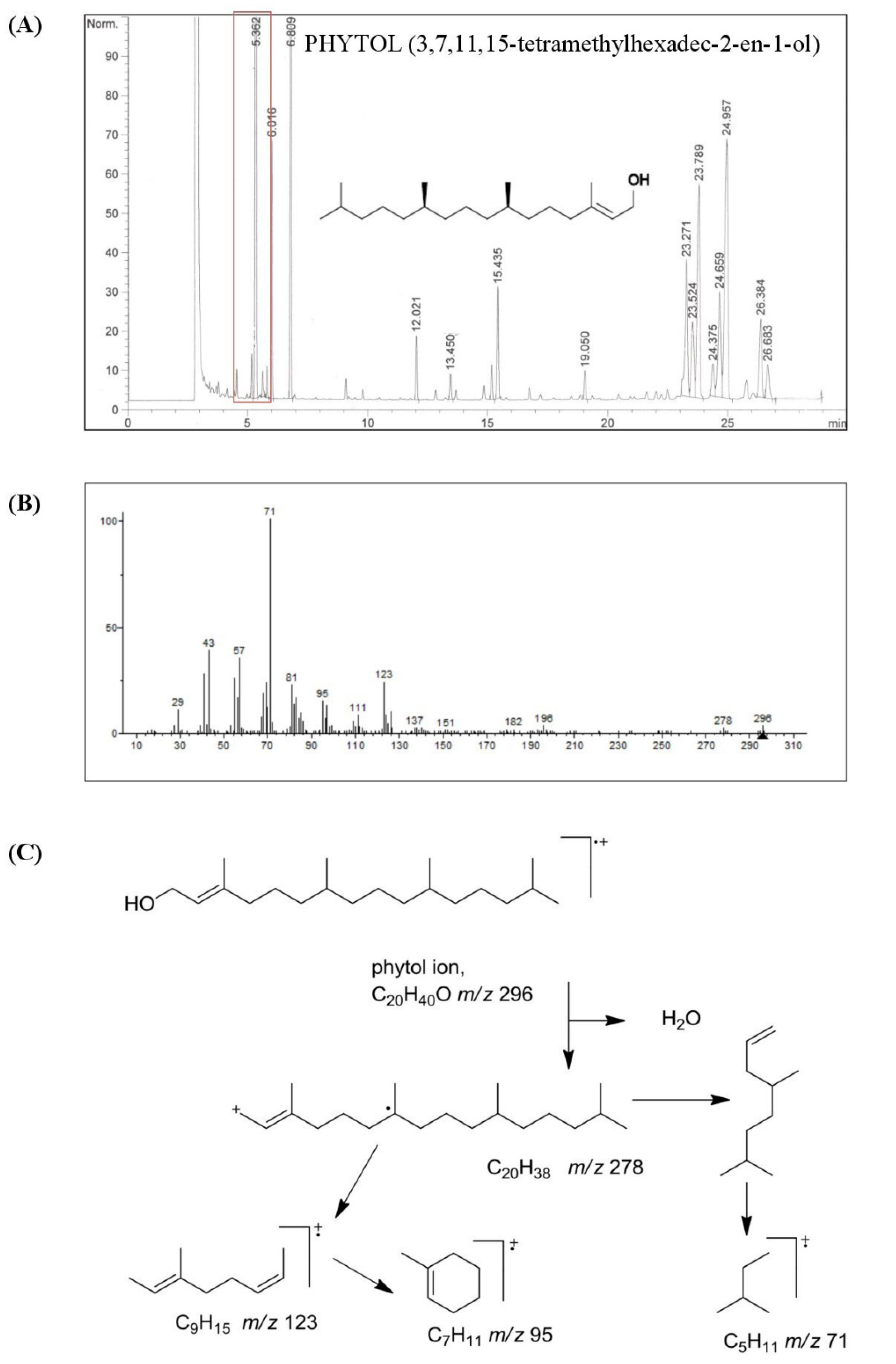
3.1. Chemical Characterization of HSO

Our study performed the isolation and characterization of the acyclic fatty alcohol fraction from an unsaponifiable matter of HSO. On the other hand, and for the first time, we explored the effect of phytol, the major compound in this fraction, on both the inflammatory response and reprogramming of functional phenotypes in human primary monocyte-derived mature macrophages. As shown in Figure 1, the main acyclic alcohols present in HSO were phytol (73.57%), followed by geranylgeraniol (11.45%) and hexacosanol (7.05%), representing 92% of the total fraction (226.94 ± 2.26 mg/kg of HSO). These components are quite different from those of policosanol, which is a commercial mixture of very long-chain alcohols containing mostly octocosanol (~60%), triacosanol (~13%), and hexacosanol (~6%) [38], and other vegetable oils such as olive oil, pomace olive oil, grape seed oil, and evening primrose oil [12,39,40].
To date, several chemical and nutritional studies have reported that acyclic fatty alcohols obtained from waxy materials of different origins exert beneficial effects on physiology [13,41]. For example, fixed oil has been described as having beneficial effects on osteoarthritis [42], hyperlipidemia [43], hyperalgesia [44], and inflammation [45,46]. However, little research has dealt with the pharmacological effects of phytol (3,7,11,15-tetramethylhexadec-2-en-1-ol), the main compound (167.59 ± 1.81 mg/kg of HSO) present in the acyclic fatty alcohol fraction isolated from HSO [16]. Phytol is scarcely present in human foods, such as asparagus, beans, spinach, or raw vegetables [47].
Free phytol in those products was reported in the range of 0.7–2 mg/kg of undried food. In dried tea or beef stock cubes, higher amounts of (12.5–14.7 mg/kg) phytol have been reported [48]. To date, previous studies have demonstrated the low toxicity and high tolerance of phytol, increasing the interest in this natural bioactive compound from plants [14,16,49]. Many data support the bioactivity of phytol in vivo and in vitro [50,51,52]. Our study demonstrates that HSO is one of the phytol-richest natural products.

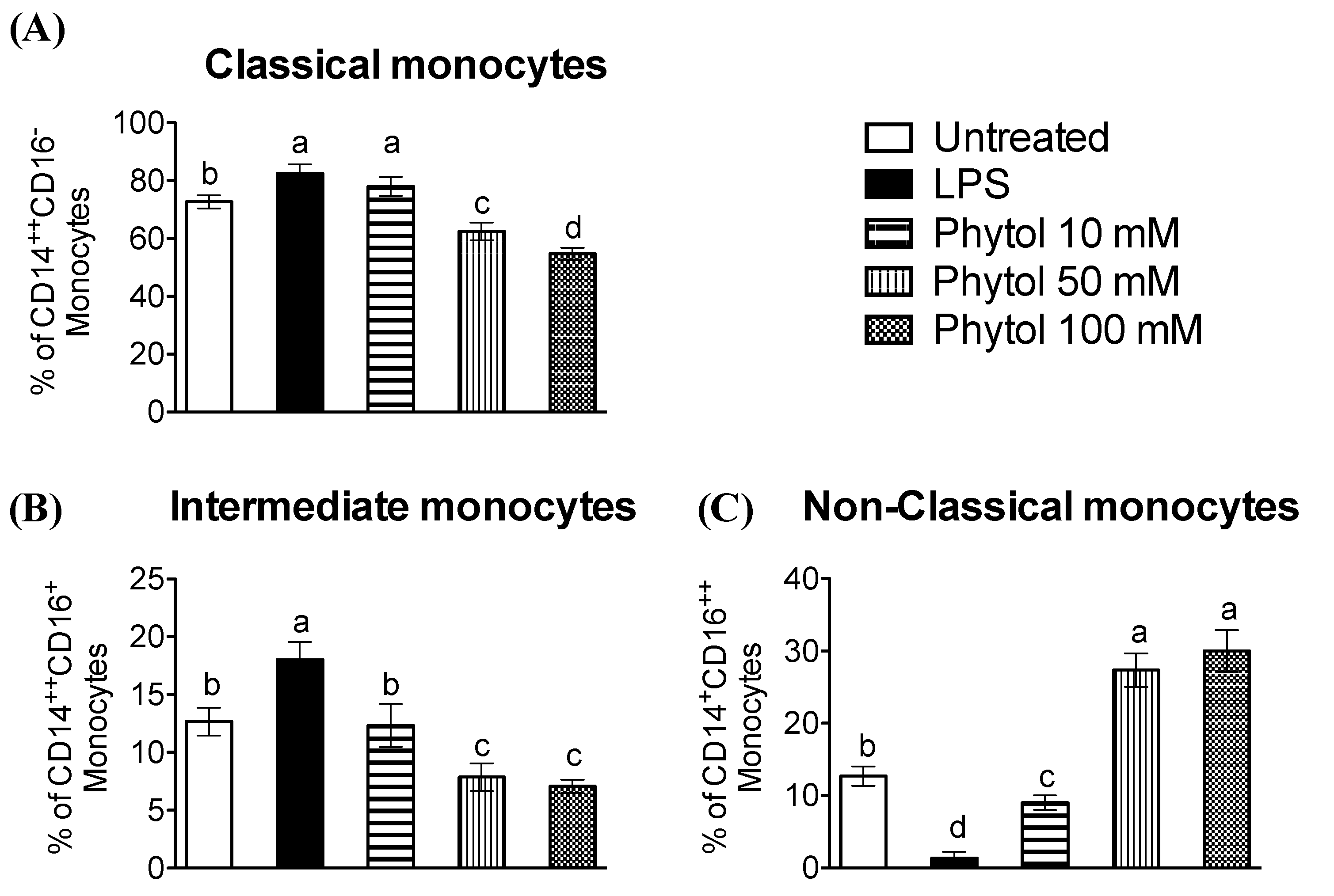
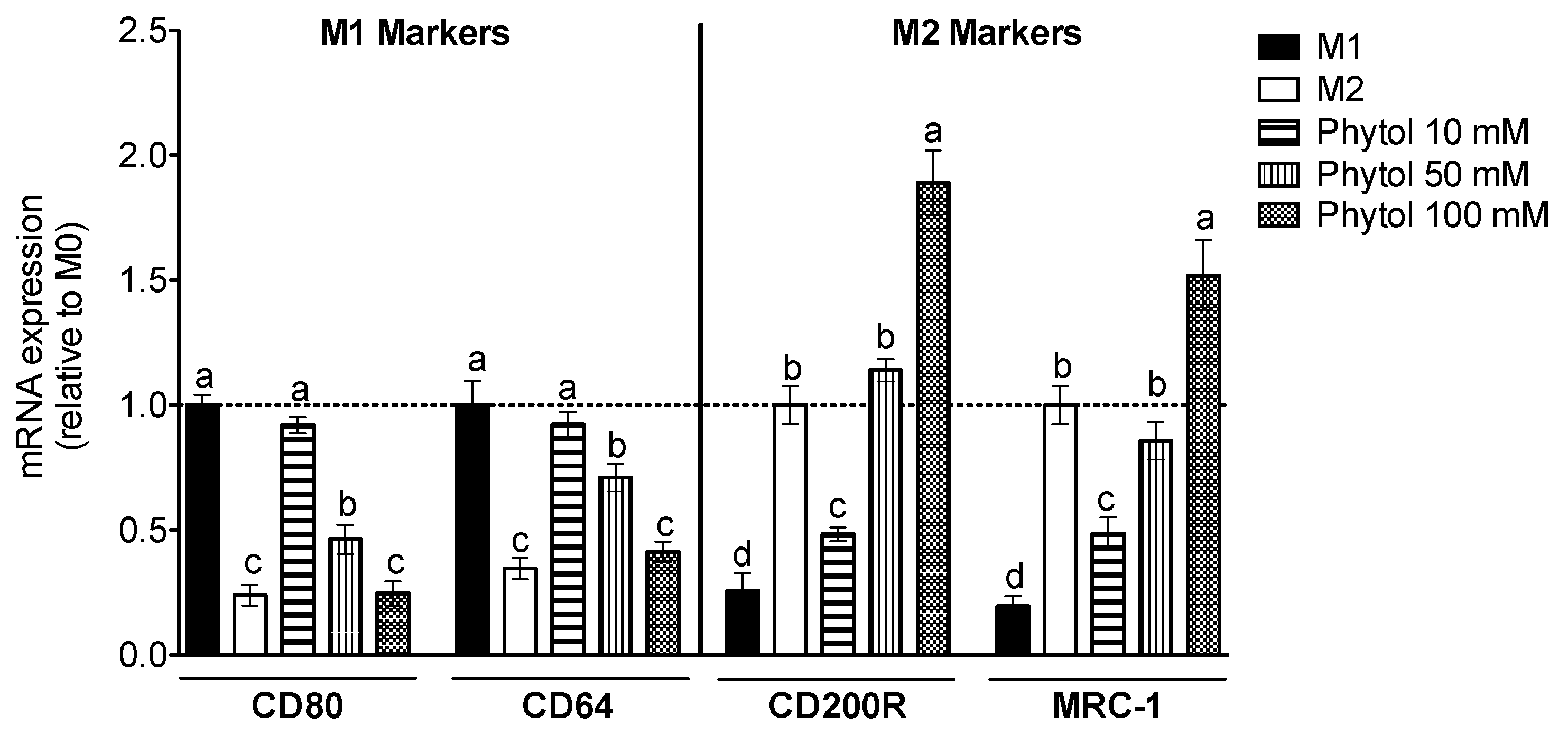
3.2. Effect of Phytol on Monocyte Differentiation and Macrophage Polarization

The literature on HSO has already highlighted the value of this natural product for the identification and medicinal use of its components, including phytol. For that reason, it is necessary to support the promising advantages of HSO in the immunology area or in inflammatory and vascular-related dysfunctions where one of the pivotal cells for the initiation of inflammation is the monocyte [53,54,55]. Most circulating monocytes are known as classical monocytes (CD14++CD16). Other subsets are non-classical monocytes (CD14+CD16++) and intermediate monocytes [26,27]. Both classical and intermediate monocytes have a pro-inflammatory phenotype that, as a response to LPS treatment, quite actively produces TNF-α, IL-1β, and IL-6, thus promoting various inflammatory disorders, including atherosclerosis [56,57]. Consequently, to avoid continuous inflammatory events and complete controlled repair, the correct balance of the monocyte cell subsets appears to be critical. There is a significant increase in CD14 expression in classical and intermediate monocytes when comparing LPS-treated cells with control untreated cells (Figure 2A,B). The addition of phytol overrode the LPS effect diminishing CD14 expression and also remarkably increasing CD16 (Figure 2C). The results of our study point, for the first time, to the feasible role of phytol as a regulator in human monocytes of the balance among the different subsets of monocytes. Classical and intermediate cells with pro-inflammatory phenotype show lower surface expression, while non-classical monocytes exhibit higher CD16 values after phytol doses compared to LPS. Therefore, phytol appears to be a promising indicator for preventing persistent inflammation and achieving controlled repair.
In response to specific local microenvironments, tissue-infiltrated monocyte-derived macrophages undergo polarization into M1, pro-inflammatory type 1, or M2, anti-inflammatory type 2 cells, both of which play a critical role in early events and subsequent resolution of the immune response [23,57]. These M1 and M2 macrophages illustrate the extremes of a continuum of functional phenotypes [27,58]. On the one hand, M1 macrophages originate from LPS and/or IFNγ, while the phenotype of M2 macrophages is influenced by IL-4. Therefore, macrophage plasticity modulation appears to be a potential strategy to control joint inflammation. The phytol has previously been observed to decrease acute inflammation in vivo and pro-inflammatory cytokine production in peritoneal exudate and neutrophils from mice [14,59,60]. Therefore, we asked whether phytol had any effect on polarization and cytokine response in human mature macrophages. For that purpose, we quantified the gene expression of phenotypic macrophage markers for M1 (CD80, CD64) and M2 (CD200R, MRC-1). As shown in Figure 3, both CD80 and CD64 genes appear to be up-regulated by LPS plus IFNγ, while CD200R and MRC-1 appear to be up-regulated by IL-4. It was noteworthy to observe that phytol skewed macrophage polarization towards M2.

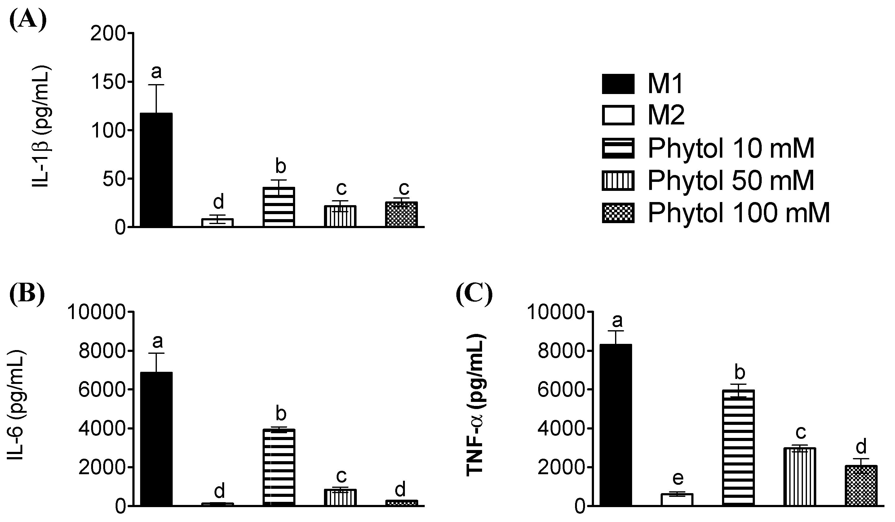
3.3. Effects of Phytol on Pro-Inflammatory Cytokine Levels

This ability of phytol to regulate the polarization of M0 macrophage cells was additionally studied by analyzing the release into the culture medium of pro-inflammatory cytokines of the M1 phenotype, such as TNFα, IL-6, or IL-1β, upon exposure to LPS. Analysis of cytokine production (Figure 4A–C) illustrated that phytol significantly decreased the inflammatory capability of mature human mature macrophages. The quantification of pro-inflammatory cytokines in culture medium showed lower values than the control M1 when IL-1β, IL-6, and TNF-α were tested. Τhis tendency correlates with phytol doses, diminishing half the average values when comparing 10-, 50-, and 100 mM treatments. IL-6 release after 50 mM phytol is the lowest decrease, well over half of the 10 mM dose. Finally, the production of pro-inflammatory cytokines compared to M1 was decreased in the lowest proportion with 10 mM phytol and dramatically diminished with 100 mM phytol dose, as shown in Figure 4C.
Previous research findings have shown that macrophage-derived cytokines are crucial in human aging [61,62] and also in a wide range of human inflammatory disorders [63,64,65,66,67,68,69,70]. This phenotype is considered to operate in dynamic interchange if there is an extreme risk of inflammation risk and a negative feedback procedure is needed, for example, to stop inflammation [71,72]. These findings support the idea that phytol ingestion may prevent macrophage activation and alter macrophage plasticity in human tissue under inflammatory conditions.

4. Conclusions

These results showed that phytol, which was isolated and identified for the first time in HSO, can help to better understand the specific mechanism by which this acyclic diterpene exerts beneficial effects on monocyte-macrophage plasticity. So far, HSO has already been demonstrated to include healthy polyunsaturated fatty acids, as well as antioxidant tocopherols and anti-inflammatory phytosterols in its unsaponifiable fraction. In recent years, inflammation has emerged as a leading pathophysiologic mechanism in atherosclerosis and other diseases, so the effects of phytol on different hallmarks of the inflammatory response contribute to the recommendation of HSO as an interesting source of functional compounds.

Author Contributions

Conceptualization, S.M.-d.l.P.; methodology, R.T. and M.C.M.-L.; investigation, E.G.-C., C.M.C.-C., M.C.M.-L. and S.M.-d.l.P.; writing—original draft preparation, S.M.-d.l.P.; writing—review and editing, M.E.M. All authors have read and agreed to the published version of the manuscript.

Funding

This study was supported by a grant from the Andalusian Government (2021/CTS-1074).

Institutional Review Board Statement

Not applicable.

Informed Consent Statement

Not applicable.

Data Availability Statement

Data is contained within the article.

Acknowledgments

Rocio Toscano has the benefit of a “Margarita Salas” postdoctoral grant from the Recovery, Transformation, and Resilience Plan (Next Generation EU). Elena Grao-Cruces has the benefit of a doctoral fellowship (BES-2012-056104) from the Andalusian Government. Maria C. Millan-Linares acknowledges the financial grant US-1381492 supported by the Andalusian Plan for Research, Development, and Innovation 2020 from the European Regional Development Fund (ERDF).

Conflicts of Interest

The authors declare no conflict of interest.

References

  1. Yan, X.; Tang, J.; Passos, C.D.S.; Nurisso, A.; Simões-Pires, C.A.; Ji, M.; Lou, H.; Fan, P. Characterization of Lignanamides from Hemp (Cannabis sativa L.) Seed and Their Antioxidant and Acetylcholinesterase Inhibitory Activities. J. Agric. Food Chem. 2015, 63, 10611–10619. [Google Scholar] [CrossRef] [PubMed]
  2. Farinon, B.; Molinari, R.; Costantini, L.; Merendino, N. The Seed of Industrial Hemp (Cannabis sativa L.): Nutritional Quality and Potential Functionality for Human Health and Nutrition. Nutrition 2020, 12, 1935. [Google Scholar] [CrossRef] [PubMed]
  3. Rupasinghe, H.P.V.; Davis, A.; Kumar, S.K.; Murray, B.; Zheljazkov, V.D. Industrial Hemp (Cannabis sativa subsp. sativa) as an Emerging Source for Value-Added Functional Food Ingredients and Nutraceuticals. Molecules 2020, 25, 4078. [Google Scholar] [CrossRef] [PubMed]
  4. Rea Martinez, J.; Montserrat-de la Paz, S.; De la Puerta, R.; Garcia-Gimenez, M.D.; Fernandez-Arche, M.A. Characterization of bioactive compounds in defatted hempseed (Cannabis sativa L.) by UHPLC-HRMS/MS and anti-inflammatory activity in primary human monocytes. Food Funct. 2020, 11, 4057–4066. [Google Scholar] [CrossRef]
  5. Rodriguez-Martin, N.M.; Montserrat-de la Paz, S.; Toscano, R.; Grao-Cruces, E.; Villanueva, A.; Pedroche, J.; Millan, F.; Millan-Linares, M.C. Hemp (Cannabis sativa L.) Protein Hydrolysates Promote Anti-Inflammatory Response in Primary Human Monocytes. Biomolecules 2020, 10, 803. [Google Scholar] [CrossRef]
  6. Rodriguez-Martin, N.M.; Toscano, R.; Villanueva, A.; Pedroche, J.; Millan, F.; Montserrat-de la Paz, S.; Millan-Linares, M.C. Neuroprotective protein hydrolysates from hemp (Cannabis sativa L.) seeds. Food Funct. 2019, 10, 6732–6739. [Google Scholar] [CrossRef]
  7. Tapia, G.; Valenzuela, R.; Espinosa, A.; Romanque, P.; Dossi, C.; Gonzalez-Mañán, D.; Videla, L.A.; D’Espessailles, A. N-3 long-chain PUFA supplementation prevents high fat diet induced mouse liver steatosis and inflammation in relation to PPAR-α upregulation and NF-κB DNA binding abrogation. Mol. Nutr. Food Res. 2014, 58, 1333–1341. [Google Scholar] [CrossRef]
  8. Montserrat-de la Paz, S.M.; Marín-Aguilar, F.; García-Giménez, M.D.; Fernández-Arche, M.A. Hemp (Cannabis sativa L.) Seed Oil: Analytical and Phytochemical Characterization of the Unsaponifiable Fraction. J. Agric. Food Chem. 2014, 62, 1105–1110. [Google Scholar] [CrossRef]
  9. Oomah, B.D.; Busson, M.; Godfrey, D.V.; Drover, J.C.G. Characteristics of hemp (Cannabis sativa L.) seed oil. Food Chem. 2002, 76, 33–43. [Google Scholar] [CrossRef]
  10. Rezapour-Firouzi, S.; Mohammadian, M.; Sadeghzadeh, M.; Mehranfar, S.; Mazloomi, E. The effects of evening primrose/hemp seed oil compared to rapamycin on the gene expression of immunological parameters in experimental autoimmune encephalomyelitis splenocytes. Iran. J. Allergy Asthma Immunol. 2020, 19, 183–192. [Google Scholar] [CrossRef]
  11. Izzo, L.; Pacifico, S.; Piccolella, S.; Castaldo, L.; Narváez, A.; Grosso, M.; Ritieni, A. Chemical Analysis of Minor Bioactive Components and Cannabidiolic Acid in Commercial Hemp Seed Oil. Molecules 2020, 25, 3710. [Google Scholar] [CrossRef] [PubMed]
  12. Montserrat-De La Paz, S.; García-Giménez, M.D.; Ángel-Martín, M.; Pérez-Camino, M.C.; Fernández Arche, A. Long-chain fatty alcohols from evening primrose oil inhibit the inflammatory response in murine peritoneal macrophages. J. Ethnopharmacol. 2014, 151, 131–136. [Google Scholar] [CrossRef] [PubMed]
  13. Giuffrè, A.M.; Capocasale, M. Policosanol in Tomato (Solanum lycopersicum L.) Seed Oil: The Effect of Cultivar. J. Oleo Sci. 2015, 64, 625–631. [Google Scholar] [CrossRef] [PubMed] [Green Version]
  14. Silva, R.O.; Sousa, F.B.M.; Damasceno, S.R.B.; Carvalho, N.S.; Silva, V.G.; Oliveira, F.R.M.A.; Sousa, D.P.; Aragão, K.S.; Barbosa, A.L.R.; Freitas, R.M.; et al. Phytol, a diterpene alcohol, inhibits the inflammatory response by reducing cytokine production and oxidative stress. Fundam. Clin. Pharmacol. 2014, 28, 455–464. [Google Scholar] [CrossRef] [PubMed] [Green Version]
  15. De Moraes, J.; de Oliveira, R.N.; Costa, J.P.; Junior, A.L.G.; de Sousa, D.P.; Freitas, R.M.; Allegretti, S.M.; Pinto, P.L.S. Phytol, a Diterpene Alcohol from Chlorophyll, as a Drug against Neglected Tropical Disease Schistosomiasis Mansoni. PLoS Negl. Trop. Dis. 2014, 8, e2617. [Google Scholar] [CrossRef] [Green Version]
  16. Islam, M.T.; Ali, E.S.; Uddin, S.J.; Shaw, S.; Islam, M.A.; Ahmed, M.I.; Chandra Shill, M.; Karmakar, U.K.; Yarla, N.S.; Khan, I.N.; et al. Phytol: A review of biomedical activities. Food Chem. Toxicol. 2018, 121, 82–94. [Google Scholar] [CrossRef]
  17. Alencar, M.V.O.B.; Islam, M.T.; Ali, E.S.; Santos, J.V.O.; Paz, M.F.C.J.; Sousa, J.M.C.; Dantas, S.M.M.M.; Mishra, S.K.; Cavalcante, A.A.C.M. Association of Phytol with Toxic and Cytotoxic Activities in an Antitumoral Perspective: A Meta-Analysis and Systemic Review. Anticancer. Agents Med. Chem. 2018, 18, 1828–1837. [Google Scholar] [CrossRef]
  18. Al-Sayed, E.; Ke, T.Y.; Hwang, T.L.; Chen, S.R.; Korinek, M.; Chen, S.L.; Cheng, Y. Bin Cytotoxic and anti-inflammatory effects of lignans and diterpenes from Cupressus macrocarpa. Bioorganic Med. Chem. Lett. 2020, 30, 127127. [Google Scholar] [CrossRef]
  19. Fang, S.M.; Liu, R.; Li, L.; Yao, J.L.; Liu, E.W.; Fan, G.W.; Zhang, H.; Gao, X.M. Anti-inflammatory diterpenes from the fruits of Vitex trifolia L. var. simplicifolia Cham. J. Asian Nat. Prod. Res. 2019, 21, 985–991. [Google Scholar] [CrossRef]
  20. Liu, Y.Y.; Liu, Y.P.; Wang, X.P.; Qiao, Z.H.; Yu, X.M.; Zhu, Y.Z.; Xie, L.; Qiang, L.; Fu, Y.H. Bioactive daphnane diterpenes from Wikstroemia chuii with their potential anti-inflammatory effects and anti-HIV activities. Bioorg. Chem. 2020, 105, 104388. [Google Scholar] [CrossRef]
  21. Campos-Xolalpa, N.; Alonso-Castro, Á.J.; Ortíz-Sanchez, E.; Zapata-Morales, J.R.; González-Chávez, M.M.; Pérez, S. Anti-inflammatory and antitumor activities of the chloroform extract and anti-inflammatory effect of the three diterpenes isolated from Salvia ballotiflora Benth. BMC Complement. Med. Ther. 2021, 21, 1–11. [Google Scholar] [CrossRef] [PubMed]
  22. Islam, M.T.; Ayatollahi, S.A.; Zihad, S.M.N.K.; Sifat, N.; Khan, M.R.; Paul, A.; Salehi, B.; Islam, T.; Mubarak, M.S.; Martins, N.; et al. Phytol anti-inflammatory activity: Pre-clinical assessment and possible mechanism of action elucidation. Cell. Mol. Biol. 2020, 66, 264–269. [Google Scholar] [CrossRef] [PubMed]
  23. Porta, C.; Riboldi, E.; Ippolito, A.; Sica, A. Molecular and epigenetic basis of macrophage polarized activation. Semin. Immunol. 2015, 27, 237–248. [Google Scholar] [CrossRef] [PubMed]
  24. Amengual, J.; Barrett, T.J. Monocytes and macrophages in atherogenesis. Curr. Opin. Lipidol. 2019, 30, 401–408. [Google Scholar] [CrossRef] [PubMed]
  25. Bermudez, B.; Dahl, T.B.; Medina, I.; Groeneweg, M.; Holm, S.; Montserrat-de la Paz, S.; Rousch, M.; Otten, J.; Herias, V.; Varela, L.M.; et al. Leukocyte Overexpression of Intracellular NAMPT Attenuates Atherosclerosis by Regulating PPARγ-Dependent Monocyte Differentiation and Function. Arterioscler. Thromb. Vasc. Biol. 2017, 37, 1157–1167. [Google Scholar] [CrossRef] [PubMed] [Green Version]
  26. Montserrat-De La Paz, S.; De La Puerta, R.; Fernandez-Arche, A.; Quilez, A.M.; Muriana, F.J.G.; Garcia-Gimenez, M.D.; Bermudez, B. Pharmacological effects of mitraphylline from Uncaria tomentosa in primary human monocytes: Skew toward M2 macrophages. J. Ethnopharmacol. 2015, 170, 128–135. [Google Scholar] [CrossRef] [Green Version]
  27. Wolf, A.A.; Yáñez, A.; Barman, P.K.; Goodridge, H.S. The ontogeny of monocyte subsets. Front. Immunol. 2019, 10, 1642. [Google Scholar] [CrossRef] [Green Version]
  28. Ożańska, A.; Szymczak, D.; Rybka, J. Pattern of human monocyte subpopulations in health and disease. Scand. J. Immunol. 2020, 92, e12883. [Google Scholar] [CrossRef]
  29. Italiani, P.; Boraschi, D. From Monocytes to M1/M2 Macrophages: Phenotypical vs. Functional Differentiation. Front. Immunol. 2014, 5, 514. [Google Scholar] [CrossRef] [Green Version]
  30. Yunna, C.; Mengru, H.; Lei, W.; Weidong, C. Macrophage M1/M2 polarization. Eur. J. Pharmacol. 2020, 877, 173090. [Google Scholar] [CrossRef]
  31. Wong, K.L.; Yeap, W.H.; Tai, J.J.Y.; Ong, S.M.; Dang, T.M.; Wong, S.C. The three human monocyte subsets: Implications for health and disease. Immunol. Res. 2012, 53, 41–57. [Google Scholar] [CrossRef] [PubMed]
  32. Benoit, M.; Desnues, B.; Mege, J.-L. Macrophage Polarization in Bacterial Infections. J. Immunol. 2008, 181, 3733–3739. [Google Scholar] [CrossRef] [PubMed] [Green Version]
  33. Montserrat-De La Paz, S.; Fernández-Arche, M.A.; Ángel-Martín, M.; García-Giménez, M.D. Phytochemical characterization of potential nutraceutical ingredients from Evening Primrose oil (Oenothera biennis L.). Phytochem. Lett. 2014, 8, 158–162. [Google Scholar] [CrossRef]
  34. Martin, M.E.; Millan-Linares, M.C.; Naranjo, M.C.; Toscano, R.; Abia, R.; Muriana, F.J.G.; Bermudez, B.; Montserrat-de la Paz, S. Minor compounds from virgin olive oil attenuate LPS-induced inflammation via visfatin-related gene modulation on primary human monocytes. J. Food Biochem. 2019, 43, e12941. [Google Scholar] [CrossRef]
  35. Montserrat-de la Paz, S.; Bermudez, B.; Lopez, S.; Naranjo, M.C.; Romero, Y.; Bando-Hidalgo, M.J.; Abia, R.; Muriana, F.J.G. Exogenous fatty acids and niacin on acute prostaglandin D 2 production in human myeloid cells. J. Nutr. Biochem. 2017, 39, 22–31. [Google Scholar] [CrossRef] [Green Version]
  36. Montserrat-De La Paz, S.; Fernández-Arche, Á.; Ángel-Martín, M.; García-Giménez, M.D. The sterols isolated from Evening Primrose oil modulate the release of proinflammatory mediators. Phytomedicine 2012, 19, 1072–1076. [Google Scholar] [CrossRef]
  37. Toscano, R.; Millan-Linares, M.C.; Lemus-Conejo, A.; Claro, C.; Sanchez-Margalet, V.; Montserrat-de la Paz, S. Postprandial triglyceride-rich lipoproteins promote M1/M2 microglia polarization in a fatty-acid-dependent manner. J. Nutr. Biochem. 2020, 75, 108248. [Google Scholar] [CrossRef]
  38. Guo, Y.L.; Xu, R.X.; Zhu, C.G.; Wu, N.Q.; Cui, Z.P.; Li, J.J. Policosanol attenuates statin-induced increases in serum proprotein convertase subtilisin/kexin type 9 when combined with atorvastatin. Evid.-Based Complement. Altern. Med. 2014, 2014. [Google Scholar] [CrossRef]
  39. Fernández-Arche, A.; Marquez-Martín, A.; Vazquez, R.D.L.P.; Perona, J.S.; Terencio, C.; Perez-Camino, C.; Ruiz-Gutierrez, V. Long-chain fatty alcohols from pomace olive oil modulate the release of proinflammatory mediators. J. Nutr. Biochem. 2009, 20, 155–162. [Google Scholar] [CrossRef] [Green Version]
  40. Millan-Linares, M.C.; Bermudez, B.; Martin, M.E.; Muñoz, E.; Abia, R.; Millan, F.; Muriana, F.J.G.; Montserrat-de la Paz, S. Unsaponifiable fraction isolated from grape (Vitis vinifera L.) seed oil attenuates oxidative and inflammatory responses in human primary monocytes. Food Funct. 2018, 9, 2517–2523. [Google Scholar] [CrossRef] [Green Version]
  41. He, X.-F.; Chen, J.-J.; Li, T.-Z.; Zhang, X.-K.; Guo, Y.-Q.; Zhang, X.-M.; Hu, J.; Geng, C.-A. Nineteen New Flavanol–Fatty Alcohol Hybrids with α-Glucosidase and PTP1B Dual Inhibition: One Unusual Type of Antidiabetic Constituent from Amomum tsao-ko. J. Agric. Food Chem. 2020, 68, 11434–11448. [Google Scholar] [CrossRef] [PubMed]
  42. Silva, R.D.F.; Barreto, A.S.; Gomes Trindade, G.G.; Lima, C.M.; Araujo, A.A.S.; Menezes, I.R.A.; Candido, E.A.F.; Santana, E.T.N.; Silva-Junior, W.M.; Quintans, J.S.S.; et al. Enhancement of the functionality of women with knee osteoarthritis by a gel formulation with Caryocar coriaceum Wittm (“Pequi”) nanoencapsulated pulp fixed oil. Biomed. Pharmacother. 2022, 150, 112938. [Google Scholar] [CrossRef] [PubMed]
  43. Figuereido, P.R.L.; Oliveira, I.B.; Neto, J.B.S.; Oliveira, J.A.; Ribeiro, L.B.; Viana, G.S.B.; Rocha, T.M.; Leal, L.K.A.M.; Kerntopf, M.R.; Felipe, C.F.B.; et al. Caryocar coriaceum Wittm. (Pequi) fixed oil presents hypolipemic and anti-inflammatory effects in vivo and in vitro. J. Ethnopharmacol. 2016, 191, 87–94. [Google Scholar] [CrossRef] [PubMed]
  44. Brito, R.G.; Dos Santos, L.P.; Quintans, J.S.S.; Lucca Junior, W.; Araujo, A.A.S.; Saravanan, S.; Menezes, I.R.A.; Countinho, H.D.M.; Quintans-Junior, L.J. Citronellol, a natural acyclic monoterpene, attenuates mechanical hyperalgesia response in mice: Evidence of the spinal cord lamina I inhibition. Chem. Biol. Interact. 2015, 239, 111–117. [Google Scholar] [CrossRef]
  45. Barreto, R.S.S.; Quintans, J.S.S.; Amarante, R.K.L.; Nascimento, T.S.; Amarante, R.S.; Barreto, A.S.; Pereira, E.W.M.; Duarte, M.C.; Countinho, H.D.M.; Menezes, I.R.A.; et al. Evidence for the involvement of TNF-α and IL-1β in the antinociceptive and anti-inflammatory activity of Stachys lavandulifolia Vahl. (Lamiaceae) essential oil and (-)-α-bisabolol, its main compound, in mice. J. Ethnopharmacol. 2016, 191, 9–18. [Google Scholar] [CrossRef] [PubMed]
  46. Vetter, W.; Schröder, M.; Lehnert, K. Differentiation of refined and virgin edible oils by means of the trans—And cis-phytol isomer distribution. J. Agric. Food Chem. 2012, 60, 6103–6107. [Google Scholar] [CrossRef]
  47. Brown, P.J.; Mei, G.; Gibberd, F.B.; Burston, D.; Mayne, P.D.; McClinchy, J.E.; Sidey, M. Diet and Refsum’s disease. The determination of phytanic acid and phytol in certain foods and the application of this knowledge to the choice of suitable convenience foods for patients with Refsum’s disease. J. Hum. Nutr. Diet. 1993, 6, 295–305. [Google Scholar] [CrossRef]
  48. Pejin, B.; Kojic, V.; Bogdanovic, G. Natural Product Research Formerly Natural Product Letters An insight into the cytotoxic activity of phytol at in vitro conditions. Nat. Prod. Res. 2014, 28, 2053–2056. [Google Scholar] [CrossRef]
  49. Costa, J.; Islam, M.; Santos, P.; Ferreira, P.; Oliveira, G.; Alencar, M.; Paz, M.; Ferreira, É.; Feitosa, C.; Citó, A.; et al. Evaluation of Antioxidant Activity of Phytol Using Non- and Pre-Clinical Models. Curr. Pharm. Biotechnol. 2016, 17, 1278–1284. [Google Scholar] [CrossRef]
  50. Wang, J.; Hu, X.; Ai, W.; Zhang, F.; Yang, K.; Wang, L.; Zhu, X.; Gao, P.; Shu, G.; Jiang, Q.; et al. Phytol increases adipocyte number and glucose tolerance through activation of PI3K/Akt signaling pathway in mice fed high-fat and high-fructose diet. Biochem. Biophys. Res. Commun. 2017, 489, 432–438. [Google Scholar] [CrossRef]
  51. Bobe, G.; Zhang, Z.; Kopp, R.; Garzotto, M.; Shannon, J.; Takata, Y. Phytol and its metabolites phytanic and pristanic acids for risk of cancer: Current evidence and future directions. Eur. J. Cancer Prev. 2020, 29, 191. [Google Scholar] [CrossRef] [PubMed]
  52. Vazquez-Madrigal, C.; Lopez, S.; Grao-Cruces, E.; Millan-Linares, M.C.; Rodriguez-Martin, N.M.; Martin, M.E.; Alba, G.; Santa-Maria, C.; Bermudez, B.; Montserrat-de la Paz, S. Dietary Fatty Acids in Postprandial Triglyceride-Rich Lipoproteins Modulate Human Monocyte-Derived Dendritic Cell Maturation and Activation. Nutrients 2020, 12, 3139. [Google Scholar] [CrossRef]
  53. Montserrat-de la Paz, S.; Rodriguez-Martin, N.M.; Villanueva, A.; Pedroche, J.; Cruz-Chamorro, I.; Millan, F.; Millan-Linares, M.C. Evaluation of Anti-Inflammatory and Atheroprotective Properties of Wheat Gluten Protein Hydrolysates in Primary Human Monocytes. Foods 2020, 9, 854. [Google Scholar] [CrossRef]
  54. Rosillo, M.A.; Montserrat-de la Paz, S.; Abia, R.; Castejon, M.L.; Millan-Linares, M.C.; Alarcon-de la Lastra, C.; Fernandez-Bolaños, J.G.; Muriana, F.J.G. Oleuropein and its peracetylated derivative negatively regulate osteoclastogenesis by controlling the expression of genes involved in osteoclast differentiation and function. Food Funct. 2020, 11, 4038–4048. [Google Scholar] [CrossRef] [PubMed]
  55. Rogacev, K.S.; Zawada, A.M.; Emrich, I.; Seiler, S.; Böhm, M.; Fliser, D.; Woollard, K.J.; Heine, G.H. Lower Apo A-I and lower HDL-C levels are associated with higher intermediate CD14++CD16+monocyte counts that predict cardiovascular events in chronic kidney disease. Arterioscler. Thromb. Vasc. Biol. 2014, 34, 2120–2127. [Google Scholar] [CrossRef] [PubMed] [Green Version]
  56. Xu, H.; Jiang, J.; Chen, W.; Li, W.; Chen, Z. Review Article Vascular Macrophages in Atherosclerosis. J. Immunol. Res. 2019, 2019. [Google Scholar] [CrossRef] [Green Version]
  57. Malyshev, I.; Malyshev, Y. Current concept and update of the macrophage plasticity concept: Intracellular mechanisms of reprogramming and M3 macrophage “switch” phenotype. Biomed. Res. Int. 2015, 2015. [Google Scholar] [CrossRef] [Green Version]
  58. Carvalho, A.M.S.; Heimfarth, L.; Pereira, E.W.M.; Oliveira, F.S.; Menezes, I.R.A.; Coutinho, H.D.M.; Picot, L.; Antoniolli, A.R.; Quintans, J.S.S.; Quintans-Júnior, L.J. Phytol, a Chlorophyll Component, Produces Antihyperalgesic, Anti-inflammatory, and Antiarthritic Effects: Possible NFκB Pathway Involvement and Reduced Levels of the Proinflammatory Cytokines TNF-α and IL-6. J. Nat. Prod. 2020, 83, 1107–1117. [Google Scholar] [CrossRef]
  59. Blum, L.; Tafferner, N.; Spring, I.; Kurz, J.; DeBruin, N.; Geisslinger, G.; Parnham, M.J.; Schiffmann, S. Dietary phytol reduces clinical symptoms in experimental autoimmune encephalomyelitis (EAE) at least partially by modulating NOX2 expression. J. Mol. Med. 2018, 96, 1131–1144. [Google Scholar] [CrossRef]
  60. Corcoran, M.P.; Meydani, M.; Lichtenstein, A.H.; Schaefer, E.J.; Dillard, A.; Lamon-Fava, S. Sex hormone modulation of proinflammatory cytokine and CRP expression in macrophages from older men and postmenopausal women. J. Endocrinol. 2010, 206, 217. [Google Scholar] [CrossRef] [Green Version]
  61. Montserrat-de la Paz, S. Active Ingredients from Foods: Biochemical and Processing Aspects. Foods 2021, 10, 1240. [Google Scholar] [CrossRef] [PubMed]
  62. Hanna, A.; Frangogiannis, N.G. Inflammatory Cytokines and Chemokines as Therapeutic Targets in Heart Failure. Cardiovasc. Drugs Ther. 2020, 34, 849–863. [Google Scholar] [CrossRef] [PubMed]
  63. Chen, Y.; Yu, C.Y.; Deng, W.M. The role of pro-inflammatory cytokines in lipid metabolism of metabolic diseases. Int. Rev. Immunol. 2019, 38, 249–266. [Google Scholar] [CrossRef] [PubMed]
  64. Zhou, M.; Li, S.; Pathak, J.L. Pro-inflammatory Cytokines and Osteocytes. Curr. Osteoporos. Rep. 2019, 17, 97–104. [Google Scholar] [CrossRef]
  65. Hu, F.; Lou, N.; Jiao, J.; Guo, F.; Xiang, H.; Shang, D. Macrophages in pancreatitis: Mechanisms and therapeutic potential. Biomed. Pharmacother. 2020, 131, 110693. [Google Scholar] [CrossRef] [PubMed]
  66. Zhang, H.; Cai, D.; Bai, X. Macrophages regulate the progression of osteoarthritis. Osteoarthr. Cartil. 2020, 28, 555–561. [Google Scholar] [CrossRef]
  67. Kishore, A.; Petrek, M. Roles of Macrophage Polarization and Macrophage-Derived miRNAs in Pulmonary Fibrosis. Front. Immunol. 2021, 12, 678457. [Google Scholar] [CrossRef]
  68. Ma, H.; Li, Y.N.; Song, L.; Liu, R.; Li, X.; Shang, Q.; Wang, Y.; Shao, C.; Shi, Y. Macrophages inhibit adipogenic differentiation of adipose tissue derived mesenchymal stem/stromal cells by producing pro-inflammatory cytokines. Cell Biosci. 2020, 10, 1–12. [Google Scholar] [CrossRef]
  69. Leuti, A.; Talamonti, E.; Gentile, A.; Tiberi, M.; Matteocci, A.; Fresegna, D.; Centonze, D.; Chiurchiù, V. Macrophage Plasticity and Polarization Are Altered in the Experimental Model of Multiple Sclerosis. Biomolecules 2021, 11, 837. [Google Scholar] [CrossRef]
  70. Fernandes, T.L.; Gomoll, A.H.; Lattermann, C.; Hernandez, A.J.; Bueno, D.F.; Amano, M.T. Macrophage: A Potential Target on Cartilage Regeneration. Front. Immunol. 2020, 11, 111. [Google Scholar] [CrossRef] [Green Version]
  71. Mesquida-Veny, F.; Del Río, J.A.; Hervera, A. Macrophagic and microglial complexity after neuronal injury. Prog. Neurobiol. 2021, 200, 101970. [Google Scholar] [CrossRef] [PubMed]
  72. Huang, T.C.; Wu, H.L.; Chen, S.H.; Wang, Y.T.; Wu, C.C. Thrombomodulin facilitates peripheral nerve regeneration through regulating M1/M2 switching. J. Neuroinflamm. 2020, 17, 1–14. [Google Scholar] [CrossRef] [PubMed]
Figure 1. Characterization of phytol isolated from HSO. (A) Gas chromatogram of acyclic aliphatic alcohols, triterpene alcohols, and 4-methylsterol fractions of unsaponifiable HSO and chemical structure of phytol (3,7,11,15-tetramethylhexadec-2-en-1-ol). (B) Mass spectrum and (C) schematic representation of the phytol mass fragmentation pattern.
Foods 11 02366 g001
Figure 2. Effect of phytol on monocytes cells. FACS analysis (MFI) data of CD14 and CD16 as monocyte surface markers after 24 h treatment in the presence or absence of 100 ng/mL of LPS and phytol at 10, 50, or 100 mM. (A) Classical monocytes CD14++CD16, (B) intermediate monocytes CD14++CD16+, and (C) non-classical monocytes CD14+CD16++. Values are shown as means ± SD (n = 3) and, when marked with different letters, are significantly different (p < 0.01).
Foods 11 02366 g002
Figure 3. Effect of Phytol on polarization into M1 or M2 macrophages. M0 macrophage cells were treated with LPS and IFNγ for the M1 control, with IL-4 for M2 control, or with 10–100 mM phytol for an additional 24 h. RT-qPCR was used to measure the relative gene expression of the markers CD80, CD64, CD200R, and MRC-1. The values are means ± SD (n = 3) and, when marked with different letters, they are significantly different (p < 0.01).
Foods 11 02366 g003
Figure 4. Effect of phytol on the release of pro-inflammatory cytokines. Cells from M0 macrophages were incubated in the presence of LPS and IFNγ, for M1 control, or with IL-4 for the M2 control, or with 10–100 mM Phytol for an additional 24 h. Using ELISA protocols, in polarized macrophage culture supernatants, the concentrations of (A) IL-1β, (B) IL-6, and (C) TNFα were measured. The values are means ± SD (n = 3) and, if marked with different letters, they are significantly different (p < 0.01).
Foods 11 02366 g004
Table 1. Sequences of RT-PCR primers for gene expression analysis.
TargetGenBank
Accession Number
DirectionSequence (5′→3′)
CD80NM_005191.3Forward
Reverse
GGGAAAGTGTACGCCCTGTA
GCTACTTCTGTGCCCACCAT
CD200RNM_138940.2Forward
Reverse
GTTGCCCTCCTATCGCATTA
TGGAAATTCCCATCAGGTGT
MRNM_002438.3Forward
Reverse
GGCGGTGACCTCACAAGTAT
ACGAAGCCATTTGGTAAACG
CD64NM_000566.3Forward
Reverse
GTCCAAATCTCCAAGTGCGG
CCCAAGTATGAGAGCAGCGT
GAPDHNM_001289746Forward
Reverse
CACATGGCCTCCAAGGAGTAAG
CCAGCAGTGAGGGTCTCTCT
Publisher’s Note: MDPI stays neutral with regard to jurisdictional claims in published maps and institutional affiliations.

