Article

Lactic Acid Bacteria Diversity and Characterization of Probiotic Candidates in Fermented Meats

by
Elvina Parlindungan
1,
Gabriele A. Lugli
2,
Marco Ventura
2,
Douwe van Sinderen
1,* and
Jennifer Mahony
1,*
1
School of Microbiology & APC Microbiome Ireland, University College Cork, Western Road, T12 YT20 Cork, Ireland
2
Laboratory of Probiogenomics, Department of Chemistry, Life Sciences and Environmental Sustainability, University of Parma, 43121 Parma, Italy
*
Authors to whom correspondence should be addressed.
Foods 2021, 10(7), 1519; https://doi.org/10.3390/foods10071519
Submission received: 24 May 2021 / Revised: 24 June 2021 / Accepted: 28 June 2021 / Published: 1 July 2021

Abstract

:
Probiotics are defined as live microorganisms which confer health benefits to the host when administered in adequate amounts. Many lactic acid bacteria (LAB) strains have been classified as probiotics and fermented foods are an excellent source of such LAB. In this study, novel probiotic candidates from two fermented meats (pancetta and prosciutto) were isolated and characterized. LAB populations present in pancetta and prosciutto were evaluated and Lactiplantibacillus plantarum was found to be the dominant species. The antagonistic ability of selected isolates against LAB and non-LAB strains was investigated, in particular, the ability to produce anti-microbial compounds including organic acids and bacteriocins. Probiotic characteristics including antibiotic susceptibility, hydrophobicity and autoaggregation capacity; and ability to withstand simulated gastric juice, bile salt, phenol and NaCl were assessed. Among the characterized strains, L. plantarum 41G isolated from prosciutto was identified as the most robust probiotic candidate compared. Results from this study demonstrate that artisanal fermented meat is a rich source of novel strains with probiotic potential.

Graphical Abstract

1. Introduction

With increasing public awareness for the need to improve human diet and lifestyle, there is growing market demand for functional foods and supplements which contain probiotics [1]. Probiotics are defined as live microorganisms that confer a health benefit on the host when administered in adequate amounts [2]. Lactic acid bacteria (LAB) have a long history of use as probiotics, many of which have a generally-recognized-as-safe (GRAS) status [3]. While probiotic LAB available on the market have primarily been isolated from humans [4,5], literature has identified fermented dairy, plant and meat products as a source of potentially novel probiotic LAB of extraintestinal origin [6,7,8]. Screening for novel probiotic candidates is desirable since probiotic features and health benefits conferred are known to be strain-specific [9].
Naturally fermented foods exhibit a rich biodiversity of microorganisms which make them a good source of potential probiotic LAB [10]. Latilactobacillus sakei, Latilactobacillus curvatus and Lactiplantibacillus plantarum are among the most prevalent LAB species associated with fermented meat products [11,12,13,14,15]. To a lesser extent, some studies have reported the presence of Lactobacillus gasseri, Lactiplantibacillus pentosus, Lacticaseibacillus rhamnosus, Lactobacillus johnsonii, Lactococcus lactis and Pediococcus pentosaceus [13,15,16]. These LAB are known to play an important role in food safety and protection through the production of antimicrobial compounds, including organic acids and bacteriocins [17,18]. The combination of different species and strains of LAB in fermented meat products has resulted in the emergence of different varieties of fermented meat products, including salami, chorizo, pancetta, prosciutto and pepperoni with an extended shelf life and unique sensory characteristics [19]. Fermented meats may be an excellent source of probiotics since they harbor high numbers of LAB [8].
Two spontaneously fermented Italian meats (pancetta and prosciutto) were acquired from a local market to screen for the presence of novel probiotic candidates. Guidelines for screening candidate probiotic LAB have been reviewed by Binda et al. [1]. Candidate probiotic strains must be (i) sufficiently characterized through evaluation of their tolerance to gastrointestinal conditions, antimicrobial activity and/or adhesion capacity; (ii) be safe, i.e., they should not be reservoirs of antibiotic resistance genes that could be transferred to pathogens; (iii) supported by at least one positive human clinical trial; and (iv) be alive in sufficient efficacious dose in the products they are applied to. This study aimed to screen for novel probiotic candidates, specifically focusing on the first two microbiological criteria.
The diversity of LAB in these two products was assessed using 16S rRNA gene-based analysis and (GTG)5 genetic fingerprinting. The susceptibility of the isolates to antibiotics as well as their ability to produce organic acids were investigated. Antagonistic activity of the isolates was assessed against a range of LAB and ESKAPE pathogens (Enterococcus faecium, Staphylococcus aureus, Klebsiella pneumonia, Acinetobacter baumannii, Pseudomonas aeruginosa, and Enterobacter species; highly virulent pathogens that could exhibit resistance to antibiotics) as well as fungal species. Subsequently, resistances towards bile salt, gastric juice, salt and phenol were assessed while their bile salt hydrolase (BSH) activity, autoaggregation capacity and hydrophobicity were also determined.

2. Materials and Methods

2.1. LAB Diversity, Organic Acid Profiling and Antibiotic Susceptibility

2.1.1. Isolation and Growth Condition

Artisanal traditionally fermented prosciutto and pancetta were purchased from a local market in Cork, Ireland. The amount of 5 g of each sample was transferred into 45 g of phosphate buffered saline (PBS, pH 7.0) (Sigma Aldrich, St. Louis, MO, USA) and pummeled for 2 min at 300 rpm in a stomacher (Stomacher Circular 400; Seward, UK). Serial dilutions of each sample were prepared and plated on De Man, Rogosa and Sharpe (MRS) agar (Oxoid, Hampshire, UK) and incubated at 30 °C and 37 °C aerobically for 48 h. The number of colonies were counted and individual colonies were isolated for further investigation. A total of 106 individual colonies were randomly selected from pancetta (n = 71) and prosciutto (n = 35) and grown on MRS broth (Oxoid) for 24 h at 30 °C and 37 °C. Stock cultures of all isolates were stored at −80 °C in MRS broth supplemented with 30% (v/v) glycerol (Thermo Fisher, Waltham, MA, USA).

2.1.2. Species Identification Using 16S rRNA Sequence Analysis

In order to identify LAB species present in pancetta and prosciutto, 16S rRNA gene amplification and sequencing were performed for 106 selected isolates using the following primers: LucF, 5′-CTTGTTACGACTTCACCC-3′ and LucR, 5′- TGCCTAATACATGCAAGT-3′ (Eurofins MWG, Ebersberg, Germany) [20]. PCR amplification of 16S rRNA genes was conducted using Taq DNA polymerase mastermix (Qiagen, Hilden, Germany) with the following PCR conditions: initial denaturation at 94 °C for 10 min, 30 cycles of 94 °C for 30 s, 40 °C for 30 s, 72 °C for 1 min and 30 s followed by a final extension at 72 °C for 10 min. PCR amplifications were performed with Applied Biosystems™ 2720 Thermal Cycler (Thermo Fisher). The amplicons were purified using the GenElute™ PCR Clean-Up Kit (Sigma Aldrich) according to the manufacturer’s instruction and subjected to Sanger sequencing (Eurofins MWG). The generated sequences were analyzed by comparative sequence analysis (BLASTN) against available sequence data on the National Center for Biotechnology Information (NCBI) database (https://blast.ncbi.nlm.nih.gov/Blast.cgi, accessed on 12 October 2020).

2.1.3. Phylogenetic Inference

The 16S rRNA gene sequences of selected isolates were aligned and processed in EditSeq software v5.01 (DNASTAR, Inc., Madison, WI, USA). A phylogenetic tree was then constructed using the neighbour-joining method and bootstrapped employing 1000 replicates. The final tree was visualized using MEGA7 [21]. Lactococcus lactis L-4 (Genbank accession number: LT853603.1) was selected as an outgroup organism representing a distinct LAB species.

2.1.4. Genetic Fingerprinting by (GTG)5 PCR

The selected isolates were subjected to PCR genomic fingerprinting with the single oligonucleotide primer (GTG)5, 5′-GTGGTGGTGGTGGTG-3′ [22]. PCR amplifications containing Taq DNA polymerase mastermix (Qiagen, Manchester, UK) were performed with Applied Biosystems™ 2720 Thermal Cycler (Thermo Fisher) with the following conditions: initial denaturation at 95 °C for 7 min; 30 cycles of 90 °C for 30 s, 40 °C for 1 min and 65 °C for 8 min; and a final extension at 65 °C for 16 min. The PCR products were applied to a 1% agarose gel containing ethidium bromide for 1 h at a constant voltage of 110 V in 1 × TAE buffer (40 mM Tris–Acetate, 1 mM EDTA, pH 8.0). The PCR profiles were visualized by UV (ultraviolet) transillumination and a digital image was captured using GeneSnap software (Syngene, MD, USA).

2.1.5. Identification and Quantification of Organic Acid Production

LAB isolates were grown at the respective temperature at which they were isolated (Table S1) aerobically for 24 h. The organic acids present in the cell free supernatant (CFS) were analyzed by injecting 20 µL of the sample into an Agilent 1200 high-performance liquid chromatography (HPLC) system with a Refractive Index Detector and a REFEX 8 μ 8% H Organic Acid Column 300 × 7.8 mmol/L (Phenomenex, CA, USA). The elution fluid was H2SO4 (5 mmol/L) at a flow rate of 0.6 mL/min with the temperature of the column retained at 65 °C. The standards used were 10 mmol/L lactate, acetate, propionate, succinate and formate. MRS broth was also analyzed as a control. Statistical analysis was performed using two-way ANOVA (analysis of variance) verified with Tukey’s multiple comparison tests. A p-value of <0.05 was considered as statistically significant, with appropriate consideration given to samples with values close to this arbitrary threshold. All statistical analyses were performed using GraphPad Prism7 (GraphPad, CA, USA). Unless otherwise stated, all results were presented as mean ± the standard deviation of the mean of three independent experiments.

2.1.6. Antibiotic Susceptibility

LAB resistance towards antibiotics was assessed by the disk diffusion method, adapted from Anisimova and Yarullina [23] with slight modifications. Briefly, isolates were grown to a concentration of 108 CFU/mL. Sterile swab (Thermo Fisher) was dipped into the culture and streaked onto MRS agar plate. Disks containing antibiotics (gentamicin—10 µg/disc; vancomycin—30 µg/disc; tetracycline—30 µg/disc; ampicillin—10 µg/disc; erythromycin—15 µg/disc; rifampicin and streptomycin—10 µg/disc; penicillin—10 µg/disc; chloramphenicol—10 µg/disc; mupirocin—200 µg/disc; nalidixic acid—30 µg/disc; oxalicin—5 µg/disc) were placed on the agar using antibiotic disk dispenser (Thermo Fisher). Plates were incubated at 37 °C for 72 h. The inhibition zones (mm diameter) were scored against the following arbitrary scale: R (resistant), MS (moderately susceptible) or S (susceptible), according to established cut-off described in Table S1.

2.2. Antagonistic Activity

2.2.1. Bacteria Indicator Strains

All indicator strains were grown in specific growth media (all supplied by Oxoid) and incubated for 24 h aerobically. They include the following: Lactococcus cremoris HP, MG1363, NZ9000 (M17 supplemented with 0.5% glucose (Merck, NJ, USA), 30 °C), Leuconostoc paramesenteroides NCDO869 (MRS, 30 °C), Leuconostoc mesenteroides NCDO2028 (MRS, 30 °C), Klebsiella aerogenes NCIMB10102 (BHI, 37 °C), Enterococcus faecium NCIMB11508 (BHI, 37 °C), Pseudomonas aeruginosa PA01 (BHI, 37 °C), Listeria innocua UCC3, DPC3565, DPC3566, DPC3567 (BHI, 37 °C) and Staphylococcus aureus NCDO949 (BHI, 37 °C).

2.2.2. Spot on Lawn Antibacterial Assay

The spot-on-lawn assay was performed to evaluate the antibacterial potential of 22 selected isolates; two isolates of the same species were selected based on the source of origin and incubation conditions they were isolated from (Table S2). This method was adapted from Crowley et al. [24]. Briefly, 2 µL of 24 h grown culture isolates were spotted onto two MRS agar (Oxoid) plates, which were incubated at 37 °C for 48 h. The plates were subjected to UV treatment for 45 min. Indicator strains at concentrations of 105 to 106 CFU/mL were mixed with 0.8% w/v semi-solid agar and poured onto the MRS agar plates containing the isolates. The plates were incubated for a further 24 h, after which zones of inhibition surrounding the LAB colony were measured. The zones were scored against the following arbitrary scale: no inhibition observed, −; 9–15 mm zone of inhibition, +; 16–22 mm zone of inhibition, + +; 23–29 mm zone of inhibition, + + +; ≥30 mm zone of inhibition, + + + +. If the zone of inhibition was present, the ‘+’ symbol is presented whereas if the zone of inhibition was hazy, this symbol will be color-coded red. Assays were performed at least in triplicate.

2.2.3. Well Diffusion Assay to Detect Bacteriocin Production

Three isolates exhibiting clear antimicrobial activity (L. plantarum 13A and P. acidilactici 40J from pancetta and L. plantarum 41P from prosciutto) were identified based on the spot assay and were selected for further bacteriocin activity analyses using well diffusion assays adapted from Tagg and McGiven [25]. Briefly, 24 h grown cultures of selected isolates were centrifuged at 4000× g for 10 min at 4 °C. The cell-free supernatant (CFS), which was the culture broth without cell pellet, was collected, filtered through 0.20 µm pore size filters (Whatman International Ltd., Maidstone, UK), neutralized to pH 7 using 5 M NaOH (Thermo Fisher) and 5 mg/mL catalase (Sigma Aldrich) was added to ascertain if pH was the inhibitory factor and to eliminate the effect of other antimicrobial compounds/factors such as H2O2. Fresh overnight cultures of each indicator strain (of 105 to 106 CFU/mL) were mixed with semi-solid (0.8% w/v) agar. Upon solidification, wells with a diameter of 8 mm were prepared using sterile pipette tips. The amount of 100 µL of cell free supernatant (CFS) was added into each well and left at room temperature until the supernatant diffused into the agar. The plates were then incubated at 37 °C. After 24 h, the zones of inhibition were measured and the results were recorded as ‘+’ where inhibition was observed and ‘−’ where no inhibition was observed. Assays were performed in at least triplicate.

2.2.4. Spot-on-Lawn Antifungal Assay

The spot-on-lawn assay was performed to evaluate the antifungal potential of the 22 isolates by employing a method adapted from Crowley et al. [24]. Briefly, 2 µL of 24 h grown culture isolates were spotted onto MRS agar (Oxoid) plates, which were incubated at 37 °C for 48 h. Fungal strains Penicillium digitatum DSM2731 and Penicillium expansum DSM1282 were obtained from the DSMZ culture collection (Braunschweig, Germany) and were cultivated on Sabouraud 4% dextrose agar (Sigma Aldrich) at 30 °C for at least 4 days or until sporulation occurred. Fungal spore suspensions were prepared by scraping spores from the surface of the mold lawn and suspending the spores in 1/4 strength Ringer’s solution containing 0.8% Tween 80. The MRS plates were overlaid with Sabouraud 4% dextrose semi-solid agar seeded with ∼104 to 105 spores/mL, containing 2.5 µg/mL ampicillin (Sigma Aldrich) to retard bacterial growth. Plates were incubated at 30 °C for 48 h and the zones of inhibition surrounding the LAB colony were measured. The zones were scored against the following arbitrary scale: no inhibition observed, −; 9–15 mm zone of inhibition, +; 16–22 mm zone of inhibition, + +; 23–29 mm zone of inhibition, + + +; ≥30 mm zone of inhibition, + + + +. Assays were performed in at least triplicate. Antifungal strains L. plantarum 16 and 62 [24], sourced from the University College Cork (UCC) culture collection, were used as positive controls for the anti-fungal assay.

2.3. Screening for Probiotic Candidates

2.3.1. Phenol and Salt Tolerance

Based on the results of the antibiotic resistance tests, 12 isolates (L. curvatus 41A, 15E and 15A; L. sakei 42C; L. plantarum 38I, 13A, 37F, 41G and 41P; P. acidilactici 40J; L. coryniformis subsp. torquens 42L and 14I) were selected for further analysis. Overnight cultures (1% v/v) were inoculated into MRS broth containing 0.2 or 0.5% (v/v) phenol and 1.5, 2.5 or 3.5% (w/v) sodium chloride. Phenols can inhibit the growth of LAB and thus resistance to phenol is important for their survival in the gastrointestinal tract [26]. While tolerance to NaCl is not a probiotic feature, the incorporation of probiotic LAB in food products presents major technical challenges if they are sensitive to salt present in the food products that they are added to. Phenol and NaCl tests were performed following the methods by Shehata et al. [27] and Mafra et al. [28]. MRS broth containing the isolates without phenol and sodium chloride was used as controls in the assay. Bacterial cells in the culture broth were measured by measuring the optical density (OD600) after 24 h incubation at 37 °C. Results were presented as relative abundance (%) according to the following formula.
% Relative abundance = ODt (test sample)/ODc (positive control) × 100%

2.3.2. Bile Salt Tolerance

Bile salt tolerance of 12 LAB isolates (those referred to in the previous section) was evaluated following a method adapted from Shehata et al. [27]. In brief, fresh overnight cultures of the isolates (1% v/v) were sub-cultured in 10 mL MRS broth for 20 h at 37 °C. The cells were washed twice in saline (0.85% w/v) and resuspended in MRS broth containing 0.3% (w/v) bile salt (Sigma Aldrich) and incubated at 37 °C for 3 h. This concentration was applied as 0.3% (w/v) is believed to be the average representative of bile salt concentration in the gastrointestinal tract [29]. Aliquots of 100 µL were removed at constant intervals (t = 0, 1, 2, 3 h) and spread on MRS agar plates; plates were incubated at 37 °C for 72 h to determine total viable cell count (CFU/mL).

2.3.3. Simulated Gastric Juice Tolerance

Simulated gastric juice tolerance of the 12 LAB isolates was tested, following a method adapted from Shehata et al. [27]. Briefly, overnight cultures of the isolates (1% v/v) were sub-cultured in 10 mL MRS broth for 20 h at 37 °C. The cells were washed three times with PBS (Sigma Aldrich) pH 7.0 and then resuspended in 1 mL of the same buffer. The amounts of 5 mL of simulated gastric juice (made by resuspending pepsin (3 g/L) in sterile saline (0.5% w/v), pH adjusted to 2.0) and 1.5 mL saline solution (0.5% w/v) were added to the resuspended cell pellet. The mixture was vortexed for 10 s and incubated at 37 °C for 3 h. Aliquots of 100 µL were removed at constant intervals (t = 0, 1, 2, 3 h) and spread on MRS agar plates; plates were incubated at 37 °C for 72 h to determine total viable cell count (CFU/mL) after exposure to the simulated gastric juice.

2.3.4. Analysis of Autoaggregation and Hydrophobicity Properties

Autoaggregation and hydrophobicity analysis were performed using the method described by Melo et al. [30] and Vinderola et al. [31], respectively, with some modifications. Briefly, isolates were cultured in 10 mL MRS broth overnight at 37 °C. The bacterial pellet was collected and resuspended in saline (0.85% w/v) solution to an optical density (OD600) of 0.40. The suspension was incubated at 37 °C for 3, 5 and 24 h and the OD600 was monitored hourly. ODi refers to initial measurement at t = 0 h; ODf refers to final measurement at t = 3, 5 or 24 h. The percentage aggregation (A) was measured as described below.
% A = [ODi − ODf]/ODi × 100%.
Hexane was used to determine the hydrophobicity of the cell surface. The amount of 600 µL of hexane (RCI Labscan, Bangkok, Thailand) was added to 3 mL of cell suspension and mixed by vortexing thoroughly for 2 min. The mixtures were incubated at 37 °C for 1 h, allowing the hydrocarbon phase to rise completely. The aqueous phase was carefully removed and the hydrophobicity (H) was measured as follows.
% H = [ODi (before hexane added) − ODf (after hexane added)]/ODi × 100%

2.4. Bioinformatic Identification of Bacteriocin Gene Clusters and Probiotic Gene Markers

2.4.1. DNA Extraction, Genome Sequencing, Assembly and Annotation

DNA was extracted from a fresh 10 mL overnight culture of selected bacteriocin producing/probiotic candidates (L. plantarum 13A, 41P, 41G, 38I; P. acidilactici 40J; L. coryniformis subsp. torquens 42L and 14I) using Invitrogen PureLink™ Genomic DNA Mini Kit (Thermo Fisher) according to the manufacturer’s instructions with some modifications. The cell pellet was resuspended and incubated in TE buffer containing 25% sucrose (Thermo Fisher) and 30 mg/mL lysozyme (Sigma Aldrich). Chromosomal DNA extracted from each strain was sequenced by the commercial sequencing service provider GenProbio srl (Parma, Italy) using an Illumina MiSeq platform. Genomic libraries were constructed using the TruSeq DNA PCR-Free LT Kit (Illumina®) and 2.5 μg of genomic DNA, which was fragmented with a Bioruptor NGS ultrasonicator (Diagenode, Denville, NJ, USA) followed by size evaluation using Tape Station 2200 (Agilent Technologies, Santa Clara, CA, USA). Library samples were loaded into a Flow Cell V3 600 cycles (Illumina®) and draft genome Illumina sequencing was performed on a MiSeq genomic platform (Illumina, Cambridge, UK) at GenProbio srl (Parma, Italy). Fastq files of the paired-end reads obtained from the genome sequencing were used as input for genome assemblies through the MEGAnnotator pipeline in default mode [32]. The MIRA program (version 4.0.2) was used for de novo assembly of genome sequence data [33]. Following final genome assembly, putative protein-encoding genes were identified using the prediction software Prodigal (version 2.0) [34]. Protein-encoding genes were automatically annotated using a BLASTP v2.2.26 (cut-off E-value of 0.0001) sequence alignment against the non-redundant protein (nr) database curated by NCBI (ftp://ftp.ncbi.nih.gov/blast/db/, accessed on 18 November 2020).
Genome sequencing of L. plantarum 41P was performed using a combination of the Illumina MiSeq platform as described above and the Pacific Bioscience (Pacbio) SMRT RSII sequencing platform (PacBio, Macrogen, Seoul, Korea). The obtained raw reads were assembled with the Hierarchical Genome Assembly Process (HGAP) pipeline using the protocol RS_Assembly.2 implemented in SMRT Smart Analysis portal v.2.3 (https://www.pacb.com/support/software-downloads/, accessed on 4 May 2021). In order to achieve a complete circular genome, paired reads from the Illumina platform and filtered subreads from the Pacbio platform were assembled using Unicycler v0.4.8-beta [35] in bold mode. For chromosomal contigs, overlapping regions were identified using BLASTN, trimmed and rotated so the genome commenced with dnaA. Automatic annotation of predicted open reading frames (ORFs) was performed using a combination of PRODIGAL v.2.6.3 (https://github.com/hyattpd/Prodigal, accessed on 4 May 2021) and BLASTP alignments to assign annotation (using an E-value cut-off of 0.0001 for hits showing at least 50% of similarity across at least 50% of the sequence length) against a non-redundant protein database provided by the National Centre for Biotechnology Information portal (http://www.ncbi.nlm.nih.gov/, accessed on 4 May 2021). Where appropriate, automatic annotation was refined with information obtained from similarity searches involving alternative databases such as protein families (Pfam) [36] and clusters of orthologous groups of proteins (COG) [37]. Ribosomal RNA (rRNA) and transfer RNA (tRNA) genes were detected using RNAMMER v1.2 (http://www.cbs.dtu.dk/services/RNAmmer/, accessed on 4 May 2021) and tRNA-scanSE v2.0 (http://lowelab.ucsc.edu/tRNAscan-SE/, accessed on 4 May 2021), respectively.

2.4.2. Identification of Bacteriocin-Encoding Gene Cluster

The genome sequences of bacteriocin-producing probiotic candidates were analyzed using BAGEL4 [38] and antiSMASH [39] to identify potential loci associated with the production of bacteriocin(s). Potential areas of interest identified by BAGEL4 and antiSMASH were manually checked and compared to previously identified bacteriocin-associated operons where appropriate.

2.4.3. Genotypic and Phenotypic Characterization of Probiotic Functions

The genome sequences of probiotic candidates were investigated for the presence of previously reported probiotic gene markers such as those encoding bile salt hydrolase (bsh), fibronectin binding protein (fbp) and mucin binding protein (mub) based on sequence homology to previously characterized equivalents available in the NCBI (ftp://ftp.ncbi.nih.gov/blast/db/, accessed on 7 May 2021) and UniProt (https://www.uniprot.org/, accessed on 7 May 2021) database. In order to phenotypically confirm BSH activity of selected LAB isolates, the isolates were grown on MRS agar plates containing 0.5% (w/v) taurodeoxycholic acid sodium salt (TDCA; Sigma Aldrich) [40] and incubated at 37 °C for 72 h. The bile conjugation activities of colonies were manifested in two forms—either through a precipitation zone formation around colonies or through the production of opaque white colonies without precipitate halos.

2.5. Statistical Analysis

Statistical analysis was performed using two-way ANOVA verified with Tukey’s multiple comparison tests. A p-value of less than 0.05 was considered as statistically significant. All statistical analyses were performed using GraphPad Prism7 (GraphPad, San Diago, CA, USA). Unless otherwise stated, all results were presented as mean ± standard deviation of three independent experiments.

2.6. Genbank Accession Numbers

Genbank accession numbers include the following: JAECZS000000000 (L. plantarum 13A), CP075330 to CP075335 (L. plantarum 41P), JAHBBQ000000000 (L. plantarum 41G), JAHBBR000000000 (L. plantarum 38I), JAHBBT000000000 (L. coryniformis subsp. torquens 42L), JAHBBS000000000 (L. coryniformis subsp. torquens 14I), JAECZQ000000000 (P. acidilactici 40J), AY705375.1 (P. acidilactici K10), X94434.2 (L. plantarum C11), NDXC01000075.1 (L. plantarum NI326) and DQ323671.2 (L. plantarum J23).

3. Results and Discussion

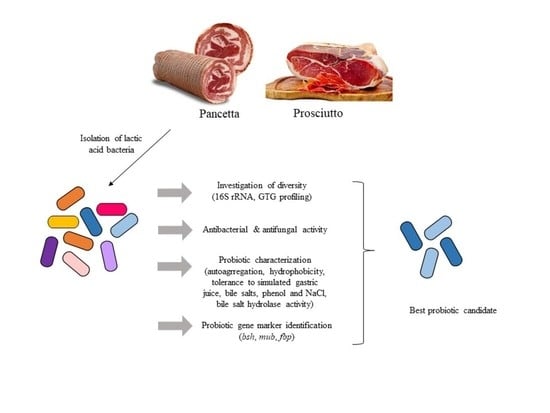
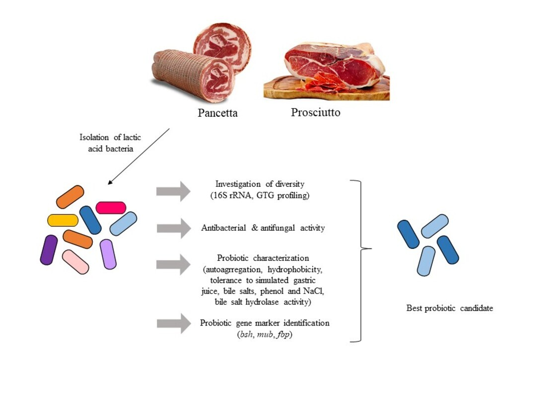
3.1. Diversity of LAB in Pancetta and Prosciutto

Fermented meats are reported to be a rich source of LAB and we therefore evaluated and compared the microbial composition of two Italian fermented meat products. This was achieved through a combination of classical microbiological and molecular approaches, permitting an evaluation of the diversity and distribution of LAB in prosciutto and pancetta. MRS was used as a selective medium to isolate potential probiotic LAB. Prior to the isolation of individual colonies, the number of viable colonies (CFU/mL) of both products was counted on MRS agar at 30 °C and 37 °C. The prosciutto sample was observed to host approximately 103 CFU/mL, while significantly higher counts were observed for pancetta at approximately 106 CFU/mL at both temperatures (Figure 1B). A total of 35 isolates from prosciutto and 71 isolates from pancetta were retrieved for 16S rRNA gene sequence analysis.
Analysis of the isolates derived from prosciutto at 30 °C suggests a dominance of L. curvatus (8/15 or 53%), in addition to L. plantarum (4/15 or 27%) and L. sakei (3/15 or 20%) (Figure 1A). At an incubation temperature of 37 °C, however, the majority of the isolated LAB population was represented by L. plantarum (15/20 or 75%) with a significantly lower percentage of P. acidilactici (3/20 or 15%) and Loigolactobacillus coryniformis subsp. torquens (2/20 or 10%). In the pancetta sample, the dominant LAB species appeared to be L. plantarum regardless of the incubation temperature (Figure 1A). There was a comparatively higher proportion of L. sakei (14/41 or 34%) compared to L. curvatus (2/41 or 5%) at 30 °C while a small subpopulation of L. coryniformis subsp. torquens (2/30 or 7%) was identified specifically at 37 °C. Overall, L. plantarum was the predominant LAB species in both meat products and prosciutto appeared to exhibit a higher diversity of organisms depending on the incubation temperature. L. coryniformis subsp. torquens and P. acidilactici were identified specifically at 37 °C, whereas L. sakei and L. curvatus were determined to be more dominant at 30 °C (Figure 1A). L. plantarum isolates were obtained at both 30 °C and 37 °C, which likely reflects this species’ versatility and adaptability to grow at different temperatures.
Long fermentation and ripening times expose LAB to prolonged stress, including acid stress, oxidation and starvation, which in turn affects their survival in specific food niches. The accumulation of lactic acid produced by LAB could exert physiological changes [41], which may cause the collapse of the proton motive forces as it acidifies the cytoplasm, resulting in cell death [42]. L. plantarum is regarded as one of the most versatile and industrially important LAB due to its useful properties, a high survivability and being functional in a range of fermented food niches [43]. L. plantarum was also observed to be among the dominant LAB species found in fermented meat products of different geographical regions, including Asia [44,45], South America [46] and Africa [47]. It is generally accepted that L. plantarum is highly tolerant against acid and alkali stress and some strains of L. plantarum were reported to be able to withstand heat, oxidative strees and starvation stress [48,49,50].
A previous study of LAB diversity in fermented meats produced in a range of European countries highlighted a dominance of L. sakei in a variety of fermented meat products [51]. The second most predominant species was P. pentosaceus (for Belgium and Germany) or L. curvatus (for Spain, France and Italy). P. acidilactici and L. coryniformis subps. torquens species have also been found in fermented meat products [52,53,54,55], although they were not commonly identified as the dominant LAB species surviving in European fermented meats. The findings in the present study concur with what has been reported in literature as Figure 1A demonstrated low percentage of L. coryniformis subps. torquens (10% in prosciutto and 5% in pancetta) and P. acidilactici (15% in prosciutto). However, it is noteworthy that the dominant LAB components identified in the present study differed from those observed in the pan-European study, suggesting a product-specific microflora.
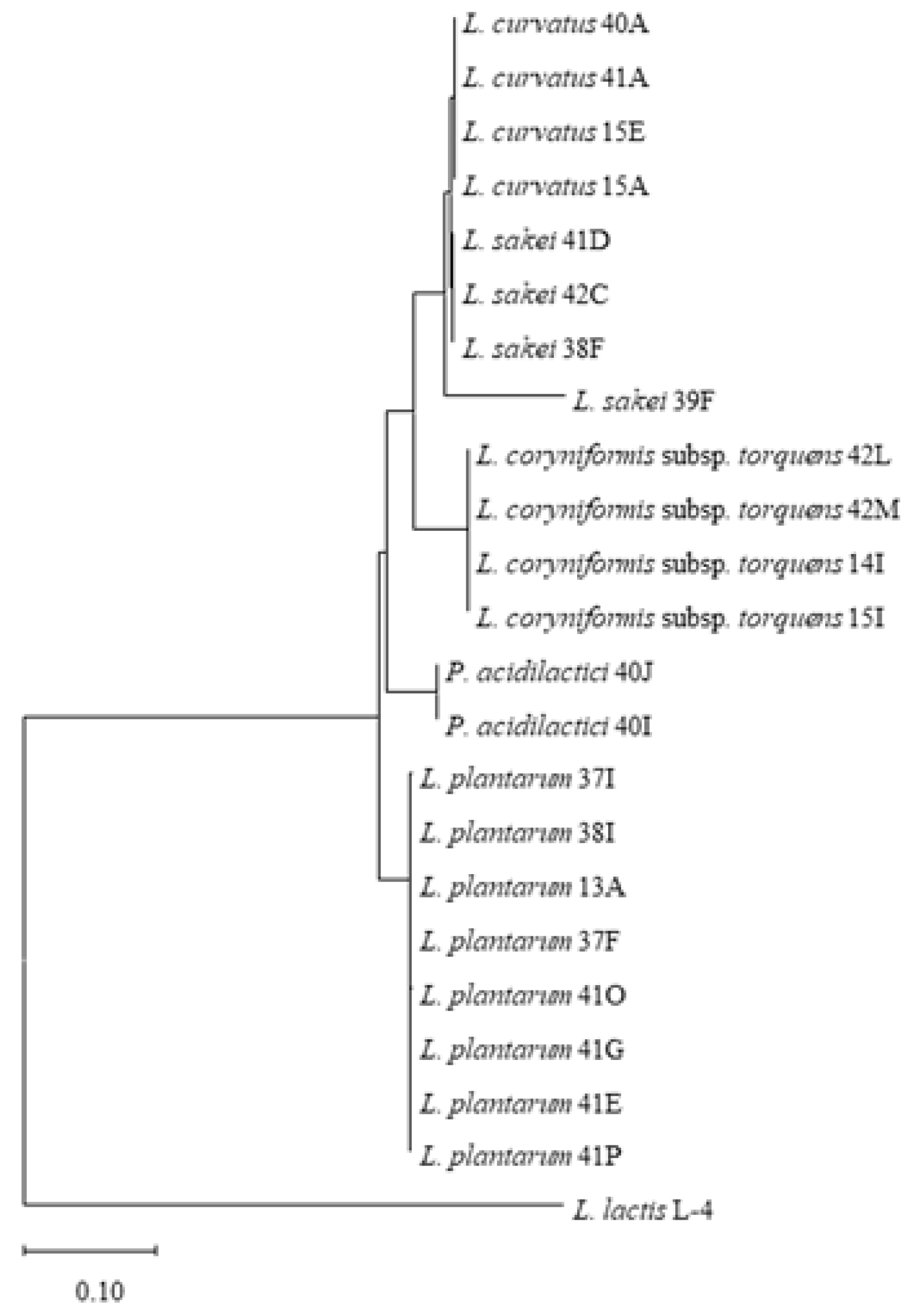
Based on the 16S rRNA gene sequence data, food source and incubation temperature (Table S2), we selected 22 representative LAB isolates and subjected these strains to genetic fingerprinting analysis (GTG)5 (Figure S1), thereby yielding species level profiles which confirmed the 16S rRNA speciation results of the selected isolates. Isolates of L. plantarum (lanes 1–8), L. coryniformis subsp. torquens (lanes 17–20) and P. acidilactici (lanes 21–22) were shown to exhibit species-specific profiles. Furthermore, (GTG)5 profiles of L. sakei strains 38F and 39F (lanes 11–12) were shown to be highly similar while that of L. sakei 41D was shown to lack a band (~1400 bp), whereas the L. sakei 42C strain produced a slightly smaller band of less than 1400 bp, suggesting that these may be distinct isolates. While the genetic fingerprinting profiles of L. curvatus 40A, 41A and 15E (lane 13, 14 and 16) were shown to be highly similar, L. curvatus 15A exhibits an additional distinct amplicon (~5000 bp). The GTG species level profiling was consistent with phylogenetic analysis of the 16S rRNA gene sequences of these strains with four distinct clusters representing the five LAB species (L. sakei and L. curvatus cluster together in the phylogenetic tree) identified in this study (Figure 2 and Figure S1).

3.2. Organic Acid Production and Antagonistic Activity

LAB may possess a competitive advantage to survive through the production of antimicrobial compounds including organic acids. LAB may produce more than one type of organic acid (although primarily lactic acid) and this may vary by species and even strain. In order to evaluate the organic acid production profiles of 22 representative isolates, the cell-free supernatant (CFS) of fresh overnight cultures of the selected strains were analyzed by HPLC. Representative isolates of L. plantarum, L. sakei, L. curvatus, L. coryniformis subsp. torquens and P. acidilactici were analyzed in this manner. Perhaps unsurprisingly, the isolates were observed to primarily produce lactic and acetic acids (Figure 3, Table S3). Among the five different species of LAB, strains of L. plantarum produced the highest concentration of lactic acid compared to other species, with L. plantarum 41O and 41P producing significantly (p < 0.001) higher lactate of 157.09 ± 0.29 mmol/L and 158.02 ± 0.22 mmol/L, respectively, compared to L. plantarum 41G, 41E, 13A, 37F, 38I and 37I (106.67 to 138.91 mmol/L). L. curvatus (71.14 to 78.07 mmol/L) and L. sakei (68.75 to 81.90 mmol/L) were significantly weaker lactic acid producers compared to P. acidilactici (~88 mM). In general, L. plantarum species also formed significantly (p < 0.001) higher acetate compared to other species, with L. plantarum 41O, 41P, 37I and 38I (ranging from 45.16 to 46.77 mmol/L) being the strongest than compared to L. plantarum 41G, 41E, 13A and 37F (ranging from 40.94 to 43.82 mmol/L). The concentration of acetate produced by L. sakei did not significantly (p > 0.05) differ compared to L. coryniformis subsp. torquens, P. acidilactici and L. curvatus. Furthermore, propionate was not produced by the tested isolates except for L. plantarum at a range of 2.81 to 4.90 mmol/L.
The inhibitory activity of the selected 22 LAB isolates was assessed using the spot-on-lawn antimicrobial assay against several indicator organisms associated with food spoilage and/or food poisoning. The inhibitory activity of LAB may be due to the production of organic acids and/or bacteriocins and thus it was aimed to discern the source of antimicrobial activity where relevant. The nature of inhibitory activity was classified into two types (shown in Figure S2): If a hazy undefined zone was observed, then the inhibitory effect was predicted to be primarily due to organic acid production, whereas if a clear defined zone was observed it was presumed to be due to bacteriocins [56]. In summary, all L. plantarum and L. sakei isolates exhibited inhibitory activity against all indicators tested in the spot-on-lawn assays (L. cremoris HP; L. paramesenteroides NCDO869; L. mesenteroides NCDO2028; K. aerogenes NCIMB10102; E. faecium NCIMB11508; P. aeruginosa PA01; L. innocua UCC3 and S. aureus NCDO949) (Table 1). L. curvatus 40A and 41A exhibited weak inhibitory activity against L. cremoris HP and L. curvatus 15A and 15E presented a weak antagonistic effect against S. aureus NCDO944 alone among the tested indicator strains. L. coryniformis subsp. torquens 42L and 42M demonstrated weak to moderate inhibitory activity against all indicators, whereas 14I and 15I showed similar activity against all indicators except for L. cremoris HP and E. aerogenes NCIMB10172 (Table 1). L. innocua UCC3, E. faecium NCIMB11508 and S. aureus NCDO944 were strongly inhibited by P. acidilactici 40J and 40I (Table 1).
The effectiveness of organic acids as a natural preservative in contributing to food protection and safety against a range of spoilage and pathogenic microorganisms has been well studied [57,58,59]. Bacterial genera associated with food spoilage, such as Lactococcus, Leuconostoc, Klebsiella and Pseudomonas [60,61], and pathogenic bacteria associated with food poisoning, such as Listeria and Staphylococcus [62,63], were used as indicators in this study (Table 1). Among the 22 strains tested, 14 isolates of L. plantarum, L. sakei and P. acidilactici inhibited all indicators tested, whereas L. curvatus and L. coryniformis subsp. torquens showed antagonistic ability at least against one indicator tested. Additionally, the antifungal potential of the isolates was evaluated. All isolates of L. plantarum, P. acidilactici and L. coryniformis subsp. torquens inhibited P. expansum DSM1282 (Table 1), but none of the isolates were active against P. digitatum DSM2731. P. expansum is recognized as one of the most dominant post-harvest pathogens in fruits and vegetables mainly in pomaceous fruits [64]. While the compound(s) that exert the anti-fungal activity are not defined within the scope of this study, previous studies have reported the anti-fungal activity of phenyllactic acid produced by L. plantarum [65] and a bacteriocin-like compound produced by P. pentosaceous [66].
Findings from this study indicate that the presence of LAB in prosciutto and pancetta play a crucial role in controlling spoilage and/or pathogenic microorganisms through the production of organic acids and, in some cases, bacteriocins. However, it is important to note that organic acids and bacteriocins may not be the sole contributing factors, as LAB may also produce a variety of antimicrobial compounds including hydrogen peroxide, carboxylic acid, diacetyl and reuterin [67].

3.3. Bacteriocin Activity and Associated Gene Clusters

In order to further validate that the inhibitory effect exerted by LAB isolates were not due to the reduced pH, well diffusion assays using pH neutralized and catalase treated (to remove H2O2 effect) CFS from selected representative isolates—L. plantarum 13A, 41G, 38I, 41P and P. acidilactici 40J (Table 2)—were performed. These representative isolates were selected based on the product origin and the incubation temperature they were isolated from, as shown in Table S2. These assays confirmed that L. plantarum 13A, 41G, 38I and 41P produced bacteriocins that were effective against L. cremoris HP, MG1363, NZ9000 and L. paramesenteroides NCDO869, while that produced by P. acidilactici 40J was effective against L. mesenteroides NCDO2028, E. faecium NCIMB11508 and L. innocua strains UCC3, DPC3565, DPC3566 and DPC3567 (Table 2). Growth of K. aerogenes NCIMB10102, P. aeruginosa PA01 and S. aureus NCDO949 was not inhibited by the three tested isolates, which suggests that the inhibitory activity against these indicators was due to organic acids (Table 1). In addition, the compounds produced by L. plantarum 13A, 41G, 38I, 41P and P. acidilactici 40J were sensitive to proteinase K, which further substantiated their proteinaceous nature.
In order to identify bacteriocin-encoding gene clusters responsible for the observed antimicrobial activity, the genomes of five isolates were sequenced and assembled. Potential bacteriocin gene clusters in these five isolates were detected based on in silico analysis of retrieved contigs with BAGEL4 and antiSMASH. Based on this analysis, it is proposed that P. acidilactici 40J produces a pediocin-like bacteriocin with genes encoding the putative structural peptide (PedA); the immunity protein PedB; the transport proteins, PedC and PedD with 99 to 100% identity to those that encode pediocin PA1 produced by P. acidilactici K10 (Figure S3). Additionally, the organization of the gene clusters in P. acidilactici 40J and K10 are identical. The bacteriocin produced by this strain (40J), which is likely pediocin, was effective against Listeria, Enterococcus and Leuconostoc (Table 2) and in agreement with the literature pertaining to pediocin PA1 [68].
Different strains of L. plantarum have been reported to produce bacteriocins with varied structural properties and antagonistic activities. For instance, L. plantarum B21 was previously demonstrated to be effective against a broad range of LAB and a few non-LAB species (Listeria monocytogenes and Clostridium perfringens) [69], whereas L. plantarum LPL-1 was effective against a broad range of non-LAB species (L. monocytogenes, S. aureus, E. faecalis, B. amyloliquefaciens and B. pumilus) and several strains of Lactococcus and Lactobacillus [70]. L. plantarum NI326 produces the cyclic bacteriocin plantaricyclin A, which shared 99 to 100% similarity to the bacteriocin encoding cluster of L. plantarum 13A, 38I and 41G isolated in the present study (Figure S3), that is effective against some strains of Lactococcus, L. bulgaricus and Alicyclobacillus acidoterrestris but is not effective against L. plantarum, L. brevis, Pediococcus, Listeria, Escherichia, Staphylococcus, Streptococcus, Klebsiella and Bacillus [71]. Based on comparative analysis of the identified bacteriocin encoding gene clusters, it is likely that 41P produces novel bacteriocin(s), which is potentially a derivative of plantaricyclin A since the only identifiable bacteriocin-encoding region in the strain’s genome (and which is plasmid-encoded) possessed only 42% similarity to plantaricyclin A. However, further investigations through purification and identification using mass spectrometry are required to confirm its potential novelty.

3.4. Antibiotic Susceptibility and Screening for Probiotic Candidates

Horizontal transfer of antibiotic resistance genes, particularly those within mobile genetic elements (transposons and plasmids) deserve particular attention due to the risk of resistance transfer to pathogenic organisms [72]. The determination and comparison of antibiotic susceptibility patterns in a number of representative strains of each species are major steps in differentiating between intrinsic and acquired antibiotic resistance in probiotic bacteria [72]. Antibiotic resistance tests were first performed as a preliminary screen for potential LAB strains amongst the 22 isolates (Table 3). It is generally known that Lactobacillus sp. are intrinsically resistant to gentamicin, streptomycin, nalidixic acid and vancomycin [73,74]. L. curvatus 40A displayed widespread antibiotic-resistance amongst the L. curvatus strains tested, while 42C was the most sensitive amongst the L. sakei isolates. L. plantarum isolates 37F and 41G were the most antibiotic-sensitive; 37I, 38I and 41P were moderately sensitive, while 13A, 41O and 41E were resistant to several antibiotics. Nonetheless, 37F, 41G, 38I, 41P and 13A, which exhibited distinct sensitivity/resistance profiles, were selected for further probiotic tests since all L. plantarum isolates were bacteriocin producers (Table 1). Additionally, P. acidilactici 40J, L. coryniformis subsp. torquens 42L, 42M and 14I were selected for further analysis.
In this study, hexane was used to evaluate hydrophobicity of LAB cell surface. This method is considered important in determining probiotic bacterial adhesion capacity to epithelial host cells [5,75]. L. coryniformis subsp. torquens and L. plantarum displayed significantly (p < 0.05) greater % hydrophobicity (ranging from 31.2 to 42.2%) compared to L. curvatus, L. sakei and P. acidilactici (ranging from 4.9 to 18.1%) (Table 4). Isolates 42L, 14I, 41P, 41G and 38I did not differ significantly (p > 0.05) and showed highest overall % hydrophobicity. According to the classification proposed by Colloca et al. [76], bacterial hydrophobicity can be considered low (0 to 34%), moderate (35 to 69%) or high (70 to 100%). Altogether, the hydrophobicity values for meat isolates could be considered moderate when compared to those previously reported.
The auto-aggregation capacity of bacteria has also been associated with higher adhesion to epithelial cells and persistence in the intestine [77]. All isolates tested in this study were shown to exhibit auto-aggregation during the first 3 h (from 9.7 to 15.6%), which increased with time after 24 h (from 28.9 to 69.0%) (Table 4). In particular, L. plantarum 41P, 38I, 37F and 41G did not significantly (p > 0.05) differ and showed the highest auto-aggregation levels (63.6 ± 2.2 to 69.0 ± 4.2%, t = 24 h) compared to all other isolates. Autoaggregation % of L. plantarum strains reported in this study was similar to several lactobacilli and L. plantarum strains reported by other studies [77,78,79]. Thus, autoaggregation levels of meat isolates in this study are generally similar or slightly higher to the probiotic candidates reported in the literature.
The abilities to tolerate gastric juice (stomach phase) and bile salt (intestinal phase) are important properties of probiotics to ensure their viability and survival in the human gastrointestinal tract [80]. It was revealed that L. curvatus and L. sakei do not survive simulated gastric juice conditions after 1 h incubation and L. coryniformis subsp. torquens is unable to withstand such conditions following 2 h exposure (Table 5). However, they are able to survive 0.3% bile salt exposure for a period of 3 h with approximately 5-fold to 9-fold viable count reduction (Table 6). In contrast, L. plantarum is able to withstand simulated gastric juice to some degree with 10-fold to 15-fold reduction after 1 h, but with declined survival (ranging from 104 to 102 CFU/mL) after 2 h and 3 h (Table 5). These isolates appear to be more robust than previous reports on this species [78]. Similarly, L. plantarum strains isolated in this study were highly tolerant to bile salt stress, remainig viable at 108 CFU/mL (5 to 6-fold reduction) after 3 h (Table 6). Furthermore, P. acidilactici remained viable at 105 to 104 CFU/mL after a challenge with simulated gastric juice for 1 h to 3 h (Table 5) and an 8-fold reduction in survival was observed after a challenge with bile salt for 3 h (Table 6). It was reported that the median of gastric emptying half time in healthy adults is approximately 1 h [81], but there are considerable variabilities among individuals [82]. Therefore, L. coryniformis subsp. torquens could still be considered as a probiotic candidate. In summation, all isolates display high tolerance to 0.3% bile salt (displayed by less than 10-fold reduction in survival after 3 h), but only L. plantarum and P. acidilactici strains are able to survive more than 2 h exposure to simulated gastric juice.
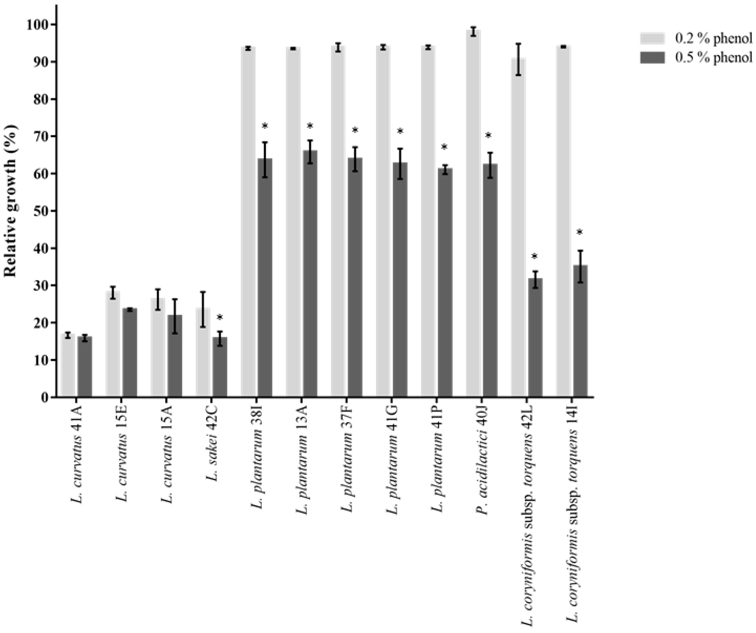
Probiotics also need to survive toxic metabolites, particularly phenols produced during the digestion process. Amino acids derived from the diet or endogenous proteins can be deaminated by bacteria in the gastrointestinal tract which results in the formation of phenols [26]. All strains of L. plantarum, P. acidilactici and L. coryniformis subsp. torquens generally exhibited almost unimpaired growth in the presence of 0.2% phenol (>90% growth compared to untreated control) (Figure 4). Upon exposure to 0.5% phenol, their growth was significantly (p < 0.05) decreased from 61 to 65% (for 38I, 13A, 37F, 41G and 41P), 62% (for 40J), 31 and 35% (for 42L and 14I, respectively). These values were arguably higher compared to what was reported in other studies [27,78]. L. curvatus and L. sakei were nonetheless shown to exhibit low survival to 0.2% phenol (from 16 to 28%) and 0.5% phenol (from 15 to 23%) stress (Figure 4). It is important to note that phenol content in foods (ranging from flour, oil, raw beans, cocoa, fruits and wine) greatly varies from 0.0002 to 3.6% (w/w) [83]. Furthermore, its bioavailability in the human gut greatly varies depending on several factors, such as the individual’s diet, the matrix of polyphenol-rich foods which may affect intestinal absorption and, likewise, the interaction of phenols with nutrients taken from the same meal could result in alteration in their absorption [83]. For these reasons, 0.2% and 0.5% phenol concentrations (selected as the average representative phenol content present in foods) were tested against a panel of probiotic candidates.
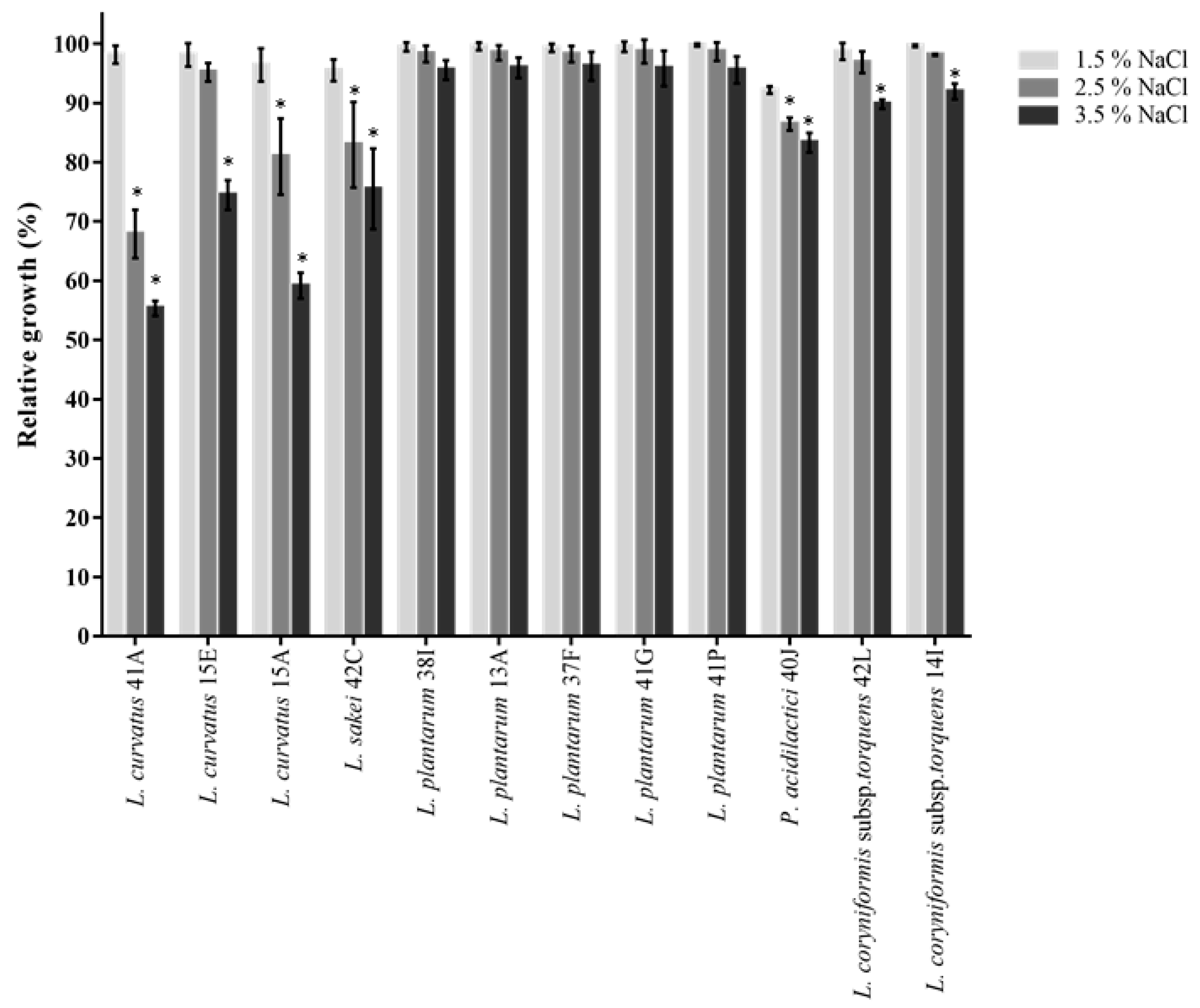
During industrial processing, probiotic LAB may encounter osmotic stress due to changes in solute concentration in the environment, which could result in cellular hydration and dehydration and this may negatively affect the survival rate and influence metabolic activities of probiotics [84]. Probiotic strains could be added to fermented foods containing varying concentrations of salt. Thus, tolerance to NaCl may offer a competitive advantage for probiotics. For example, spontaneous sauerkraut fermentations operate optimally with a concentration of 2.5% (w/w) NaCl [85], while 2 to 3% salt in meat fermentation was demonstrated to improve meat texture [86]. All tested isolates were resistant to 1.5% NaCl, with L. plantarum being the most resistant to higher NaCl concentrations (Figure 5). Relatively high tolerance to 2.5% and 3.5% NaCl exposure was also observed for all isolates of L. coryniformis subsp. torquens (89 to 98%), P. acidilactici 40J (83 to 86%) and L. curvatus 15E (74 to 95%). Overall, L. curvatus 41A, 15A and L. sakei 42C demonstrated moderate tolerance to 2.5% (67 to 83% growth) and 3.5% (55 to 75% growth).
Current literature asserts that BSH activity offers a selective advantage for probiotic strains to survive and perform in the intestinal milieu [87]. However, it is debatable whether BSH activity provides desirable effects for the human host, as there is concern over the safety of administering BSH-positive probiotics. However, research has shown that in the case of bifidobacteria and lactobacilli, BSH-positive probiotics may not be able to dehydroxylate deconjugated bile salts and thus the majority of products resulting from BSH activity may be precipitated and excreted in feces [87]. BSH activity was assessed on selected probiotic candidates in the present study by growing them on MRS agar containing 0.5% TDCA and it was found that strains 38I, 41G, 42L and 14I produced BSH and bsh gene homologues were identified in the genome of these strains (Table S4). However, a bsh gene homologue was also identified in the genome of P. acidilactici 40J, which indicates that there could be a low level of BSH expression under the circumstances the bacteria were grown in or that the plate assay containing TDCA might not be the most appropriate method to test BSH activity in this strain. Other studies suggested the use of thin layer chromatography (TLC) assay as a more sensitive qualitative method to identify BSH-positive LAB [88]. The presence of other probiotic gene markers such as mub (mucin binding protein) and fbp (fibronectin binding protein), which are known to play a role in promoting probiotic adherence to the host gastrointestinal tract, was also investigated [89]. Strains 38I, 41G, 40J, 42L and 14I were found to possess homologues of the mub and fbp genes (Table S4).

4. Conclusions

The probiotics market is expected to grow at a rate of 5.6% annually from 2020 to 2027, reaching an estimated economic value of USD 75.9 billion by 2027 [90]. This growth is attributed to increasing public awareness regarding the health benefits of probiotics that are known to be strain-specific [91]. These probiotics could aid in the prevention of antibiotic-associated diarrhea, Clostridium difficile infection, nosocomial infection and travelers’ diarrhea and could also help in the treatment of inflammatory bowel syndrome, pediatric acute diarrhea and Helicobacter pylori infection [91]. With the increasing trend of public interest towards probiotics, it becomes crucial to characterize and discover novel probiotic strains. In this study, probiotic candidates sourced from fermented meats were assessed and identified, with L. plantarum 41G being the strongest candidate, considering all factors assessed in this study. Overall, this strain was the most susceptible to antibiotics (compared to other isolates belonging to the same species), with moderate hydrophobicity and auto-aggregation value, high tolerance to bile salt, phenol and NaCl stress, while it was also able to survive simulated gastric juice conditions for 2 to 3 h incubation. Additionally, but to a lesser extent, P. acidilactici 40J and L. coryniformis subsp. torquens 4L may be considered good probiotic candidates since the former showed inhibitory activity due to bacteriocin production, which was mainly targeted against Listeria, whereas the latter demonstrated moderate hydrophobicity and auto-aggregation ability similar to L. plantarum strains; both candidates exhibited high survivability to bile salt and NaCl stress and phenol tolerance. Nevertheless, further experiments utilizing microencapsulation are needed to explore their application potential. Finally, L. plantarum 41P was identified to produce a novel bacteriocin(s), while L. plantarum, P. acidilactici and L. coryniformis subsp. torquens isolates demonstrated antifungal capacity. The identification of a potentially novel antimicrobial(s)-producing strain combined with the broad anti-fungal and anti-bacterial activity of strains isolated in this study highlights the untapped potential of artisanal meat products as a source of novel probiotic strains.

Supplementary Materials

The following are available online at https://www.mdpi.com/article/10.3390/foods10071519/s1, Table S1: Standards for interpreting the inhibition zone diameters for antibiotics used in this study. Results were recorded as: R (resistant), MS (moderately susceptible) or S (susceptible), Table S2: The source of origin along with the incubation condition where the 22 LAB isolated from, Table S3: Average mean concentration of organic acid (mmol/L) and standard deviation (SD) of organic acids produced by LAB, obtained from three independent experiments, Table S4: Amino acid sequences that encode bsh, mub and fbp genes in probiotic candidate strains, Figure S1: (GTG)5-PCR DNA fingerprints of LAB isolates obtained from prosciutto and pancetta. Lane 1–8: L. plantarum strains 41G; 41E; 41O; 41P; 13A 37F; 38I; and 37I. Lane 9–12: L. sakei strains 41D; 42C; 38F; and 39F. Lane 13–16: L. curvatus strains 40A; 41A; 15A; and 15E. Lane 17–20: L. coryniformis subsp. torquens strains 42L; 42M; 14I; and 15I. Lane 21–22: P. acidilactici 40J and 40I. The first lane was GeneRuler DNA Ladder Mix (Thermo Fisher), Figure S2: Representative spot-on-lawn agar plate of LAB isolates against L. innocua UCC3 indicator. The zone of inhibition formed by P. acidilactici 40 J and 40I were defined, whereas the zone of inhibition produced by L. plantarum 41P, 41O, 38I and 37I and L. coryniformis subsp. torquens 42M, 42L, 14I and 15I were hazy or less defined, Figure S3: Schematic representation of the gene clusters involved in the production of previously studied strains of LAB (L. plantarum NI326 and P. acidilactici K10) with bacteriocin producing strains isolated in this study (L. plantarum 41G, 38I and 13A and P. acidilactici 40J).

Author Contributions

Conceptualization, J.M.; methodology, E.P. and G.A.L.; software, G.A.L.; validation, E.P. and G.A.L.; formal analysis, E.P., J.M. and G.A.L.; investigation, E.P.; resources, J.M., D.v.S. and M.V.; data curation, E.P.; writing—original draft preparation, E.P.; writing—review and editing, J.M. and D.v.S.; supervision, J.M., D.v.S. and M.V.; project administration, J.M. and D.v.S.; funding acquisition, J.M. and D.v.S. All authors have read and agreed to the published version of the manuscript.

Funding

This publication has emanated from research conducted with the financial support of/supported in part by a grant from the Science Foundation Ireland under Starting Investigator Research Grant (SIRG) (Ref. No. 15/SIRG/3430) awarded to JM and the Principal Investigator award (Ref. No. 13/IA/1953) awarded to DvS. For the purpose of Open Access, the author has applied a CC BY public copyright license to any Author Accepted Manuscript version arising from this submission.

Institutional Review Board Statement

Not applicable.

Informed Consent Statement

Not applicable.

Data Availability Statement

Not applicable.

Acknowledgments

The authors thank Dan Walsh (University College Cork) for his assistance in running the HPLC; Philip Kelleher (University College Cork) for his help in genome sequence deposition; Ian O’Neill (University College Cork) for his assistance in PacBio genome assembly; and John O’Callaghan (University College Cork) for providing indicator strains used in this study.

Conflicts of Interest

The authors declare no conflict of interest.

References

  1. Binda, S.; Hill, C.; Johansen, E.; Obis, D.; Pot, B.; Sanders, M.E.; Tremblay, A.; Ouwehand, A.C. Criteria to qualify microorganisms as “probiotic” in foods and dietary supplements. Front. Microbiol. 2020, 11, 1662. [Google Scholar] [CrossRef]
  2. Hill, C.; Guarner, F.; Reid, G.; Gibson, G.R.; Merenstein, D.J.; Pot, B.; Morelli, L.; Canani, R.B.; Flint, H.J.; Salminen, S.; et al. The International Scientific Association for Probiotics and Prebiotics consensus statement on the scope and appropriate use of the term probiotic. Nat. Rev. Gastroenterol. Hepatol. 2014, 11, 506–514. [Google Scholar] [CrossRef] [Green Version]
  3. Ricci, A.; Allende, A.; Bolton, D.; Chemaly, M.; Davies, R.; Girones, R.; Herman, L.; Koutsoumanis, K.; Lindqvist, R.; Nørrung, B.; et al. Scientific opinion on the update of the list of QPS-recommended biological agents intentionally added to food or feed as notified to EFSA. EFSA J. 2017, 15, 3. [Google Scholar] [CrossRef] [Green Version]
  4. Pessoa, W.F.B.; Melgaço, A.C.C.; de Almeida, M.E.; Ramos, L.P.; Rezende, R.P.; Romano, C.C. In vitro activity of Lactobacilli with probiotic potential isolated from cocoa fermentation against Gardnerella vaginalis. BioMed Res. Int. 2017, 2017, 1–10. [Google Scholar] [CrossRef] [PubMed] [Green Version]
  5. Falah, F.; Vasiee, A.; Behbahani, B.A.; Yazdi, F.T.; Moradi, S.; Mortazavi, S.A.; Roshanak, S. Evaluation of adherence and anti-infective properties of probiotic Lactobacillus fermentum strain 4-17 against Escherichia coli causing urinary tract infection in humans. Microb. Pathog. 2019, 131, 246–253. [Google Scholar] [CrossRef]
  6. Choi, A.-R.; Patra, J.K.; Kim, W.J.; Kang, S.-S. Antagonistic activities and probiotic potential of lactic acid bacteria derived from a plant-based fermented food. Front. Microbiol. 2018, 9, 1963. [Google Scholar] [CrossRef]
  7. Bin Masalam, M.S.; Bahieldin, A.; Alharbi, M.G.; Al-Masaudi, S.; Al-Jaouni, S.K.; Harakeh, S.M.; Al-Hindi, R.R. Isolation, molecular characterization and probiotic potential of lactic acid bacteria in saudi raw and fermented milk. Evid. Based Complement. Altern. Med. 2018, 2018, 1–12. [Google Scholar] [CrossRef] [Green Version]
  8. Munekata, P.E.S.; Pateiro, M.; Zhang, W.; Domínguez, R.; Xing, L.; Fierro, E.M.; Lorenzo, J.M. Autochthonous probiotics in meat products: Selection, identification, and their use as starter culture. Microorganisms 2020, 8, 1833. [Google Scholar] [CrossRef]
  9. Jampaphaeng, K.; Cocolin, L.; Maneerat, S. Selection and evaluation of functional characteristics of autochthonous lactic acid bacteria isolated from traditional fermented stinky bean (Sataw-Dong). Ann. Microbiol. 2016, 67, 25–36. [Google Scholar] [CrossRef]
  10. Rezac, S.; Kok, C.R.; Heermann, M.; Hutkins, R. Fermented foods as a dietary source of live organisms. Front. Microbiol. 2018, 9, 1785. [Google Scholar] [CrossRef]
  11. Urso, R.; Comi, G.; Cocolin, L. Ecology of lactic acid bacteria in Italian fermented sausages: Isolation, identification and molecular characterization. Syst. Appl. Microbiol. 2006, 29, 671–680. [Google Scholar] [CrossRef] [PubMed]
  12. Van Ba, H.; Seo, H.-W.; Seong, P.-N.; Kang, S.-M.; Kim, Y.-S.; Cho, S.-H.; Park, B.-Y.; Ham, J.-S.; Kim, J.-H. Lactobacillus plantarum (KACC 92189) as a potential probiotic starter culture for quality improvement of fermented sausages. Food Sci. Anim. Resour. 2018, 38, 189–202. [Google Scholar] [CrossRef]
  13. Połka, J.; Rebecchi, A.; Pisacane, V.; Morelli, L.; Puglisi, E. Bacterial diversity in typical Italian salami at different ripening stages as revealed by high-throughput sequencing of 16S rRNA amplicons. Food Microbiol. 2015, 46, 342–356. [Google Scholar] [CrossRef]
  14. Geeraerts, W.; De Vuyst, L.; Leroy, F. Mapping the dominant microbial species diversity at expiration date of raw meat and processed meats from equine origin, an underexplored meat ecosystem, in the Belgian retail. Int. J. Food Microbiol. 2019, 289, 189–199. [Google Scholar] [CrossRef] [PubMed]
  15. Zheng, J.; Wittouck, S.; Salvetti, E.; Franz, C.M.A.P.; Harris, H.M.B.; Mattarelli, P.; O’Toole, P.W.; Pot, B.; Vandamme, P.; Walter, J.; et al. A taxonomic note on the genus Lactobacillus: Description of 23 novel genera, emended description of the genus Lactobacillus Beijerinck 1901, and union of Lactobacillaceae and Leuconostocaceae. Int. J. Syst. Evol. Microbiol. 2020, 70, 2782–2858. [Google Scholar] [CrossRef]
  16. Federici, S.; Ciarrocchi, F.; Campana, R.; Ciandrini, E.; Blasi, G.; Baffone, W. Identification and functional traits of lactic acid bacteria isolated from Ciauscolo salami produced in central Italy. Meat Sci. 2014, 98, 575–584. [Google Scholar] [CrossRef]
  17. Kuley, E.; Özyurt, G.; Özogul, I.; Boga, M.; Akyol, I.; Rocha, J.M.; Özogul, F. The role of selected lactic acid bacteria on organic acid accumulation during wet and spray-dried fish-based silages. Contributions to the winning combination of microbial food safety and environmental sustainability. Microorganisms 2020, 8, 172. [Google Scholar] [CrossRef] [PubMed] [Green Version]
  18. Tarman, A.A.; Ramli, N.N.; Ridla, M.; Yaman, M.A.; Setiyono, A.A. Effects of organic acids on salmonella enteritidis growth inhibition and ileum surface area in laying ducks fed anaerobically fermented feed. Int. J. Poult. Sci. 2017, 16, 98–104. [Google Scholar] [CrossRef]
  19. Kroöckel, L. The role of lactic acid bacteria in safety and flavour development of meat and meat products. In Lactic Acid Bacteria—R&D for Food, Health and Livestock Purposes; Kongo, M., Ed.; IntechOpen: London, UK, 2013. [Google Scholar]
  20. Corsetti, A.; Settanni, L.; Van Sinderen, D. Characterization of bacteriocin-like inhibitory substances (BLIS) from sourdough lactic acid bacteria and evaluation of their in vitro and in situ activity. J. Appl. Microbiol. 2004, 96, 521–534. [Google Scholar] [CrossRef] [Green Version]
  21. Kumar, S.; Stecher, G.; Li, M.; Knyaz, C.; Tamura, K. MEGA X: Molecular evolutionary genetics analysis across computing platforms. Mol. Biol. Evol. 2018, 35, 1547–1549. [Google Scholar] [CrossRef]
  22. Vec, P.Å.; Vancanneyt, M.; Seman, M.; Snauwaert, C.; Lefebvre, K.; Sedláček, I.; Swings, J. Evaluation of (GTG)5-PCR for identification of Enterococcus spp. FEMS Microbiol. Lett. 2005, 247, 59–63. [Google Scholar] [CrossRef] [Green Version]
  23. Anisimova, E.A.; Yarullina, D.R. Antibiotic resistance of Lactobacillus strains. Curr. Microbiol. 2019, 76, 1407–1416. [Google Scholar] [CrossRef]
  24. Crowley, S.; Mahony, J.; Van Sinderen, D. Broad-spectrum antifungal-producing lactic acid bacteria and their application in fruit models. Folia Microbiol. 2012, 58, 291–299. [Google Scholar] [CrossRef] [PubMed]
  25. Tagg, J.R.; McGiven, A.R. Assay system for bacteriocins. Appl. Microbiol. 1971, 21, 943. [Google Scholar] [CrossRef]
  26. Sathyabama, S.; Vijayabharathi, R.; Devi, P.B.; Kumar, M.R.; Priyadarisini, V.B. Screening for probiotic properties of strains isolated from feces of various human groups. J. Microbiol. 2012, 50, 603–612. [Google Scholar] [CrossRef]
  27. Shehata, M.; El Sohaimy, S.; El-Sahn, M.A.; Youssef, M. Screening of isolated potential probiotic lactic acid bacteria for cholesterol lowering property and bile salt hydrolase activity. Ann. Agric. Sci. 2016, 61, 65–75. [Google Scholar] [CrossRef] [Green Version]
  28. Mafra, J.F.; Cruz, A.I.C.; Santana, T.S.D.; Ferreira, M.A.; Araujo, F.M.; Evangelista-Barreto, N.S. Probiotic characterization of a commercial starter culture used in the fermentation of sausages. Food Sci. Technol. 2021, 41, 240–246. [Google Scholar] [CrossRef]
  29. Gilliland, S.; Staley, T.; Bush, L. Importance of bile tolerance of Lactobacillus acidophilus used as a dietary adjunct. J. Dairy Sci. 1984, 67, 3045–3051. [Google Scholar] [CrossRef]
  30. Melo, T.A.; Dos Santos, T.F.; Pereira, L.R.; Passos, H.M.; Rezende, R.P.; Romano, C.C. Functional profile evaluation of Lactobacillus fermentum TCUESC01: A new potential probiotic strain isolated during cocoa fermentation. BioMed Res. Int. 2017, 2017, 1–7. [Google Scholar] [CrossRef] [Green Version]
  31. Vinderola, C.; Medici, M.; Perdigon, G. Relationship between interaction sites in the gut, hydrophobicity, mucosal immunomodulating capacities and cell wall protein profiles in indigenous and exogenous bacteria. J. Appl. Microbiol. 2004, 96, 230–243. [Google Scholar] [CrossRef]
  32. Lugli, G.A.; Milani, C.; Mancabelli, L.; Van Sinderen, D.; Ventura, M. MEGAnnotator: A user-friendly pipeline for microbial genomes assembly and annotation. FEMS Microbiol. Lett. 2016, 363. [Google Scholar] [CrossRef] [PubMed] [Green Version]
  33. Chevreux, B.; Wetter, T.; Suhai, S. Genome sequence assembly using trace signals and additional sequence information. In Proceedings of the German Conference on Bioinformatics (GCB), Hannover, Germany, 4–6 October 1999; pp. 45–56. [Google Scholar]
  34. Hyatt, D.; Chen, G.-L.; Locascio, P.F.; Land, M.L.; Larimer, F.W.; Hauser, L.J. Prodigal: Prokaryotic gene recognition and translation initiation site identification. BMC Bioinform. 2010, 11, 119. [Google Scholar] [CrossRef] [Green Version]
  35. Wick, R.R.; Judd, L.M.; Gorrie, C.L.; Holt, K.E. Unicycler: Resolving bacterial genome assemblies from short and long sequencing reads. PLoS Comput. Biol. 2017, 13, e1005595. [Google Scholar] [CrossRef] [PubMed] [Green Version]
  36. Punta, M.; Coggill, P.C.; Eberhardt, R.; Mistry, J.; Tate, J.; Boursnell, C.; Pang, N.; Forslund, K.; Ceric, G.; Clements, J.; et al. Pfam: The protein families database. Nucleic Acids Res. 2011, 40, D290–D301. [Google Scholar] [CrossRef]
  37. Tatusov, R.L.; Fedorova, N.D.; Jackson, J.D.; Jacobs, A.R.; Kiryutin, B.; Koonin, E.V.; Krylov, D.M.; Mazumder, R.; Mekhedov, S.L.; Nikolskaya, A.N.; et al. The COG database: An updated version includes eukaryotes. BMC Bioinform. 2003, 4, 41. [Google Scholar] [CrossRef] [PubMed] [Green Version]
  38. Van Heel, A.J.; De Jong, A.; Song, C.; Viel, J.; Kok, J.; Kuipers, O.P. BAGEL4: A user-friendly web server to thoroughly mine RiPPs and bacteriocins. Nucleic Acids Res. 2018, 46, W278–W281. [Google Scholar] [CrossRef]
  39. Blin, K.; Shaw, S.; Steinke, K.; Villebro, R.; Ziemert, N.; Lee, S.Y.; Medema, M.H.; Weber, T. AntiSMASH 5.0: Updates to the secondary metabolite genome mining pipeline. Nucleic Acids Res. 2019, 47, W81–W87. [Google Scholar] [CrossRef] [Green Version]
  40. Dashkevicz, M.P.; Feighner, S.D. Development of a differential medium for bile salt hydrolase-active Lactobacillus spp. Appl. Environ. Microbiol. 1989, 55, 11–16. [Google Scholar] [CrossRef] [Green Version]
  41. Even, S.; Lindley, N.D.; Loubière, P.; Cocaign-Bousquet, M. Dynamic response of catabolic pathways to autoacidification in Lactococcus lactis: Transcript profiling and stability in relation to metabolic and energetic constraints. Mol. Microbiol. 2002, 45, 1143–1152. [Google Scholar] [CrossRef]
  42. Papadimitriou, K.; Alegría, Á.; Bron, P.A.; de Angelis, M.; Gobbetti, M.; Kleerebezem, M.; Lemos, J.A.; Linares, D.M.; Ross, P.; Stanton, C.; et al. Stress physiology of lactic acid bacteria. Microbiol. Mol. Biol. Rev. 2016, 80, 837–890. [Google Scholar] [CrossRef] [Green Version]
  43. Guidone, A.; Zotta, T.; Ross, R.P.; Stanton, C.; Rea, M.; Parente, E.; Ricciardi, A. Functional properties of Lactobacillus plantarum strains: A multivariate screening study. LWT Food Sci. Technol. 2014, 56, 69–76. [Google Scholar] [CrossRef]
  44. Tran, K.T.; May, B.K.; Smooker, P.; Van, T.T.H.; Coloe, P.J. Distribution and genetic diversity of lactic acid bacteria from traditional fermented sausage. Food Res. Int. 2011, 44, 338–344. [Google Scholar] [CrossRef]
  45. Wanangkarn, A.; Liu, D.-C.; Swetwiwathana, A.; Jindaprasert, A.; Phraephaisarn, C.; Chumnqoen, W.; Tan, F.-J. Lactic acid bacterial population dynamics during fermentation and storage of Thai fermented sausage according to restriction fragment length polymorphism analysis. Int. J. Food Microbiol. 2014, 186, 61–67. [Google Scholar] [CrossRef]
  46. Albano, H.; Van Reenen, C.A.; Todorov, S.D.; Cruz, D.; Fraga, L.; Hogg, T.; Dicks, L.M.; Teixeira, P. Phenotypic and genetic heterogeneity of lactic acid bacteria isolated from “Alheira”, a traditional fermented sausage produced in Portugal. Meat Sci. 2009, 82, 389–398. [Google Scholar] [CrossRef]
  47. Mejri, L.; Hassouna, M. Characterization and selection of Lactobacillus plantarum species isolated from dry fermented sausage reformulated with camel meat and hump fat. Appl. Biol. Chem. 2016, 59, 533–542. [Google Scholar] [CrossRef]
  48. Parente, E.; Ciocia, F.; Ricciardi, A.; Zotta, T.; Felis, G.E.; Torriani, S. Diversity of stress tolerance in Lactobacillus plantarum, Lactobacillus pentosus and Lactobacillus paraplantarum: A multivariate screening study. Int. J. Food Microbiol. 2010, 144, 270–279. [Google Scholar] [CrossRef]
  49. Parlindungan, E.; May, B.K.; Jones, O.A.H. Metabolic insights into the effects of nutrient stress on Lactobacillus plantarum B21. Front. Mol. Biosci. 2019, 6, 75. [Google Scholar] [CrossRef] [Green Version]
  50. Heunis, T.; Deane, S.; Smit, S.; Dicks, L. Proteomic profiling of the acid stress response in Lactobacillus plantarum 423. J. Proteome Res. 2014, 13, 4028–4039. [Google Scholar] [CrossRef] [PubMed]
  51. Van Reckem, E.; Geeraerts, W.; Charmpi, C.; Van Der Veken, D.; De Vuyst, L.; Leroy, F. Exploring the link between the geographical origin of European fermented foods and the diversity of their bacterial communities: The case of fermented meats. Front. Microbiol. 2019, 10, 2302. [Google Scholar] [CrossRef] [PubMed]
  52. Anastasiadou, S.; Papagianni, M.; Filiousis, G.; Ambrosiadis, I.; Koidis, P. Pediocin SA-1, an antimicrobial peptide from Pediococcus acidilactici NRRL B5627: Production conditions, purification and characterization. Bioresour. Technol. 2008, 99, 5384–5390. [Google Scholar] [CrossRef] [PubMed]
  53. Mandal, V.; Sen, S.K.; Mandal, N.C. Isolation and characterization of pediocin NV 5 producing Pediococcus acidilactici LAB 5 from vacuum-packed fermented meat product. Indian J. Microbiol. 2011, 51, 22–29. [Google Scholar] [CrossRef] [Green Version]
  54. Morishita, Y.; Shiromizu, K. Characterization of Lactobacilli isolated from meats and meat products. Int. J. Food Microbiol. 1986, 3, 19–29. [Google Scholar] [CrossRef]
  55. Landeta, G.; Curiel, J.; Carrascosa, A.; Muñoz, R.; Rivas, B.D.L. Technological and safety properties of lactic acid bacteria isolated from Spanish dry-cured sausages. Meat Sci. 2013, 95, 272–280. [Google Scholar] [CrossRef] [Green Version]
  56. Domínguez-Manzano, J.; Jimenez-Diaz, R. Suppression of bacteriocin production in mixed-species cultures of lactic acid bacteria. Food Control 2013, 30, 474–479. [Google Scholar] [CrossRef]
  57. Peh, E.; Kittler, S.; Reich, F.; Kehrenberg, C. Antimicrobial activity of organic acids against Campylobacter spp. and development of combinations—A synergistic effect? PLoS ONE 2020, 15, e0239312. [Google Scholar] [CrossRef] [PubMed]
  58. Theron, M.M.; Lues, J.F. Organic acids and meat preservation: A review. Food Rev. Int. 2007, 23, 141–158. [Google Scholar] [CrossRef]
  59. Lepaus, B.M.; Rocha, J.S.; José, J.F.B.D.S. Organic acids and hydrogen peroxide can replace chlorinated compounds as sanitizers on strawberries, cucumbers and rocket leaves. Food Sci. Technol. 2020, 40, 242–249. [Google Scholar] [CrossRef] [Green Version]
  60. Arslan, S.; Eyi, A.; Özdemir, F. Spoilage potentials and antimicrobial resistance of Pseudomonas spp. isolated from cheeses. J. Dairy Sci. 2011, 94, 5851–5856. [Google Scholar] [CrossRef] [Green Version]
  61. Andreevskaya, M.; Jääskeläinen, E.; Johansson, P.; Ylinen, A.; Paulin, L.; Björkroth, J.; Auvinen, P. Food spoilage—Associated Leuconostoc, Lactococcus, and Lactobacillus species display different survival strategies in response to competition. Appl. Environ. Microbiol. 2018, 84, e00554-18. [Google Scholar] [CrossRef] [PubMed] [Green Version]
  62. Oxaran, V.; Dittmann, K.K.; Lee, S.H.I.; Chaul, L.T.; Oliveira, C.; Corassin, C.H.; Alves, V.F.; De Martinis, E.C.P.; Gram, L. Behavior of foodborne pathogens Listeria monocytogenes and Staphylococcus aureus in mixed-species biofilms exposed to biocides. Appl. Environ. Microbiol. 2018, 84, e02038-18. [Google Scholar] [CrossRef] [Green Version]
  63. Miranda, R.C.; Schaffner, D.W. Longer contact times increase cross-contamination of Enterobacter aerogenes from surfaces to food. Appl. Environ. Microbiol. 2016, 82, 6490–6496. [Google Scholar] [CrossRef] [Green Version]
  64. Tannous, J.; Barda, O.; Luciano-Rosario, D.; Prusky, D.B.; Sionov, E.; Keller, N.P. New insight into pathogenicity and secondary metabolism of the plant pathogen Penicillium expansum through deletion of the epigenetic reader SntB. Front. Microbiol. 2020, 11, 610. [Google Scholar] [CrossRef] [PubMed] [Green Version]
  65. Lavermicocca, P.; Valerio, F.; Visconti, A. Antifungal activity of phenyllactic acid against molds isolated from bakery products. Appl. Environ. Microbiol. 2003, 69, 634–640. [Google Scholar] [CrossRef] [PubMed] [Green Version]
  66. Azevedo, P.O.D.S.D.; Mendonça, C.M.N.; Moreno, A.C.R.; Bueno, A.V.I.; De Almeida, S.R.Y.; Seibert, L.; Converti, A.; Watanabe, I.-S.; Gierus, M.; Oliveira, R.P.D.S. Antibacterial and antifungal activity of crude and freeze-dried bacteriocin-like inhibitory substance produced by Pediococcus pentosaceus. Sci. Rep. 2020, 10, 1–14. [Google Scholar] [CrossRef]
  67. Stoyanova, L.G.; Ustyugova, E.A.; Netrusov, A. Antibacterial metabolites of lactic acid bacteria: Their diversity and properties. Appl. Biochem. Microbiol. 2012, 48, 229–243. [Google Scholar] [CrossRef]
  68. Bédard, F.; Hammami, R.; Zirah, S.; Rebuffat, S.; Fliss, I.; Biron, E. Synthesis, antimicrobial activity and conformational analysis of the class IIa bacteriocin pediocin PA-1 and analogs thereof. Sci. Rep. 2018, 8, 1–13. [Google Scholar] [CrossRef] [Green Version]
  69. Golneshin, A.; Gor, M.-C.; Williamson, N.; Vezina, B.; Van, T.T.H.; May, B.K.; Smith, A.T. Discovery and characterisation of circular bacteriocin plantacyclin B21AG from Lactiplantibacillus plantarum B21. Heliyon 2020, 6, e04715. [Google Scholar] [CrossRef]
  70. Wang, Y.; Qin, Y.; Xie, Q.; Zhang, Y.; Hu, J.; Li, P. Purification and characterization of plantaricin LPL-1, a novel class IIa bacteriocin produced by Lactobacillus plantarum LPL-1 isolated from fermented fish. Front. Microbiol. 2018, 9, 2276. [Google Scholar] [CrossRef]
  71. Borrero, J.; Kelly, E.; O’Connor, P.M.; Kelleher, P.; Scully, C.; Cotter, P.; Mahony, J.; Van Sinderen, D. Plantaricyclin A, a novel circular bacteriocin produced by Lactobacillus plantarum NI326: Purification, characterization, and heterologous production. Appl. Environ. Microbiol. 2017, 84, e01801. [Google Scholar] [CrossRef] [Green Version]
  72. Gueimonde, M.; Sánchez, B.; Reyes-Gavilán, C.G.D.L.; Margolles, A. Antibiotic resistance in probiotic bacteria. Front. Microbiol. 2013, 4, 202. [Google Scholar] [CrossRef] [Green Version]
  73. Blandino, G.; Milazzo, I.; Fazio, D. Antibiotic susceptibility of bacterial isolates from probiotic products available in Italy. Microb. Ecol. Heal. Dis. 2008, 20, 199–203. [Google Scholar] [CrossRef] [Green Version]
  74. Kastner, S.; Perreten, V.; Bleuler, H.; Hugenschmidt, G.; Lacroix, C.; Meile, L. Antibiotic susceptibility patterns and resistance genes of starter cultures and probiotic bacteria used in food. Syst. Appl. Microbiol. 2006, 29, 145–155. [Google Scholar] [CrossRef]
  75. De Souza, B.M.S.; Borgonovi, T.F.; Casarotti, S.; Todorov, S.; Penna, A.L.B. Lactobacillus casei and Lactobacillus fermentum strains isolated from mozzarella cheese: Probiotic potential, safety, acidifying kinetic parameters and viability under gastrointestinal tract conditions. Probiotics Antimicrob. Proteins 2019, 11, 382–396. [Google Scholar] [CrossRef]
  76. Colloca, M.E.; Ahumada, M.C.; López, M.E.; Nader-Macías, M.E. Surface properties of Lactobacilli isolated from healthy subjects. Oral Dis. 2008, 6, 227–233. [Google Scholar] [CrossRef]
  77. Krausova, G.; Hyrslova, I.; Hynstova, I. In vitro evaluation of adhesion capacity, hydrophobicity, and auto-aggregation of newly isolated potential probiotic strains. Fermentation 2019, 5, 100. [Google Scholar] [CrossRef] [Green Version]
  78. Fonseca, H.C.; Melo, D.D.S.; Ramos, C.L.; Dias, D.R.; Schwan, R.F. Probiotic properties of Lactobacilli and their ability to inhibit the adhesion of enteropathogenic bacteria to Caco-2 and HT-29 cells. Probiotics Antimicrob. Proteins 2021, 13, 102–112. [Google Scholar] [CrossRef]
  79. Ng, S.Y.; Koon, S.S.; Padam, B.S.; Chye, F.Y. Evaluation of probiotic potential of lactic acid bacteria isolated from traditional Malaysian fermented Bambangan (Mangifera pajang). Cyta J. Food 2015, 13, 1–10. [Google Scholar] [CrossRef] [Green Version]
  80. Succi, M.; Tremonte, P.; Reale, A.; Sorrentino, E.; Grazia, L.; Pacifico, S.; Coppola, R. Bile salt and acid tolerance of Lactobacillus rhamnosus strains isolated from Parmigiano Reggiano cheese. FEMS Microbiol. Lett. 2005, 244, 129–137. [Google Scholar] [CrossRef] [PubMed] [Green Version]
  81. Vasavid, P.; Chaiwatanarat, T.; Pusuwan, P.; Sritara, C.; Roysri, K.; Namwongprom, S.; Kuanrakcharoen, P.; Premprabha, T.; Chunlertrith, K.; Thongsawat, S.; et al. Normal solid gastric emptying values measured by scintigraphy using asian-style meal: A multicenter study in healthy volunteers. J. Neurogastroenterol. Motil. 2014, 20, 371–378. [Google Scholar] [CrossRef] [PubMed]
  82. Degen, L.P.; Phillips, S.F. Variability of gastrointestinal transit in healthy women and men. Gut 1996, 39, 299–305. [Google Scholar] [CrossRef] [PubMed] [Green Version]
  83. Valdés, L.; Cuervo, A.; Salazar, N.; Ruas-Madiedo, P.; Gueimonde, M.; González, S. The relationship between phenolic compounds from diet and microbiota: Impact on human health. Food Funct. 2015, 6, 2424–2439. [Google Scholar] [CrossRef]
  84. Wood, J.M. Bacterial responses to osmotic challenges. J. Gen. Physiol. 2015, 145, 381–388. [Google Scholar] [CrossRef]
  85. Yang, X.; Hu, W.; Xiu, Z.; Jiang, A.; Sarengaowa, G.; Ji, Y.; Guan, Y.; Feng, K. Effect of salt concentration on microbial communities, physicochemical properties and metabolite profile during spontaneous fermentation of Chinese northeast sauerkraut. J. Appl. Microbiol. 2020, 129, 1458–1471. [Google Scholar] [CrossRef]
  86. Elias, M.; Laranjo, M.; Santos, A.C.A.; Potes, M.E. The role of salt on food and human health. Salt Earth 2020, 19. [Google Scholar] [CrossRef] [Green Version]
  87. Begley, M.; Hill, C.; Gahan, C.G.M. Bile salt hydrolase activity in probiotics. Appl. Environ. Microbiol. 2006, 72, 1729–1738. [Google Scholar] [CrossRef] [Green Version]
  88. Guo, C.; Zhang, L.W.; Han, X.; Li, J.; Du, M.; Yi, H.; Feng, Z.; Zhang, Y.; Xu, X. Short communication: A sensitive method for qualitative screening of bile salt hydrolase-active lactobacilli based on thin-layer chromatography. J. Dairy Sci. 2011, 94, 1732–1737. [Google Scholar] [CrossRef]
  89. Devi, S.M.; Archer, A.C.; Halami, P.M. Screening, characterization and in vitro evaluation of probiotic properties among lactic acid bacteria through comparative analysis. Probiotics Antimicrob. Proteins 2015, 7, 181–192. [Google Scholar] [CrossRef]
  90. Meticulous Market Research. Probiotics Market by Strain (Bacillus), by Product Type (Dairy Food {Yogurt}, Baked Food}, Animal Feed), By Form (Liquid), By Sales Channel (Super Markets, Pharmacies), By End-User (Human {Adults, Senior}, Animal), and Geography- Global Forecast To 2027; Meticulous Market Research Pvt. Ltd.: Pune, India, 2021; p. 325. [Google Scholar]
  91. McFarland, L.V.; Evans, C.T.; Goldstein, E.J.C. Strain-specificity and disease-specificity of probiotic efficacy: A systematic review and meta-analysis. Front. Med. (Lausanne) 2018, 5, 124. [Google Scholar] [CrossRef]
Figure 1. (A) LAB species populations identified based on 16S rRNA gene sequence analysis. A total of 35 isolates (n = 15 at 30 °C and n = 20 at 37 °C) from prosciutto and 71 isolates (n = 41 at 30 °C and n = 30 at 37 °C) were selected from pancetta. (B) Bacterial counts (CFU/mL) on MRS agar after 48 h incubation at 30 °C and 37 °C incubation from prosciutto and pancetta.
Foods 10 01519 g001
Figure 2. Phylogenetic tree based on the 16S rRNA gene sequences of 22 selected LAB species. Lactococcus lactis L-4 was applied as the outgroup.
Foods 10 01519 g002
Figure 3. Concentration of organic acids (mmol/L) in the CFS produced by 22 selected LAB species. The data indicates mean ± standard deviation of three independent experiments.
Foods 10 01519 g003
Figure 4. Effect of phenol on the growth of LAB isolates. Asterix (*) showed significantly different (p < 0.05) mean ± SD (of three independent experiments) compared to lower phenol concentration (0.2%).
Foods 10 01519 g004
Figure 5. Effect of NaCl on the growth of LAB isolates. Asterisk (*) indicates significantly different (p < 0.05) mean ± SD (of three independent experiments) compared to lower NaCl concentration (1.5%).
Foods 10 01519 g005
Table 1. Spot-on-lawn antimicrobial and antifungal assays of LAB isolates against a range of indicators: L. cremoris HP; L. paramesenteroides NCDO869; L. mesenteroides NCDO2028; K. aerogenes NCIMB10102; E. faecium NCIMB11508; P. aeruginosa PA01; L. innocua UCC3; S. aureus NCDO949; and fungal strain P. expansum DSM1282. + = zone of inhibition present (color-coded red if the zone of inhibition against bacteria indicators looked hazy); − = no zone of inhibition.
L. cremoris HPL. paramesenteroides NCDO869L. mesenteroides NCDO2028K. aerogenes NCIMB10102E. faecium NCIMB11508P. aeruginosa PA01L. innocua UCC3S. aureus NCDO949P. expansum DSM1282
L. plantarum 41G+ + ++ ++++++ +++ + +
L. plantarum 41E+ + ++ ++++++ + +++ + +
L. plantarum 41O+ + ++ ++++++ + + ++ + + ++ + +
L. plantarum 41P+ + ++ +++ ++++ + ++ + ++ + +
L. plantarum 13A+ + ++ ++++ + ++ ++ + ++ + ++ + +
L. plantarum 37F+ + ++ ++++ + +++ ++ + ++ +
L. plantarum 38I+ + ++ +++ ++ ++ + ++ + + ++ + ++ + +
L. plantarum 37I+ + ++ +++ ++ ++ ++ + ++ + ++ +
L. sakei 41D+++++++ ++
L. sakei 42C+++++++ ++
L. sakei 38F+++++ +++ + ++ +
L. sakei 39F+++++++ + ++ +
L. curvatus 40A+
L. curvatus 41A+
L. curvatus 15A+
L. curvatus 15E+
L. coryniformis subsp. torquens 42L+ +++++ ++ ++
L. coryniformis subsp. torquens 42M+++ +++ ++ + ++
L. coryniformis subsp. torquens 14I+++++
L. coryniformis subsp. torquens 15I++ ++ ++ ++
P. acidilactici 40J++ + + +++ + + +++ + + ++ + + ++ +
P. acidilactici 40I++ + + ++ ++ + + +++ + + ++ + ++ +
Table 2. Well diffusion assay of CFS (pH neutralized to 6–7, catalase added to eliminate the effect of H2O2) of L. plantarum 13A, 41G, 38I and 41P and P. acidilactici 40J against a range of indicators. Presence of zone of inhibition was presented as ‘+’ and absence of zone of inhibition was presented as ‘−‘.
Indicators13A41G38I41P40J
L. cremoris HP a++++
L. cremoris MG1363 d++++
L. cremoris NZ9000 d++++
L. paramesenteroides NCDO869 b ++++
L. mesenteroides NCDO2028 b+
K. aerogenes NCIMB10102 c
E. faecium NCIMB11508 c+
P. aeruginosa PA01 a
L. innocua UCC3 d+
L. innocua DPC3565 d+
L. innocua DPC3566 d+
L. innocua DPC3567 d+
S. aureus NCDO949 b
a Strains obtained from American Type Culture Collection (ATCC); b strains obtained from the National Collection of Dairy Organism (NCDO); c strains obtained from the National Collection of Industrial and Marine Bacteria (NCIMB); d strains obtained from the University College Cork (UCC) culture collection.
Table 3. Antibiotics susceptibility of LAB. Results were expressed as R (resistant), MS (moderately susceptible) or S (susceptible).
PenicillinOxacilinAmpicillinVancomycinGentamicinTetracyclineErythromycinStreptomycinChloramphenicolMupirocinRifampicinNalidixic Acid
L. curvatus 40ASRSRRSSRRSSR
L. curvatus 41ARRSRSSSRSSSR
L. curvatus 15ESRSRRSSRSSSR
L. curvatus 15ASRSRRSSRSSSR
L. sakei 41DMSRSRRSSRRSSR
L. sakei 42CSRSRRSSRSSSR
L. sakei 38FRRSRRSSRRSSR
L. sakei 39FRRSRRSSRRSSR
L. plantarum 37IRRSRRSRRSSSR
L. plantarum 38 IRRSRRSSRSSRR
L. plantarum 13ARRSRRSSRRSRR
L. plantarum 37FRRSRRSSRMSSSR
L. plantarum 41ORRSRRSRRRSSR
L. plantarum 41GRRSRRSSRSSSR
L. plantarum 41ERRSRRSRRMSSRR
L. plantarum 41PRRSRRSSRRSSR
P. acidilactici 40JSRSRRSSRMSSSR
P. acidilactici 40ISRSRRSRRRSSR
L. coryniformis subsp. torquens 42LSRSRSSSMSSSSR
L. coryniformis subsp. torquens 42MSRSRSSRMSSSSR
L. coryniformis subsp. torquens 14ISRSRSSSRSSSR
L. coryniformis subsp. torquens 15ISRSRSRRRSSSR
Table 4. Hydrophobicity (%) and autoaggregation (%) of LAB isolates. Results presented as mean ± standard deviation (SD) of three independent experiments.
StrainHydrophobicity (%)Autoaggregation (%)
3 h5 h24 h
L. curvatus 41A8.2 ± 0.610.5 ± 0.413.9 ± 1.128.9 ± 1.5
L. curvatus 15E7.8 ± 0.710.2 ± 0.912.3 ± 1.332.0 ± 6.2
L. curvatus 15A18.1 ± 2.49.7 ± 2.915.1 ± 1.450.7 ± 5.8
L. sakei 42C4.9 ± 0.512.6 ± 0.314.0 ± 0.445.1 ± 5.0
L. plantarum 38I37.1 ± 0.311.4 ± 0.119.0 ± 1.066.9 ± 5.2
L. plantarum 13A34.5 ± 0.410.6 ± 0.718.1 ± 0.456.9 ± 2.8
L. plantarum 37F31.2 ± 0.511.8 ± 0.320.4 ± 0.963.6 ± 2.2
L. plantarum 41G37.4 ± 0.711.9 ± 0.419.7 ± 0.767.6 ± 4.1
L. plantarum 41P39.9 ± 2.011.4 ± 0.119.5 ± 0.469.0 ± 4.2
P. acidilactici 40J11.5 ± 1.410.6 ± 0.714.4 ± 0.828.7 ± 3.8
L. coryniformis subsp. torquens 42L38.8 ± 2.315.4 ± 1.122.5 ± 0.748.0 ± 3.6
L.coryniformis subsp. torquens 14I42.2 ± 0.715.6 ± 1.322.5 ± 0.652.6 ± 4.5
Table 5. Survival of selected LAB isolates under simulated gastric juice conditions at 37 °C. Results presented as mean ± SD from three independent experiments. No survival was presented as ‘−‘.
StrainMean ± SD (log10 CFU/mL)
0 h1 h2 h3 h
L. curvatus 41A8.16 ± 0.17
L. curvatus 15E7.62 ± 0.45
L. curvatus 15A7.53 ± 0.59
L. sakei 42C7.46 ± 0.23
L. plantarum 38I9.45 ± 0.028.03 ± 0.413.96 ± 0.742.09 ± 0.13
L. plantarum 13A9.40 ± 0.207.94 ± 0.434.32 ± 0.823.11 ± 0.97
L. plantarum 37F9.57 ± 0.117.90 ± 0.624.41 ± 0.422.98 ± 0.35
L. plantarum 41G9.42 ± 0.227.84 ± 0.433.59 ± 1.212.00 ± 1.41
L. plantarum 41P9.34 ± 0.207.10 ± 1.175.78 ± 0.553.31 ± 0.93
P. acidilactici 40J9.56 ± 0.065.57 ± 0.424.40 ± 0.383.99 ± 0.43
L. coryniformis subsp. torquens 42L8.79 ± 0.242.69 ± 0.53
L.coryniformis subsp. torquens 14I8.40 ± 0.442.91 ± 0.20
Table 6. Survival of selected LAB isolates in MRS broth supplemented with 0.3% bile salts at 37 °C. Results presented as mean ± SD from three independent experiments.
StrainMean ± SD (log10 CFU/mL)
0 h1 h2 h3 h
L. curvatus 41A7.59 ± 0.227.36 ± 0.117.08 ± 0.146.72 ± 0.18
L. curvatus 15E7.57 ± 0.207.21 ± 0.237.07 ± 0.116.83 ± 0.34
L. curvatus 15A7.22 ± 0.327.08 ± 0.236.90 ± 0.206.64 ± 0.21
L. sakei 42C7.41 ± 0.117.06 ± 0.036.96 ± 0.236.52 ± 0.14
L. plantarum 38I8.90 ± 0.188.77 ± 0.148.57 ± 0.208.41 ± 0.08
L. plantarum 13A8.81 ± 0.038.86 ± 0.128.47 ± 0.148.22 ± 0.08
L. plantarum 37F8.83 ± 0.158.70 ± 0.088.48 ± 0.168.23 ± 0.12
L. plantarum 41G8.89 ± 0.158.81 ± 0.228.60 ± 0.108.38 ± 0.11
L. plantarum 41P9.01 ± 0.088.61 ± 0.14*8.55 ± 0.148.39 ± 0.09
P. acidilactici 40J9.01 ± 0.128.92 ± 0.048.48 ± 0.068.25 ± 0.05
L. coryniformis subsp. torquens 42L8.59 ± 0.138.20 ± 0.267.94 ± 0.267.78 ± 0.26
L.coryniformis subsp. torquens 14I8.73 ± 0.058.33 ± 0.408.09 ± 0.157.91 ± 0.22
Publisher’s Note: MDPI stays neutral with regard to jurisdictional claims in published maps and institutional affiliations.

