Article

DFT Investigation of the Molecular Properties of the Dimethylglyoximato Complexes [M(Hdmg)2] (M = Ni, Pd, Pt)

Department of Chemistry, Faculty for Mathematics and Natural Sciences, Institute for Inorganic Chemistry, University of Cologne, Greinstraße 6, D-50939 Köln, Germany
*
Author to whom correspondence should be addressed.
Inorganics 2021, 9(6), 47; https://doi.org/10.3390/inorganics9060047
Submission received: 27 April 2021 / Revised: 21 May 2021 / Accepted: 29 May 2021 / Published: 7 June 2021
(This article belongs to the Section Coordination Chemistry)

Abstract

:
Important applications of the NiII, PdII and PtII complexes [M(Hdmg)2] (H2dmg = dimethylglyoxime) stem from their metal...metal stacked virtually insoluble aggregates. Given the virtual insolubility of the materials, we postulated that the rare reports on dissolved species in solution do not represent monomolecular species but oligomers. We thus studied the structural and spectral properties of the monomolecular entities of these compounds using density functional theory (DFT) and time-dependent DFT computations in dimethyl sulfoxide (DMSO) as a solvent. The molecular geometries, IR and UV-vis spectra, and frontier orbitals properties were computed using LANL2DZ ecp and def2TZVP as basis sets and M06-2X as the functional. The results are compared with the available experimental and other calculated data. The optimised molecular geometries proved the asymmetric character of the two formed O–HO bonds which connect the two Hdmg ligands in the completely planar molecules. Calculated UV-vis spectra revealed the presence of three absorptions in the range 180 to 350 nm that are red-shifted along the series Ni–Pd–Pt. They were assigned to essentially ligand-centred π−π* transitions in part with metal(d) to ligand(π*) charge transfer (MLCT) contributions. The notorious d‒p transitions dominating the colour and electronics of the compounds in the solid-state and oligomeric stacks are negligible in our monomolecular models strongly supporting the idea that the previously reported spectroscopic observations or biological effects in solutions are not due to monomolecular complexes but rather to oligomeric dissolved species.

Graphical Abstract

1. Introduction

The three compounds [M(Hdmg)2] (M = Pt, Pd, Ni) owe most of their specific properties to the peculiar dimethylglyoximato ligand Hdmg (= singly deprotonated 2,3-butane-2,3-dione dioxime) [1,2,3,4,5,6,7,8,9,10,11,12,13,14,15,16,17,18,19,20,21,22,23,24,25]. Two Hdmg ligands in a square planar coordination allows two very strong O‒H...O hydrogen bonds (H bonds) of the two neighbouring oxime and oximate functions (Scheme 1) and the localisation of the proton was discussed as to lie close to a symmetric H bond [1,2,3,4,5,9,10,11,12,13,21,24]. As a result of these H bonds, the NiII complex is square planar.
Contrary to an earlier report which considered the short H bond in [Ni(Hdmg)2] as responsible for the low solubility [24], Ni...Ni stacking interactions of the square planar structures are causing effective aggregation and low solubility in various solvents in line with the slightly higher solubility of the ethylmethylglyoximate [Ni(Hemg)2] and other derivatives [9,10,15,18,23]. Even though [Ni(Hemg)2] has a shorter O...O distance of 2.33 Å [18] compared with [Ni(Hdmg)2] (2.40 Å or longer; Table S1) and probably stronger hydrogen bonding, the solubility is markedly higher. The poor solubility alongside the bright red colour is well-established as a probe for NiII in elemental analyses [16,17,22,23]. Even without the H bonds, the geometry of the PdII and PtII derivatives would be square planar, due to the intrinsically strong ligand field of 4d and 5d metals, and the metal...metal stacking is facilitated for these two elements due to the larger size of their frontier d orbitals [9]. Thus, all three complexes form extensive stacks with relatively short M...M distances of 3.24 (Ni) [4,10,11,14,19,24], 3.25 (Pd) [13,19], or 3.26 Å (Pt) [10,12,13,20], respectively in their solid structures with neighbouring molecules being rotated by almost 90° to allow room for the CH3 groups (collected structural data in Table S1, Supplementary Materials).
Pressure experiments have shown that the M...M interactions account also for the compressibility of these structures and the pressure-dependent changes of colour and electronic properties [3,4,8,12,26,27,28,29,30,31]. These virtually insoluble metal...metal stacked solids are the basis for applications of the three compounds [M(Hdmg)2] (M = Pt, Pd, Ni) for the extraction or removal of these elements [32,33,34,35], as components of solid catalysts [36,37,38,39,40], as solid but easily evaporable precursors [37,38,39,40,41,42,43,44,45,46,47], and in optical devices (e.g., pressure calibrants/indicators) [3,4,12,26,27,28,29,30,31,48,49,50,51].
On the other hand, some applications as the analytical photometric determination of traces of Ni, Pd or Pt [32,35,52] or their use in (medical) coordination chemistry [52,53,54,55,56] seem to make use of the properties of the molecular units and UV-vis absorption spectra of [Ni(Hdmg)2] in solution have been discussed as to stem from monomolecular units [15,16,46,57]. However, other spectroscopic data from species in solution is not reported and the frequently reported virtual insolubility of the materials sheds doubts on the question of the monomolecular species cause the absorptions of [Ni(Hdmg)2] [15,16,52,57] and are responsible for the biological effects of [Ni(Hdmg)2] [53] and [Pt(Hdmg)2] [54]. Recent quantum chemical calculations on [Ni(Hdmg)2] gave absorption bands at 180, 220, 248sh, and 359 nm for the monomer and marked shifted bands (183, 218, 254sh, 368 nm) for the dimer in the gas phase letting us assume that the reported solution spectra represent dissolved, but very probably oligomeric species.
We thus embarked on a study of the three complexes [M(Hdmg)2] (M = Ni, Pd, Pt) using density functional theory (DFT). Starting from the results of the previous DFT study on the [Ni(Hdmg)2] complex, which used the range-separated hybrid functional ωB97X-D, augmented with dispersion correction and split-valence LANL2DZ effective core potentials (ecp) for Ni and 6-31G (d,p) basis set for all non-metallic elements [1], we chose the M06-2X as nonlocal functional without any symmetry constraint and split-valence LANL2DZ ecp (all metals) and def2TZVP (other atoms) basis sets. In contrast to the previous calculation that was carried out in the gas-phase, we applied the conductor-like polarizable continuum model (CPCM) for DMSO as a solvent to study the effect of this very polar solvent in comparison to the non-polar gas-phase study. The def2TZVP basis set instead of the more sophisticated 6-31G (d,p) was chosen to save computational time. For the same reason, we used the Hay and Wadt effective core potentials for the metal atoms. We thus calculated molecular structures, IR data, frontier orbital compositions, and UV-vis absorption spectra. We compared our calculated results with available experimental data and results from similar DFT studies with different functionals and basis sets and thus also provide an overview of the present state of knowledge.
Recently reported DFT and TD-DFT calculations on the molecular and electronic structures of planar d8 configured NiII, PdII, and PtII complexes have shown that the reliability of calculated data with experimental data [58,59,60,61,62,63,64,65,66] is very much dependent on functionals and basis sets. Furthermore, comparative DFT and experimental studies for these three metals were recently reported [62,63,64,65,66,67] allowing to gauge for the differences of these three metals.

2. Experimental Section

Computational Methods. All calculations were performed using the Gaussian 09 suite of programs [68]. Geometry optimisations and calculations of all complexes were carried out in DMSO as solvent using density functional theory (DFT) employing the conductor-like polarizable continuum model (CPCM) [69,70]. The functional for this study is M06-2X [71] as nonlocal functional, which is applied without any symmetry constraint. We used split-valence basis sets def2TZVP [72] for all non-metallic elements and LANL2DZ [73] for metals (M = Ni, Pd, and Pt) the latter applying the effective core potentials ecp(10/18) for Ni, ecp(28/18) for Pd, and ecp(60/18)for Pt by Hay and Wadt [74,75,76]. TD-DFT calculations for UV-vis transitions were run at the same level of theory and basis sets with DMSO as a solvent model for 45 singlet excited states.

3. Results and Discussion

3.1. DFT-Optimised Molecular Geometries

The DFT-calculated geometries show centrosymmetric planar molecules with two identical O–HO bonds (Figure 1). In contrast to earlier reports [4,5,12,15,18] we did not find different torsion angles of the four CH3 groups. In a recent experimental diffraction study using high-quality single crystals of [Ni(Hdmg)2] up to pressures of 5 GPa [4] interactions with the two different oxime O atoms on one Hdmg ligand led to different torsion angles of the two CH3 groups. We suppose that our calculations underestimate these small torsional energies [4].
The calculated M‒N bond lengths increase from 1.912 to 2.015 Å along the series Ni << Pt < Pd which reflects the size of the central atom (Tables S2–S4, Supplementary). In many experimental and theoretical studies, PdII has been found slightly larger than PtII [63,64,65,66,77,78,79,80,81], in keeping with the effects of the lanthanide contraction [82]. While for the Ni complex (Table S2) the two calculated M‒N bonds show identical length, for Pd the M‒N6(oximate) bond is slightly shorter than M‒N3(oxime) (Table S3). For Pt M‒N3 is markedly shorter than M‒N6 (Table S4). Experimentally M‒N6 is found slightly shorter than M‒N3 for both Pt and Pd, while for Ni the experimental data is ambiguous. A shorter M‒N6 bond compared to the M‒N3 bond is in line with a Coulombic contribution to the M‒L bond which is probably not correctly reproduced by the DFT calculations which usually overestimate the covalent character of a bond.
The oxime/oximato N3–O2/N6–O7 bonds are calculated in excellent agreement with the experimental values showing a markedly shorter N6‒O7 bonds in all cases. Also the chelate N3–M–N6 angles about 82° for the Ni complex and ~79° for the Pt and Pd derivatives agree very well with the experimental data. The same is true for the N6–M–N10 angles (~97° for Ni; ~100 for Pd and Pt) which, together with the size of the metal set the stage for the O‒H...O hydrogen bond (Figure 1).
In agreement with the experimental data, the computations reproduced the strong asymmetric O–HO hydrogen bonds [83]. Most of our calculated geometries of these H bonds agree very well with the experimental data. The maximum differences between recent X-ray data [4,11] and our results for Ni complex in angles and bond lengths are just 2.4° and 0.1 Å, respectively. As expected the O...H distance increases from Ni (1.45 Å) to Pt (1.67 Å) and Pd (1.70 Å) in keeping with the increasing ionic radii. The O‒H bond length decreased slightly from 1.05 (Ni) to 1.01 Å (Pd and Pt). Both findings agree also very well with IR data (see later) and the markedly weaker H bonds in [Pd(Hdmg)2] and [Pt(Hdmg)2] were supported by both previous X-ray diffraction [12,13] and computational studies [1,3,5].
The essence of the O–HO bond in [Ni(Hdmg)2] has been under debate. Some early X-ray diffraction and spectroscopic studies [16,20,24,84,85], suggested a symmetric H bond in which the H atoms occupies a central position between the two O atoms and a very recent theoretical study found a symmetric relaxed potential energy curve (PEC) [1]. However, the same DFT study could show that the rigid PEC exhibits asymmetric double-well potentials with two clear minima [1] in keeping with other experimental work from 1959 to date [11,19,28,86,87,88,89,90], and further theoretical studies [3,5], thus proving the asymmetric nature of the mentioned bond. Our calculated O–H and OH geometries showed the clear orientation of the proton towards one of the oxygen atoms and thus a clearly unsymmetric H bond.
It is interesting to note that previous calculations on the molecular geometries of the isolated [M(Hdmg)2] complexes (M = Ni, Pd, Pt) using B3P86 exchange-correlation density functional in conjunction with a 6-311+(+)G** basis set (in the gas phase) [5] gave generally shorter M‒N bonds, slightly shorter N‒O bonds, very similar N–M–N angles and rather identical geometries for the O–H...O hydrogen bond and their surroundings compared with our data calculated with the M06-2X functional LANL2DZ ecp and def2TZVP as basis sets in DMSO solution. The authors of this study report that the values calculated with the B3LYP functional gave lower agreement with experimental data. However, the main problem is that experimental data is only available for the stacked molecules in the solid making comparison with data calculated in the gas phase [5] or in solution (our study) impossible. In a very recent study on [M(Hdmg)2] (M = Ni, Cu), the range-separated hybrid functional ωB97X-D, augmented with dispersion correction, was used alongside with split-valence 6-31G (d,p) basis set for all non-metallic elements and LANL2DZ ecp for Ni and Cu [1]. The geometry of the [Ni(Hdmg)2] complex does not vary largely from ours. This method allowed also to calculate the Ni...Ni distances to 3.17 Å, which is markedly shorter than the reported experimental values of about 3.24 Å (Table S1) [1]. In a similar way, the Pt derivative [Pt(Hdmg)2] was geometry-optimised using the mPW1PW91 functional with 6-31G(d,p) and LANL2DZ basis sets, again with very similar molecular geometries [3]. The Pt...Pt distance at ambient pressure was calculated to 3.3 Å which compares reasonably well with about 3.25 Å found in experimental structures (Table S1).

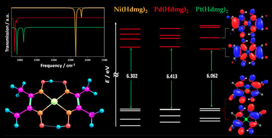
3.2. Infrared (IR) Spectroscopy

The DFT-calculated IR spectra of [M(Hdmg)2] (M = Ni, Pd, Pt) complexes contain characteristic absorption bands for the O–H, C=N, and N–O vibrations (Figure 2).
Our spectral assignments (Table 1, Table 2 and Table 3) are in keeping with an early detailed report on the IR spectra of Ni-, Pd-, Pt- and Cu-dimethylglyoximate complexes, including deuteration experiments [21], DFT-calculated and experimental data on [Ni(Hdmg)2] [87], and recent DFT-calculated IR spectra for [Ni(Hdmg)2] [1]. The marked deviation of the calculated values from experimental data is very probably because all these experiments have been carried out on solid samples. Thus, the experimental data represents the molecular stacks and not isolated molecules. Therefore, instead of comparing our calculated data with experimental data, we use it solely for benchmarking our computational method.
Compared with the recently applied range-separated hybrid functional ωB97X-D, augmented with dispersion correction and split-valence 6-31G (d,p) basis set for all non-metallic elements and LANL2DZ ecp [1], our results for the N‒O stretching vibration of the [Ni(Hdmg)2] gave markedly lower values, while the C=N stretch are quite similar and the O‒H bending is calculated at far higher energy (Table 1). Compared with the results from early calculations using the B3LYP functional and 6-311+(+)G**basis set for all atoms [87] our introduction of split basis had only a small impact on the N‒O stretching and O‒H bending energies, while for the C=N stretch our values lie markedly higher. Compared with the experimental data from solid samples, our calculated values and those of the recent DFT study using also split basis sets [1] are much higher. We tentatively ascribe this to the more sophisticated modelling of the metal atoms using split basis sets and believe that values of about 1650 cm‒1 represent the C=N stretch in isolated molecules, while the stacking in the solid reduces the energy of this resonance markedly.
Three transitions in the calculated spectra can be assigned to N–O stretching. They show very different intensities for the three complexes, while the energies are rather invariable (Figure 2, Table 1, Table 2 and Table 3).
For the C=N stretching modes the calculated spectra show two transitions for Ni (Table 1) and Pd (Table 2) and two merged bands for Pt (Table 3). They have similar intensities and similar energies for Pd and Pt but far higher values for Ni. We ascribe the energy differences between the metals to higher metal d to π*CN backbonding for Pt and Pd compared to Ni, in keeping with the larger 5d and 4d orbitals compared with 3d. The O–H bending vibrations showed a characteristic band in Ni complex at 1810 cm‒1, while for Pd and Pt complexes these transitions are extremely weak and at the same energy (1786 cm‒1).
The O–H symmetric and asymmetric stretching modes in the calculated IR spectra of [Ni(Hdmg)2] are far lower (2174 and 2240 cm‒1) than those of [Pd(Hdmg)2] (2939 and 2964 cm‒1) and [Pt(Hdmg)2] (2857 and 2934 cm‒1) (Figure 2, Table 1, Table 2 and Table 3). The calculated values are perfectly in keeping with our calculated structural data and in line with increasing ionic radii for the metals (Pd2+ > Pt2+ >> Ni2+) [63,64,65,67] weakening the H bond. For the asymmetric O–HO interactions this would lead to a shorter O–H bond and higher frequency [86]. Akin to a recent theoretical study [1], we also found that the asymmetric O–H stretching vibration in comparison with the symmetric stretching is very intense. This is in line with the bigger change of molecular dipole moment for the asymmetric O–H stretching mode.

3.3. Energies and Compositions of the Frontier Orbitals

The energies and compositions of the lowest unoccupied molecular orbital (LUMO) and highest occupied molecular orbitals (HOMO) were calculated for the three complexes based on the S0 and T1 geometry-optimised structures (Figures S1–S3 and Tables S5–S7 in the Supplementary Materials). In the electronic S0 ground state, most of the frontier orbitals including HOMO-3 to LUMO+3 differ from the Ni complex to the Pd and Pt derivatives, with the exception of the LUMOs which are highly similar in all three complexes with large coefficients on the metal-ligand bonds and the dmg π system (Figure 3 and Figure S2). The HOMO for the Ni complex is essentially centred on the ligands (100%) and has high similarities with the HOMO-1 for Pt (Figure 3), while the HOMO for the Pt complex obtains some metal d contributions (18%). For Pd these two orbitals are almost degenerate and the HOMO also obtains some metal contributions (8%). All three HOMO-3 orbitals have predominant metal dz2 character with the dz2 character getting more and more dominant along the series Ni << Pd < Pt. For all three complexes, the LUMO and LUMO+1 have small to negligible metal contributions. The LUMO+2 for Pt and the LUMO+3 for Pd resemble the essentially metal-centred LUMO+3 of the Ni complex (Figure S2) and clearly represent the metal pz orbital.
The calculated HOMO–LUMO energy gaps are 6.302 and 6.413 eV for the Ni and Pd complexes, respectively and reduced to 6.062 eV for the Pt complex, which is caused by a marked stabilisation of the LUMO (Figure 3). This series Pd > Ni > Pt agrees quite well with the experimental binding energies and electrochemical or optical HOMO-LUMO gaps of comparable series of Pt, Pd, and Ni complexes [63,64,65,67,77].
The XPS determined Ni 2p binding energies for [Ni(Hdmg)2] are reported at about 853 (2p3/2) to 870 (2p1/2) eV [38] while for the ligands N(1s) and O(1s) binding energies the sequence is Pt ~ Pd > Ni [91] which is in keeping with the size of the metal valence orbitals for the π-backbonding.
Gas-phase DFT calculations of [Ni(Hdmg)2] gave a value of 7.32 eV decreasing to 6.97 eV upon dimerisation [1] and very similar HOMO and LUMO compositions. Adding one or two H2O or NH3 molecules as additional axial ligand increased the value for the monomer and also changed the orbital composition of HOMO and LUMO [1].
The frontier orbitals of the T1 excited state (LSOMO: Lowest Singly Occupied Molecular Orbital, and HSOMO: Highest Singly Occupied Molecular Orbital) showed predominant contributions of the ligand to the HSOMO and HSOMO+1, while the metal participation increases in the LSOMO and LSOMO-1 for Pd and Pt complexes (Figure S3 and Table S7, Supplementary). So, again the Ni complex differs from the Pd and Pt derivatives. The HSOMO and LSOMO are mostly located in the ligand for Ni complex and metal contribution increases in the HSOMO+2 and HSOMO+3.
When comparing the HOMO–LUMO (S0) and LSOMO–HSOMO (T1) energy level diagrams for the three complexes (Figure S4, Supplementary) a significant stabilisation in the energy of the HSOMO compared with the LUMO is found, while the LSOMO for the T1 state remained close to the HOMO of the S0 state in all three cases.

3.4. UV-vis Absorption Spectroscopy

TD-DFT calculated absorption bands are very similar for all three complexes and show three main band systems containing several transitions (Figure 4). A closer look shows that the intense bands in the range 150–180 nm are very similar with maxima at 170, 171 and 176 nm for Ni(II), Pd(II), and Pt(II), respectively. The band system ranging from 180 to 280 nm shows more pronounced differences in energy (211, 231 and 250 nm) and band shape. The long-wavelength envelopes found from 270 to 400 nm are very similar for Ni and Pd, with maxima at 310 and 312 nm, respectively. For the Pt complex, the long-wavelength absorption is markedly red-shifted to the range 307–440 nm with a maximum of 350 nm. The intensity of these maxima decreases along the series from 6178 (Ni) to 4016 (Pd) and 2835 ε/M‒1cm‒1 (Pt) (Table 4).
The broad long-wavelength absorption band for [Ni(Hdmg)2] is essentially composed of the HOMO-1→LUMO (79%) and HOMO→LUMO+1 (12%) transitions (Table S8, Supplementary). The same band for the Pd complex correspond to a mixture of the HOMO-1→LUMO (48%) and HOMO→LUMO (35%) transitions (Table S9). Visual plots of the molecular orbitals (Figure S1, SI) show that both bonds have almost pure π−π* character. For the Pt complex, this band is essentially described by the HOMO→LUMO (90%) transition (Table S10) with has essentially ligand-centred π−π* character, but with a marked metal-to-ligand charge transfer (MLCT) contribution (Figure S1).
Further absorption bands at higher energy have predominant π−π* character which is in line with most of the states being essentially ligand centred (Figure S1 and Tables S6 and S7). Marked MLCT contributions for the Ni complex are found for transitions from HOMO-8 and HOMO-9 to the LUMO (Table S8). Corresponding transitions from the Pd and Pt derivatives include the HOMO-7 and HOMO-6 states, respectively (Tables S9 and S10). This is in line with the increasing ligand field energy and π-backbonding in the series Ni–Pd–Pt leading to a de-stabilisation of the low-lying dπ orbitals. No transitions with pronounced ligand field (d‒d*) character were found for the three complexes. However for Pd and Pt transitions with marked d‒p character (HOMO-3→LUMO+3 (Pd) or HOMO-3→LUMO+2 (Pt)) were found at quite high energies corresponding to 167 and 181 nm for Pd and Pt, respectively. The p-type HOMO+3 orbital for Ni is less involved in electronic transitions.
The calculated spectra for [Pd(Hdmg)2] and [Pt(Hdmg)2] show the expected red-shifted bands compared with the Ni derivative (Table 4) in keeping with experimental spectra on all three complexes in CHCl3 solution [15,16,57] and spectra recorded on thin films [27,28,29,30,51] or solids [22,51,57,92,93]. Also, the underlying transitions were quite similar for all three complexes as has been suggested before based on experimental data [17,27,28,29,30,51,90,93].
The recently calculated absorption spectra of monomeric and dimeric (Ni...Ni stacked) [Ni(Hdmg)2] are on first view similar to our spectrum absorption maxima [1]. A closer look shows that the energies of the two UV bands with maxima at 180 and 220 nm do not differ much between monomer and dimer. The calculated energies of these gas-phase calculations using the range-separated hybrid functional ωB97X-D, augmented with dispersion correction and split-valence 6-31G (d,p) basis set for all non-metallic elements and LANL2DZ ecp for Ni are only slightly lower than our numbers (Table 4). However, for the long-wavelength bands, this method gave a very different band maximum of 359 nm compared with 310 nm from our calculations in DMSO solvent and dimerisation shifted the band to 368 nm [1]. In the same study the long-wavelength band is described to have d‒p character although their main contributions come from the HOMO-1→LUMO and HOMO-2→LUMO transitions [1] and their HOMO-1, HOMO-2 and LUMO show higher metal contributions than those calculated in our study.
Experimental UV-vis absorption spectra reported for [Ni(Hdmg)2] in CHCl3 solution showed pronounced bands at 253, 328, 373 and 420 nm [15,16,57] but also features at lower energies [16,57]. For other solvents, very similar absorptions were reported [16,17,46,52,57] and for the Pd and Pt derivatives, these bands are markedly red-shifted [57,92]. The bands ranging from 200 to 300 nm and the long-wavelength bands at 300–400 nm have been previously assigned to either π‒π*, MLCT or mixed π‒π*/MLCT transitions [1,15,16,17,46,48,51,57,92,93]. Importantly, no solvatochromic effect was found on the spectra [Ni(Hdmg)2] in solvents ranging from benzene to water [16] and a band at 266 nm is also observed for the Hdmg anion in solution [17], which is rather counter-indicative for charge transfer transitions but supports the idea of solvent-invariant π‒π* transitions [94,95].
In films and bulk solids broad absorption bands with maxima ranging from 500 to 700 nm dominate the visible part of the spectrum while the UV to visible part looks similar to the spectra in solution [15,16,46,57,92,93]. The broad long-wavelength bands were assigned to the so-called d‒p (3dz2→4pz) transitions [1,2,3,6,15,46,48,49,50,51,52,57]. The energy of these transitions is very dependent on the metal...metal proximity and stronger interactions lead to lower absorption energies [1,2,3,6,9,15,26,27,28,29,30,46,47,48,49,50,51,57]. The appearance of this band in studies reporting spectra of dissolved complexes is thus a clear indication for intermolecular stacking and formation of oligomeric stacks in such solutions [15,16,46,57]. This is strongly supported by the above mentioned calculated red-shift for a long-wavelength band for the Ni...Ni stacked dimer [Ni(Hdmg)2]2 compared with the monomer while UV are not affected from dimerisation [1]. This makes it also difficult to compare our calculated spectra with experimental data.
In summary, contrasting to previous assignments of the absorption bands ranging from 200 to 450 nm to essentially MLCT with some π‒π* character we found a predominant π‒π* character for all these bands. In contrast to previous studies, we also found that admixture of metal contributions of MLCT or d‒p type occurs rather for transitions at higher energies, while the long-wavelength bands retain rather pure π‒π* character. However, this picture rapidly changes when axial ligands were added or dimers were formed [1,2]. Then metal-contributions both of MLCT and d‒p type get more pronounced for the long-wavelength absorptions of the monomeric species and dominate for the stacked molecules in the solid the visible spectral range with the typical d‒p transitions (mixed with MLCT type of transition) [1,2,3,6,15,46,48,49,50,51,52]. In our calculations, we used the conductor-like polarizable continuum model (CPCM) for the inclusion of polarisation effects from the solvent (here: DMSO). However, this method does not include real solvent molecules which are potentially coordinating but provides a polarisable surrounding (continuum). Compared with calculations on monomers in the gas phase [1] our calculation might thus overestimate delocalisation effects. However, our model definitely seems to be able to circumvent the dramatic impact of any species (ligands, other complexes) in the axial positions of the planar complexes on the dz2 and pz orbitals leading to massive metal contributions to optical transitions.

4. Conclusions

We have studied the three NiII, PdII and PtII complexes [M(Hdmg)2] (H2dmg = dimethylglyoxime) by density functional DFT and TD-DFT methods to explore the structural and IR and UV-vis absorption spectroscopic properties of the molecular species of these complexes. The three compounds have a very high tendency to form metal...metal directed stacks and thus aggregate rapidly in solution. This has previously hampered spectroscopic characterisation of these complexes in solution and most of the studies, including theoretical studies, and most reported applications have been focussing on the solid-state. Still, some applications such as catalysis, trace element detection and biomedicine might be based on monomolecular species. Studies reporting spectroscopic properties of the complexes in solution are rare and mainly restricted to UV-vis absorption spectra. These studies reported bands in the visible range (>400 nm), but recent quantum chemical calculations did not agree with these observations and we supposed that instead of monomolecular species these solutions represent monomolecular together with oligomeric species. Thus, spectroscopic data of the monomolecular species is probably not reported yet. Thus, we computed the molecular geometries, IR spectra, frontier orbital compositions and energy together with UV-vis spectra using the M06-2X functional and LANL2DZ ecp (metals) and def2TZVP (other atoms) as basis sets. DMSO as solvent was included employing the conductor-like polarizable continuum model (CPCM). The optimised molecular geometries proved the asymmetric character of the two formed O–HO bonds in the compounds, which connect the adjacent Hdmg ligands in the completely planar complexes. IR frequencies of the O–H bond were higher in the PdII and PtII complexes than the NiII derivative in line with weaker HO interactions. Comparison of calculated data of [M(Hdmg)2] from three DFT studies showed that the use of split-valence basis sets is superior to the previously reported use of one type of basis sets for the metals and the light atoms (B3LYP functional and 6-311+(+)G**basis set). Calculated compositions of frontier orbitals gave essentially ligand-centred lowest unoccupied molecular orbitals (LUMOs) and highest occupied molecular orbitals (HOMO) except for the LUMO+3 (Ni, Pd) or LUMO+2 (Pt) with clear metal pz and the HOMO-3 orbitals with predominant metal dz2 character. Calculated UV-vis spectra gave three bands in the range 180 to 350 nm. These bands are similar for all three complexes but show a marked red-shift in the series going from Ni to Pt. We assign them to predominantly ligand-centred π‒π* transitions, in contrast to previous assignments to mainly metal(d) to ligand(π*) charge transfer (MLCT) transitions. For our calculations metal contributions to the π‒π* transitions occur only at higher energies for the monomolecular species, while the long-wavelength bands have almost pure π‒π* character. These metal contributions comprise MLCT and also the notorious so-called d‒p transitions that dominate the visible spectral range and the colours and other opto-electronic properties of the three compounds in the solid. Although, these d‒p transitions have previously been assigned to absorption bands in the UV to vis range (200–450 nm) for the isolated molecules in solution, we found significant d‒p contributions only for bands at around 180 for Pt and Pd and none for Ni. Other calculations on monomers, dimers and oligomers showed that further ligands added to the two axial positions, dimerisation or oligomerisation has a strong impact on both the 3dz2 and the 4pz orbitals dramatically increasing their contributions to the electronic transitions. Together with these calculations, our results support our idea that spectroscopic data of these three complexes does not represent the isolated molecules but a mixture of monomeric and oligomeric species. In a way, we looked for the first time at the isolated molecules in a surrounding-isolating solvent model.

Supplementary Materials

The following figures and tables are available online at https://www.mdpi.com/article/10.3390/inorganics9060047/s1. Table S1: Essential experimental structural parameters of [M(Hdmg)2] (M = Ni, Pd, Pt). Table S2: Selected experimental and computed geometries of [Ni(Hdmg)2], Table S3: Selected experimental and computed geometries of [Pd(Hdmg)2], Table S4: Selected (experimental and computed) geometries of [Pt(Hdmg)2], Figure S1: DFT-optimised structures in the S0 ground state for [M(Hdmg)2] (M = Ni, Pd, Pt), Table S5: The XYZ coordinates of the optimised ground state for [M(Hdmg)2] (M = Ni, Pd, Pt), Table S6: Compositions (%) of frontier MOs in the S0 ground state for [M(Hdmg)2] (M = Ni, Pd, Pt), Figure S2: Frontier orbital landscape in the S0 ground state for [M(Hdmg)2] (M = Ni, Pd, Pt), Table S7: Compositions (%) of frontier MOs in the T1 excited state for [M(Hdmg)2] (M = Ni, Pd, Pt), Figure S3: Frontier orbital landscape in the T1 excited state for [M(Hdmg)2] (M = Ni, Pd, Pt), Figure S4: Energy diagrams of frontier MOs for [M(Hdmg)2] (M = Ni, Pd, Pt) at the optimised S0 and T1 geometry, Table S8: Wavelengths and character of calculated transitions for [Ni(Hdmg)2], Table S9: Wavelengths and character of calculated transitions for [Pd(Hdmg)2], Table S10: Wavelengths and character of calculated transitions for [Pt(Hdmg)2].

Author Contributions

M.N. carried out the DFT calculations, provided figures and tables and wrote the draft manuscript. A.K. supervised the project, did the data curation and revised the manuscript. Both authors have read and agreed to the published version of the manuscript.

Funding

M.N. acknowledges the German Academic Exchange Service (DAAD)—Funding programme/-ID: Research Grants—Doctoral Programmes in Germany, 2020/21 (grant No. 57507871).

Institutional Review Board Statement

Not applicable.

Informed Consent Statement

Not applicable.

Data Availability Statement

Not applicable.

Acknowledgments

We thank Simon Schmitz and Alexander Haseloer (both Department of Chemistry, University of Cologne) for support with the DFT calculations. The Regional Computing Centre of the University of Cologne (RRZK) is acknowledged for providing computing time on the DFT-funded High-Performance Computing (HPC) system CHEOPS as well as for the support.

Conflicts of Interest

The authors declare no conflict of interest.

References

  1. Ali, S.Y.; Reddy, K.D.; Manna, A.K. Structural, Electronic, and Spectral Properties of Metal Dimethylglyoximato [M(DMG)2; M = Ni2+, Cu2+] Complexes: A Comparative Theoretical Study. J. Phys. Chem. A 2019, 123, 9166–9174. [Google Scholar] [CrossRef] [PubMed]
  2. Patra, S.G.; Mandal, N.; Datta, A.; Datta, D. On bonding in bis(dimethylglyoximato)nickel(II). Comput. Theor. Chem. 2017, 1114, 118–124. [Google Scholar] [CrossRef]
  3. Liu, K.; Orimoto, Y.; Aoki, Y. Theoretical investigation of the pressure-induced insulator-to-metal-to-insulator transitions in one-dimensional bis(dimethylglyoximato) platinum(II), Pt(dmg)2. Polyhedron 2015, 87, 141–146. [Google Scholar] [CrossRef] [Green Version]
  4. Bruce-Smith, I.F.; Zakharov, B.A.; Stare, J.; Boldyreva, E.V.; Pulham, C.R. Structural Properties of Nickel Dimethylglyoxime at High Pressure: Single-Crystal X-ray Diffraction and DFT Studies. J. Phys. Chem. C 2014, 118, 24705–24713. [Google Scholar] [CrossRef]
  5. Kovacs, A. Theoretical study of the strong intramolecular hydrogen bond and metal–ligand interactions in group 10 (Ni, Pd, Pt) bis(dimethylglyoximato) complexes. J. Organomet. Chem. 2007, 692, 5383–5389. [Google Scholar] [CrossRef]
  6. Di Bella, S.; Casarin, M.; Fragala, I.; Granozzi, G.; Marks, T.J. Electronic Structure of Tetracoordinate Transition-Metal Complexes. 2. Comparative Theoretical ab Initio/Hartree-Fock-Slater and UV-Photoelectron Spectroscopic Studies of Building Blocks for Low-Dimensional Conductors: Glyoximate Complexes of Palladium(II) and Platinum(II). Inorg. Chem. 1988, 27, 3993–4002. [Google Scholar]
  7. Yoshida, K. Epitaxial Growth of Bis(dimethylglyoximato)platinum(II) Accompanied by Hole Formation. Cryst. Growth Des. 2020, 20, 7271–7275. [Google Scholar] [CrossRef]
  8. Takeda, K.; Sasaki, T.; Hayashi, J.; Kagami, S.; Shirotani, I.; Yakushi, K. X-ray and optical studies of one-dimensional bis(dimethylglyoximato)Pd(II), Pd(dmg)2 at high pressures. J. Phys. Conf. Ser. 2010, 215, 012065. [Google Scholar] [CrossRef]
  9. Thomas, T.W.; Underhill, A.E. Metal–metal interactions in transition-metal complexes containing infinite chains of metal atoms. Chem. Soc. Rev. 1972, 1, 99–120. [Google Scholar] [CrossRef]
  10. Tauzher, G.; Dreos, R.; Felluga, A.; Nardin, G.; Randaccio, L.; Stener, M. Intramolecular and intermolecular O‒H‒O hydrogen bond in some nickel(II) complexes with tridentate amino‒oxime ligands. Inorg. Chim. Acta 2003, 355, 361–367. [Google Scholar] [CrossRef]
  11. Li, D.-X.; Xu, D.-J.; Xu, Y.-Z. Redetermination of bis(dimethylglyoximato-[κ]2-N,N’)nickel(II). Acta Crystallogr. Sect. E Struct. Rep. Online 2003, 59, m1094–m1095. [Google Scholar] [CrossRef] [Green Version]
  12. Konno, M.; Okamoto, T.; Shirotani, I. Structure changes and proton transfer between O...O in bis(dimethylglyoximato)platinum(II) at low temperature (150 K) and at high pressures (2.39 and 3.14 GPa). Acta Crystallogr. Sect. B Struct. Sci. 1989, 45, 142–147. [Google Scholar] [CrossRef] [Green Version]
  13. Hussain, M.S.; Salinas, B.E.V.; Schlemper, E.O. Three-dimensional determinations of the crystal structures of bis(dimethylglyoximato)palladium(II) and bis(dimethylglyoximato)platinum(II). Acta Crystallogr. Sect. B Struct. Crystallogr. Cryst. Chem. 1979, 35, 628–633. [Google Scholar] [CrossRef]
  14. Murmann, R.K.; Schlemper, E.O. On the crystal structure of bis(glyoximato)nickel(II). Acta Crystallogr. 1967, 23, 667–669. [Google Scholar] [CrossRef]
  15. Martin, J.D.; Hogan, P.; Abboud, K.A.; Dahmen, K.-H. Variations on Nickel Complexes of the vic-Dioximes: An Understanding of Factors Affecting Volatility toward Improved Precursors for Metal-Organic Chemical Vapor Deposition of Nickel. Chem. Mater. 1998, 10, 2525–2532. [Google Scholar] [CrossRef]
  16. Caton, J.E.; Banks, C.V. Studies of the spectra of copper dimethyl-glyoxime, nickel dimethylglyoxime and nickel ethylmethylglyoxime in various solvents. Talanta 1966, 13, 967–977. [Google Scholar] [CrossRef]
  17. Burger, K.; Ruff, I.; Ruff, F. Some theoretical and practical problems in the use of organic reagents in chemical analysis—IV: Infra-red and ultra-violet spectrophotometric study of the dimethylglyoxime complexes of transition metals. J. Inorg. Nucl. Chem. 1965, 27, 179–190. [Google Scholar] [CrossRef]
  18. Frasson, E.; Panattoni, C. X-ray studies on the metal complexes with the glyoximes. IV. Structure of Ni-methyl-ethyl-glyoxime. Acta Crystallogr. 1960, 13, 893–898. [Google Scholar] [CrossRef]
  19. Williams, D.E.; Wohlauer, G.; Rundle, R.E. Crystal Structures of Nickel And Palladium Dimethylglyoximes. J. Am. Chem. Soc. 1959, 81, 755–756. [Google Scholar] [CrossRef]
  20. Frasson, E.; Panattoni, C.; Zannetti, R. X-ray studies on the metal complexes with the glyoximes. II. Structure of the Pt-dimethylglyoxime. Acta Crystallogr. 1959, 12, 1027–1031. [Google Scholar] [CrossRef]
  21. Blinc, R.; Hadži, D. Infrared spectra and hydrogen bonding in the nickel–dimethylglyoxime and related complexes. J. Chem. Soc. 1958, 4536–4540. [Google Scholar] [CrossRef]
  22. Huila, M.F.G.; Lukin, N.; Parussulo, A.L.A.; Oliveira, P.V.; Kyohara, P.K.; Araki, K.; Toma, H.E. Unraveling the Mysterious Role of Palladium in Feigl bis(dimethylglyoximate)nickel(II) Spot Tests by Means of Confocal Raman Microscopy. Anal. Chem. 2012, 84, 3067–3069. [Google Scholar] [CrossRef]
  23. Sharpe, A.G.; Wakefield, D.B. The basis of the selectivity of dimethylglyoxime as a reagent in gravimetric analysis. J. Chem. Soc. 1957, 281–285. [Google Scholar] [CrossRef]
  24. Godycki, L.E.; Rundle, R.E. The structure of nickel dimethylglyoxime. Acta Crystallogr. 1953, 6, 487–495. [Google Scholar] [CrossRef] [Green Version]
  25. Gerasimchuk, N. Editorial (Thematic Issue: Recent Advances in Chemistry and Applications of Oximes and their Metal Complexes: Part I). Curr. Inorg. Chem. 2015, 5, 3–4. [Google Scholar] [CrossRef]
  26. Takeda, K.; Shirotani, I.; Yakushi, K. Pressure-Induced Insulator-to-Metal-to-Insulator Transitions in One-Dimensional Bis(dimethylglyoximato)platinum(II), Pt(dmg)2. Chem. Mater. 2000, 12, 912–916. [Google Scholar] [CrossRef]
  27. Shirotani, I.; Suzuki, K.; Yagi, T. Pressure-sensitive Optical Properties of Bis(1,2-dionedioximato)-Palladium(II) Complexes. Proc. Jpn. Acad. Ser. B 1992, 68, 57–62. [Google Scholar] [CrossRef] [Green Version]
  28. Takeda, K.; Hayashi, J.; Shirotani, I.; Fukuda, H.; Yakushi, K. Structural, Optical, and Electrical Properties of One-Dimensional Bis(Dimethylglyoximato)nickel(II), Ni(dmg)2 at High Pressure. Mol. Cryst. Liq. Cryst. 2006, 460, 131–144. [Google Scholar] [CrossRef]
  29. Shirotani, I.; Suzuki, K.; Suzuki, T.; Yagi, T.; Tanaka, M. Absorption Spectra and Electrical Resistivities of Bis(1,2-dione dioximato)nickel(II) Complexes at High Pressures. Bull. Chem. Soc. Jpn. 1992, 65, 1078–1083. [Google Scholar] [CrossRef]
  30. Shirotani, I.; Inagaki, Y.; Utsumi, W.; Yagi, T. Pressure-sensitive absorption spectra of thin films of bis(diphenylglyoximato)platinum(II), Pt(dpg)2: Potential application as an indicator of pressure. J. Mater. Chem. 1991, 1, 1041–1043. [Google Scholar] [CrossRef]
  31. Ferraro, J.R. Some possible new internal pressure calibrants. Inorg. Nucl. Chem. Lett. 1970, 6, 823–825. [Google Scholar] [CrossRef]
  32. Kamnoet, P.; Aeungmaitrepirom, W.; Menger, R.F.; Henry, C.S. Highly selective simultaneous determination of Cu(II), Co(II), Ni(II), Hg(II), and Mn(II) in water samples using microfluidic paper-based analytical de-vices. Analyst 2021, 146, 2229–2239. [Google Scholar] [CrossRef]
  33. Mirhashemi, A.; Ghorbani, Y.; Sadighi, S. Synthesis and evaluation of Fe3O4–Al2O3/SDS–DMG adsorbent for extraction and preconcentration of Pd(II) from real samples. J. Iran. Chem. Soc. 2020, 17, 2073–2081. [Google Scholar] [CrossRef]
  34. Olorundare, F.O.G.; Nkosi, D.; Arotiba, O.A. Voltammetric Determination of Nitrophenols at a Nickel Dimethylglyoxime Complex—Gold Nanoparticle Modified Glassy Carbon Electrode. Int. J. Electrochem. Sci. 2016, 11, 7318–7332. [Google Scholar] [CrossRef]
  35. Andris, B.; Prazsky, M.; Sebesta, F. Rapid separation and determination of 107Pd in radioactive waste produced during NPP A-1 decommissioning. J. Radioanal. Nucl. Chem. 2015, 304, 123–126. [Google Scholar] [CrossRef]
  36. Ma, J.; Meng, W.; Zhang, L.; Li, F.; Li, T. Effective oil–water mixture separation and photocatalytic dye de-contamination through nickel-dimethylglyoxime microtubes coated superhydrophobic and superoleophilic films. RSC Adv. 2021, 11, 5035–5043. [Google Scholar] [CrossRef]
  37. Qi, P.; Gu, Y.; Sun, H.; Lian, Y.; Yuan, X.; Hu, J.; Deng, Z.; Yao, H.-C.; Guo, J.; Peng, Y. Active Nickel Derived from Coordination Complex with Weak Inter/Intra-molecular Interactions for Efficient Hydrogen Evolution via a Tandem Mechanism. J. Catal. 2020, 389, 29–37. [Google Scholar] [CrossRef]
  38. Sarkar, B.; Kwek, W.; Verma, D.; Kim, J. Effective vacuum residue upgrading using sacrificial nickel(II) dimethylglyoxime complex in supercritical methanol. Appl. Catal A Gen. 2017, 545, 148–158. [Google Scholar] [CrossRef]
  39. Titova, Y.Y.; Belykh, L.B.; Shmidt, F.K. Formation and properties of Ziegler systems based on nickel bis(dimethylglyoximate) in catalysis of hydrogenation reactions. Russ. J. Gen. Chem. 2014, 84, 2413–2420. [Google Scholar] [CrossRef]
  40. Feng, J.-J.; Zhou, D.-L.; Xi, H.-X.; Chen, J.-R.; Wang, A.-J. Facile synthesis of porous worm-like Pd nanotubes with high electrocatalytic activity and stability towards ethylene glycol oxidation. Nanoscale 2013, 5, 6754–6757. [Google Scholar] [CrossRef] [PubMed]
  41. Yuan, M.; Li, Q.; Zhang, J.; Wu, J.; Zhao, T.; Liu, Z.; Zhou, L.; He, H.; Li, B.; Zhang, G. Engineering Surface Atomic Architecture of NiTe Nanocrystals Toward Efficient Electrochemical N2 Fixation. Adv. Funct. Mater. 2020, 30, 2004208. [Google Scholar] [CrossRef]
  42. Wang, T.; Li, F.; Huang, H.; Yin, S.; Chen, P.; Jin, P.; Chen, Y. Porous Pd‒PdO Nanotubes for Methanol Electrooxidation. Adv. Funct. Mater. 2020, 30, 2000534. [Google Scholar] [CrossRef]
  43. Dong, X.; Li, J.; Wei, D.; Li, R.; Qu, K.; Wang, L.; Xu, S.; Kang, W.; Li, H. Pd(II)/Ni(II)-dimethylglyoxime derived Pd‒Ni‒P@N-doped carbon hybrid nanocatalysts for oxygen reduction reaction. Appl. Surf. Sci. 2019, 479, 273–279. [Google Scholar] [CrossRef]
  44. Kordatos, K.; Vlasopoulos, A.; Strikos, S.; Ntziouni, A.; Gavela, S.; Trasobares, S.; Kasselouri-Rigopoulou, V. Synthesis of carbon nanotubes by pyrolysis of solid Ni(dmg)2. Electrochim. Acta 2009, 54, 2466–2472. [Google Scholar] [CrossRef]
  45. Dakhel, A.; Ali-Mohamed, A.Y. Characterisation and ac-electrical investigation of sublimated bis(dimethylglyoximato)palladium(II) thin films. J. Organomet. Chem. 2006, 691, 3760–3764. [Google Scholar] [CrossRef]
  46. Dakhel, A.A.; Ahmed, Y.A.-M.; Henari, F. Structural and optical studies of evaporated bis(dimethylglyoximato)nickel(II) thin films. Opt. Mater. 2006, 28, 925–929. [Google Scholar] [CrossRef]
  47. Suzuki, I.; Honjo, T.; Terada, K. Vacuum Sublimation Behavior of Nickel(II), Palladium(II), and Platinum(II) Chelates with Dimethylglyoxime. Bull. Chem. Soc. Jpn. 1990, 63, 3686–3688. [Google Scholar] [CrossRef] [Green Version]
  48. Iwai, S.; Kamata, T.; Murata, S.; Yamamoto, K.; Ohta, T. Wavepacket motion during thermalization of self-trapped exciton driven by an intramolecular vibration in one-dimensional platinum dimethylglyoxime complex. J. Lumin. 2000, 87–89, 629–632. [Google Scholar] [CrossRef]
  49. Kamata, T.; Fukaya, T.; Matsuda, H.; Mizukami, F. Reversible control of the nonlinear optical activity of one-dimensional metal complexes. Appl. Phys. Lett. 1994, 65, 1343–1345. [Google Scholar] [CrossRef]
  50. Kamata, T.; Curran, S.; Roth, S.; Fukaya, T.; Matsuda, H.; Mizukami, F. Third-order nonlinear optical prop-erties of evaporated thin films of platinum-alkyldionedioxime complexes: Effects of metal—Metal distance. Synth. Met. 1996, 83, 267–271. [Google Scholar] [CrossRef]
  51. Kamata, T.; Fukaya, T.; Kodzasa, T.; Matsuda, H.; Mizukami, F. Third-order nonlinear optical properties of bis(dimethylglyoximato)metal(II). Synth. Met. 1995, 71, 1725–1726. [Google Scholar] [CrossRef]
  52. Yang, B.; Li, J.; Zhang, L.; Xu, G. A molecularly imprinted electrochemiluminescence sensor based on the mimetic enzyme catalytic effect for ultra-trace Ni2+ determination. Analyst 2016, 141, 5822–5828. [Google Scholar] [CrossRef] [PubMed]
  53. Benoit, S.L.; Maier, R.J. The nickel-chelator dimethylglyoxime inhibits human amyloid beta peptide in vitro aggregation. Sci. Rep. 2021, 11, 1–11. [Google Scholar] [CrossRef] [PubMed]
  54. Biancalana, L.; Batchelor, L.K.; Dyson, P.J.; Zacchini, S.; Schoch, S.; Pampaloni, G.; Marchetti, F. α-Diimine homologues of cisplatin: Synthesis, speciation in DMSO/water and cytotoxicity. New J. Chem. 2018, 42, 17453–17463. [Google Scholar] [CrossRef]
  55. Kluge, T.; Bette, M.; Rüffer, T.; Bruhn, C.; Wagner, C.; Ströhl, D.; Schmidt, J.; Steinborn, D. Activation of Acetyl Ligands through Hydrogen Bonds: A New Way to Platinum(II) Complexes Bearing Protonated Imino-acetyl Ligands. Organometallics 2013, 32, 7090–7106. [Google Scholar] [CrossRef]
  56. Schwieger, S.; Heinemann, F.W.; Wagner, C.; Kluge, R.; Damm, C.; Israel, G.; Steinborn, D. A Bis(acetyl)-Bridged Platinum(II) Coordination Polymer as a Building Block for Diacetylplatinum(II) Complexes and Platina-β-diketones. Organometallics 2009, 28, 2485–2493. [Google Scholar] [CrossRef]
  57. Nagakura, S.; Ohashi, Y.; Hanazaki, I. Spectroscopic study of the interaction between the central metal ions in the crystals of bis(dimethylglyoximato)nickel(II) and related complexes. Inorg. Chem. 1970, 9, 2551–2556. [Google Scholar] [CrossRef]
  58. Kurz, H.; Schötz, K.; Papadopoulos, I.; Heinemann, F.W.; Maid, H.; Guldi, D.M.; Hörner, G.; Weber, B. A Flu-orescence-Detected Coordination-Induced Spin State Switch. J. Am. Chem. Soc. 2021, 143, 3466–3480. [Google Scholar] [CrossRef]
  59. Berkefeld, A.; Fröhlich, M.; Kordan, M.; Hörner, G.; Schubert, H. Selective metalation of phenol-type proligands for preparative organometallic chemistry. Chem. Commun. 2020, 56, 3987–3990. [Google Scholar] [CrossRef]
  60. Heil, A.; Marian, C.M. Structure—Emission Property Relationships in Cyclometalated Pt(II) β-Diketonate Complexes. Inorg. Chem. 2019, 58, 6123–6136. [Google Scholar] [CrossRef]
  61. Garbe, S.; Krause, M.; Klimpel, A.; Neundorf, I.; Lippmann, P.; Ott, I.; Brünkink, D.; Strassert, C.A.; Doltsinis, N.L.; Klein, A. Cyclometalated Pt Complexes of CNC Pincer Ligands: Luminescence and Cytotoxic Properties. Organometallics 2020, 39, 746–756. [Google Scholar] [CrossRef]
  62. Föller, J.; Friese, D.H.; Riese, S.; Kaminski, J.M.; Metz, S.; Schmidt, D.; Wuerthner, F.; Lambert, C.; Marian, C.M. On the photophysical properties of IrIII, PtII, and PdII (phenylpyrazole) (phenyldipyrrin) complexes. Phys. Chem. Chem. Phys. 2020, 22, 3217–3233. [Google Scholar] [CrossRef]
  63. Mews, N.M.; Reimann, M.; Hörner, G.; Kaupp, M.; Schubert, H.; Berkefeld, A. A four-parameter system for rationalising the electronic properties of transition metal–radical ligand complexes. Dalton Trans. 2020, 49, 9735–9742. [Google Scholar] [CrossRef] [PubMed]
  64. Haseloer, A.; Denkler, L.M.; Jordan, R.; Reimer, M.; Olthof, S.; Schmidt, I.; Meerholz, K.; Hörner, G.; Klein, A. Ni, Pd, and Pt complexes of a tetradentate dianionic thiosemicarbazone-based O^N^N^S ligand. Dalton Trans. 2021, 50, 4311–4322. [Google Scholar] [CrossRef] [PubMed]
  65. Eskelinen, T.; Buss, S.; Petrovskii, S.K.; Grachova, E.V.; Krause, M.; Klein, A.; Strassert, C.A.; Koshevoy, I.O.; Hirva, P. Photophysics and Excited State Dynamics of Cyclometalated [M(C^N^N)(CN)] (M = Ni, Pd, Pt) Complexes: A Theoretical and Experimental Study. Inorg. Chem. 2021. [Google Scholar] [CrossRef]
  66. Krause, M.; von der Stück, R.; Brünink, D.; Buss, S.; Doltsinis, N.L.; Strassert, C.A.; Klein, A. Platinum and palladium complexes of tridentate C^N^N (phen-ide)-pyridine-thiazol ligands—A case study involving spectroelectrochemistry, photoluminescence spectroscopy and TD-DFT calculations. Inorg. Chim. Acta 2021, 518, 120093. [Google Scholar] [CrossRef]
  67. Poirier, S.; Lynn, H.; Reber, C. Variation of M·H–C Interactions in Square-Planar Complexes of Nickel(II), Palladium(II), and Platinum(II) Probed by Luminescence Spectroscopy and X-ray Diffraction at Variable Pressure. Inorg. Chem. 2018, 57, 7713–7723. [Google Scholar] [CrossRef] [Green Version]
  68. Frisch, M.J.; Trucks, G.W.; Schlegel, H.B.; Scuseria, G.E.; Robb, M.A.; Cheeseman, J.R.; Scalmani, G.; Barone, V.; Mennucci, B.; Petersson, G.A.; et al. Gaussian 09, revision A.02; Gaussian, Inc.: Wallingford, CT, USA, 2016. [Google Scholar]
  69. Barone, V.; Cossi, M.; Tomasi, J. A new definition of cavities for the computation of solvation free energies by the polarizable continuum model. J. Chem. Phys. 1997, 107, 3210–3221. [Google Scholar] [CrossRef]
  70. Cossi, M.; Scalmani, G.; Rega, N.; Barone, V. New developments in the polarizable continuum model for quantum mechanical and classical calculations on molecules in solution. J. Chem. Phys. 2002, 117, 43–54. [Google Scholar] [CrossRef]
  71. Zhao, Y.; Truhlar, D.G. The M06 suite of density functionals for main group thermochemistry, thermochemical kinetics, noncovalent interactions, excited states, and transition elements: Two new functionals and systematic testing of four M06-class functionals and 12 other functionals. Theor. Chem. Acc. 2008, 120, 215–241. [Google Scholar] [CrossRef] [Green Version]
  72. Weigend, F.; Ahlrichs, R. Balanced basis sets of split valence, triple zeta valence and quadruple zeta valence quality for H to Rn: Design and assessment of accuracy. Phys. Chem. Chem. Phys. 2005, 7, 3297–3305. [Google Scholar] [CrossRef]
  73. Roy, L.E.; Hay, P.J.; Martin, R.L. Revised Basis Sets for the LANL Effective Core Potentials. J. Chem. Theory Comput. 2008, 4, 1029–1031. [Google Scholar] [CrossRef] [PubMed]
  74. Rohlfing, C.M.; Hay, P.J.; Martin, R.L. An effective core potential investigation of Ni, Pd, and Pt and their monohydrides. J. Chem. Phys. 1986, 85, 1447–1455. [Google Scholar] [CrossRef]
  75. Hay, P.J.; Wadt, W.R. Ab initio effective core potentials for molecular calculations. Potentials for K to Au including the outermost core orbitals. J. Chem. Phys. 1985, 82, 299–310. [Google Scholar] [CrossRef]
  76. Hay, P.J.; Wadt, W.R. Ab initio effective core potentials for molecular calculations. Potentials for the transition metal atoms Sc to Hg. J. Chem. Phys. 1985, 82, 270–283. [Google Scholar] [CrossRef]
  77. Potocny, A.M.; Pistner, A.J.; Yap, G.P.A.; Rosenthal, J. Electrochemical, Spectroscopic, and 1O2 Sensitization Characteristics of Synthetically Accessible Linear Tetrapyrrole Complexes of Palladium and Platinum. Inorg. Chem. 2017, 56, 12703–12711. [Google Scholar] [CrossRef]
  78. Kar, P.; Yoshida, M.; Kobayashi, A.; Routaboul, L.; Braunstein, P.; Kato, M. Colour tuning by the stepwise synthesis of mononuclear and homo- and hetero-dinuclear platinum(II) complexes using a zwitterionic quinonoid ligand. Dalton Trans. 2016, 45, 14080–14088. [Google Scholar] [CrossRef]
  79. Phadnis, P.P.; Jain, V.K.; Schurr, T.; Klein, A.; Lissner, F.; Schleid, T.; Kaim, W. Synthesis, spectroscopy, structure and photophysical properties of dinaphthylmethylarsine complexes of palladium(II) and platinum(II). Inorg. Chim. Acta 2005, 358, 2609–2617. [Google Scholar] [CrossRef]
  80. Dey, D.; Kumbhare, L.B.; Jain, V.K.; Klein, A.; Schurr, T.; Kaim, W.; Belaj, F. Structural Varieties in 1-Dimethylaminopropyl-2-chalcogenolate and 2-Dimethylaminopropyl-1-chalcogenolate (S, Se, Te) Complexes of Palladium(II) and Platinum(II): Synthesis, Spectroscopy and Structures. Eur. J. Inorg. Chem. 2004, 22, 4510–4520. [Google Scholar] [CrossRef]
  81. Phadnis, P.P.; Jain, V.K.; Klein, A.; Weber, M.; Kaim, W. Configurational selectivity in benzyldimethylarsine complexes of palladium(II) and platinum(II): Synthesis, spectroscopy and structures. Inorg. Chim. Acta 2003, 346, 119–128. [Google Scholar] [CrossRef]
  82. Huheey, J.E.; Huheey, C.L. Anomalous properties of elements that follow “long periods” of elements. J. Chem. Educ. 1972, 49, 227–230. [Google Scholar] [CrossRef]
  83. Steiner, T. The Hydrogen Bond in the Solid State. Angew. Chem. Int. Ed. 2002, 41, 48–76. [Google Scholar] [CrossRef]
  84. Rundle, R.E.; Parasol, M. O–H Stretching Frequencies in Very Short and Possibly Symmetrical Hydrogen Bonds. J. Chem. Phys. 1952, 20, 1487–1488. [Google Scholar] [CrossRef]
  85. Caton, J.E.; Banks, C.V. Hydrogen bonding in some copper(II) and nickel(II) vic-dioximes. Inorg. Chem. 1967, 6, 1670–1675. [Google Scholar] [CrossRef]
  86. Orel, B.; Penko, M.; Hadzi, D. Infrared and resonance enhanced Raman spectra of some metal vic-dioximes with short hydrogen bonds. Spectrochim. Acta Part A Mol. Spectrosc. 1980, 36, 859–864. [Google Scholar] [CrossRef]
  87. Szabó, A.; Kovács, A. Vibrational analysis of the bis(dimethylglyoximato)nickel(II) complex. J. Mol. Struct. 2003, 651–653, 547–553. [Google Scholar] [CrossRef]
  88. Várhelyi, C.; Kovács, A.; Gömöry, A.; Várhelyi, C.; Pokol, G.; Farkas, G.; Sohár, P. Comparative spectral and thermal studies of [Pt(DioxH)2] chelates. J. Coord. Chem. 2009, 62, 2429–2437. [Google Scholar] [CrossRef]
  89. Bigotto, A.; Galasso, V.; De Alti, G. Infrared spectra of transition metal glyoximates. Spectrochim. Acta Part A Mol. Spectrosc. 1971, 27, 1659–1670. [Google Scholar] [CrossRef]
  90. Kohler, U.; Hausen, H.-D.; Weidlein, J. Bis(dimethylmetall(III)glyoximato)metallate(II) (metall(III) = Al, Ga, In, metall(II) = Ni, Pd, Pt, Cu). J. Organomet. Chem. 1984, 272, 337–350. [Google Scholar] [CrossRef]
  91. Yoshida, T. An X-Ray Photoelectron Spectroscopic Study of Dioxime Metal Complexes. Bull. Chem. Soc. Jpn. 1978, 51, 3257–3260. [Google Scholar] [CrossRef] [Green Version]
  92. Basu, G.; Cook, G.M.; Belford, R.L. The Green Band of Crystalline Nickel Dimethylglyoxime. I. Mixed Crystals with Palladium, Platinum, and Copper and the Questions of Nonlocalized or d-p Transitions. Inorg. Chem. 1964, 3, 1361–1368. [Google Scholar] [CrossRef]
  93. Nishida, Y.; Kozuka, M.; Nakamoto, K. Resonance raman spectra of [Ni(dmg)2], [Pd(dmg)2] and [Pt(dmg)2] in the solid state (dmgH = dimethylglyoxime). Inorg. Chim. Acta 1979, 34, L273–L275. [Google Scholar] [CrossRef]
  94. Kaim, W.; Kohlmann, S.; Ernst, S.; Olbrich-Deussner, B.; Bessenbacher, C.; Schulz, A. What determines the solvatochromism of metal-to-ligand charge transfer transitions? A demonstration involving 17 tungsten carbonyl complexes. J. Organomet. Chem. 1987, 321, 215–226. [Google Scholar] [CrossRef]
  95. Manuta, D.M.; Lees, A.J. Solvatochromism of the Metal to Ligand Charge-Transfer Transitions of Zerovalent Tungsten Carbonyl Complexes. Inorg. Chem. 1986, 25, 3212–3218. [Google Scholar] [CrossRef]
Scheme 1. Schematic sketch of the three complexes [M(Hdmg)2].
Inorganics 09 00047 sch001
Figure 1. General schematic of the structures of [M(Hdmg)2] (M = Ni, Pd, Pt), with numbering.
Inorganics 09 00047 g001
Figure 2. (a) DFT calculated IR spectra of [M(Hdmg)2] (M = Ni, Pd, Pt), solvent: DMSO, functional: M06-2X, basis set: For C, H, N, O: def2TZVP, and for Ni, Pd, and Pt: LANL2DZ ecp. (b) Magnified view (1850–100 cm‒1) of the IR spectra of [M(Hdmg)2] (M = Ni, Pd, Pt).
Inorganics 09 00047 g002
Figure 3. DFT calculated energies of occupied MOs (black) and unoccupied MOs (red) for [M(Hdmg)2]; frontier MOs are given for M = Pt.
Inorganics 09 00047 g003
Figure 4. TD-DFT-calculated UV-vis absorption spectra of [Ni(Hdmg)2] (a), [Pd(Hdmg)2] (b), and [Pt(Hdmg)2] (c), (solvent: DMSO, functional: M06-2X, basis set: def2TZVP For C, H, N, O, LANL2DZ ecp for Ni, Pd and Pt.
Inorganics 09 00047 g004
Table 1. Selected (experimental and computed) IR data for [Ni(Hdmg)2] a.
O‒HO‒HC=NN‒ON‒OReference
StretchingBendingStretchingStretchingStretching
2174 (sym) 2240 (asym)18101647 and 16771222 and 12971121Our Calc.
2429 (sym) 2540 (asym)1600–182316371336 (sym) 1365 (asym)1261 (sym) 1261 (asym)Calc. [1] b
258517821529 and 159012491120Calc. [85] c
23501780156012351100Exp. [21]
2350-157612411103Exp. [17]
-17901550 and 157212401101Exp. [87]
a Calculated transition energies or observed band maxima in cm‒1. Our results: Solvent: DMSO, functional: M06-2X, basis set: def2TZVP for C, H, N, O, LANL2DZ ecp for Ni; Calc.: calculated data; Exp.: experimental data. b functional: ωB97X-D, basis sets 6-31G (d,p) for C, H, N, O and LANL2DZ ecp for Ni. c Functional: B3LYP, basis set: 6-311+(+)G**.
Table 2. Selected (experimental and computed) IR data for [Pd(Hdmg)2] a.
O‒HO‒HC=NN‒ON‒OReference
StretchingBendingStretchingStretchingStretching
2939 (sym)
2964 (asym)
17861611 and 16211217 and 13131114Our results
234017101550 and 150012501090Exp. [21]
2340-155212591091Exp. [17]
a Calculated transition energy or observed band maxima in cm‒1. Our results: Solvent: DMSO, functional: M06-2X, basis set: def2TZVP for C, H, N, O, LANL2DZ ecp for Pd; Exp.: experimental data.
Table 3. Selected (experimental and computed) IR data for [Pt(Hdmg)2] a.
O‒HO‒HC=NN‒ON‒OReference
StretchingBendingStretchingStretchingStretching
2857 (sym)
2934 (asym)
178816191217 and 13201119Our results
2350-155012621089Exp. [17]
a Calculated transition energy or observed band maxima in cm‒1. Our results: Solvent: DMSO, functional: M06-2X, basis set: def2TZVP for C, H, N, O, LANL2DZ ecp for Pt; Exp.: experimental data.
Table 4. DFT-calculated absorption maxima of the complexes [M(Hdmg)2] (M = Ni, Pd, Pt) a,b.
[M(Hdmg)2][Ni(Hdmg)2][Pd(Hdmg)2][Pt(Hdmg)2]
λ3 (ε) b170 (4.3) 171 (4.1)176 (6.5)
λ2 (ε) b211 (4.1)231 (1.9)250 (2.5)
λ1 (ε) b310 (6.2)312 (4.0)350 (2.8)
a Solvent: DMSO, functional: M06-2X, basis set: For C, H, N, O: def2TZVP, for Ni, Pd, Pt: LANL2DZ ecp. b Absorption maxima λ are given in nm, and molar absorption coefficients ε are given in 104 L mol‒1 cm‒1.
Publisher’s Note: MDPI stays neutral with regard to jurisdictional claims in published maps and institutional affiliations.

