Article

Synthesis of Polystyrene@TiO2 Core–Shell Particles and Their Photocatalytic Activity for the Decomposition of Methylene Blue

1
Department of Sustainable Engineering, College of Industrial Technology, Nihon University, 1-2-1, Izuni-cho, Narashino 275-8575, Chiba, Japan
2
Department of Engineering for Future Innovation, National Institute of Technology, Ichinoseki College, Takanashi, Hagisho, Ichinoseki 021-8511, Iwate, Japan
*
Author to whom correspondence should be addressed.
Inorganics 2023, 11(8), 343; https://doi.org/10.3390/inorganics11080343
Submission received: 31 May 2023 / Revised: 14 August 2023 / Accepted: 17 August 2023 / Published: 21 August 2023
(This article belongs to the Special Issue 10th Anniversary of Inorganics: Inorganic Materials)

Abstract

:
In this study, we investigated the preparation conditions of polystyrene (PS)@TiO2 core–shell particles and their photocatalytic activity during the decomposition of methylene blue (MB). TiO2 shells were formed on the surfaces of PS particles using the sol–gel method. Homogeneous PS@TiO2 core–shell particles were obtained using an aqueous NH3 solution as the promoter of the sol–gel reaction and stirred at room temperature. This investigation revealed that the temperature and amount of the sol–gel reaction promoter influenced the morphology of the PS@TiO2 core–shell particles. The TiO2 shell thickness of the PS@TiO2 core–shell particles was approximately 5 nm, as observed using transmission electron microscopy. Additionally, Ti elements were detected on the surfaces of the PS@TiO2 core–shell particles using energy-dispersive X-ray spectroscopy analysis. The PS@TiO2 core–shell particles were used in MB decomposition to evaluate their photocatalytic activities. For comparison, we utilized commercial P25 and TiO2 particles prepared using the sol–gel method. The results showed that the PS@TiO2 core–shell particles exhibited higher activity than that of the compared samples.

1. Introduction

As environmental pollution continues to worsen, technologies and materials that mitigate this issue have become widely researched topics [1,2]. Organic dyes primarily released from industrial plants are among such pollutants that form part of effluents in various water bodies [3,4]. Dyes such as methylene blue (MB) are toxic and nonbiodegradable, and they remain in the water for long periods of time [5]. Therefore, developing methods for removing dyes from water bodies is crucial.
Many approaches have been reported for dye removal, such as flocculation, chemical oxidation, membrane filtration, chemical coagulation, photochemical degradation, and biological degradation [4,5,6]. Notably, photocatalysis is an effective method for dye degradation because of its advantages, such as its eco-friendliness, high efficiency, low cost, and reusability [7,8]. Recently, hydrogen evolution was promoted in the presence of photocatalysts such as metal compound/g-C3N4 systems and flower-like carbon quantum dots/BiOBr composites [9,10,11,12,13].
Titanium dioxide (TiO2) is well known for being an excellent photocatalyst. This oxide has attracted attention for use in pollutant removal because it offers high conversion efficiency, good chemical stability, and low cost [14,15,16]. The sol–gel reaction has been reported as a synthetic method for generating TiO2 particles [17,18]. In general, TiO2 particles with a small particle size can efficiently act as a photocatalyst; however, these tend to agglomerate due to their high energy and reduce the number of available active sites [19]. To solve this problem, TiO2 particles have been immobilized on polymer substrates such as polymers, including polyethlene [20,21] and poly (dimethylsiloxane) beads, [22] and so on [23]. Moreover, it is difficult to obtain TiO2 particles with uniform size due to the fast sol–gel reaction rates exhibited by Ti precursors [24,25]. Therefore, the challenge is to immobilize TiO2 particles uniformly on the surfaces of substrates. Additionally, many complex processes are required in order to obtain substrate-supported TiO2 particles due their long synthesis time [26,27,28]. We propose a facile method for the immobilization of TiO2 particles on the surfaces of polystyrene (PS) particles.
Monodisperse PS particles with a positive charge were prepared using emulsifier-free emulsion polymerization using 2′2-azodiisobutyramidine dihydrochloride as the initiator and poly (vinylpyrrolidone) as the stabilizer [29,30,31]. Previous studies found that SiO2-Al2O3 nanoparticles could be collected on the surfaces of PS particles. In this method, the SiO2-Al2O3 nanoparticles were prepared using the sol–gel method, and the nanoparticles with negative charges were attracted to the surfaces of the PS particles because of their positive charges, thus forming SiO2-Al2O3 shells [32]. With this knowledge in mind, we attempted to collect TiO2 particles on the surfaces of PS particles using this simple method.
Herein, we report PS@TiO2 core–shell particles synthesized using the sol–gel method. In this method, TiO2 shells were formed on the surfaces of PS particles. Homogeneous spherical particles were obtained by adjusting the reaction temperature and amount of the promoter aqueous NH3 solution. A schematic illustration of the synthesis of the PS@TiO2 core–shell particles is shown in Figure 1. Experimental observations confirmed that the diameters of PS@TiO2 core–shell particles were approximately 220 nm. Furthermore, TiO2 was confirmed to be present on the surfaces of the PS@TiO2 core–shell particles. The photocatalytic activity of the PS@TiO2 core–shell particles was evaluated using MB decomposition. The PS@TiO2 core–shell particles showed better performance than that of the commercial P25.

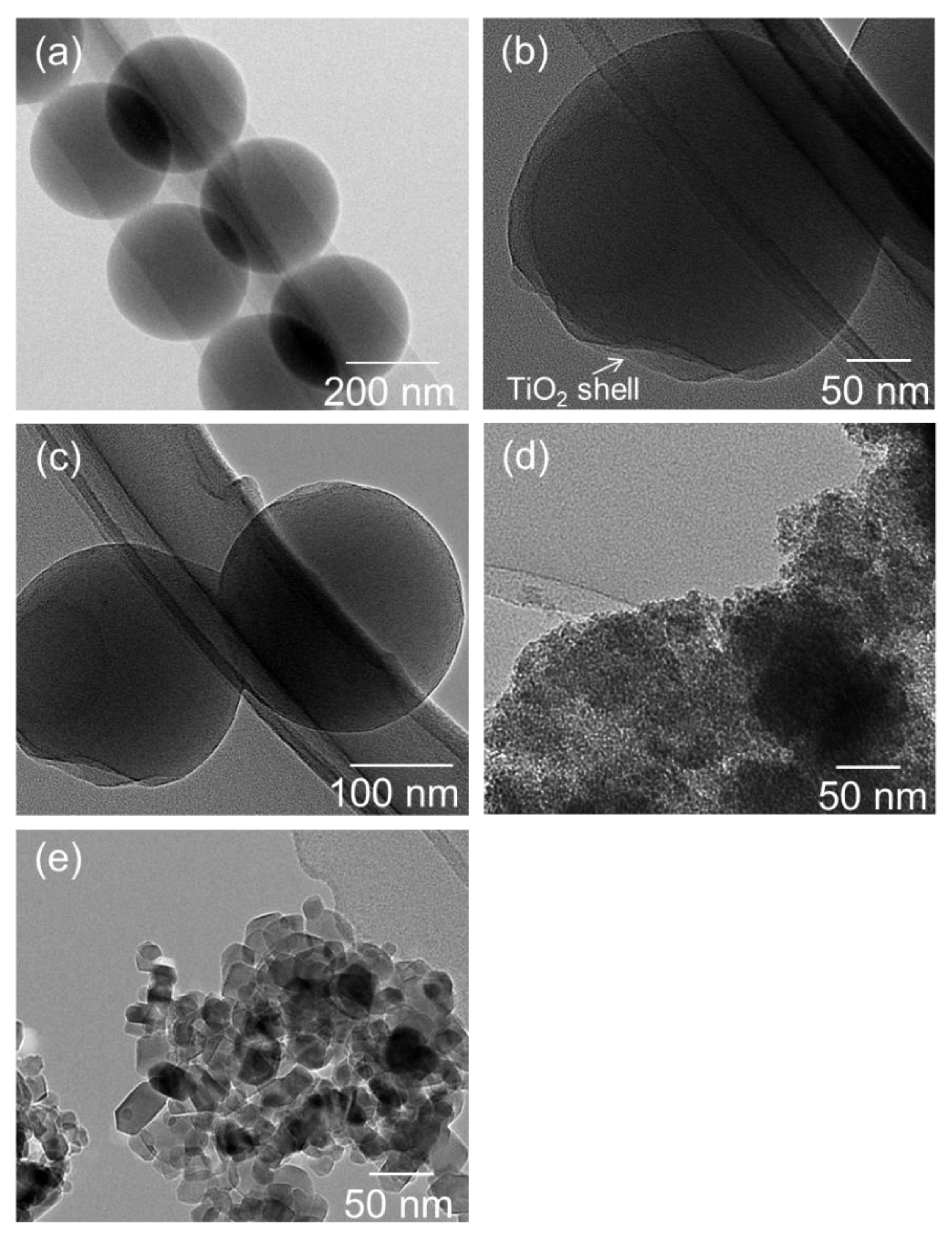
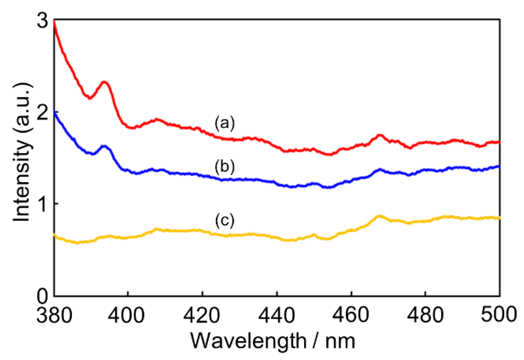
2. Results and Discussion

First, we investigated the influence of preparation conditions such as the preparation temperature and amount of aqueous NH3 solution on the morphology of PS@TiO2 core–shell particles. Figure 2a–c show scanning electron microscopy (SEM) images of PS@TiO2 core–shell particles prepared at various preparation temperatures and with varying amounts of aqueous NH3 solution. Uniform spherical particles were prepared using 300 μL of aqueous NH3 solution at room temperature (Figure 2a), whereas spherical particles and particles with irregular shapes were prepared using 300 μL of aqueous NH3 solution at 323 K (Figure 2b). Meanwhile, the rough layers, including spherical particles on the surface, were observed in SEM images of Figure 2c when prepared using 1000 μL of aqueous NH3 solution at room temperature. Using this process, the TiO2 nanoparticles prepared via the sol–gel reaction were collected on the surfaces of PS particles. Reportedly, the sol–gel reaction rate enhances higher temperatures and increases the amounts of promoters such as NH3 [33]. Consistent with the above reports, a faster sol–gel reaction rate promoted TiO2 particle growth, leading to the formation of particles with irregular shapes and rough layers, as shown in Figure 2b,c. Moreover, we confirmed the presence of TiO2 on the surfaces of uniform PS@TiO2 core–shell particles prepared using 300 μL of aqueous NH3 solution at room temperature through energy-dispersive X-ray spectroscopy (EDX) measurements. Figure 3 shows elemental mapping images of O, Ti, and C for the prepared PS@TiO2 core–shell particles. O and Ti were observed in the overall images, and the ratio of O:Ti was approximately 1:1. Additionally, the areas of C elemental mapping overlapped the areas of Ti and O elemental mapping. This result suggests that very thin TiO2 shells can be formed on the surfaces of PS particles. Meanwhile, we measured the specific surface area of the PS@TiO2 core–shell particles using the Brunauer–Emmett–Teller (BET) method. The results showed that the specific surface area of the PS@TiO2 core–shell particles was 125 m2 g−1.
Next, the morphology of PS@TiO2 core–shell particles were observed in detail using transmission electron microscopy (TEM). For comparison, we used TiO2 particles prepared using sol–gel reactions and commercial P25. Figure 4a shows a TEM image of PS particles. In this image, the diameters of the PS particles are approximately 280 nm. Figure 4b,c show TEM images of the PS@TiO2 core–shell particles. The obtained images show that the diameters of the spherical particles were approximately 290 nm. The presence of the TiO2 shell was confirmed by the different contrasts between core and shell. As shown in Figure 4a–c, the shell thickness of the PS@TiO2 core–shell particles could be ~5 nm. This result is also consistent with the results of the SEM/energy-dispersive X-ray spectroscopy (EDX) element mapping images, which show the detection of C (Figure 3). Meanwhile, the presence of prepared TiO2 particles and P25 was also confirmed using the TEM images. In these results, the prepared TiO2 is presented in random shapes (Figure 4d), whereas crystal particles with diameters of 20–50 nm are observed in the TEM image of P25 (Figure 4e).
The crystallinity of the samples was measured using powder X-ray diffraction (XRD). Figure 5 shows the XRD patterns of the PS@TiO2 core–shell particles, the prepared TiO2, and the commercial P25. The PS@TiO2 core–shell particles showed a broad diffraction peak, whereas the prepared TiO2 showed no peak, indicating that both samples were composed of an amorphous phase. Meanwhile, the commercial P25 showed sharp peaks, indicating that it was composed of rutile and anatase phases [34].
Finally, the photocatalytic activities of the PS@TiO2 core–shell particles, the prepared TiO2, and the commercial P25 were evaluated through MB decomposition. The main peak of MB was observed at 664 nm. The UV visible (UV-Vis) absorption spectra of the PS@TiO2 core–shell particles, the prepared TiO2, the commercial P25, and the PS particles monitored at different times are shown in Figure 6a–d. As seen in these results, the intensity of the absorption peak gradually decreased with the reaction time in the presence of the PS@TiO2 core–shell particles, the prepared TiO2, and the commercial P25. However, the intensity of the absorption peak was not changed in the presence of the PS particles. Figure 6e shows the reaction time dependence of MB decomposition in the presence of these samples with UV irradiation. The relative intensity of the absorbance at 664 nm was used to calculate the value of C/C0, where C0 and C are the MB concentrations of the aqueous MB solution at the beginning and reaction time, respectively. In the presence of the PS@TiO2 core–shell particles and the prepared TiO2, 84% and 38% of MB degraded within 3 min. Meanwhile, 84% and 60% of MB degraded within 24 min, indicating that both samples did not promote MB decomposition after 4 min. In the case of the commercial P25, the intensity of the main peak gradually decreased with the increase in reaction time, and 58% of MB degraded after 24 min. On the other hand, the PS particles showed no activity. These results indicate that the PS@TiO2 core–shell particles demonstrated a faster reaction rate than those of the prepared TiO2 and the commercial P25.
The PS@TiO2 core–shell particles were used in MB decomposition with and without UV irradiation to evaluate their photocatalytic activity. Figure 7a shows the UV-Vis absorption spectra of the reaction solutions with the PS@TiO2 core–shell particles without UV irradiation. The intensity of the absorption peak at 664 nm was unchanged without UV irradiation. Figure 7b shows the reaction time dependence in the presence of the PS@TiO2 core–shell particles with and without UV irradiation. With UV irradiation, 87% of MB degraded after 24 min, as described in the above results, whereas without UV irradiation, MB decomposition was not promoted.
Herein, we discussed the differences in the photocatalytic activities of the PS@TiO2 core–shell particles, the prepared TiO2, and the commercial P25. First, we noted that PS@TiO2 core–shell particles exhibited the highest MB decomposition activity. In this activity evaluation, the masses of the samples were equal, whereas the masses of TiO2 were lower than the practical masses of the samples because PS@TiO2 core–shell particles contained PS particles as the core. Nevertheless, the PS@TiO2 core–shell particles exhibited the highest MB decomposition activity among all samples. This may have been because TiO2 shells with a shell thickness of about 5 nm were uniformly coated on surface of PS particles leading to efficient activity promotion. Therefore, the PS@TiO2 core–shell particles, the prepared TiO2, and the PS particles were investigated via photoluminescence (PL) measurements. Figure 8 shows the PL spectra of the PS@TiO2 core–shell particles, the prepared TiO2, and the PS particles. From this result, a broad peak was observed at around 390 nm for the PS@TiO2 core–shell particles and the prepared TiO2, while no peak was observed for the PS particles. In addition, the PS@TiO2 core–shell particles exhibited a higher peak intensity than the prepared TiO2. This peak was attributed to band-to-band transition [35]. Previous studies have reported that a peak was not observed for amorphous TiO2 because of the lack of fluorescence [36], whereas for the TiO2 with anatase crystalline phases, a peak was observed [35]. It may be that the TiO2 shells on the surface of the PS@TiO2 core–shell particles formed very tiny crystal particles with poor crystalline phases. The broad peaks observed at 20° in the XRD results may imply the existence of poor crystalline phases. In other reports, the peak intensity of the amorphous TiO2 coating on the surface of ZnO particles increased compared with that of ZnO particles, indicating that the fine control of TiO2 shell thickness on the surface of ZnO nanoparticles was very important for the enhancement of PL intensity [37]. Considering this, the crystallinity and thickness of the TiO2 shell of PS@TiO2 core–shell particles may play an important role in MB decomposition.
Next, a comparison of the MB decomposition obtained in this study with those of other previously reported studies [38,39,40,41,42] is listed in Table 1. It was found that PS@TiO2 core–shell particles show higher activity than most TiO2-polymers and their related catalysts. However, the MB degradation rate per dosage of the PS@TiO2 core–shell particles (0.379% min−1mg−1) was lower than that of TiO2/graphene porous composites (0.64% min−1mg−1). This result might be ascribed to the different crystallinity of TiO2. The TiO2 shells of the PS@TiO2 core–shell particles were probably amorphous phases, as shown in Figure 5a. On the other hand, the TiO2 on the graphene porous composites was an anatase phase [42]. Indeed, the band gap of the amorphous TiO2 is higher than that of the rutile and anatase TiO2 [43]. Moreover, there may also be a difference in the mechanism of photocatalysis.
In addition, we discussed the differences in reaction rates between the PS@TiO2 core–shell particles and the commercial P25. The reaction rates of MB decomposition in the presence of the PS@TiO2 core–shell particles and the commercial P25 from 0 to 4 min were 21.3% min−1 and 3.7% min−1, respectively. Meanwhile, the reaction rates of MB decomposition from 4 to 24 min were 0.13% min−1 and 2.1% min−1, respectively. The significantly faster reaction rate of the PS@TiO2 core–shell particles might be related to the zeta potential of the samples. TiO2 samples such as P25 reportedly have a negatively charged surface at a pH value of greater than 7 [44]. Meanwhile, the PS@TiO2 core–shell particles have a negatively charged surface at a pH value of more than 4–5 [45,46]. Because the pH of the reaction solution was 6 in this study, the surface of the PS@TiO2 core–shell particles was negatively charged, whereas the surface of the commercial P25 was positively charged. MB which have cationic ions were electrochemically attracted to the surface of the PS@TiO2 core–shell particles, leading to an increase in the initial reaction rate. Meanwhile, the MB was electrochemically repelled from the surface of the commercial P25. The photocatalytic activity decreased after 4 min, probably because the rate of MB adsorption on the surface was faster than the rate of decomposition induced by the photocatalyst, and the MB covered the TiO2 layer as it prevented the blocking of UV irradiation. In order to solve this problem, we suggest that TiO2 hollow spheres are prepared via calcination of the PS@TiO2 core–shell particles. The hollow spheres have high specific surface areas because of the void spaces, leading to an increase in contact frequency with the MB. We found that the PS particles can be decomposed via calcination at 723–873 K [47]. The crystallinity of the TiO2 simultaneously grows during calcination. Based on this knowledge, we expect that TiO2 hollow spheres with crystalline shells can efficiently act as photocatalysts.

3. Materials and Methods

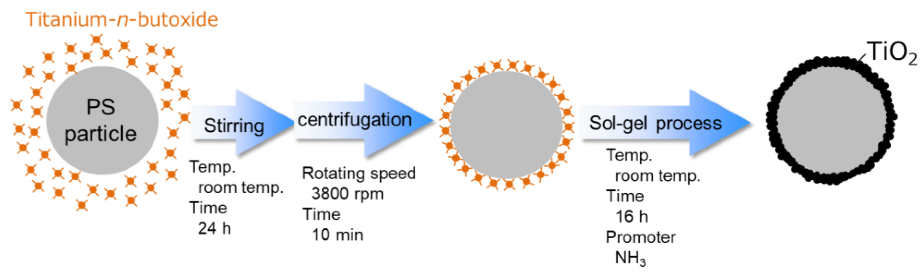
3.1. Synthesis of PS@TiO2 Core–Shell Particles and TiO2 particles

PS particles were prepared with reference to a previous study [29]. The collected PS was washed with ethanol (10 mL, Kanto Chemical Co., Tokyo, Japan, >99.5%) via centrifugation at 3800 rpm for 10 min, twice. The final PS contents were dispersed in ethanol (15 mL). This PS suspension (10 g), titanium-n-butoxide (1000 µL, Kanto Chemical Co., >98.0%), and ethanol (40 mL) were stirred at room temperature for 24 h. The obtained suspension was centrifuged at 3800 rpm for 10 min and washed with ethanol to obtain the precursor of the PS@TiO2 core–shell particles. The precursor was spread in ethanol (20 mL). After that, ethanol and an aqueous NH3 solution (300 or 1000 µL, Kanto Chemical Co., 28.0–30.0% NH3) were added to the precursor suspension, which was then stirred at room temperature or 323 K for 17 h. Then, the suspension was centrifuged at 3800 rpm for 10 min, and the collected white contents were dried in a desiccator overnight to obtain the PS@TiO2 core–shell particles. For comparison, we utilized commercial P25 (Kanto Chemical Co.) and TiO2 particles prepared using the sol–gel method. The TiO2 particles were prepared as follows: The titanium-n-butoxide (1000 μL), aqueous NH3 solution (300 μL), and ethanol were stirred at room temperature for 1 h. After that, the white suspension was separated via filtration. The collected white powders were dried in a desiccator overnight to obtain the TiO2 particles.

3.2. Characterization

The morphologies of the samples were observed via TEM (JEM 2010F, JEOL), operating at an accelerating voltage of 200 kV, and SEM (SU3500, JEOL), operating at an accelerating voltage of 15 kV. The chemical compositions and mapping images of the samples were determined using EDX spectroscopy. The specific surface areas of the samples were measured using N2 sorption at 77 K using the BET method (Belsorp-mini, MicrotracBEL). The crystallites of the samples were determined using XRD (MultiFlex X-ray diffractometer, Rigaku) at 30 kV and 15 mA of CuKα radiation. PL measurement was performed to evaluate the samples using a fluorospectrophotometer with a Xe lamp light source (HITACHI, F-4500). The measurement excitation wavelength was set to 365 nm. All measurements of the sample were carried out at room temperature.

3.3. Photocatalytic Activity in MB Decomposition

The photocatalytic activities of the PS@TiO2 core–shell particles, the prepared TiO2, the commercial P25, and the PS particles were evaluated using MB decomposition. Samples (10 mg) were added to 7 ppm aqueous MB solutions (100 mL). Thereafter, this suspension was stirred at 30 min in a black box (40 cm × 40 cm × 40 cm). Then, the photocatalytic reaction was started after irradiation with a UV lamp (365 nm) with the irradiation power (150 mW/cm2) in the black box. A total of 3 mL of the mixture was withdrawn, and the pure aqueous MB solution was separated from the suspension by centrifugation at 3800 rpm for 5 min. The adsorption spectra of these solutions were recorded on spectrophotometer (UV-1800, Shimazu).

4. Conclusions

We synthesized PS@TiO2 core–shell particles and demonstrated their photocatalytic activity in MB decomposition. In this method, TiO2 nanoparticles prepared using the sol–gel method were collected on the surfaces of PS particles. Homogeneous PS@TiO2 core–shell particles were obtained using 300 μL of an aqueous NH3 solution and stirred at room temperature. TEM images showed that the TiO2 shell thickness on the surfaces of the PS particles was approximately 5 nm. Additionally, Ti elements were confirmed to be present on the surfaces of the PS@TiO2 core–shell particles using EDX analysis. The photocatalytic activities of the PS@TiO2 core–shell particles were evaluated using MB decomposition. For comparison, we utilized TiO2, prepared using the sol–gel method, and commercial P25. The MB decompositions of the PS@TiO2 core–shell particles, the prepared TiO2, and the commercial P25 after 24 min were 87%, 60%, and 58%, respectively. The crystallinity and shell thickness of TiO2 on the surface of PS particles might play important role in MB decomposition. On the other hand, the reaction rates of the PS@TiO2 core–shell particles between 0 and 4 min (21.3% min−1) were faster than those of the commercial P25 (3.7% min−1). Meanwhile, the rate of MB decomposition from 4 to 24 min was likely promoted in the presence of the PS@TiO2 core–shell particles, probably because the surface of the PS@TiO2 core–shell particles may have adsorbed the MB and its related decomposition products, leading to a decrease in photocatalytic activity. As one of the approaches to solve this problem, PS@TiO2 core–shell particles can be synthesized via calcination to obtain TiO2 hollow spheres.

Author Contributions

Conceptualization: N.T. (Naoki Toyama); formal analysis: N.T. (Naoki Toyama) and T.T.; investigation: N.T. (Naoki Toyama), T.T., N.T. (Norifumi Terui), and S.F.; writing—original draft preparation: N.T. (Naoki Toyama); writing—review and editing: N.T. (Norifumi Terui) and S.F.; supervision: N.T. (Norifumi Terui) and S.F. All authors have read and agreed to the published version of the manuscript.

Funding

This research received no external funding.

Data Availability Statement

Not applicable.

Acknowledgments

This work was supported by the Nanotechnology Platform of the University of Tokyo, supported by “Nanotechnology Platform” of the Ministry of Education, Culture, Sports, Science and Technology (MEXT), Japan (grant number A-21-UT-0367). We are grateful to Oshikawa (University of Tokyo) for the TEM measurements. We also thank Teshima, Sato, and Chiba (South Iwate Research Center of Technology) for the SEM measurements.

Conflicts of Interest

The authors declare no conflict of interest.

References

  1. Bastakoti, B.P.; Kuila, D.; Salomon, C.; Konarova, M.; Eguchi, M.; Na, J.; Yamauchi, Y. Metal–incorporated mesoporous oxides: Synthesis and applications. J. Hazard. Mater. 2021, 401, 123348. [Google Scholar] [CrossRef]
  2. Olatidoye, O.; Thomas, D.; Bastakoti, B.P. Facile synthesis of a mesoporous TiO2 film templated by a block copolymer for photocatalytic applications. New J. Chem. 2021, 45, 15761. [Google Scholar] [CrossRef]
  3. Mahanta, U.; Khandelwal, M.; Deshpande, A.S. TiO2@SiO2 nanoparticles for methylene blue removal and photocatalytic degradation under natural sunlight and low–power UV light. Appl. Sur. Sci. 2022, 576, 151745. [Google Scholar] [CrossRef]
  4. Priya, R.; Stanly, S.; Kavitharani, K.; Mohammad, F.; Sagadevan, S. Highly effective photocatalytic degradation of methylene blue using PrO2–MgO nanocomposites under UV light. Optik 2020, 206, 164318. [Google Scholar]
  5. Din, M.I.; Khalid, R.; Najeeb, J.; Hussain, Z. Fundamentals and photocatalysis of methylene blue dye using various nanocatalytic assemblies—A critical review. J. Clean. Prod. 2021, 298, 126567. [Google Scholar] [CrossRef]
  6. Kumar, S.; Ahlawat, W.; Bhanjana, G.; Heydarifard, S.; Nazhad, M.M.; Dilbaghi, N. Nanotechnology–based water treatment strategies. J. Nanosci. Nanotechnol. 2014, 14, 1838–1858. [Google Scholar] [CrossRef] [PubMed]
  7. Gaya, U.I.; Abdullah, A.H. Heterogeneous photocatalytic degradation of organic contaminants over titanium dioxide: A review of fundamentals, progress and problems. J. Photochem. Photobiol. C Photochem. Rev. 2008, 9, 1–12. [Google Scholar] [CrossRef]
  8. Che Ramli, Z.A.; Asim, N.; Isahak, W.N.R.W.; Emdadi, Z.; Ahmad–Ludin, N.; Yarmo, M.A.; Sopian, K. Photocatalytic degradation of methylene blue under UV light irradiation on prepared carbonaceous TiO2. Sci. World J. 2014, 2014, 13–15. [Google Scholar] [CrossRef]
  9. Shen, R.; Hao, L.; Chen, Q.; Zheng, Q.; Zhang, P.; Li, X. P-Doped g-C3N4 Nanosheets with Highly Dispersed Co0.2Ni1.6Fe0.2PCocatalyst for Efficient Photocatalytic Hydrogen Evolution. Acta Phys.-Chim. Sin. 2022, 38, 2110014. [Google Scholar]
  10. Lei, Z.; Ma, X.; Hu, X.; Fan, J.; Liu, E. Enhancement of photocatalytic H2-evolution kinetics through the dual cocatalyst activity of Ni2P-NiS-decorated g-C3N4 heterojunctions. Acta Phys.-Chim. Sin. 2022, 38, 2110049. [Google Scholar]
  11. Lu, N.; Jing, X.; Xu, Y.; Lu, W.; Liu, K.; Zhang, Z. Effective cascade modulation of charge-carrier kinetics in the well-designed multi-component nanofiber system for highly-efficient photocatalytic hydrogen generation. Acta Phys.-Chim. Sin. 2023, 39, 2207045. [Google Scholar]
  12. Lu, N.; Jing, X.; Zhang, J.; Zhang, P.; Qiao, Q.; Zhang, Z. Photo-assisted self-assembly synthesis of all 2D-layered heterojunction photocatalysts with long-range spatial separation of charge-carriers toward photocatalytic redox reactions. Chem. Eng. J. 2022, 431, 134001. [Google Scholar] [CrossRef]
  13. Yan, X.; Wang, B.; Ji, M.; Jiang, Q.; Liu, G.; Liu, P.; Yin, S.; Li, H.; Xia, J. In-situ synthesis of CQDs/BiOBr material via mechanical ball milling with enhanced photocatalytic performances. Chin. J. Struct. Chem. 2022, 41, 2208044–2208051. [Google Scholar]
  14. Hashimoto, K.; Irie, H.; Fujishima, A. TiO2 Photocatalysis: A Historical Overview and Future Prospects. Jpn. J. Appl. Phys. 2005, 44, 8269. [Google Scholar] [CrossRef]
  15. Bahnemann, D. Photocatalytic water treatment: Solar energy applications. Sol. Energy 2004, 77, 445–459. [Google Scholar] [CrossRef]
  16. Ertuş, E.B.; Vakifahmetoglu, C.; Öztür, A. Enhanced methylene blue removal efficiency of TiO2 embedded porous glass. J. Eur. Ceram. Soc. 2021, 41, 1530–1536. [Google Scholar] [CrossRef]
  17. Legrand-Buscema, C.; Malibert, C.; Bach, S. Elaboration and characterization of thin films of TiO2 prepared by sol–gel process. Thin Solid Films 2002, 418, 79–84. [Google Scholar] [CrossRef]
  18. Lopez, T.; Sanchez, E.; Bosch, P.; Meas, Y.; Gomez, R. FTIR and UV-Vis (diffuse reflectance) spectroscopic characterization of TiO2 sol-gel. Mater. Chem. Phys. 1992, 32, 141–152. [Google Scholar] [CrossRef]
  19. Schneider, J.; Matsuoka, M.; Takeuchi, M.; Zhang, J.; Horiuchi, Y.; Anpo, M.; Bahneman, D.W. Understanding TiO2 Photocatalysis: Mechanisms and Materials. Chem. Rev. 2014, 114, 9919–9986. [Google Scholar] [CrossRef] [PubMed]
  20. Kasanen, J.; Salstela, J.; Suvanto, M.; Pakkanen, T.T. Photocatalytic degradation of methylene blue in water solution by multilayer TiO2 coating on HDPE. Appl. Sur. Sci. 2011, 258, 1738–1743. [Google Scholar] [CrossRef]
  21. Abd El-Rehim, H.A.; Hegazy, E.-S.A.; Diaa, D.A. Photo-catalytic degradation of Metanil Yellow dye using TiO2 immobilized into polyvinyl alcohol/acrylic acid microgels prepared by ionizing radiation. React. Funct. Polym. 2012, 72, 823–831. [Google Scholar] [CrossRef]
  22. Bertram, J.R.; Nee, M.J. A buoyant, microstructured polymer substrate for photocatalytic degradation applications. Catalysis 2018, 10, 482. [Google Scholar] [CrossRef]
  23. Shigh, S.; Mahalingam, H.; Kumar Singh, P. Polymer-supported titanium dioxide photocatalysts for environmental remediation: A review. Appl. Catal. A 2013, 462–463, 178–195. [Google Scholar]
  24. Mahshid, S.; Askari, M.; Sasani Ghamsari, M.; Afshar, M.; Lahuti, S. Mixed-phase TiO2 nanoparticles preparation using sol–gel method. J. Alloys Compd. 2009, 478, 586–589. [Google Scholar] [CrossRef]
  25. Luo, M.-L.; Tang, W.; Zhao, J.-Q.; Pu, C.-S. Hydrophilic modification of poly(ether sulfone) used TiO2 nanoparticles by a sol–gel process. J. Mater. Proc. Technol. 2006, 172, 431–436. [Google Scholar] [CrossRef]
  26. Wang, X.-J.; Li, F.-T.; Hao, Y.-J.; Liu, S.-J.; Yang, M.-L. TiO2/SBA–15 composites prepared using H2TiO3 by hydrothermal method and its photocatalytic activity. Mater. Lett. 2013, 99, 38–41. [Google Scholar] [CrossRef]
  27. Jiang, C.; Lee, K.Y.; Parlett, C.M.A.; Bayazit, M.K.; Lau, C.C.; Ruan, Q.; Moniz, S.J.A.; Lee, A.F.; Tang, J. Size–controlled TiO2 nanoparticles on porous hosts for enhanced photocatalytic hydrogen production. Appl. Catal. A 2016, 521, 113–139. [Google Scholar] [CrossRef]
  28. Wu, F.; Liu, W.; Qiu, J.; Li, J.; Zhou, W.; Fang, Y.; Zhang, S.; Li, X. Enhanced photocatalytic degradation and adsorption of methylene blue via TiO2 nanocrystals supported on graphene–like bamboo charcoal. Appl. Sur. Sci. 2015, 358, 425–435. [Google Scholar] [CrossRef]
  29. Deng, Z.; Chen, M.; Zhou, S.; You, B.; Wu, L. A Novel Method for the Fabrication of Monodisperse Hollow Silica Spheres. Langmuir 2006, 22, 6403–6407. [Google Scholar] [CrossRef]
  30. Song, X.; Gao, L. Fabrication of Hollow Hybrid Microspheres Coated with Silica/Titania via Sol−Gel Process and Enhanced Photocatalytic Activities. J. Phys. Chem. C 2007, 111, 8180–8187. [Google Scholar] [CrossRef]
  31. Leng, W.; Chen, M.; Zhou, S.; Wu, L. Capillary Force Induced Formation of Monodisperse Polystyrene/Silica Organic−Inorganic Hybrid Hollow Spheres. Langmuir 2010, 26, 14271–14275. [Google Scholar] [CrossRef]
  32. Toyama, N.; Ohki, S.; Tansho, M.; Shimizu, T.; Umegaki, T.; Kojima, Y. Influence of alcohol solvents on morphology of hollow silica–alumina composite spheres and their activity for hydrolytic dehydrogenation of ammonia borane. J. Sol-Gel Sci. Technol. 2017, 82, 92–100. [Google Scholar] [CrossRef]
  33. Rao, K.S.; El–Hami, K.; Kodaki, T.; Matsushige, K.; Makino, K. A novel method for synthesis of silica nanoparticles. J. Colloid Inter. Sci. 2005, 289, 125–131. [Google Scholar] [CrossRef]
  34. Markowska–Szczupak, A.; Wang, K.; Rokicka, P.; Endo, M.; Wei, Z.; Ohtani, B.; Morawski, A.W.; Kowalska, E. The effect of anatase and rutile crystallites isolated from titania P25 photocatalyst on growth of selected mould fungi. J. Photochem. Photobiol. 2015, 151, 54–62. [Google Scholar] [CrossRef] [PubMed]
  35. Zou, J.; Gao, J.; Xie, F. Anamorphous TiO2 sol sensitized with H2O2 with the enhancement of photocatalytic activity. J. Alloys Compd. 2010, 497, 420–427. [Google Scholar] [CrossRef]
  36. Wang, Z.; Zhang, F.; Yang, Y.; Xue, B.; Cui, J.; Guan, N. Facile Postsynthesis of Visible-Light-Sensitive Titanium Dioxide/Mesoporous SBA-15. Chem. Mater. 2007, 19, 3286–3293. [Google Scholar] [CrossRef]
  37. Liaoa, M.-H.; Hsu, C.-H.; Chen, D.-H. Preparation and properties of amorphous titania-coated zinc oxide nanoparticles. J. Solid State Chem. 2006, 179, 2020–2026. [Google Scholar] [CrossRef]
  38. Yang, J.-H.; Han, Y.-S.; Choy, J.-H. TiO2 thin-films on polymer substrates and their photocatalytic activity. Thin Solid Films 2006, 495, 266–271. [Google Scholar] [CrossRef]
  39. Metanawin, T.; Panutumrong, P.; Metanawin, S. Synthesis of polyurethane/TiO2 hybrid with high encapsulation efficiency using one-step miniemulsion polymerization for methylene blue degradation and its antibacterial applications. ChemistrySelect 2023, 8, e202204522. [Google Scholar] [CrossRef]
  40. Salehi Taleghani, M.; Salman Tabrizi, N.; Sangpour, P. Enhanced visible-light photocatalytic activity of titanium dioxide doped CNT-C aerogel. Chem. Eng. Res. Design 2022, 179, 162–174. [Google Scholar] [CrossRef]
  41. Yang, Y.; Xu, L.; Wang, H.; Wang, W.; Zhang, L. TiO2/graphene porous composite and its photocatalytic degradation of methylene blue. Mater. Design 2016, 108, 632–639. [Google Scholar] [CrossRef]
  42. Li, X.; Lin, H.; Chen, X.; Niu, H.; Zhang, T.; Liu, J. Fabrication of TiO2/porous carbon nanofibers with superior visible photocatalytic activity. New J. Chem. 2015, 39, 7863–7872. [Google Scholar] [CrossRef]
  43. Kaur, K.; Singh, C.V. Amorphous TiO2 as a photocatalyst for hydrogen production: A DFT study of structural and electronic properties. Energy Procedia 2012, 29, 291–299. [Google Scholar] [CrossRef]
  44. Zhao, J.; Hidaka, H.; Takamura, A.; Pelizzetti, E.; Serpone, N. Photodegradation of surfactants. 11. zeta.–Potential measurements in the photocatalytic oxidation of surfactants in aqueous titania dispersions. Langmuir 1993, 9, 1646–1650. [Google Scholar] [CrossRef]
  45. Imhof, A. Preparation and characterization of titania–coated polystyrene spheres and hollow titania shells. Langmuir 2001, 17, 3579–3585. [Google Scholar] [CrossRef]
  46. Jang, I.B.; Sung, J.H.; Choi, H.J.; Chin, I. Synthesis and characterization of titania coated polystyrene core–shell spheres for electronic ink. Synth. Met. 2005, 152, 9–12. [Google Scholar] [CrossRef]
  47. Toyama, N.; Umegaki, T.; Kojima, Y. Fabrication of hollow silica–alumina composite spheres and their activity for hydrolytic dehydrogenation of ammonia borane. Int. J. Hydrogen Energy 2014, 39, 17136–17143. [Google Scholar] [CrossRef]
Figure 1. Schematic illustration of the formation process of PS@TiO2 core–shell particles.
Inorganics 11 00343 g001
Figure 2. SEM images of PS@TiO2 core–shell particles prepared at (a) room temperature, (b) 323 K using 300 μL of aqueous NH3 solution, and (c) room temperature using 1000 μL of aqueous NH3 solution.
Inorganics 11 00343 g002
Figure 3. SEM/EDX images of (a) PS@TiO2 core–shell particles and element mappings of (b) Ti, (c) O, and (d) C.
Inorganics 11 00343 g003
Figure 4. TEM images of (a) PS particles, (b,c) PS@TiO2 core–shell particles, (d) prepared TiO2, and (e) commercial P25.
Inorganics 11 00343 g004
Figure 5. XRD patterns of (a) the PS@TiO2 core–shell particles, (b) the prepared TiO2, and (c) the commercial P25.
Inorganics 11 00343 g005
Figure 6. UV-Vis absorption spectra of the reaction solutions with (a) PS@TiO2 core–shell particles, (b) prepared TiO2, (c) commercial P25, and (d) PS particles. (e) Relationship between C/C0 and the reaction time for MB decomposition.
Inorganics 11 00343 g006
Figure 7. UV-Vis absorption spectra of the reaction solutions with (a) PS@TiO2 core–shell particles without UV irradiation. (b) Relationship between C/C0 and the reaction time for MB decomposition in the presence of the PS@TiO2 core–shell particles with and without UV irradiation.
Inorganics 11 00343 g007
Figure 8. PL spectra of (a) PS@TiO2 core–shell particles, (b) prepared TiO2, and (c) PS particles.
Inorganics 11 00343 g008
Table 1. Comparison of the MB decomposition of TiO2-polymer photocatalysts in previous reports.
SamplesLight SourceMB ConcentrationDosage/mgDegradation Rate/% min−1References
PS@TiO2 core–shell particlesUV lamp
(150 mW cm−2)
7 ppm
(1.9 × 10⁻⁵ M)
103.67This study
TiO2 firm/ABSXenon lamp
(300 W)
1.0 × 10⁻⁶ MNo data
(3 × 3 cm)
0.6738
PU/aTiO2 Hybrid (10 wt%)UV-A irradiation10 ppmNo data3.1939
CAT-1 *1Xenon lamp
(300 W)
50 ppm200.2940
TiO2/graphene porous compositeXenon lamp
(50 W)
10 ppm10.6441
TiO2/porous carbon nanofibersXenon lamp
(800 W)
10 ppm501.8842
*1 CAT-1: TiO2-MWCNT-C 1 hybrid aerogel.
Disclaimer/Publisher’s Note: The statements, opinions and data contained in all publications are solely those of the individual author(s) and contributor(s) and not of MDPI and/or the editor(s). MDPI and/or the editor(s) disclaim responsibility for any injury to people or property resulting from any ideas, methods, instructions or products referred to in the content.

