
Synthesis, Crystal Structure, Hirshfeld Surfaces Analysis, Interaction with DNA and Comparation of Different Bases in Hirshfeld Atom Refinement of New Polymorph of Chlorido(η6-p-cymene)(diclofenac)Ruthenium(II) Organometallic Compound

Abstract
:
1. Introduction
2. Results
2.1. Synthesis and Spectral Properties
2.2. Crystal Structure
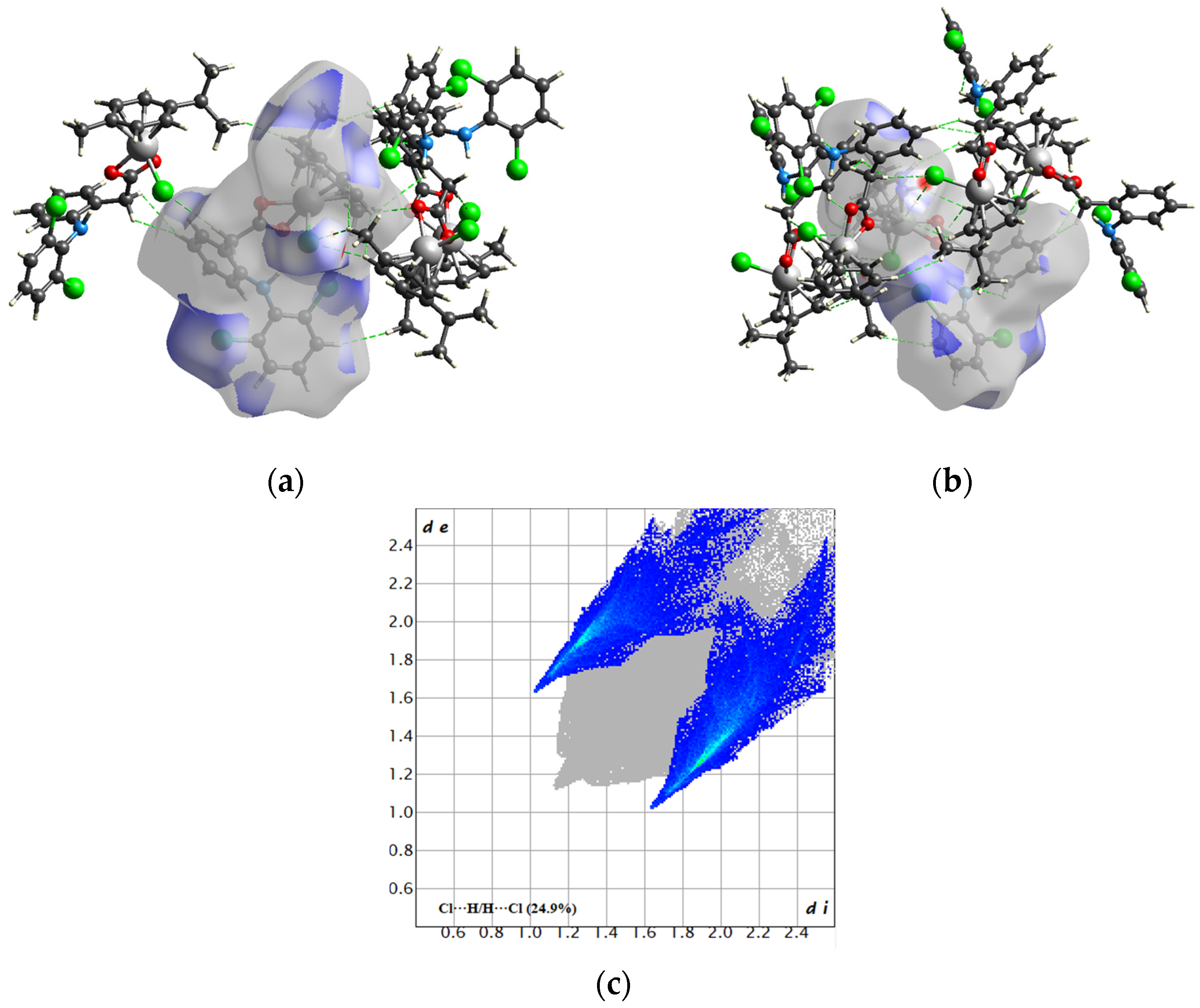
2.3. Hirshfeld Surfaces Analysis
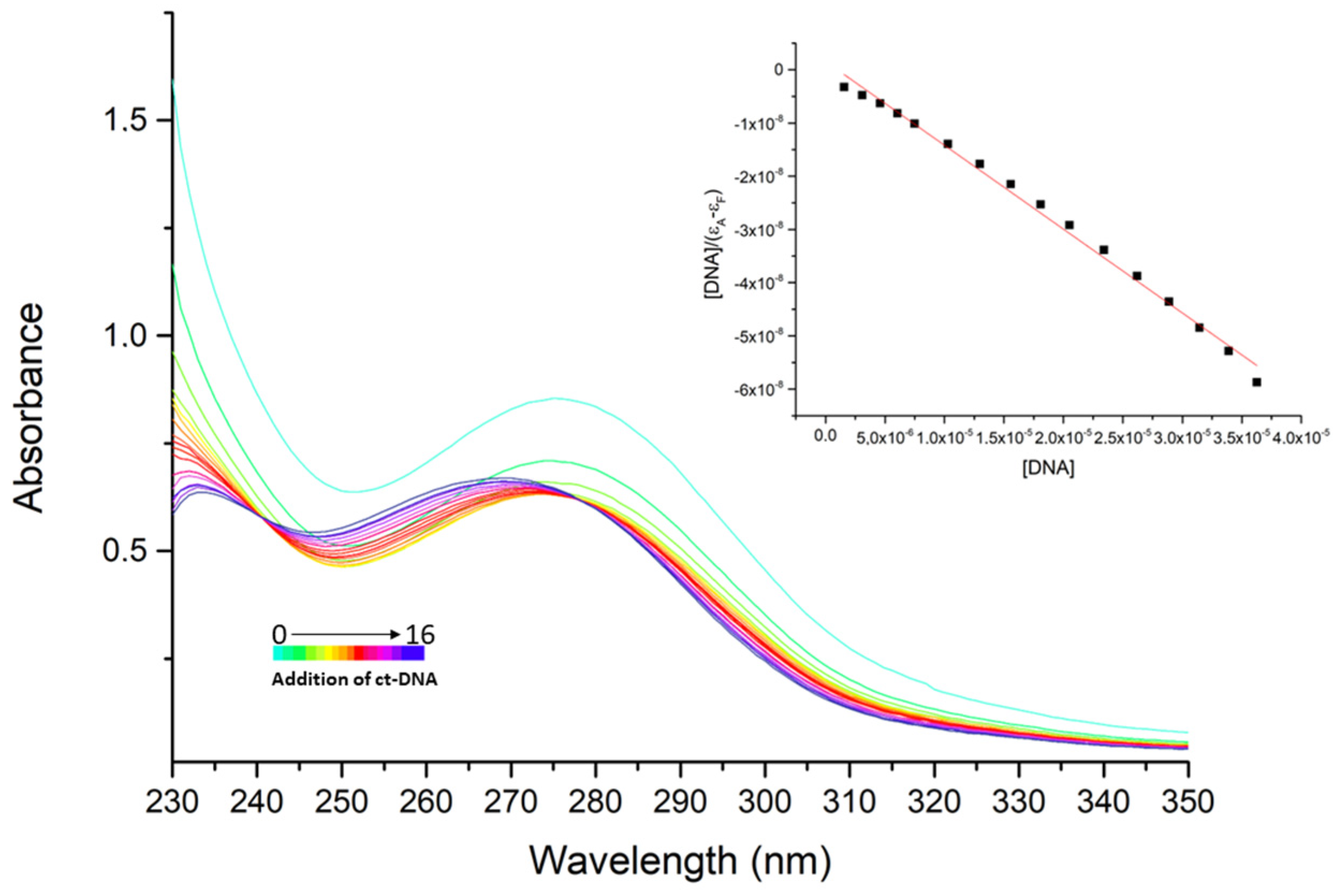
2.4. Interaction with DNA
3. Materials and Methods
3.1. Materials and Synthesis
3.2. Single-Crystal Crystallography
3.3. Hirshfeld Surface Analysis
3.4. Spectral Methods
3.5. DNA Binding Studies
4. Discussion
Supplementary Materials
Author Contributions
Funding
Institutional Review Board Statement
Informed Consent Statement
Data Availability Statement
Conflicts of Interest
References
- Gan, T.J. Diclofenac: An update on its mechanism of action and safety profile. Curr. Med. Res. Opin. 2010, 7, 1715–1731. [Google Scholar] [CrossRef] [PubMed]
- Sallman, A.R. The history of diclofenac. Am. J. Med. 1986, 80, 29–33. [Google Scholar] [CrossRef] [PubMed]
- Vane, J.R.; Botting, R.M. Mechanism of action of nonsteroidal anti-inflammatory drugs. Am. J. Med. 1998, 104, 2S–8S. [Google Scholar] [CrossRef]
- Srivastava, P.; Mishra, R.; Verma, M.; Sivakumar, S.; Patra, A.K. Cytotoxic ruthenium(II) polypyridyl complexes with naproxen as NSAID: Synthesis, biological interactions and antioxidant activity. Polyhedron 2019, 172, 132–140. [Google Scholar] [CrossRef]
- Kumar, P.; Swagatika, A.; Dasari, S.; Tomar, R.S.; Patra, A.K. Modulation of ruthenium anticancer drugs analogs with tolfenamic acid: Reactivity, biological interactions and growth inhibition of yeast cell. J. Inorg. Biochem. 2019, 199, 110769. [Google Scholar] [CrossRef] [PubMed]
- Kovala-Demertzi, D. Transition metal complexes of diclofenac with potentially interesting anti-inflammatory activity. J. Inorg. Biochem. 2000, 79, 153–157. [Google Scholar] [CrossRef]
- Biswas, P.; Datta, H.K.; Dastidar, P. Multi-NSAID-based Zn(II) coordination complex-derived metallogelators/metallogels as plausible multi-drug self-delivery systems. Chem. Commun. 2022, 58, 969–972. [Google Scholar] [CrossRef]
- Tarushi, A.; Hatzimitriou, A.G.; Estrader, M.; Kessissoglou, D.P.; Tangoulis, V.; Psomas, G. Toward multifunctional materials incorporating stepladder manganese(III) inverse-[9-MC-3[-metallacrowns and anti-inflammatory drugs. Inorg. Chem. 2017, 56, 7048–7057. [Google Scholar] [CrossRef]
- Kumar, S.; Sharma, R.P.; Venugopalan, P.; Ferretti, V.; Perontsis, S.; Psomas, G. Copper(II) diclofenac complexes: Synthesis, structural studies and interaction with albumins and cald-thymus DNA. J. Inorg. Biochem. 2018, 187, 97–108. [Google Scholar] [CrossRef]
- Perontsis, S.; Dimitriou, A.; Fotiadou, P.; Hatzidimitriou, A.G.; Papadopoulos, A.N.; Psomas, G. Cobalt(II) complexes with the non-steroidal anti-inflammatory drug diclofenac and nitrogen-donor ligands. J. Inorg. Biochem. 2019, 196, 110688. [Google Scholar] [CrossRef]
- Bera, S.; Chowdhury, A.; Sarkar, K.; Dastidar, P. Design and synthesis of ZnII-coordination polymers anchored with NSAIDs: Metallovesicle formation and multi-drug delivery. Chem. Asian J. 2020, 15, 503–510. [Google Scholar] [CrossRef]
- Lu, C.; Laws, K.; Eskandari, A.; Suntharalingam, K. A reactive oxygen species-generating, cyclooxygenase-2 inhibiting, cancer stem cell-potent tetranuclear copper(II) cluster. Dalton Trans. 2017, 46, 12785–12789. [Google Scholar] [CrossRef] [PubMed]
- Garcia-Garcia, A.; Mendez-Arriaga, J.M.; Martin-Escolano, R.; Cepeda, J.; Gomez-Ruiz, S.; Salinas-Castillo, A.; Seco, J.M.; Sanchez-Moreno, M.; Choquesillo, A.; Ruiz-Muelle, A.B.; et al. In vitro evaluation of leishmanicidal properties of a new family of monodimensional coordination polymers based on diclofenac ligand. Polyhedron 2020, 184, 114570. [Google Scholar] [CrossRef]
- Alisir, S.H.; Bariboga, B.; Caglar, S.; Buyukgungor, O. Synthesis, characterization, photoluminescent bproperties ant antimicrobial activities of two novel polymeric silver(I) complexes with diclofenac. J. Mol. Struct. 2017, 1130, 156–164. [Google Scholar] [CrossRef]
- Mondal, S.; Dastidar, P. Designing metallogelators derived from NSAID-basef Zn(II) coordination complexes for drug-delivery applications. Chem. Asian J. 2020, 15, 3558–3567. [Google Scholar] [CrossRef]
- Zampakou, M.; Tangoulis, V.; Raptopoulou, C.P.; Psycharism, V.; Papadopoulos, A.N.; Psomas, G. Structurally diverse manganese(II)-diclofenac complex showing enhanced antioxidant activity and affinity to serum albumins in comparison to sodium dichlofenac. Eur. J. Inorg. Chem. 2015, 13, 2285–2294. [Google Scholar] [CrossRef]
- Sayen, S.; Carlier, A.; Tarpin, M.; Guillon, E. A novel copper(II) mononuclear complex with the non-steroidl anti-inflammatory drug diclofenac: Structural characterization and biological activity. J. Inorg. Biochem. 2013, 120, 39–43. [Google Scholar] [CrossRef]
- Banti, C.N.; Jatzidimitriou, A.G.; Koirkoumelis, N.; Hadjikakou, S.K.; Tarpin, M.; Guillon, E. Diclofenac conjugates wuth biocides through silver(I) ions (CoMeD’s); development of a reliable model for the prediction of anti-proliferation of NSAID’s-silver formulations. J. Inorg. Biochem. 2019, 194, 7–18. [Google Scholar] [CrossRef]
- Kakoulidou, C.; Gritzapis, P.S.; Hatzidimitriou, A.G.; Fylaktakidou, K.C.; Psomas, G. Zn(II) complexes of (E)-4-(2-(2-(pyridin-2-ylmethylene)hydrazinyl)quinazoline in combination with non-steroidal anti-inflammatory drug sodium diclofenac: Structure, DNA binding and photo-cleavage studies, antioxidant activity and interaction with albumin. J. Inorg. Biochem. 2020, 211, 111194. [Google Scholar] [CrossRef]
- Caglar, S.; Aydernir, I.E.; Adiguzel, E.; Caglar, B.; Buyukgungor, O. Four copper(II) diclofenac complexes with pyridine derivatives: Synthesis, crystal structures, spectroscopic properties, thermal analysis and catechol oxidase activities. Inorg. Chim. Acta 2013, 408, 131–138. [Google Scholar] [CrossRef]
- Biswas, P.; Dastidar, P. Anchoring drugs to a zinc(II) coordintion polymer network: Exploiting structural rationale toward the desigb if metallogels for drug-delivery applications. Inorg. Chem. 2021, 60, 3218–3231. [Google Scholar] [CrossRef] [PubMed]
- Dos Santos, P.R.; Pitch, C.T.; Back, D.; Smiderle, F.; Dumas, F.; Moura, S. Synthesis, chemical characterization and DNA interaction study of new diclofenac and ibuprofen zinc(II)-nicotinamide ternary complexes as cyclooxygense inhibitor prototypes. J. Inorg. Biochem. 2020, 206, 111046. [Google Scholar] [CrossRef] [PubMed]
- Ashouri, F.; Faraji, A.R.; Molaeian, S.; Fall, M.A.; Butcher, R.J. The novel cobalt and manganese polymeric complex with the non-steroidal anti-inflammatory drug diclofenac: Synthesis, characterization and antivacterial studies. J. Mol. Struct. 2020, 1204, 127483. [Google Scholar] [CrossRef]
- Paul, M.; Sarkar, K.; Deb, J.; Dastidar, P. Hand-ground nanoscale ZnII-based coordination polymers derived from NSAIDs: Cell migration inhibition of human breast cancer cells. Chem. Eur. J. 2017, 23, 5736–5747. [Google Scholar] [CrossRef] [PubMed]
- Johnson, A.; Iffland-Muhlhaus, L.; Northcote-Smith, J.; Singh, K.; Ortu, F.; Apfel, U.P.; Sunthealingam, K. A bioinspired redox-modulating copper(II)-macroxyclic complex bearing non-steroidal anti-inflammatory drugs with anti-cancer stem cell activity. Dalton Trans. 2022, 54, 5904–5912. [Google Scholar] [CrossRef]
- Tarushi, A.; Raptopoulou, C.P.; Psychris, V.; Kontos, C.K.; Kessissoglou, D.P.; Scorillas, A.; Tangoulis, V.; Psomas, G. Copper(II) inverse-[9-metallacrown-3] compounds accommodating nitrato or diclofenac ligands: Structure, magnetism, and biological activity. Eur. J. Inorg. Chem. 2016, 2, 219–231. [Google Scholar] [CrossRef]
- Altay, A.; Caglar, S.; Caglar, B.; Sahin, O. Synthesis, structural, thermal elucidation and in vitro anticancer activity of novel silver(I) complexes with non-steroidal anti-inflammatory drugs diclofenac and mefenamic acid including picoline derivatives. Polyhedron 2018, 151, 160–170. [Google Scholar] [CrossRef]
- Alisir, S.H.; Dege, N.; Tapramaz, R. Synthesis, crystal structures and characterizations of three new copper(II) complexes including anti-inflammatory diclofenac. Acta Crystallogr. 2019, C75, 388–397. [Google Scholar] [CrossRef]
- Sayen, S.; Guillon, E. Tetra kis{2-[2-(2,6-dichloro anilino)phen yl]ethano ato-κ2O:O’}bis [(dimethyl sulfoxide-κO)copper(II)](Cu-Cu): A binuclear CuII complex with the non-steroidal anti-inflammatory drug diclofenac. Acta Crystallogr. 2012, E68, m474–m475. [Google Scholar] [CrossRef]
- Antonenko, T.A.; Gracheva, Y.A.; Shpakovsky, D.B.; Vorobyev, M.A.; Tafeenko, V.A.; Mazur, D.M.; Milaeva, E.R. Cytotoxic activity of organotin compounds containing non-steroidal anti-inflammatory drugs. J. Organomet. Chem. 2022, 960, 122191. [Google Scholar] [CrossRef]
- Kourkoumelis, N.; Kovala-Demertzi, D.; Tiekink, E.R.T. Crystal and molecular structure of 1,2:3,4-di-μ2-[2,6-dichlorophenyl)amino]benzeneacetato-O,O-l,3-bis[(2,6-dichlorophenyl)amino]-benzeneacetato-O-1,2,4:2,3,4-di-μ3-oxo-tetrakis[di-n-butyltin(IV)]: {[nBu2Sn(O2CCH2C6H4N(H)C6H3Cl2)]2O}2. Z. Kristallogr. 1999, 214, 758–762. [Google Scholar] [CrossRef]
- Li, S.-H.; Zhao, Y.; Zhang, L. Crystal structure of catena-poly-[(μ2-2-(2-((2,6-dimethylphenyl)amino)phenyl)acetato-κ2O:O′)(μ2-2-(2-((2,6-dimethylphenyl)amino)phenyl)acetate-κ3O,O′:O′)cadmium(II)])].C28H20N2Cl4O4Cd. Z. Kristallogr. NCS 2016, 231, 1009–1011. [Google Scholar] [CrossRef]
- Oliveira, K.M.; Honorato, J.; Gonçalves, G.R.; Cominetti, M.R.; Batista, A.A.; Correa, R.S. Ru(II)/diclofenac-based complexes: DNA, BSA interaction and their anticancer evaluation against lung and breast tumor cells. Dalton Trans. 2020, 49, 12643–12652. [Google Scholar] [CrossRef] [PubMed]
- Khan, R.A.; Al-Lohedan, H.A.; Farah, M.A.; Ali, M.S.; Alsalme, A.; Al-Anazi, K.M.; Tabassum, S. Evaluation of (ɳ6-p-cymene) ruthenium diclofenac complex as anticancer chemotherapeutic agent: Interaction with biomolecules, cytotoxicity assays. J. Biomol. Struct. Dyn. 2019, 37, 3905–3913. [Google Scholar] [CrossRef]
- Mandal, P.; Kundu, B.K.; Vyas, K.; Sabu, V.; Helen, A.; Dhankar, S.S.; Nagaraja, C.M.; Bhattacherjee, D.; Bhabak, K.P.; Mukhopadhyay, S. Ruthenium(II) Arene NSAID Complexes: Inhibition of cyclooxygenase and antiproliferative activity against cancer cell lines. Dalton Trans. 2018, 47, 517–527. [Google Scholar] [CrossRef]
- Nakamoto, I. Infrared and Raman Spectra of Inorganic and Coordination Compounds, Part B, 6th ed.; Wiley: Hoboken, NJ, USA, 2009. [Google Scholar]
- Janiak, C. A critical account on π-π stacking in metal complexes with aromatic nitrogen-containing ligands. J. Chem. Soc. Dalton Trans. 2020, 21, 3885–3896. [Google Scholar] [CrossRef]
- Spackman, M.A.; McKinnon, J.J.; Jayalitaka, D. Electrostatic potentials mapped on Hirshfeld surfaces provide direct insight into intermolecular interactions in crystals. Cryst. Eng. Comm. 2008, 10, 377–388. [Google Scholar] [CrossRef]
- Koziskova, J.; Hahn, F.; Richter, J.; Kožíšek, J. Comparison of different absorption corrections on the model structure of tetrakis(μ2-acetato)-diaqua-di-copper(II). Acta Chim. Slovaca 2016, 9, 136–140. [Google Scholar] [CrossRef]
- Sheldrick, G.M. SHELXT–Integrated space-group and crystal-structure determination. Acta Crystallogr. 2015, A71, 3–8. [Google Scholar] [CrossRef]
- Sheldrick, G.M. Crystal structure refinement with SHELXL. Acta Crystallogr. 2015, C71, 3–8. [Google Scholar] [CrossRef]
- Capelli, S.C.; Bürgi, H.-B.; Dittrich, B.; Grabowsky, S.; Jayatilaka, D. Hirshfeld atom refinement. IUCrJ 2014, 1, 361–379. [Google Scholar] [CrossRef] [PubMed]
- Chrappová, J.; Pateda, Y.R.; Rakovský, E. Molecular acidity: Synthesis and crystal structure analysis of NH4[Zn(cma)(H2O)2]∙H2O using IAM and HAR approaches. J. Chem. Crystallogr. 2022; in press. [Google Scholar] [CrossRef]
- Kleemiss, F.; Dolomanov, O.V.; Bodensteiner, M.; Peyerimhoff, N.; Midgley, L.; Bourhis, L.J.; Genoni, A.; Malaspina, L.A.; Jayatilaka, D.; Spencer, J.L.; et al. Accurate crystal structures and chemical properties from NoSpherA2. Chem. Sci. 2021, 12, 1675–1692. [Google Scholar] [CrossRef] [PubMed]
- Bourhis, L.J.; Dolomanov, O.V.; Gildea, R.J.; Howard, J.A.K.; Puschmann, H. The anatomy of a comprehensive constrained, restrained refinement program for the modern computing environment—Olex2 dissected. Acta Crystallogr. 2015, A71, 59–75. [Google Scholar] [CrossRef]
- Neese, F. Software update: The ORCA program system, version 4.0. WIREs Comput. Mol. Sci. 2018, 8, e1327. [Google Scholar] [CrossRef]
- Adamo, C.; Barone, V. Toward reliable density functional methods without adjustable parameters: The PBE0 model. J. Chem. Phys. 1999, 110, 6158–6169. [Google Scholar] [CrossRef]
- Pollak, P.; Weigend, F. Segmented contracted error-consistent basis sets of double- and Triple-ζ valence quality for one- and two-component relativistic all-electron calculations. J. Chem. Theory Comput. 2017, 13, 3696–3705. [Google Scholar] [CrossRef]
- Barros, C.L.; de Oliveira, P.J.P.; Jorge, F.E.; Canal Neto, A.; Campos, M. Gaussian basis set of double zeta quality for atoms Rb through Xe: Application in non-relativistic and relativistic calculations of atomic and molecular properties. Mol. Phys. 2010, 108, 1965–1972. [Google Scholar] [CrossRef]
- Campos, C.T.; Jorge, F.E. Triple zeta quality basis sets for atoms Rb through Xe: Application in CCSD(T) atomic and molecular property calculations. Mol. Phys. 2013, 111, 167–173. [Google Scholar] [CrossRef]
- Andrae, D.; Häußermann, U.; Dolg, M.; Stoll, H.; Preuß, H. Energy-adjustedab initio pseudopotentials for the second and third row transition elements. Theor. Chim. Acta 1990, 77, 123–141. [Google Scholar] [CrossRef]
- Weigend, F.; Ahlrichs, R. Balanced basis sets of split valence, triple zeta valence and quadruple zeta valence quality for H to Rn: Design and assessment of accuracy. Phys. Chem. Chem. Phys. 2005, 7, 3297. [Google Scholar] [CrossRef]
- Dolomanov, O.V.; Bourhis, L.J.; Gildea, R.J.; Howard, J.A.K.; Puschmann, H. OLEX2: A complete structure solution, refinement and analysis program. J. Appl. Crystallogr. 2009, 42, 339–341. [Google Scholar] [CrossRef]
- Spackman, P.R.; Turner, M.J.; McKinnon, J.J.; Wolff, S.K.; Grimwood, D.J.; Jayalitaka, D.; Spackman, M.A. CrystalExplorer: A program for Hirshfeld surface analysis, visualization and quantitative analysis of molecular crystals. J. Appl. Crystallogr. 2021, 54, 1006–1011. [Google Scholar] [CrossRef] [PubMed]
- Hirshfeld, F.L. Vobded-atom fragments for describing molecular charge densities. Theor. Chim. Acta. 1977, 44, 129–138. [Google Scholar] [CrossRef]
- Spackman, M.A.; Jayalitaka, D. Hirhfeld surface analysis. Cryst. Eng. Comm. 2009, 11, 19–32. [Google Scholar] [CrossRef]
- Spackman, M.A.; McKinnon, J.J. Fingerprinting intermolecular interactions in molecular crystals. Cryst. Eng. Comm. 2002, 4, 378–392. [Google Scholar] [CrossRef]
- Parkin, A.; Barr, G.; Dong, W.; Gilmore, C.J.; Jayalitaka, D.; McKinnon, J.J.; Spackman, M.A.; Wilson, C.C. Comparing entire crystal structures: Structural genetic fingerprinting. Cryst. Eng. Comm. 2007, 9, 648–652. [Google Scholar] [CrossRef]
- McKinnon, J.J.; Jayalitaka, D.; Spackman, M.A. Towards quantitative analysis of intermolecular interactions with Hirshfeld surfaces. Chem. Commun. 2007, 3814–3816. [Google Scholar] [CrossRef]
- Wang, K.; He, X.; Rong, C.; Zhong, A.; Liu, S.; Zhao, D. On the origin and nature of internal methyl rotation barriers: An information-theoretic approach study. Theor. Chem. Acc. 2022, 141, 68. [Google Scholar] [CrossRef]
- Liu, S.; Pederse, L.G. Estimation of Molecular Acidity via Electrostatic Potential at the Nucleus and Valence Natural Atomic Orbitals. J. Phys. Chem. A 2019, 123, 6751–6760. [Google Scholar] [CrossRef]
- Cao, X.; Rong, C.; Zhong, A.; Lu, T.; Liu, S. Molecular acidity: An accurate description with information-theoretic approach in density functional reactivity theory. J. Comput. Chem. 2018, 39, 117–129. [Google Scholar] [CrossRef]
- Marmur, J. Thermal renaturation of deoxyribonucleic acids. J. Mol. Biol. 1961, 3, 208–211. [Google Scholar] [CrossRef]
- Reichmann, M.F.; Rice, S.A.; Thomas, C.A.; Doty, P.J. A Further Examination of the Molecular Weight and Size of Desoxypentose Nucleic Acid. J. Am. Chem. Soc. 1954, 76, 3047–3053. [Google Scholar] [CrossRef]


| 1 | 1a | 1b | 1c | 1d | 1e | 1f | 1g | 1h | |
|---|---|---|---|---|---|---|---|---|---|
| Base set | IAM | x2c-SVP | Jorge-DZP-DKH | Jorge-TZP-DKH | x2c-TZVP | x2c-TZVPP | ECP-def2-SVP | ECP-def2-TZVP | ECP-def2-TZVPP |
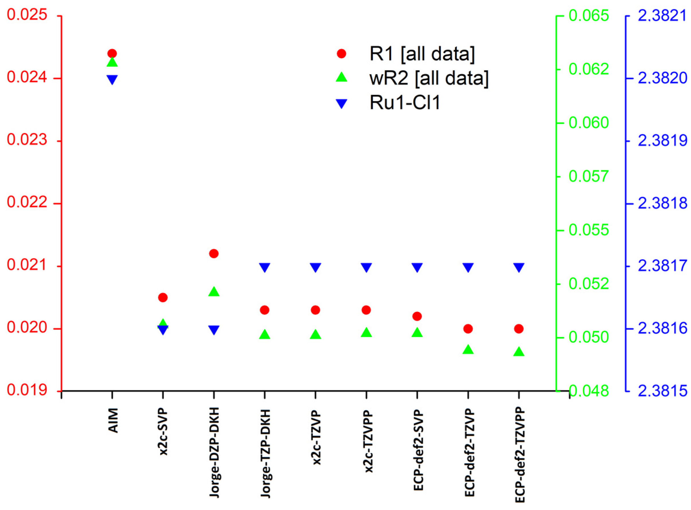
| R1 [I ≥ 2σ(I)] | 0.0226 | 0.0187 | 0.0195 | 0.0186 | 0.0186 | 0.0186 | 0.0185 | 0.0183 | 0.0183 |
| wR2 [I ≥ 2σ(I)] | 0.0616 | 0.0496 | 0.0511 | 0.0492 | 0.0492 | 0.0493 | 0.0492 | 0.0484 | 0.0484 |
| R1 [all data] | 0.0244 | 0.0205 | 0.0212 | 0.0203 | 0.0203 | 0.0203 | 0.0202 | 0.0200 | 0.0200 |
| wR2 [all data] | 0.0628 | 0.0506 | 0.0521 | 0.0501 | 0.0501 | 0.0502 | 0.0502 | 0.0494 | 0.0493 |
| Ru1-Cl1 | 2.3820(6) | 2.3816(5) | 2.3816(5) | 2.3817(5) | 2.3817(5) | 2.3817(5) | 2.3817(5) | 2.3817(5) | 2.3817(5) |
| Ru1-O1 | 2.1711(15) | 2.1705(12) | 2.1709(12) | 2.1705(12) | 2.1705(12) | 2.1705(12) | 2.1702(12) | 2.1702(12) | 2.1702(12) |
| Ru1-O2 | 2.1434(15) | 2.1425(12) | 2.1429(12) | 2.1426(12) | 2.1426(12) | 2.1426(12) | 2.1422(12) | 2.1422(12) | 2.1422(12) |
| GOF | 1.051 | 1.088 | 1.089 | 1.106 | 1.106 | 1.108 | 1.089 | 1.094 | 1.094 |
| Largest difference peak/hole | 1.18/−0.61 | 1.11/−0.69 | 1.10/−0.70 | 1.12/−0.69 | 1.13/−0.68 | 1.13/−0.69 | 1.12/−0.66 | 1.13/−0.66 | 1.13/−0.67 |
| λ (nm) | Δλ (nm) | ΔA/A0 (%) | Kb (M−1) |
|---|---|---|---|
| 275 | −6 | 22 a | 9.99 (±3.21) × 105 |
| 233 | −1 | 49 | 5.35 (±0.61) × 105 |
| Compound | Pbca | 1 |
|---|---|---|
| Chemical formula | C24H24Cl3NO2Ru | C24H24Cl3NO2Ru |
| Bases set | -(IAM) | -(IAM) |
| Mr | 607.89 | 607.89 |
| Crystal system | Orthorombic | Triclinic |
| Space group | Pbca | P |
| T/K | 298(2) | 100 |
| a/Å | 16.8227(12) | 9.4171(2) |
| b/Å | 11.3329(8) | 10.8706(2) |
| c/Å | 25.1614(17) | 12.7090(2) |
| α/° | 90 | 90.2240(10) |
| β/° | 90 | 107.2210(10) |
| γ/° | 90 | 112.2920(10) |
| V/Å3 | 4797.0(6) | 1139.83(4) |
| Z | 8 | 2 |
| Crystal size/mm | 0.36 × 0.27 × 0.24 | 0.11 × 0.063 × 0.03 |
| ρcalc/g∙cm−3 | 1.567 | 1.771 |
| S | 1.071 | 1.051 |
| R1 [I > 2σ(I)] | 0.0282 | 0.0237 |
| wR2 [All data] | 0.0741 | 0.0657 |
| REFCODE/CCDC | TESSAO [35] | 2235444 |
Disclaimer/Publisher’s Note: The statements, opinions and data contained in all publications are solely those of the individual author(s) and contributor(s) and not of MDPI and/or the editor(s). MDPI and/or the editor(s) disclaim responsibility for any injury to people or property resulting from any ideas, methods, instructions or products referred to in the content. |
© 2023 by the authors. Licensee MDPI, Basel, Switzerland. This article is an open access article distributed under the terms and conditions of the Creative Commons Attribution (CC BY) license (https://creativecommons.org/licenses/by/4.0/).
Share and Cite
Schoeller, M.; Piroš, M.; Lušpai, K.; Braniša, J.; Moncol, J. Synthesis, Crystal Structure, Hirshfeld Surfaces Analysis, Interaction with DNA and Comparation of Different Bases in Hirshfeld Atom Refinement of New Polymorph of Chlorido(η6-p-cymene)(diclofenac)Ruthenium(II) Organometallic Compound. Inorganics 2023, 11, 190. https://doi.org/10.3390/inorganics11050190
Schoeller M, Piroš M, Lušpai K, Braniša J, Moncol J. Synthesis, Crystal Structure, Hirshfeld Surfaces Analysis, Interaction with DNA and Comparation of Different Bases in Hirshfeld Atom Refinement of New Polymorph of Chlorido(η6-p-cymene)(diclofenac)Ruthenium(II) Organometallic Compound. Inorganics. 2023; 11(5):190. https://doi.org/10.3390/inorganics11050190
Chicago/Turabian StyleSchoeller, Martin, Milan Piroš, Karol Lušpai, Jana Braniša, and Ján Moncol. 2023. "Synthesis, Crystal Structure, Hirshfeld Surfaces Analysis, Interaction with DNA and Comparation of Different Bases in Hirshfeld Atom Refinement of New Polymorph of Chlorido(η6-p-cymene)(diclofenac)Ruthenium(II) Organometallic Compound" Inorganics 11, no. 5: 190. https://doi.org/10.3390/inorganics11050190