Article

3′-Aminothiocyclohexanespiro-5′-hydantoin and Its Pt(II) Complex—Synthesis, Cytotoxicity and Xanthine Oxidase Inhibitory Activity

by
Emiliya Cherneva
1,2,
Mariyana Atanasova
1,
Žaklina Šmelcerović
3,
Katarina Tomović
4,
Rossen Buyukliev
1,
Andrija Šmelcerović
5,* and
Adriana Bakalova
1,*
1
Department of Chemistry, Faculty of Pharmacy, Medical University of Sofia, 2 Dunav Str., 1000 Sofia, Bulgaria
2
Institute of Organic Chemistry with Centre of Phytochemistry, Bulgarian Academy of Sciences, Acad. G. Bonchev Str., Build 9, 1113 Sofia, Bulgaria
3
Center for Biomedicinal Science, Faculty of Medicine, University of Niš, Bulevar Dr Zorana Đinđića 81, 18000 Niš, Serbia
4
Department of Pharmacy, Faculty of Medicine, University of Niš, Bulevar Dr Zorana Đinđića 81, 18000 Niš, Serbia
5
Department of Chemistry, Faculty of Medicine, University of Niš, Bulevar Dr Zorana Đinđića 81, 18000 Niš, Serbia
*
Authors to whom correspondence should be addressed.
Inorganics 2022, 10(10), 175; https://doi.org/10.3390/inorganics10100175
Submission received: 8 August 2022 / Revised: 11 October 2022 / Accepted: 18 October 2022 / Published: 20 October 2022

Abstract

:
Herein, we report the synthesis of platinum(II) complex bearing 3′-aminothiocyclohexanespiro-5′-hydantoin as ligand. The complex was characterized by IR, NMR spectral analyses, elemental analyses and density functional theory (DFT) calculations. Cytotoxicity and inhibitory potential on xanthine oxidase (XO) were evaluated by performed docking calculations. The cytotoxic activities of the 3′-aminothiocyclohexanespiro-5′-hydantoin (1), its Pt(II) complex (2), thiocyclohexanespiro-5′-hydantoin (3), and its platinum complex (4) were assessed against HL-60 and MDA-MB-231 cells in comparison with the antiproliferative activity of cisplatin as a referent. The ligands (1 and 3) did not exhibit in vitro antitumor efficacy on either of the human tumor cell lines. Complex 2 showed higher antitumor activity (IC50 = 42.1 ± 2.8 μM on HL-60 and 97.8 ± 7.5 μM against MDA-MB-231 cells) than complex 4 (IC50 = 89.6 ± 2.8 μM on HL-60 and 112.5 ± 4.2 μM in MDA-MB-231 cells). IC50 values of cisplatin as referent were 8.7 ± 2.4 μM on HL-60 and 31.6 ± 5.4 μM on MDA-MB-231 cell lines. The inhibitory activity of ligands and complexes against XO, evaluated in vitro, were compared with allopurinol (IC50 = 1.70 ± 0.51 μM) as standard inhibitor. The platinum(II) complexes (2 and 4) inhibited the activity of XO, with IC50 values 110.33 ± 26.38 μM and 115.45 ± 42.43 μM, respectively, while the ligands 1 and 3 did not show higher degrees of inhibition at concentrations lower than 150 μM. The inhibitory potential against XO might be a possible precedent resulting in improved profile and anticancer properties.

1. Introduction

Metal complexes are considered to be an extremely important class of compounds in medicine and medicinal chemistry because of their diverse pharmacological applications. Many complexes act as antioxidant, antibacterial, antiviral, analgesic, antimicrobial and anticancer agents [1,2,3]. Transition metal complexes have been extensively investigated as chemotherapeutic agents. Platinum-based anticancer agents have become the most common class used in chemotherapy worldwide [4,5]. Anticancer drugs that have been used in cancer therapy have serious well-known side effects [6,7,8]. To increase bioavailability, enhance cytotoxicity, and reduce side effects in comparison to cisplatin, a new class of Pt(II) complexes [9,10] was synthesized.
Hydantoins (imidazolidinediones) are small molecules that possess different biological and pharmaceutical applications, including as anticonvulsants, antiarrhythmics, antibacterial and antitumor agents [11,12,13]. A series of spirohydantoins were studied and evaluated as promising anticancer compounds in our earlier research [14,15].
The tumor growth causes tissue damage that provokes the inflammatory response and possible increase in the activity and the expression of xanthine oxidase (XO) in plasma. The activation of carcinogenic compounds can be catalyzed by this oxidase. Xanthine oxidase might also act tumorigenically by producing reactive oxygen and nitrogen species [16]. Despite being rare [17], there are data on metal complex-based inorganic XO inhibitors [18,19,20,21,22,23].
The present paper describes the synthesis, characterization, cytotoxic activity and inhibitory potential on XO of a new platinum complex (complex 2). The biological activities of 3′-aminothiocyclohexanespiro-5′-hydantoin (1) and thiocyclohexanespiro-5′-hydantoin (3) were compared with those of their platinum complexes (2 and 4) (Figure 1). To define the docking interactions of Pt complex–XO complex, docking calculations were performed.

2. Results and Discussion
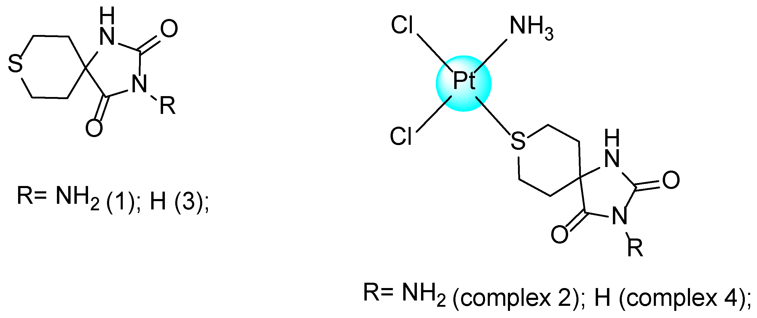
2.1. Synthesis

On the basis of our previous investigations [24], the structure of thiocyclohexanespiro-5′-hydantoin was essential for the platinum complexes to possess the potent XO inhibitory activity. Therefore, we synthesized a new Pt(II) complex with 3′-aminothiocyclohexanespiro-5′-hydantoin as ligand. The complex was synthesized via one-step reaction by mixing K[Pt(NH3)Cl3] with the ligand in a stoichiometric ratio of 1:1 under mild conditions and good chemical yield.

2.2. Spectral Characterization and Geometry Optimization

2.2.1. Spectral Analysis

The coordination of the ligand with platinum ion was demonstrated by comparing of the IR spectra of the ligand 1 and complex 2. The stretching vibrations corresponding to the C–S bond are observed at 655 cm−1 in ligand 1, while in complex 2, ν(C–S) is at 625 cm−1. This decrease of 30 cm−1 is due to the fact that the ligand bonds with the metal ion via the sulphur atom from the thiocyclohexane moiety. The NH and ν(C═O) stretching are slightly affected upon coordination. The IR spectra of the new complex 2 and the ligand 1 in the far IR below 400 cm−1 region were also recorded, where metal–halogen stretching vibrations occur. Two absorption bands at 330 and 319 cm−1 are assignable to Pt-Cl stretching. The doublet character of these bands is an indication of the cis-configuration of the complex [25,26,27]. Moreover, the method for obtaining of the new complex supposed the synthesis of a cis-isomer according to the literature [28]. For example, for the synthesis of the cis-isomer, K[Pt(NH3)Cl3] as a starting complex is used, while for the synthesis of the trans-isomer, the other starting complex—cis-[Pt(NH3)2Cl(NO3)]—is used. Starting from K[PtCl3NH3], the reaction with ligand 1 leads to substitution of one of the two trans-chlorides (both cis to the ammine), which are mutually trans-labilized [29] (Supplementary Materials Figures S1 and S2).
A comparison of the 1H NMR spectra of pure ligand 1 and complex 2 indicates a chemical shift difference of 0.20 ppm for the axial protons from CH2 groups connected to the sulphur atom. This chemical change is proof of the coordinative bonding between the sulphur atom and platinum ion (Supplementary Materials Figures S3 and S4).
There is no great difference between the chemical shifts of the carbon atoms in the 13C NMR spectra of the metal-free ligand 1 and complex 2 (Supplementary Materials Figures S5 and S6).

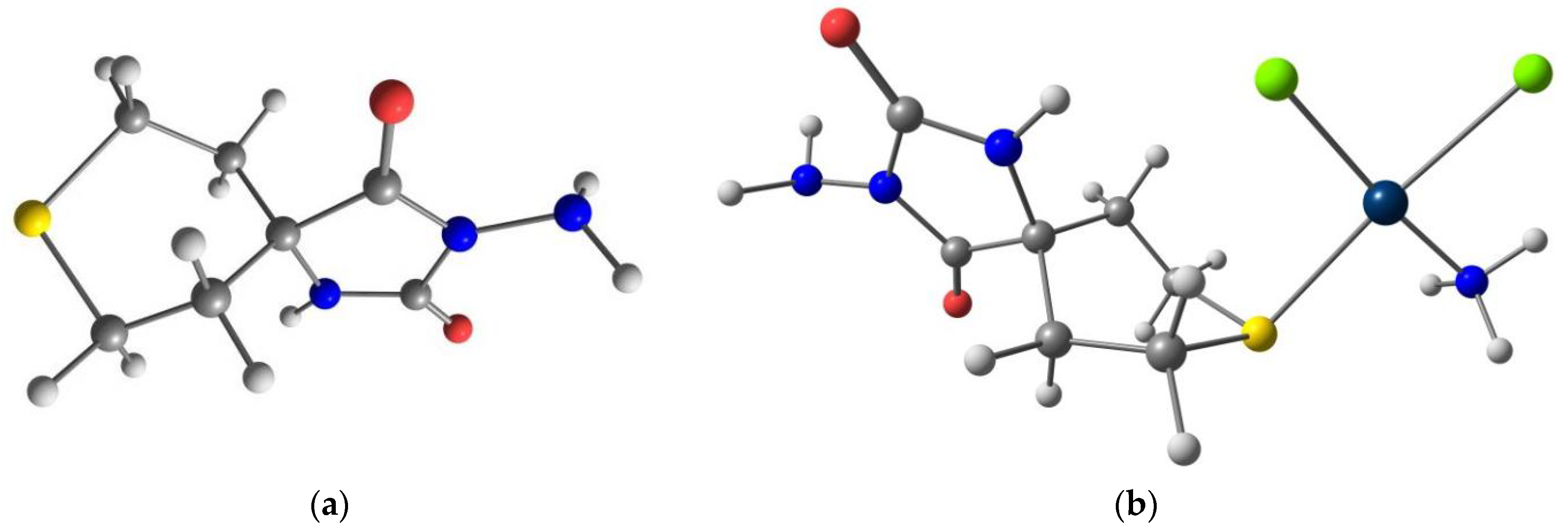
2.2.2. Quantum Chemical Modeling

Being unable to grow single crystals, quantum chemical studies were applied to explore the structure and the way of bonding of the Pt ion with the ligand using the B3LYP/6-31+G(d) level for nonmetals and LANL2DZ for platinum. In Figure 2, the optimized structures of the studied compounds are presented. In our previously published work [30], the optimized molecular geometry of ligand 1 was described in detail. To investigate the geometrical structure of the complex, two possible cis-conformations are optimized, namely boat and chair, by taking into account the position of spiro C-atom around platinum center. The calculations in the gaseous state show that the boat conformation is energetically more favorable by 16 kJ/mol than chair. This result is similar to that found in our previous calculations on the thiocyclohexanespiro-5′-hydantoin complex [30].
Pt(II) ion is located in the center of a slightly distorted square planar coordination geometry. The Pt-Cl (2.41 Å), Pt-S (2.45 Å) and Pt-Nammonia (2.09 Å) bond lengths are similar or close to values observed for other optimized square planar complexes [31,32,33,34,35]. After coordination of the ligand with the platinum ion, the ligand geometry will not change significantly, according to the theoretical analysis. The lengthening of the C-S bonds by 0.07–0.08 Å is probably due to the bonding of the ligand with the Pt ion.
To prove the cis-configuration of complex 2, IR spectra of cis- and trans-isomers were calculated. In comparing these two spectra, small differences can be observed. However, when comparing of the calculated IR spectra for cis-and trans-isomers with the experimental data, it can be observed that the experimental IR spectrum is close to the theoretical spectrum of the cis-isomer. The fact that the theoretical spectrum was made in the gas phase, while the experimental spectrum was made in the solid phase, should be taken into account (Supplementary Materials Figure S7)

2.3. Biological Evaluation

2.3.1. Antiproliferative Activity

In vitro cytotoxicity of the new complex 2 proved to be significant within this series of compounds. Complex 2 showed higher antitumor efficacy against HL-60 and MDA-MB-231 than complex 4 and the ligands (1 and 3). IC50 values were 42.1 ± 2.8 μM for complex 2 and 89.6 ± 2.8 μM for complex 4 on the HL-60 cell line and 97.8 ± 7.5 μM for complex 2, and 112.5 ± 4.2 μM for complex 4 on the MDA-MB-231 cell line. Ligands 1 and 3 did not exhibit in vitro antiproliferative activity on either of the human tumor cell lines. IC50 values of cisplatin as a referent were 8.7 ± 2.4 μM on HL-60 and 31.6 ± 5.4 μM against MDA-MB-231 cell lines.

2.3.2. XO Activity

The platinum(II) complexes (2 and 4) inhibited the activity of XO, with IC50 values 110.33 ± 26.38 μM and 115.45 ± 42.43 μM, respectively, while ligands 1 and 3 did not show a higher degree of inhibition at concentrations lower than 150 μM. The IC50 value of allopurinol as a standard inhibitor was 1.70 ± 0.51 μM.

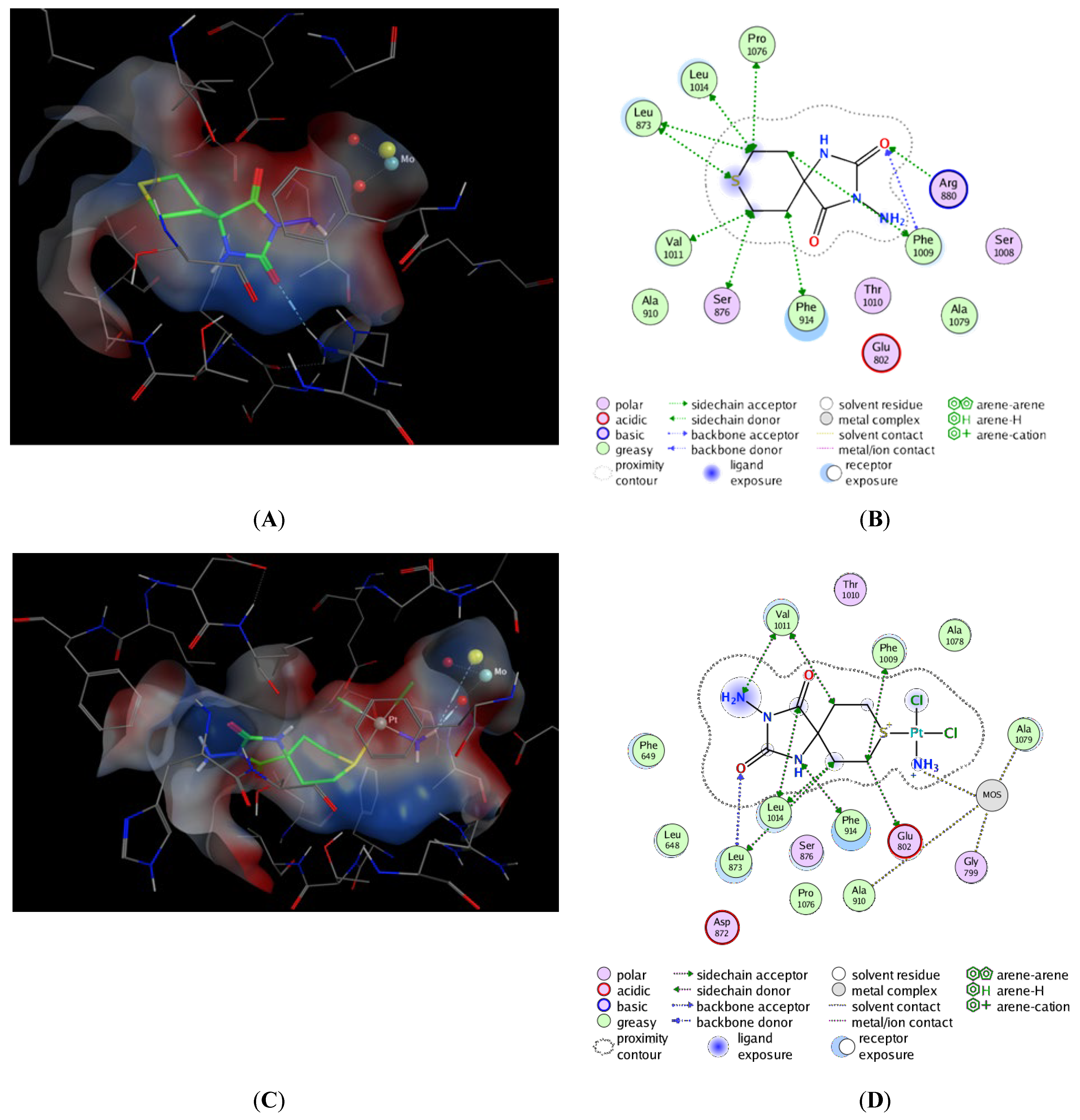
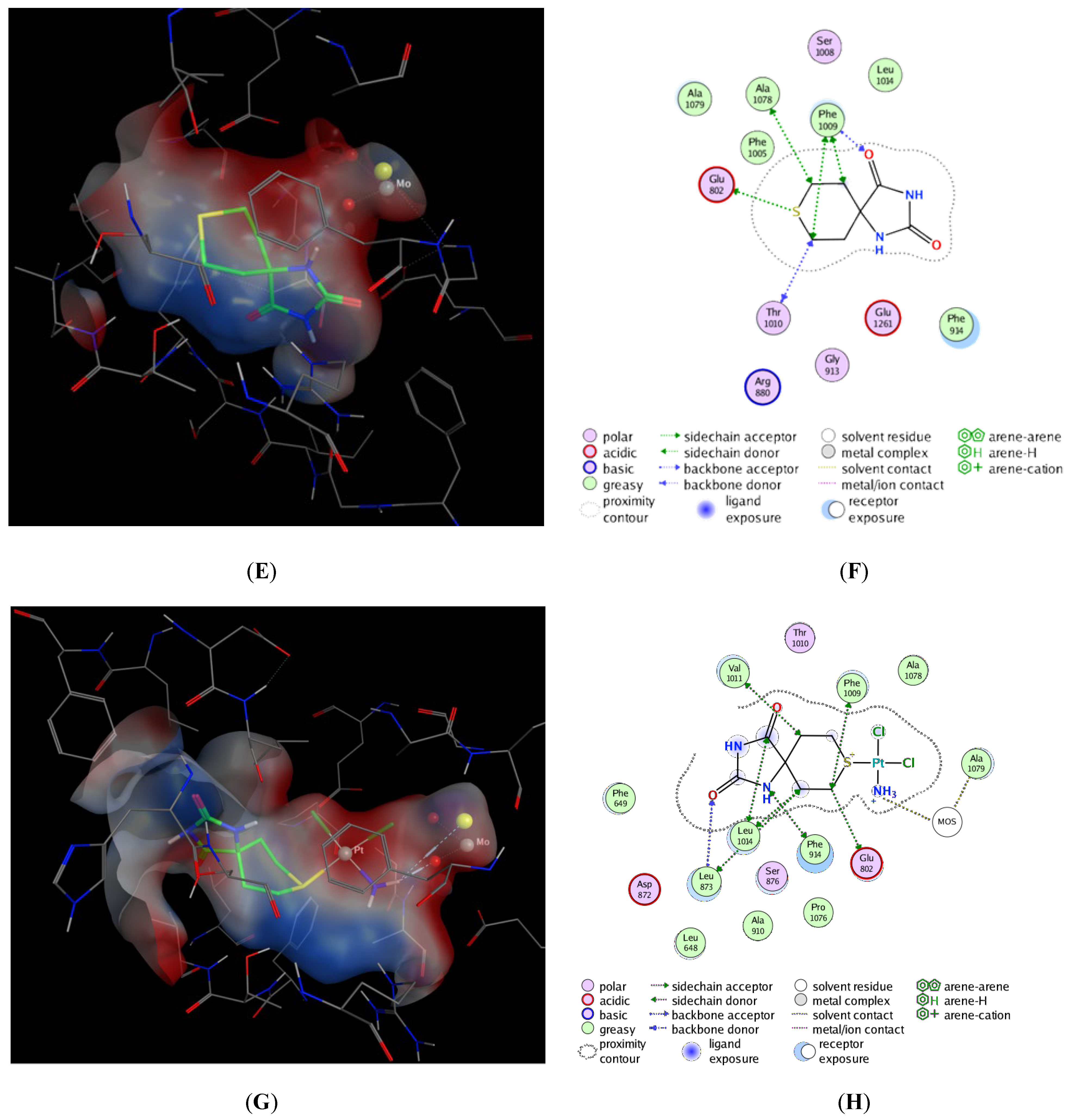
3. Molecular Docking Study

The best docking scores with the lowest Affinity dG value of ligands 1 and 3 and Pt(II) complexes 2 and 4 are given in Table 1. The corresponding binding poses at the XO binding site are depicted in Figure 3. Ligand 1 forms a hydrogen bond between the O atom of CO group at the fourth position and the hydrogen atom of the guanidino group of Arg880 (Figure 3A,B). Additionally, Van der Waals interactions stabilize the predicted pose. In the case of ligand 3 molecule, only weak Van der Waals interactions occur (Figure 3E,F). Both molecules are characterized by the low Affinity dG score of the docking calculations (Table 1). This is possibly due to the fact that both ligands 1 and 3 are placed close to the entrance, rather than deep in the binding pocket, where the molybdenum center is located.
The best predicted poses of complexes 2 and 4 insert deep into the pocket, close to the molybdenum atom (Figure 3C,D,G,H). The coordinative bound of ammonium group links with oxygen and sulfur atoms of MOS. In addition, Van der Waals interactions stabilize the complexes. This results in higher Affinity dG values in both molecules (Table 1).

4. Materials and Methods

Thiocycolhexanespiro-5′-hydantoin is available by means of Bucherer–Berg reaction from (NH4)2CO3, NaCN and tetrahydro-4H-thiopyran-4-one (Aldrich). Potassium amminethrechloroplatinate(II) was purchased from Sigma Aldrich USA. All other chemicals used were of analytical reagent grade. The carbon, nitrogen and hydrogen content of the new compound were determined by elemental analyses on a EuroEA 3000—Single, “Euro Vector SpA”. IR spectra were recorded in the range of 4000–100 cm−1 on Bruker Invenio R spectrophotometer, ATR (attenuated total reflectance) mode with a diamond crystal accessory. The spectra were referenced to air as a background by accumulating 100 scans, at a resolution of 2 cm−1. The 1H NMR and 13C spectra were recorded on Bruker WM 500 (500 MHz) spectrometer in dimethyl sulfoxide as a solvent. Chemical shifts are expressed to tetramethylsilan and are represented in δ (ppm). Splitting patterns were indicated by the symbols s (singlet), m (multiplet) and bs (broad singlet). The solution of complex 2 was freshly prepared in DMSO-d6. Melting point was determined as the phase transition from solid to liquid at atmospheric pressure, on a Buchi 540 apparatus.

4.1. Synthesis

Cis-Amminedichlorido-(3′-aminothiocyclohexanespiro-5′-hydantoin) Platinum (II)—Cis-[PtL(NH3)Cl2], Complex 2

To aqueous solution of K[Pt(NH3)Cl3] (0.1605 g, 0.0449 mmol) was added an aqueous ethanol solution of ligand (ϕr = 1:1) (0.1079 g, 0.437 mmol). The reaction mixture was stirred for 10 h at ambient temperature and the solution cooled to 4 °C. The lemon-yellow precipitate was filtered off and dried in a vacuum. The purity was checked by elemental analyses. Thin-layer chromatography (TLC) is used for identifying the compound. The eluent is CH3COOC2H5/C2H5OH—φr = 2:1. Yield: ca. 73%, m. p.: 276–277 °C (dec.). Anal. Calc. for C7H14N4O2SCl2Pt: C, 17.36; N, 11.57; H, 2.89. Found: C, 17.88; N, 10.91; H, 3.31. IR (ATR, cm−1): 3501, 3265, 3187, 1773, 1705, 625, 330, 319. 1H-NMR (500MHz, DMSO-d6, δ, ppm): 8.64 (s, 1H, NH); 4.66 (bs, 2H, NH2); 4.27–4.24 (m, 3H, NH3); 2.82–2.79 (m, 2H, CH2-S(ax)); 2.62–2.61 (m, 2H, CH2-S(eq)); 1.92–1.87 (m, 2H, CH2-C(ax)); 1.74–1.71 (m, 2H, CH2-C(eq)).

4.2. Quantum Chemical Modeling

The Gaussian 03 software package was used for theoretical calculations [36]. The geometry of the ligand and its complex was performed using a gradient analytical technique. The results were obtained by B3LYP (Becke’s three-parameter non-local exchange) [37,38] functional correlation, 6–311++G(g,p) basis set for all non-metal atoms, and LANL2DZ basis set for the platinum center. The optimized structures were also characterized by analytical calculations of harmonic vibrational frequencies at the same level of theory.

4.3. Biological Evaluation

A preliminary evaluation of the cytotoxic activity of complex 2, as well as of complex 4, ligands 1 and 3 and cisplatin as a referent drug, was performed on two human tumor cell lines: HL-60 and MDA-MB-231.
HL-60 is a promyelocytic leukemia cell line, while the MDA-MB-231 is a triple negative breast cancer cell line. The solid tumor cell line (MDA-MB-231) was grown as monolayer-adherent cultures in 90% RPMI-1640 medium (Cat. No. F7524, Sigma® Life Science, Steinheim, Germany) supplemented with 10% fetal bovine serum (FBS, Cat. No. F7524, Sigma® Life Science, Steinheim, Germany), non-essential amino acids, 1 mM sodium pyruvate and 10 mg/mL human insulin. The other cell line (HL-60) was grown as a suspension-type culture under standard conditions—RPMI-1640 liquid medium supplemented with 10% FBS and 2 mM L-glutamine, in cell culture flasks, housed at 37 °C in an incubator “BB 16- Function Line” Heraeus with humidified atmosphere and 5% CO2. The cells were kept in log phase by supplementation with a fresh medium after removal of cell suspension aliquots, two or three times a week.
The cell lines were obtained from DSMZ German Collection of Microorganisms and Cell Cultures and were validated in the Laboratory of Pharmacology and Toxicology (in the Faculty of Pharmacy, Medical University of Sofia, Bulgaria) as being suitable to test metal complexes. Their DSMZ catalog numbers are HL-60 (ACC 3) and MDA-MB-231 (ACC 73). The cytotoxic effects of ligands 1 and 3 and complex 4 of the same human tumor cell lines were described in our previous study [30,33]. The cytotoxicity of the new complex 2 was assessed using the MTT assay, as described by Mossman [39] with some modifications [40]. The method is based on the reduction of the yellow tetrazolium salt MTT [3-(4,5-dimethylthiazol-2-yl)-2,5-diphenyltetrazolium bromide] (Cat. No. M5655-1G, Sigma®Life Science, Steinheim, Germany) to a violet formazan via the mitochondrial succinate dehydrogenase in viable cells.
For the treatment procedures, the tested compound was freshly dissolved in DMSO, and thereafter diluted with RPMI-1640 growth medium. Our experience with water insoluble metal complexes has showed that there is no significant modulation of chemosensitivity of the individual cell lines. For each concentration, 8 separate wells were used. Every assay was carried out in three separate microplates. The MTT-formazan absorption was determined using a microprocessor-controlled microplate reader (Labexim LMR-1) at 580 nm.

4.4. Evaluation of XO Inhibition

The inhibitory properties of ligands 1 and 3 and their platinum complexes 2 and 4 on XO were evaluated in vitro on the commercial bovine milk enzyme by spectrophotometric measurement of uric acid formation at 293 nm, with allopurinol as a reference inhibitor, as described in our previous study [41].

4.5. Molecular Docking to Xanthine Oxidase

The ab initio optimized geometries of ligands 1 and 3 and Pt(II) complexes 2 and 4 were subject to docking calculations to the crystallographic structure of xanthine oxidase from bovine milk (pdb id: 3NRZ, R = 1.8 Å) [42]. The docking was performed using MOE2016.0801 [43] with the following settings: Placement method: triangle Matcher and scoring function AlphaHB; Refinement: induced fit with free movement of side chains and scoring function Affinity dG. In the placement step, 1000 poses of each ligand were generated using the Triangle Matcher method and 100 of them with best Alpha HB score proceeded to the next step of refinement. The five best Affinity dG-scored complexes for each ligand were kept.

5. Conclusions

The synthesis and characterization of platinum(II) complex bearing 3′-aminothiocyclohexanespiro-5′-hydantoin as ligand was reported. The experimental and theoretical data suggest coordination of Pt ion via S-donor ligand. The studied complex was documented to have promising cytotoxicity with an inhibitory activity against XO that might be an additional mechanism in the anticancer activity.

Supplementary Materials

The following supporting information can be downloaded at: https://www.mdpi.com/article/10.3390/inorganics10100175/s1, Figure S1: Experimental IR spectra of the ligand 1 (black) and complex 2(red) in the range 4000–100 cm−1; Figure S2: Experimental IR spectra of the ligand 1 (black) and complex 2 (red) in the range 1800–100 cm−1; Figure S3: 1H NMR spectrum of the ligand 1; Figure S4: 1H NMR spectrum of complex 2; Figure S5: 13C NMR spectrum of ligand 1, Figure S6: 13C NMR spectrum of complex 2; Figure S7: Theoretical IR spectra of the cis- and trans-isomers of the complex 2.

Author Contributions

Conceptualization, E.C., A.Š. and A.B.; methodology, A.B., M.A., Ž.Š., K.T., R.B., E.C. and A.Š.; software, E.C. and M.A.; formal analysis, Ž.Š., K.T., R.B., A.Š. and A.B.; investigation, E.C., M.A., A.Š. and A.B.; writing—original draft preparation, E.C., A.Š., M.A. and A.B.; writing—review and editing, M.A., Ž.Š., K.T., R.B., E.C., A.Š. and A.B.; visualization, E.C., M.A. and A.Š.; supervision, E.C., A.Š. and A.B. All authors have read and agreed to the published version of the manuscript.

Funding

This research was funded by the Ministry of Education Science and Technological Development of the Republic of Serbia grant number 451-03-68/2022-14/200113 and Faculty of Medicine of the University of Niš Internal project No. 40.

Institutional Review Board Statement

Not applicable.

Informed Consent Statement

Not applicable.

Data Availability Statement

Not applicable.

Acknowledgments

The financial support of this work by the project of the Ministry of Education, Science and Technological Development of the Republic of Serbia (number 451-03-68/2022-14/200113) and Internal project No. 40 of the Faculty of Medicine of the University of Niš is gratefully acknowledged.

Conflicts of Interest

The authors declare no conflict of interest. The funders had no role in the design of the study, in the collection, analyses, or interpretation of data, in the writing of the manuscript, or in the decision to publish the results.

Sample Availability

A sample of complex 2 is available from the authors.

References

  1. Mohammed, H.S.; Tripathi, V.D. Medicinal applications of coordination complexes. J. Phys. Conf. Ser. 2020, 1664, 012070. [Google Scholar] [CrossRef]
  2. Hossain, S.; Zakaria, C.M.; Kudrat-E-Zahan, M. Metal complexes as potential antimicrobial agent: A Review. Am. J. Heterocycl. Chem. 2018, 4, 1. [Google Scholar] [CrossRef] [Green Version]
  3. Abdel-Rahman, L.H.; Abu-Dief, A.M.; El-Khatib, R.M.; Abdel-Fatah, S.M.; Seleem, A.A. New Cd(II), Mn(II) and Ag(I) Schiff base complexes: Synthesis, characterization, DNA binding and antimicrobial activity. Int. J. Nanomater. Chem. 2016, 2, 83–91. [Google Scholar] [CrossRef]
  4. Jia, P.; Ouyang, R.; Cao, P.; Tong, X.; Zhou, X.; Lei, T.; Zhao, Y.; Guo, N.; Chang, H.; Miao, Y.; et al. Review: Recent advances and future development of metal complexes as anticancer agents. J. Coord. Chem. 2017, 70, 2175–2201. [Google Scholar] [CrossRef]
  5. Ramadan, A.M.; Alshehri, A.A.; Bondock, S. Synthesis, physico-chemical studies and biological evaluation of new metal complexes with some pyrazolone derivatives. J. Saudi Chem. Soc. 2019, 23, 1192–1205. [Google Scholar] [CrossRef]
  6. Wheate, N.J.; Walker, S.; Craig, G.E.; Oun, R. The status of platinum anticancer drugs in the clinic and in clinical trials. Dalton Trans. 2010, 39, 8113–8127. [Google Scholar] [CrossRef] [Green Version]
  7. Hartmann, J.T.; Lipp, H.-P. Toxicity of platinum compounds. Expert Opin. Pharmacother. 2003, 4, 889–901. [Google Scholar] [CrossRef]
  8. Kosmider, B.; Wyszynska, K.; Janik-Spiechowicz, E.; Osiecka, R.; Zyner, E.; Ochocki, J.; Ciesielska, E.; Wasowicz, W. Corrigendum to “Evaluation of the genotoxicity of cis-bis (3-aminoflavone) dichloroplatinum (II) in comparison with cis-DDP”. Mutat. Res. Gen. Toxic. Environment. Mutag. 2004, 558, 93–110. [Google Scholar] [CrossRef]
  9. Jakupec, M.A.; Galanski, M.; Keppler, B.K. Tumour-inhibiting platinum complexes—State of the art and future perspectives. Rev. Physiol. Biochem. Pharmacol. 2003, 146, 1–53. [Google Scholar] [CrossRef]
  10. Kalinowska-Lis, U.; Ochocki, J.; Matlawska-Wasowska, K. Trans geometry in platinum antitumor complexes. Coord. Chem. Rev. 2008, 252, 1328–1345. [Google Scholar] [CrossRef]
  11. Rajic, Z.; Zorc, B.; Raic-Malic, S.; Ester, K.; Kralj, M.; Pavelic, K.; Balzarini, J.; De Clercq, E.; Mintas, M. Hydantoin derivatives of L- and D-amino acids: Synthesis and evaluation of their antiviral and antitumoral activity. Molecules. 2006, 11, 837–848. [Google Scholar] [CrossRef] [Green Version]
  12. Kleemann, A.; Engel, J.; Kutscher, B.; Reichert, D. Pharmaceutical Substances, Synthesis, Patents, Applications, 4th ed.; Thieme: Stuttgart, Germany, 2001. [Google Scholar]
  13. Puszyńska-Tuszkanow, M.; Daszkiewicz, M.; Maciejewska, G.; Adach, A.; Cieslak-Golonka, M. Interaction of hydantoins with transition metal ions: Synthesis, structural, spectroscopic, thermal and magnetic properties of [M(H2O)4(phenytoinate)2] M = Ni(II), Co(II). Struct. Chem. 2009, 21, 315–321. [Google Scholar] [CrossRef]
  14. Bakalova, A.; Buyukliev, R.; Momekov, G.; Ivanov, D.; Todorov, D.; Konstantinov, S.; Karaivanova, M. Synthesis, physicochemical and in vitro pharmacological investigation of new platinum (II) complexes with some cycloalkanespiro-5′-hydantoins. Eur. J. Med. Chem. 2005, 40, 590–596. [Google Scholar] [CrossRef]
  15. Bakalova, A.; Buyukliev, R.; Tcholakova, I.; Momekov, G.; Konstantinov, S.; Karaivanova, M. Synthesis, physicochemical investigation and cytotoxic activity of new Pt(II) complexes with hydantoin ligands. Eur. J. Med. Chem. 2003, 38, 627–632. [Google Scholar] [CrossRef]
  16. Battelli, M.G.; Polito, L.; Bortolotti, M.; Bolognesi, A. Xanthine oxidoreductase in cancer: More than a differentiation marker. Cancer Med. 2015, 5, 546–557. [Google Scholar] [CrossRef] [Green Version]
  17. Šmelcerović, A.; Tomović, K.; Šmelcerović, Ž.; Petronijević, Ž.; Kocić, G.; Tomašič, T.; Jakopin, Ž.; Anderluh, M. Xanthine oxidase inhibitors beyond allopurinol and febuxostat; an overview and selection of potential leads based on in silico calculated physico-chemical properties, predicted pharmacokinetics and toxicity. Eur. J. Med. Chem. 2017, 135, 491–516. [Google Scholar] [CrossRef]
  18. You, Z.-L.; Shi, D.-H.; Zhu, H.-L. The inhibition of xanthine oxidase by the Schiff base zinc(II) complex. Inorg. Chem. Commun. 2006, 9, 642–644. [Google Scholar] [CrossRef]
  19. Li, Y.-G.; Shi, D.-H.; Zhu, H.-L.; Yan, H.; Ng, S.W. Transition metal complexes (M=Cu, Ni and Mn) of Schiff-base ligands: Syntheses, crystal structures, and inhibitory bioactivities against urease and xanthine oxidase. Inorg. Chim. Acta 2007, 360, 2881–2889. [Google Scholar] [CrossRef]
  20. You, Z.-L.; Shi, D.-H.; Xu, C.; Zhang, Q.; Zhu, H.-L. Schiff base transition metal complexes as novel inhibitors of xanthine oxidase. Eur. J. Med. Chem. 2008, 43, 862–871. [Google Scholar] [CrossRef]
  21. Leigh, M.; Castillo, C.E.; Raines, D.J.; Duhme-Klair, A.K. Synthesis, activity testing and molybdenum(VI) complexation of Schiff bases Derived from 2,4,6-trihydroxybenzaldehyde investigated as xanthine oxidase inhibitors. ChemMedChem 2010, 6, 612–616. [Google Scholar] [CrossRef]
  22. Ikram, M.; Rehman, S.; Khan, A.; Baker, R.J.; Hofer, T.S.; Subhan, F.; Qayum, M.; Faridoon; Schulzke, C. Synthesis, characterization, antioxidant and selective xanthine oxidase inhibitory studies of transition metal complexes of novel amino acid bearing Schiff base ligand. Inorg. Chim. Acta 2015, 428, 117–126. [Google Scholar] [CrossRef]
  23. Özerkan, D.; Ertik, O.; Kaya, B.; Kuruca, S.E.; Yanardag, R.; Ülküseven, B. Novel palladium (II) complexes with tetradentate thiosemicarbazones. Synthesis, characterization, in vitro cytotoxicity and xanthine oxidase inhibition. Investig. New Drugs 2019, 37, 1187–1197. [Google Scholar] [CrossRef] [PubMed]
  24. Cherneva, E.; Atanasova, M.; Buyukliev, R.; Tomovic, K.; Smelcerovic, Z.; Bakalova, A.; Smelcerovic, A. 3′-Methyl-4-thio-1 H -tetrahydropyranspiro-5′-hydantoin platinum complex as a novel potent anticancer agent and xanthine oxidase inhibitor. Arch. Der Pharm. 2020, 353, e2000039. [Google Scholar] [CrossRef] [PubMed]
  25. Kushev, D.; Gorneva, G.; Taxirov, S.; Spassovska, N.; Grancharov, K. Synthesis, Cytotoxicity and antitumor activity of platinum(II) complexes of cyclopentanecarboxylic acid hydrazide. Biol. Chem. 1999, 380, 1287–1294. [Google Scholar] [CrossRef] [PubMed]
  26. Nakamoto, K. Infrared and Raman Spectra of Inorganic and Coordination Compounds; Part III; Wiley: New York, NY, USA, 1978. [Google Scholar]
  27. Nuttall, R. Characteristic metal-halogen vibrational frequencies of complexes with bivalent metal halides. Talanta 1968, 15, 157–169. [Google Scholar] [CrossRef]
  28. Bednarski, P.J.; Ehrensperger, E.; Schoenenberger, H.; Burgemeister, T. Aqueous chemistry of mixed-amine cis- and transplatin analogues. Intramolecular preference for a kinetic six-membered ring over a thermodynamic five-membered ring ortho-platination product. Inorg. Chem. 1991, 30, 3015–3025. [Google Scholar] [CrossRef]
  29. Pracharova, J.; Saltarella, T.; Muchova, T.R.; Scintilla, S.; Novohradsky, V.; Novakova, O.; Intini, F.P.; Pacifico, C.; Natile, G.; Ilik, P.; et al. Novel antitumor cisplatin and transplatin derivatives containing 1-methyl-7-azaindole: Synthesis, characterization, and cellular responses. J. Med. Chem. 2014, 58, 847–859. [Google Scholar] [CrossRef]
  30. Cherneva, E.; Buyukliev, R.; Burdzhiev, N.; Michailova, R.; Bakalova, A. Synthesis, physico-chemical inves-tigation, DFT calculations and cytotoxic activity of palladium complexes with 3’-amino-4-thio-1H-tetrahydropyranspiro-5’-hydantoin. Bulg. Chem. Commun. 2017, 49, 164–170. [Google Scholar]
  31. Melník, M.; Mikuš, P. Structural aspects of monomeric platinum coordination complexes. Mater. Sci. Appl. 2014, 5, 512–547. [Google Scholar] [CrossRef] [Green Version]
  32. Pereira, A.K.D.S.; Manzano, C.M.; Nakahata, D.H.; Clavijo, J.C.T.; Pereira, D.H.; Lustri, W.R.; Corbi, P.P. Synthesis, crystal structures, DFT studies, antibacterial assays and interaction assessments with biomolecules of new platinum(II) complexes with adamantane derivatives. New J. Chem. 2020, 44, 11546–11556. [Google Scholar] [CrossRef]
  33. Bakalova, A.; Nikolova-Mladenova, B.; Buyukliev, R.; Cherneva, E.; Momekov, G.; Ivanov, D. Synthesis, DFT calculations and characterisation of new mixed Pt(II) complexes with 3-thiolanespiro-5’-hydantoin and 4-thio-1H-tetrahydropyranspiro-5’-hydantoin. Chem. Pap. 2016, 70, 93–100. [Google Scholar] [CrossRef]
  34. Ahmad, S.; Nadeem, S.; Anwar, A.; Hameed, A.; Tirmizi, S.A.; Zierkiewicz, W.; Abbas, A.; Isab, A.A.; Alotaibi, M.A. Synthesis, characterization, DFT calculations and antibacterial activity of palladium(II) cyanide complexes with thioamides. J. Mol. Struct. 2017, 1141, 204–212. [Google Scholar] [CrossRef]
  35. Subhajit Mukherjee, S.; Reddy, V.P.; Mitra, I.; Moi, S.C. In vitro kinetic based adduct formation mechanism of a cytotoxic Pt(II) complex with sulfur containing bio-relevant molecules and a theoretical approach. Polyhedron 2017, 124, 251–261. [Google Scholar] [CrossRef]
  36. Frisch, M.J.; Trucks, G.W.; Schlegel, H.B.; Scuseria, G.E.; Robb, M.A.; Cheeseman, J.R.; Montgomery, J.A.; Vreven, T.; Kudin, K.N.; Burant, J.C.; et al. Gaussian 03, Revision C.02; Gaussian Inc.: Wallingford, CT, USA, 2004. [Google Scholar]
  37. Stephens, P.J.; Devlin, F.J.; Chabalowski, C.F.; Frisch, M.J. Ab Initio calculation of vibrational absorption andcircular dichroism spectra using Density Functional Force Fields. J. Phys. Chem. 1994, 98, 11623–11627. [Google Scholar] [CrossRef]
  38. Lee, C.T.; Yang, W.T.; Parr, R.G. Development of the Colle-Salvetti correlation-energy formula into a func-tional of the electron density. Physics 1988, 37, 785–789. [Google Scholar]
  39. Mosmann, T. Rapid colorimetric assay for cellular growth and survival: Application to proliferation and cytotoxicity assays. J. Immunol. Methods 1983, 65, 55–63. [Google Scholar] [CrossRef]
  40. Konstantinov, S.M.; Eibl, H.; Berger, M.R. BCR-ABL influences the antileukaemic efficacy of alkylphosphocholines. Br. J. Haematol. 1999, 107, 365–374. [Google Scholar] [CrossRef]
  41. Smelcerovic, Z.; Veljkovic, A.; Kocic, G.; Yancheva, D.; Petronijevic, Z.; Anderluh, M.; Smelcerovic, A. Xanthine oxidase inhibitory properties and anti-inflammatory activity of 2-amino-5-alkylidene-thiazol-4-ones. Chem. Interact. 2015, 229, 73–81. [Google Scholar] [CrossRef]
  42. Cao, H.; Pauff, J.M. Hille, R. Substrate orientation and catalytic specificity in the action of xanthine oxidase: The sequential hydroxylation of hypoxanthine to uric acid. J. Biol. Chem. 2010, 285, 28044–28053. [Google Scholar] [CrossRef]
  43. Molecular Operating Environment (MOE), version MOE 2016.0801; Chemical Computing Group Inc.: Montreal, QC, Canada, 2016.
Figure 1. Structural formulas of ligands 1 and 3 and complexes 2 and 4.
Inorganics 10 00175 g001
Figure 2. Optimized structures of ligand 1 (a) and complex 2 (b).
Inorganics 10 00175 g002
Figure 3. Docking poses and interaction diagrams of ligand (1) (A,B), (complex 2) (C,D), ligand (3) (E,F) and (complex 4) (G,H). Visualization of coordinative bonds in the MOE package is denoted as positively charged atoms.
Inorganics 10 00175 g003aInorganics 10 00175 g003b
Table 1. The lowest Affinity dG scores of the studied compounds docked in bovine xanthine oxidase enzyme.
CompoundAffinity dG
Ligand 1−3.9795
Complex 2−6.5197
Ligand 3−3.4150
Complex 4−6.7772
Publisher’s Note: MDPI stays neutral with regard to jurisdictional claims in published maps and institutional affiliations.

