1. Introduction
Imaging through nonstatic and optically thick turbid media is important in industrial, military, and biomedical applications, but it is always challenging due to multiple light scattering for a long time [
1,
2,
3]. For example, analyzing and characterizing the highly optically scattered fuel spray field are critical to optimizing the engine nozzle structure to improve combustion efficiency and reduce pollution. However, direct imaging of the details of the fuel spray is difficult due to the scattered light from surrounding liquid droplets [
4,
5,
6]. Similarly, the identification of microscopic lesions within intense scattering biological tissue allows for the early detection and intervention in the development of lesions [
7,
8]. However, overcoming the multiple light scattering is a constant problem in the field of bioimaging [
9].
When photons travel through turbid media, only a few photons pass through in a straight line, without being scattered. These photons are image-bearing ballistic photons that explicitly carry the imaging information. Naturally, a major category of the scattering imaging methods, such as ballistic imaging by using spatial filtering and the time-gated ballistic imaging [
10,
11], is to separate the scattered light from the ballistic light. When there are insufficient ballistic photons through intense turbid media, some slightly scattered photons are often also used in these imaging methods at the expense of the spatial resolution. In general, lasers are the common light sources because of their high intensity, good direction. However, the high photon degeneracy of lasers also causes speckle noise during image formation, which comes from random interference between scattered photons.
In the past few years, various methods have been introduced to preclude the formation of speckles in laser images [
12]. For example, Manni et al. suggested suppressing the speckles by averaging the multiple images obtained by using different wavelengths, polarizations, and the illumination angle of the object [
13]. Cui et al. proposed to reduce the speckles by modulating the wavelength of an illumination source to reduce the temporal coherence [
14]. Later, several works have shown that the random lasers can also provide speckle-free imaging [
15,
16,
17,
18,
19]. Surprisingly, Cao et al. demonstrated that the identifiability of the object in the intense turbid medium was improved drastically just by using this spatially incoherent laser source [
15]. Namely, the object hidden in a turbid medium might be seen just by effectively suppressing the speckles. This study provides a simple and effective way for the direct imaging of the target hidden in scattering medium. However, the random laser is still in the laboratory research stage, and there is a lack of general commercial random laser. Meanwhile, the research on random laser mainly focuses on the continuous light mechanism at present, and the research on pulsed random laser is relatively rare. These limit its direct imaging targets in scattering media, especially fast-moving targets. The supercontinuum induced by femtosecond laser is a kind of light source with short pulse duration and low spatial coherence, which might also be suitable for direct illumination imaging of targets in scattering medium by suppressing speckles. In our previous work, we successfully demonstrated direct imaging of the features in a high-pressure, fast-moving, and scattering diesel spray by using a low spatially coherent SC induced by femtosecond lasers [
20]. The speckles were effectively reduced by using the SC illumination to form a uniform background and the imaging objects became identifiable. However, the image contrast was seriously degraded and even unidentifiable due to the background noise of scattered photons when the optical depth (OD) of the turbid medium was too high. To solve this problem, we then proposed an improved ballistic imaging method by using a supercontinuum with a roundabout spatial gate, in which a roundabout spatial gate was used to reduce the background noise [
21]. However, this method does not allow for real-time imaging because of the need for capturing a raw image firstly and then removing the background image. Thus, it is not suitable for imaging fast-moving objects in the turbid media.
In this study, we proposed an improved scattering imaging method by using a combination of supercontinuum illumination and Fourier spatial filtering, in which the speckles are suppressed by using the supercontinuum illumination and lots of scattered photons are simultaneously filtered by the Fourier spatial gate. A comparative study of speckle-free imaging using SC illumination, ballistic imaging by spatial filtering, and our method was performed. The results confirmed the feasibility of direct imaging through strongly scattering media by using a combination of supercontinuum illumination and spatial filtering without complex image process, especially suitable for single-shot imaging moving objects owing to the short pulse duration of the SC. This method may offer an alternative solution for real-time imaging of objects hidden in scattering medium.
2. Experiment Setup
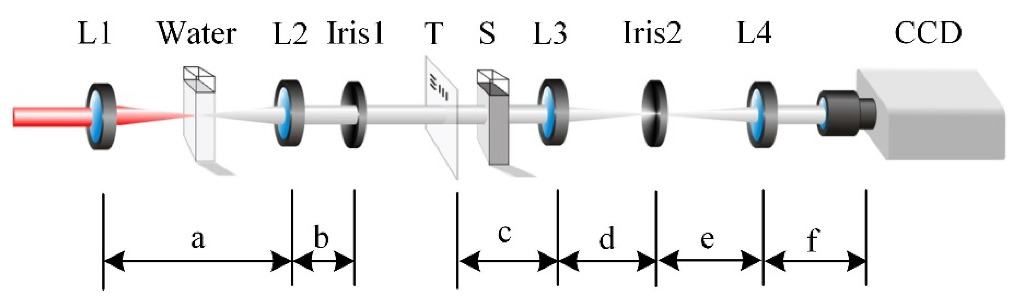
A schematic of the imaging system is shown in
Figure 1. An amplified Ti: sapphire femtosecond laser system (Libra-USP-HE, Coherent Inc., Santa Clara, CA, USA) was used in our experiments; it emits 800 nm, 50 fs, and 3.5 mJ laser pulses at a repetition rate of 1 kHz. Then the femtosecond laser pulses were introduced into the imaging system through some relay optical elements such as mirrors. Because of the dispersion of these optical elements, the pulse width was broadened to less than 200 fs in our experiment. A convex lens (L1) with a focal length of 150 mm was used to focus the laser pulse into a 5 cm cuvette filled with distilled water to generate the SC. Since the peak power and the spatial distribution of the laser pulse can affect the stability of the SC, the pulse energy of the incident femtosecond laser was adjusted to less than 1 mJ, and the focus spot of the laser was also carefully adjusted to ensure the stability of the SC. The SC was then collimated by another convex lens (L2) with a focal length of 100 mm and filtered through an aperture (Iris1) to remove the associated conical emission. The collimated SC was then introduced onto the image target. In order to facilitate the subsequent quantitative characterization of imaging quality, a resolution test chart (RT-MIL-TP2001, RealLight, Beijing, China) was used as the imaging target. A suspension of 3.13 μm polystyrene spheres was used as the scattering medium, the optical depth (OD) of which was measured by using the optical Kerr gate method [
22]. Here, OD is defined as −ln (
I/
I0), where
I is the intensity of the light exiting the suspension and
I0 is the intensity of the light entering the suspension.
The imaging target was then imaged with a 4f imaging setup, which consisted of two achromatic lenses, L3 and L4, with focal lengths of 160 mm and 200 mm, respectively. An adjustable aperture Iris2 was located at the front focus plane of the lens L3, namely the Fourier spectral plane of the 4f imaging setup, as the spatial gate. In the 4f imaging setup, the ballistic photons appear in the center region of the Fourier spectral plane, whereas most of the scattering photons appear away from the central region. The spatial gate located in the Fourier spectral plane can filter the scattering photons and allow most of the ballistic photons to pass through. A charge-coupled device (CCD) camera (INFINITY3-1M-NS-TPM, Lumenera Corporation, Ottawa, ON, Canada) was then placed on the image plane of the 4f imaging setup for image acquisition. The exposure time of the CCD camera was set as 0.5 ms. Because the repetition frequency of the femtosecond laser used here is 1 kHz and the pulse width of the supercontinuum pulse induced by femtosecond laser is mostly in the order of picosecond or sub-picosecond, all imaging results in this paper were obtained under single-pulse imaging conditions.
For comparison, the direct imaging using the SC and the 800 nm femtosecond laser was also performed. The setup of the direct imaging just using the SC was nearly identical to
Figure 1, except that no Iris2 was placed at the back focal plane of the L3. The setup of the direct imaging using the 800 nm femtosecond laser further removes the water-filled cuvette.
3. Results and Discussion
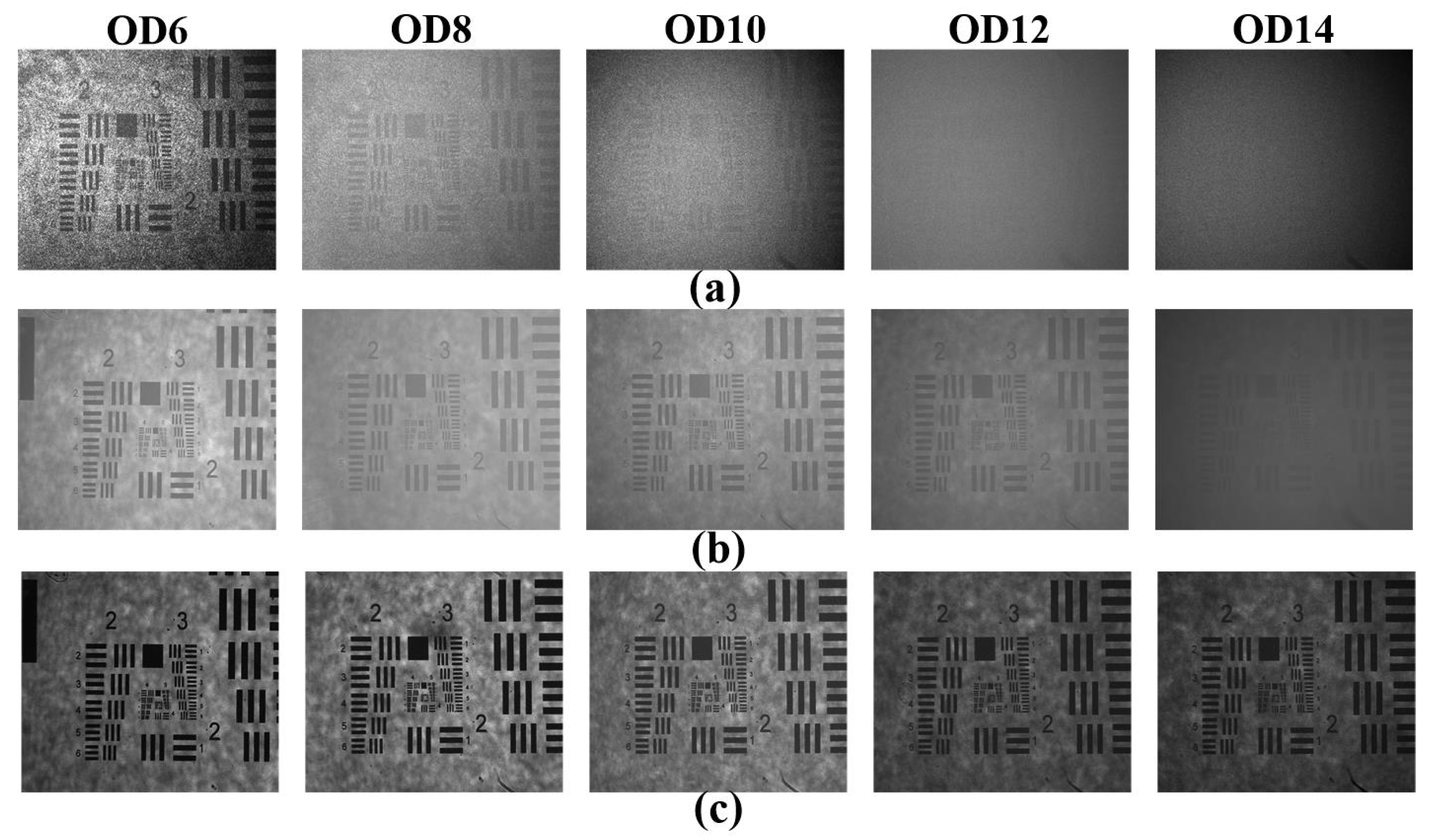
To intuitively demonstrate the differences of the three scattering imaging methods mentioned above, the images of the test patterns under different scattering conditions were obtained firstly and shown in
Figure 2. The OD of the turbid suspensions varied from about 6 to 14. The Fourier spatial gate size used here was about 5 mm. From
Figure 2a, we can see that the images have serious multiple scattering noise by using the femtosecond laser illumination directly. When the OD of the turbid medium is merely equal to about 6, lots of speckles are visible and corrupt the image seriously. As the OD increases, the multiple scattering noise gradually masks the imaging information of the test patterns.
Figure 2b shows the images using SC illumination without the Iris2. The speckle patterns are obviously suppressed to form a uniform imaging background due to the low spatial coherence of the SC source. Thus, the test patterns in the image are visible just by using the SC illumination even at OD = 12 in our experiment. Of course, the image contrast is seriously degraded now due to the remaining background noise of scattered photons. When the OD of the turbid medium increases to 14, the test patterns become nearly unidentifiable due to the further deteriorated image contrast. From
Figure 2c, we can see that by using a combination of supercontinuum illumination and Fourier spatial filtering, the speckles are still suppressed by the supercontinuum illumination and lots of scattered photons are simultaneously filtered by the spatial gate. The test patterns become visible again due to the increased image contrast.
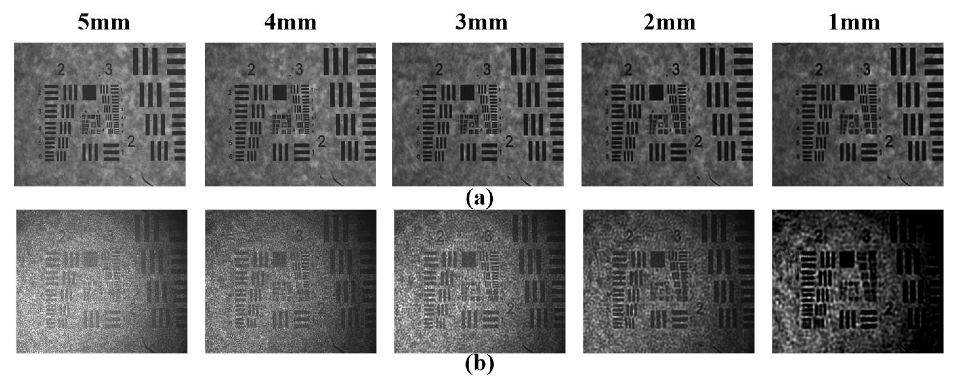
Now that the spatial gate can filter many scattered photons to offer an improved image contrast, we further studied the influence of the spatial gate sizes on the image quality of ballistic imaging by using a combination of supercontinuum illumination and spatial filtering. The ballistic imaging just using the Fourier spatial filtering was also performed for comparison. The images are shown in
Figure 3. Five different sizes of spatial gates were used here, and the OD of the turbid suspension was about 10.
From
Figure 3a, we can see that when the combination of supercontinuum illumination and spatial filtering was adopted, much of the speckle-suppressed scattering noise was filtered out. The image identifiability was significantly higher than that for ballistic imaging using only Fourier spatial filtering. The smaller spatial gate size brings a higher image contrast because of the better filtering of the scattered photons. Although the test patterns also become visible with the femtosecond laser illumination using Fourier spatial filtering, lots of speckles are still visible in
Figure 3b and corrupt the images significantly because of the interference of the residual scattered photons. In addition, the spatial gate size cannot be reduced further to filter scattered photons, because this will seriously blur the image edges and details due to the low-pass filter effects. A more detailed analysis is given later.
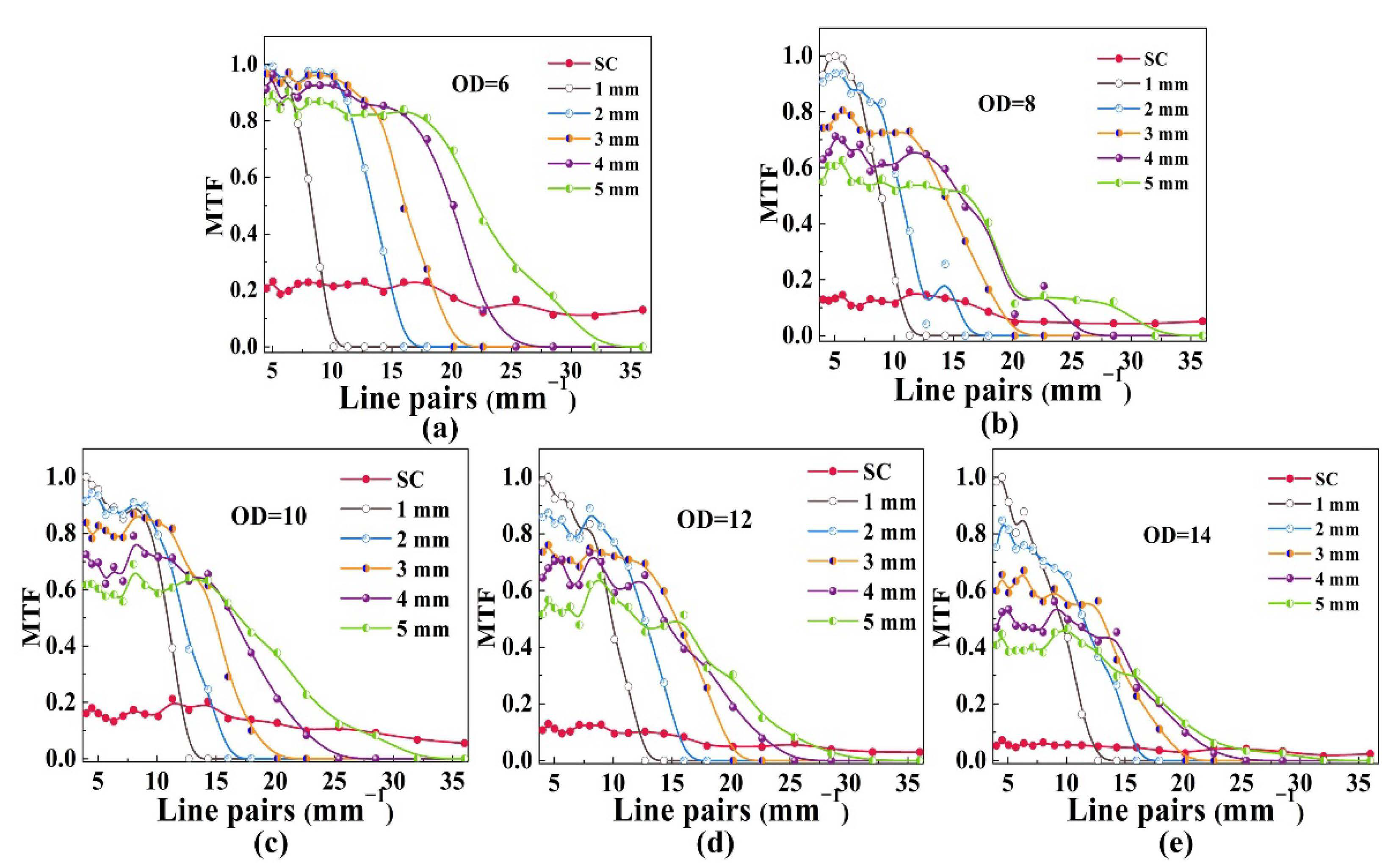
Furthermore, to quantitatively compare the image contrasts by using SC illumination with or without Fourier spatial gate, we calculated their modulation transfer function (MTF) curves under different OD conditions. The MTF is calculated as (
Imax−
Imin)/(
Imax+
Imin), where
Imax and
Imin are the maximum and minimum image intensities of the test pattern regions, respectively. The MTFs are normalized by the maximum contrast of all experimental data. The relative image contrast for the imaging system by using SC illumination only is generally low, which drops from about 0.2 to less than 0.1 as the ODs increases from 6 to 14, as shown in
Figure 4. As the speckles are effectively suppressed by using SC illumination, the image contrast degradation is attributed to the incoherent scattered photons background.
When the Fourier spatial filtering is further introduced, many scattered photons are excluded. The relative image contrasts for ballistic imaging by using a combination of supercontinuum illumination and Fourier spatial filtering are significantly improved. Although the relative image contrasts also decrease with increasing the ODs of the turbid media due to the residual scattered photons, the relative image contrasts are always four times higher than those using SC illumination only as shown in
Figure 4. This result demonstrates the advantage of improving the image contrast of our method. Of course, because the Iris2 was placed at the Fourier spectral plane of the 4f imaging setup, as shown in
Figure 1, some high spatial spectral components of the imaging target might also be filtered by the Fourier spatial gate. The relative image contrasts for the image features with these high spatial frequencies decrease accordingly, as shown in
Figure 4. Thus, an appropriately sized Fourier spatial gate should balance filtering scattered photons and retaining more spatial spectral components of the imaging target.
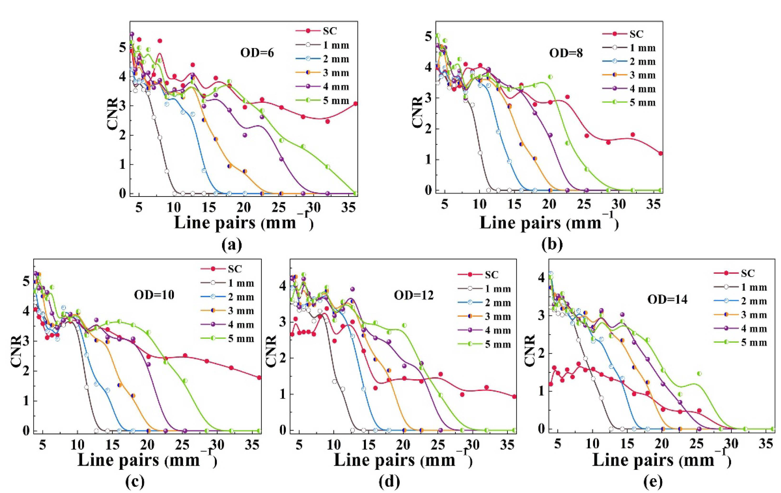
Additionally, to quantitatively compare the image quality of SC illumination with or without space gates, we computed the contrast-to-noise ratio (CNR) of the two methods, as shown in
Figure 5. The CNR is a parameter that can describe the image identifiability of the target. The CNR is defined as (<
I1> − <
I2>)/((
σ1 +
σ2)/2), where
I1 and
I2 are the intensities of signal-producing structures one (a bar in a test pattern) and two (the surrounding background), and
σ is the standard deviation of the pixel intensity [
15]. The numerator of the CNR accordingly represents the difference in intensity within different regions of the characteristic structure of the target object, while the denominator represents the fluctuate of the entire random noise. When the CNR is less than 1, the structure is hardly identifiable. As shown in
Figure 5a–c, when the ODs of the turbid media are not very high, the SC illumination imaging system without Fourier spatial gate already has an acceptable identifiability. In these conditions, we can see that ballistic imaging by using a combination of supercontinuum illumination and Fourier spatial filtering mainly offers a higher image contrast by sacrificing some image details, as shown above in
Figure 4a–c. However, when the ODs of the turbid media further increased above 10, the CNRs of image features whose spatial frequency are higher than 15 mm
−1 drop to around 1 at OD = 12 and even below 1 at OD = 14 due to the increasing residual scattered photons. At this moment, introducing the Fourier spatial gate is necessary for ensuring the image identifiability. For example, in our experiment, the image features with spatial frequencies of 25 mm
−1 is identifiable by using a 5 mm spatial gate at OD = 14, as shown in
Figure 5e. However, the image features with spatial frequencies of 5 mm
−1 are barely identifiable without using the Fourier spatial gate. Overall, the system with spatial gates is able to improve image identifiability by about two times, in this case, larger ODs. The results demonstrate that ballistic imaging through strongly scattering media by using a combination of supercontinuum illumination and Fourier spatial filtering provides a significant improvement of the contrast and identifiability of the image.
It should be noted that according to the Mie scattering theory, the optical characteristics of scattered light, such as its spatial distribution, depend on the relative dielectric coefficient, size, and other factors of scattered particles. For a certain wide-field imaging system, the number and distribution of scattered photons entering the imaging system may also be different when imaging targets are hidden in different scattering media with the same optical density. Thus, the specific experimental data in this paper may change due to the change of the specific experimental parameters, such as the numerical aperture of the imaging system and the scattering characteristics of the scattering medium. However, compared with the speckle-free imaging using SC illumination and ballistic imaging by Fourier spatial filtering, the composite method proposed in this paper has the same tendency to improve the imaging contrast and recognition of the target hidden behind the scattering medium.
In addition, although the static discrimination target is used in this paper, the imaging results are obtained under single-pulse supercontinuum illumination. Meanwhile, the supercontinuum pulse width induced by a femtosecond laser is about a picosecond or sub-picosecond. With this kind of pulse light source, single-pulse illumination imaging can freeze the motion process of most macro-objects and avoid the blurring of imaging results due to target motion. In other words, the imaging system in this paper is very suitable for imaging moving targets in a strong scattering environment as compared with the previous imaging systems with SC illumination [
20,
21].


