A Mini-Review on Functional Near-Infrared Spectroscopy (fNIRS): Where Do We Stand, and Where Should We Go?


Abstract
:1. Introduction
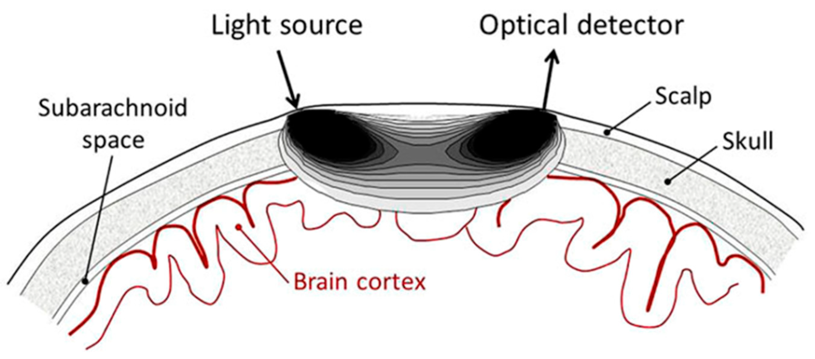
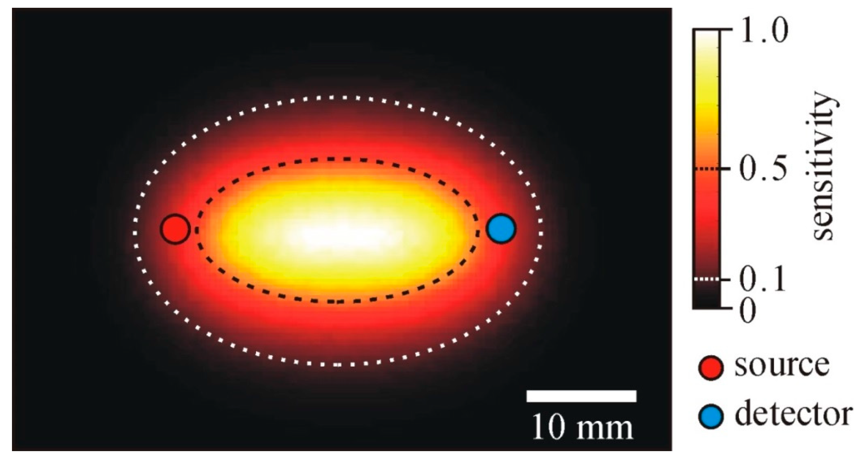
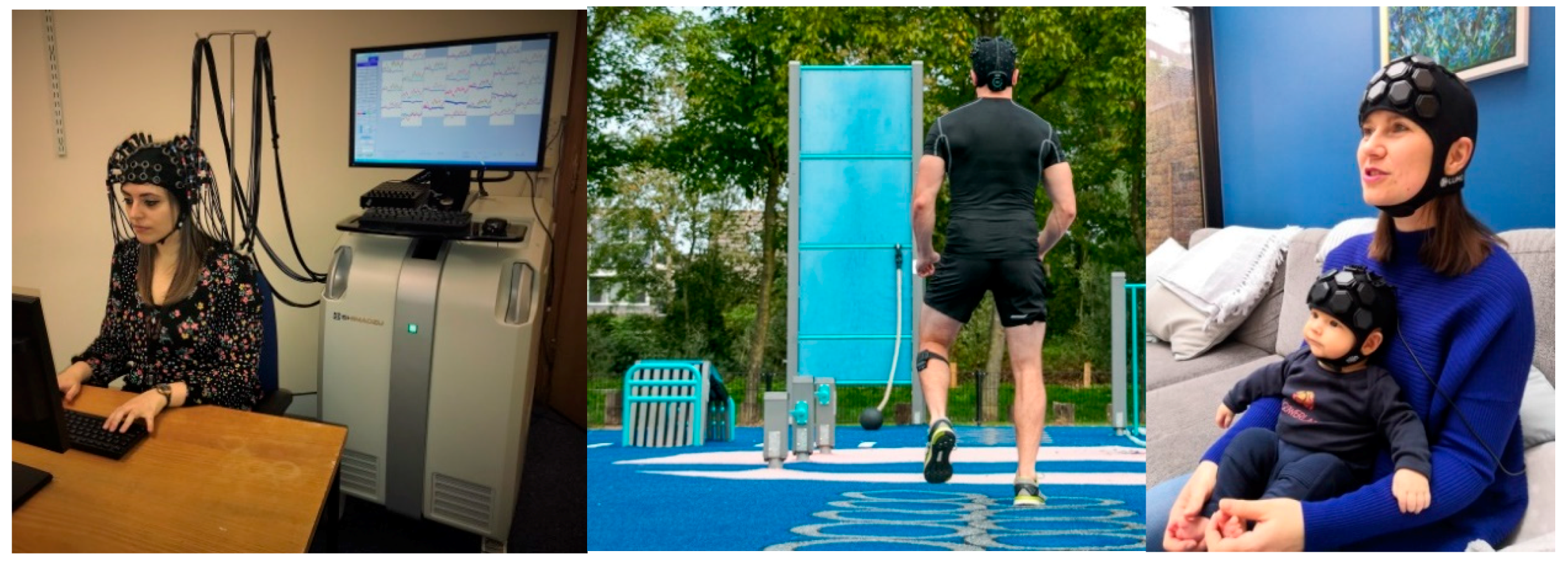
2. Where Do We Stand
3. Where Should We Go?
4. Conclusions
Author Contributions
Funding
Conflicts of Interest
References
- Hutchinson, M.R.; Stoddart, P.R.; Mahadevan-Jansen, A. Challenges and opportunities in neurophotonics discussed at the International Conference on Biophotonics. Neurophotonics 2018, 5, 040402. [Google Scholar]
- Tanner, K.; D’Amico, E.; Kaczmarowski, A.; Kukreti, S.; Malpeli, J.; Mantulin, W.W.; Gratton, E. Spectrally resolved neurophotonics: A case report of hemodynamics and vascular components in the mammalian brain. J. Biomed. Opt. 2005, 10, 064009. [Google Scholar] [CrossRef]
- Jöbsis, F.F. Noninvasive, infrared monitoring of cerebral and myocardial oxygen sufficiency and circulatory parameters. Science 1977, 198, 1264–1267. [Google Scholar] [CrossRef]
- Bigio, I.J.; Fantini, S. Quantitative Biomedical Optics Theory, Methods, and Applications, 1st ed.; Cambridge University Press: Cambridge, UK, 2016. [Google Scholar]
- Fantini, S.; Frederick, B.; Sassaroli, A. Perspective: Prospects of non-invasive sensing of the human brain with diffuse optical imaging. APL Photonics 2018, 3, 110901. [Google Scholar] [CrossRef] [Green Version]
- Wang, L.; Jacques, S.L.; Zheng, L. MCML—Monte Carlo modeling of light transport in multi-layered tissues. Comput. Methods Programs Biomed. 1995, 47, 131–146. [Google Scholar] [CrossRef]
- Sakakibara, Y.; Kurihara, K.; Okada, E. Evaluation of improvement of diffuse optical imaging of brain function by high-density probe arrangements and imaging algorithms. Opt. Rev. 2016, 23, 346–353. [Google Scholar] [CrossRef]
- Ferrari, M.; Norris, K.H.; Sowa, M.G. Medical near infrared spectroscopy 35 years after the discovery. J. Near Infrared Spectrosc. 2012, 20, vii–ix. [Google Scholar] [CrossRef]
- Ferrari, M.; Quaresima, V. Review: Near infrared brain and muscle oximetry: From the discovery to current applications. J. Near Infrared. Spectrosc. 2012, 20, 1–14. [Google Scholar] [CrossRef]
- Garvey, A.A.; Dempsey, E.M. Applications of near infrared spectroscopy in the neonate. Curr. Opin. Pediatr. 2018, 30, 209–215. [Google Scholar] [CrossRef]
- La Cour, A.; Greisen, G.; Hyttel-Sorensen, S. In vivo validation of cerebral near-infrared spectroscopy: A review. Neurophotonics 2018, 5, 040901. [Google Scholar] [CrossRef]
- Serraino, G.F.; Murphy, G.J. Effects of cerebral near-infrared spectroscopy on the outcome of patients undergoing cardiac surgery: A systematic review of randomised trials. BMJ Open. 2017, 7, e016613. [Google Scholar] [CrossRef]
- Yu, Y.; Zhang, K.; Zhang, L.; Zong, H.; Meng, L.; Han, R. Cerebral near-infrared spectroscopy (NIRS) for perioperative monitoring of brain oxygenation in children and adults. Cochrane Database Syst. Rev. 2018, 1, CD010947. [Google Scholar] [CrossRef]
- Lecrux, C.; Bourourou, M.; Hamel, E. How reliable is cerebral blood flow to map changes in neuronal activity? Auton. Neurosci. 2019, 217, 71–79. [Google Scholar] [CrossRef]
- Shetty, P.K.; Galeffi, F.; Turner, D.A. Cellular links between neuronal activity and energy homeostasis. Front. Pharmacol. 2012, 3, 43. [Google Scholar] [CrossRef]
- Tsao, A.; Galwaduge, P.T.; Kim, S.H.; Shaik, M.; Hillman, E.M.C. Measuring the thermodynamic effects of neurovascular coupling in the awake, behaving mouse brain. In Biomedical Optics 2016, OSA Technical Digest; Optical Society of America: Washington, DC, USA, 2016. [Google Scholar]
- Nakada, T.; Kwee, I.L. Fluid dynamics inside the brain barrier: Current concept of interstitial flow, glymphatic flow, and cerebrospinal fluid circulation in the brain. Neuroscientist 2019, 25, 155–166. [Google Scholar] [CrossRef]
- Scholkmann, F.; Kleiser, S.; Metz, A.J.; Zimmermann, R.; Pavia, J.M.; Wolf, U.; Wolf, M. A review on continuous wave functional near-infrared spectroscopy and imaging instrumentation and methodology. Neuroimage 2014, 85, 6–27. [Google Scholar] [CrossRef]
- Tachtsidis, I.; Scholkmann, F. False positives and false negatives in functional near-infrared spectroscopy: Issues, challenges, and the way forward. Neurophotonics 2016, 3, 031405. [Google Scholar] [CrossRef]
- Martelli, F. An ABC of near infrared photon migration in tissues: The diffusive regime of propagation. J. Near Infrared Spectrosc. 2012, 20, 29–42. [Google Scholar] [CrossRef]
- Ferrari, M.; Quaresima, V. A brief review on the history of human functional near-infrared spectroscopy (fNIRS) development and fields of application. Neuroimage 2012, 63, 921–935. [Google Scholar] [CrossRef]
- Yücel, M.A.; Selb, J.J.; Huppert, T.J.; Franceschini, M.A.; Boas, D.A. Functional near infrared spectroscopy: Enabling routine functional brain imaging. Curr. Opin. Biomed. Eng. 2017, 4, 78–86. [Google Scholar] [CrossRef]
- Nosrati, R.; Vesely, K.; Schweizer, T.A.; Toronov, V. Event-related changes of the prefrontal cortex oxygen delivery and metabolism during driving measured by hyperspectral fNIRS. Biomed. Opt. Express 2016, 7, 1323–1335. [Google Scholar] [CrossRef] [Green Version]
- Phan, P.; Highton, D.; Lai, J.; Smith, M.; Elwell, C.; Tachtsidis, I. Multi-channel multi-distance broadband near-infrared spectroscopy system to measure the spatial response of cellular oxygen metabolism and tissue oxygenation. Biomed. Opt. Express 2016, 7, 4424–4440. [Google Scholar] [CrossRef]
- Giannoni, L.; Lange, F.; Tachtsidis, I. Hyperspectral imaging solutions for brain tissue metabolic and hemodynamic monitoring: Past, current and future developments. J. Opt. 2018, 20, 044009. [Google Scholar] [CrossRef]
- Torricelli, A.; Contini, D.; Pifferi, A.; Caffini, M.; Re, R.; Zucchelli, L.; Spinelli, L. Time domain functional NIRS imaging for human brain mapping. Neuroimage 2014, 85, 28–50. [Google Scholar] [CrossRef]
- Yamada, Y.; Suzuki, H.; Yamashita, Y. Time-domain near-infrared spectroscopy and imaging: A review. Appl. Sci. 2019, 9, 1127. [Google Scholar] [CrossRef]
- Lange, F.; Tachtsidis, I. Clinical brain monitoring with time domain NIRS: A review and future perspectives. Appl. Sci. 2019, 9, 1612. [Google Scholar] [CrossRef]
- Hoshi, Y.; Yamada, Y. Overview of diffuse optical tomography and its clinical applications. J. Biomed. Opt. 2016, 21, 091312. [Google Scholar] [CrossRef]
- Lee, C.W.; Cooper, R.J.; Austin, T. Diffuse optical tomography to investigate the newborn brain. Pediatr. Res. 2017, 82, 376–386. [Google Scholar] [CrossRef] [Green Version]
- Zhao, H.; Cooper, R.J. Review of recent progress toward a fiberless, whole-scalp diffuse optical tomography system. Neurophotonics 2018, 5, 011012. [Google Scholar] [CrossRef]
- Strangman, G.E.; Ivkovic, V.; Zhang, Q. Wearable brain imaging with multimodal physiological monitoring. J. Appl. Physiol. 2018, 124, 564–572. [Google Scholar] [CrossRef]
- Pinti, P.; Aichelburg, C.; Gilbert, S.; Hamilton, A.; Hirsch, J.; Burgess, P.; Tachtsidis, I. A review on the use of wearable functional near-infrared spectroscopy in naturalistic environments. Jpn. Psychol. Res. 2018, 60, 347–373. [Google Scholar] [CrossRef]
- Orihuela-Espina, F.; Leff, D.R.; James, D.R.C.; Darzi, A.W.; Yang, G.Z. Quality control and assurance in functional near infrared spectroscopy (fNIRS) experimentation. Phys. Med. Biol. 2010, 55, 3701–3724. [Google Scholar] [CrossRef]
- Kassab, A.; Le Lan, J.; Vannasing, P.; Sawan, M. Functional near-infrared spectroscopy caps for brain activity monitoring: A review. Appl. Opt. 2015, 54, 576–586. [Google Scholar] [CrossRef]
- Brigadoi, S.; Cooper, R. How short is short? Optimum source–detector distance for short-separation channels in functional near-infrared spectroscopy. Neurophotonics 2015, 2, 025005. [Google Scholar] [CrossRef]
- Yücel, M.A.; Selb, J.; Aasted, C.M.; Lin, P.Y.; Borsook, D.; Becerra, L.; Boas, D.A. Mayer waves reduce the accuracy of estimated hemodynamic response functions in functional near-infrared spectroscopy. Biomed. Opt. Express 2016, 7, 3078–3088. [Google Scholar] [CrossRef] [Green Version]
- Pinti, P.; Scholkmann, F.; Hamilton, A.; Burgess, P.; Tachtsidis, I. Current status and issues regarding pre-processing of fNIRS neuroimaging data: An investigation of diverse signal filtering methods within a general linear model framework. Front. Hum. Neurosci. 2019, 12, 505. [Google Scholar] [CrossRef]
- Tsuzuki, D.; Dan, I. Spatial registration for functional near-infrared spectroscopy: From channel position on the scalp to cortical location in individual and group analyses. Neuroimage 2014, 85, 92–103. [Google Scholar] [CrossRef]
- Aasted, C.M.; Yücel, M.A.; Cooper, R.J.; Dubb, J.; Tsuzuki, D.; Becerra, L.; Petkov, M.P.; Borsook, D.; Dan, I.; Boas, D.A. Anatomical guidance for functional near-infrared spectroscopy: AtlasViewer tutorial. Neurophotonics 2015, 2, 020801. [Google Scholar] [CrossRef]
- Tak, S.; Ye, J.C. Statistical analysis of fNIRS data: A comprehensive review. Neuroimage 2014, 85, 72–91. [Google Scholar] [CrossRef]
- De Roever, I.; Bale, G.; Mitra, S.; Meek, J.; Robertson, N.J.; Tachtsidis, I. Investigation of the pattern of the hemodynamic response as measured by functional near-infrared spectroscopy (fNIRS) studies in newborns, less than a month old: A systematic review. Front. Hum. Neurosci. 2018, 12, 371. [Google Scholar] [CrossRef]
- Issard, C.; Gervain, J. Variability of the hemodynamic response in infants: Influence of experimental design and stimulus complexity. Dev. Cognit. Neurosci. 2018, 33, 182–193. [Google Scholar] [CrossRef]
- Chiarelli, A.M.; Zappasodi, F.; Di Pompeo, F.; Merla, A. Simultaneous functional near-infrared spectroscopy and electroencephalography for monitoring of human brain activity and oxygenation: A review. Neurophotonics 2017, 4, 041411. [Google Scholar] [CrossRef]
- Scarapicchia, V.; Brown, C.; Mayo, C.; Gawryluk, J.R. Functional magnetic resonance imaging and functional near-infrared spectroscopy: Insights from combined recording studies. Front. Hum. Neurosci. 2017, 11, 419. [Google Scholar] [CrossRef]
- Curtin, A.; Tong, S.; Sun, J.; Wang, J.; Onaral, B.; Ayaz, H. A systematic review of integrated functional near-infrared spectroscopy (fNIRS) and transcranial magnetic stimulation (TMS) studies. Front. Hum. Neurosci. 2019, 13, 84. [Google Scholar] [CrossRef]
- Pinti, P.; Tachtsidis, I.; Hamilton, A.; Hirsch, J.; Aichelburg, C.; Gilbert, S.; Burgess, P.W. The present and future use of functional near-infrared spectroscopy (fNIRS) for cognitive neuroscience. Ann. N. Y. Acad. Sci. 2018. [Google Scholar] [CrossRef]
- Quaresima, V.; Ferrari, M. Functional near-infrared spectroscopy (fNIRS) for assessing cerebral cortex function during human behavior in natural/social situations: A concise review. Organ. Res. Methods 2019, 22, 46–68. [Google Scholar] [CrossRef]
- Boas, D.A.; Elwell, C.E.; Ferrari, M.; Taga, G. Twenty years of functional near-infrared spectroscopy: Introduction for the special issue. Neuroimage 2014, 85, 1–5. [Google Scholar] [CrossRef]
- Val-Laillet, D.; Aarts, E.; Weber, B.; Ferrari, M.; Quaresima, V.; Stoeckel, L.E.; Alonso-Alonso, M.; Audette, M.; Malbert, C.H.; Stice, E. Neuroimaging and neuromodulation approaches to study eating behavior and prevent and treat eating disorders and obesity. Neuroimage Clin. 2015, 8, 1–31. [Google Scholar] [CrossRef]
- Aslin, R.N.; Shukla, M.; Emberson, L.L. Hemodynamic correlates of cognition in human infants. Annu. Rev. Psychol. 2015, 66, 349–379. [Google Scholar] [CrossRef]
- Vanderwert, R.E.; Nelson, C.A. The use of near-infrared spectroscopy in the study of typical and atypical development. Neuroimage 2014, 85, 264–271. [Google Scholar] [CrossRef]
- Wilcox, T.; Biondi, M. fNIRS in the developmental sciences. Wiley Interdiscip. Rev. Cognit. Sci. 2015, 6, 263–283. [Google Scholar] [CrossRef] [Green Version]
- Soltanlou, M.; Sitnikova, M.A.; Nuerk, H.C.; Dresler, T. Applications of functional near-infrared spectroscopy (fNIRS) in studying cognitive development: The case of mathematics and language. Front. Psychol. 2018, 9, 277. [Google Scholar] [CrossRef]
- Bendall, R.C.; Eachus, P.; Thompson, C. A brief review of research using near-infrared spectroscopy to measure activation of the prefrontal cortex during emotional processing: The importance of experimental design. Front. Hum. Neurosci. 2016, 10, 529. [Google Scholar] [CrossRef]
- Herold, F.; Wiegel, P.; Scholkmann, F.; Müller, N.G. Applications of functional near-infrared spectroscopy (fNIRS) neuroimaging in exercise–cognition science: A systematic, methodology-focused review. J. Clin. Med. 2018, 7, 466. [Google Scholar] [CrossRef]
- Homae, F. A brain of two halves: Insights into interhemispheric organization provided by near-infrared spectroscopy. Neuroimage 2014, 85, 354–362. [Google Scholar] [CrossRef]
- Cutini, S.; Basso Moro, S.; Bisconti, S. Functional near infrared optical imaging in cognitive neuroscience: An introductory review. J. Near Infrared Spectrosc. 2012, 20, 75–92. [Google Scholar] [CrossRef]
- McDonald, N.M.; Perdue, K.L. The infant brain in the social world: Moving toward interactive social neuroscience with functional near-infrared spectroscopy. Neurosci. Biobehav. Rev. 2018, 87, 38–49. [Google Scholar] [CrossRef]
- Kopton, I.M.; Kenning, P. Near-infrared spectroscopy (NIRS) as a new tool for neuroeconomic research. Front. Hum. Neurosci. 2014, 8, 549. [Google Scholar] [CrossRef] [Green Version]
- Quaresima, V.; Bisconti, S.; Ferrari, M. A brief review on the use of functional near-infrared spectroscopy (fNIRS) for language imaging studies in human newborns and adults. Brain Lang. 2012, 121, 79–89. [Google Scholar] [CrossRef]
- Rossi, S.; Telkemeyer, S.; Wartenburger, I.; Obrig, H. Shedding light on words and sentences: Near-infrared spectroscopy in language research. Brain Lang. 2012, 121, 152–163. [Google Scholar] [CrossRef]
- Curtin, A.; Ayaz, H. The age of neuroergonomics: Towards ubiquitous and continuous measurement of brain function with fNIRS. Jpn. Psychol. Res. 2018, 60, 374–386. [Google Scholar] [CrossRef]
- Zhu, Y.; Rodriguez-Paras, C.; Rhee, J.; Mehta, R.K. Methodological approaches and recommendations for functional near-infrared spectroscopy applications in HF/E research. Hum. Factors 2019. [Google Scholar] [CrossRef]
- Naseer, N.; Hong, K.S. fNIRS-based brain-computer interfaces: A review. Front. Hum. Neurosci. 2015, 9, 3. [Google Scholar] [CrossRef]
- Liu, T.; Pelowski, M.; Pang, C.; Zhou, Y.; Cai, J. Near-infrared spectroscopy as a tool for driving research. Ergonomics 2016, 59, 368–379. [Google Scholar] [CrossRef]
- Lohani, M.; Payne, B.R.; Strayer, D.L. A review of psychophysiological measures to assess cognitive states in real-world driving. Front. Hum. Neurosci. 2019, 13, 57. [Google Scholar] [CrossRef]
- Ahn, S.; Jun, S.C. Multi-modal integration of EEG-fNIRS for brain-computer interfaces—Current limitations and future directions. Front. Hum. Neurosci. 2017, 11, 503. [Google Scholar] [CrossRef]
- Hong, K.S.; Khan, M.J.; Hong, M.J. Feature extraction and classification methods for hybrid fNIRS-EEG brain-computer interfaces. Front. Hum. Neurosci. 2018, 12, 246. [Google Scholar] [CrossRef]
- Scholkmann, F.; Holper, L.; Wolf, U.; Wolf, M. A new methodical approach in neuroscience: Assessing inter-personal brain coupling using functional near-infrared imaging (fNIRI) hyperscanning. Front. Hum. Neurosci. 2013, 7, 813. [Google Scholar] [CrossRef]
- Minagawa, Y.; Xu, M.; Morimoto, S. Toward interactive social neuroscience: Neuroimaging real-world interactions in various populations. Jpn. Psychol. Res. 2018, 60, 374–386. [Google Scholar] [CrossRef]
- Wang, M.Y.; Luan, P.; Zhang, J.; Xiang, Y.T.; Niu, H.; Yuan, Z. Concurrent mapping of brain activation from multiple subjects during social interaction by hyperscanning: A mini-review. Quant. Imaging Med. Surg. 2018, 8, 819–837. [Google Scholar] [CrossRef]
- Herold, F.; Wiegel, P.; Scholkmann, F.; Thiers, A.; Hamacher, D.; Schega, L. Functional near-infrared spectroscopy in movement science: A systematic review on cortical activity in postural and walking tasks. Neurophotonics 2017, 4, 041403. [Google Scholar] [CrossRef]
- Niu, H.; He, Y. Resting-state functional brain connectivity: Lessons from functional near-infrared spectroscopy. Neuroscientist 2014, 20, 173–188. [Google Scholar] [CrossRef]
- Vitorio, R.; Stuart, S.; Rochester, L.; Alcock, L.; Pantall, A. Fnirs response during walking - Artefact or cortical activity? A systematic review. Neurosci. Biobehav. Rev. 2017, 83, 160–172. [Google Scholar] [CrossRef]
- Pelicioni, P.H.S.; Tijsma, M.; Lord, S.R.; Menant, J. Prefrontal cortical activation measured by fNIRS during walking: Effects of age, disease and secondary task. Peer J. 2019, 7, e6833. [Google Scholar] [CrossRef]
- Stuart, S.; Vitorio, R.; Morris, R.; Martini, D.N.; Fino, P.C.; Mancini, M. Cortical activity during walking and balance tasks in older adults and in people with Parkinson’s disease: A structured review. Maturitas 2018, 113, 53–72. [Google Scholar] [CrossRef]
- Mauri, M.; Nobile, M.; Bellina, M.; Crippa, A.; Brambilla, P. Light up ADHD: I. Cortical hemodynamic responses measured by functional near infrared spectroscopy (fNIRS). J. Affect. Disord. 2018, 234, 358–364. [Google Scholar] [CrossRef]
- Basura, G.J.; Hu, X.S.; Juan, J.S.; Tessier, A.M.; Kovelman, I. Human central auditory plasticity: A review of functional near-infrared spectroscopy (fNIRS) to measure cochlear implant performance and tinnitus perception. Laryngoscope Investig. Otolaryngol. 2018, 3, 463–472. [Google Scholar] [CrossRef] [Green Version]
- Liu, T.; Liu, X.; Yi, L.; Zhu, C.; Markey, P.S.; Pelowski, M. Assessing autism at its social and developmental roots: A review of autism spectrum disorder studies using functional near-infrared spectroscopy. Neuroimage 2019, 185, 955–967. [Google Scholar] [CrossRef]
- Zhang, F.; Roeyers, H. Exploring brain functions in autism spectrum disorder: A systematic review on functional near-infrared spectroscopy (fNIRS) studies. Int. J. Psychophysiol. 2019, 137, 41–53. [Google Scholar] [CrossRef] [Green Version]
- Agbangla, N.F.; Audiffren, M.; Albinet, C.T. Use of near-infrared spectroscopy in the investigation of brain activation during cognitive aging: A systematic review of an emerging area of research. Ageing Res. Rev. 2017, 38, 52–66. [Google Scholar] [CrossRef]
- Grazioli, S.; Mauri, M.; Crippa, A.; Maggioni, E.; Molteni, M.; Brambilla, P.; Nobile, M. Light up ADHD: II. Neuropharmacological effects measured by near infrared spectroscopy: Is there a biomarker? J. Affect. Disord. 2019, 244, 100–106. [Google Scholar] [CrossRef]
- Peng, K.; Pouliot, P.; Lesage, F.; Nguyen, D.K. Multichannel continuous electroencephalography-functional near-infrared spectroscopy recording of focal seizures and interictal epileptiform discharges in human epilepsy: A review. Neurophotonics 2016, 3, 031402. [Google Scholar] [CrossRef]
- Gramigna, V.; Pellegrino, G.; Cerasa, A.; Cutini, S.; Vasta, R.; Olivadese, G.; Martino, I.; Quattrone, A. Near-infrared spectroscopy in gait disorders: Is it time to begin? Neurorehabilit. Neural Repair 2017, 31, 402–412. [Google Scholar] [CrossRef]
- Beishon, L.; Haunton, V.J.; Panerai, R.B.; Robinson, T.G. Cerebral hemodynamics in mild cognitive impairment: A systematic review. J. Alzheimers Dis. 2017, 59, 369–385. [Google Scholar] [CrossRef]
- Ehlis, A.; Barth, B.; Hudak, J.; Storchak, H.; Weber, L.; Kimmig, A.S.; Kreifelts, B.; Dresler, T.; Fallgatter, A.J. Near-infrared spectroscopy as a new tool for neurofeedback training: Applications in psychiatry and methodological considerations. Jpn. Psychol. Res. 2018, 60, 225–241. [Google Scholar] [CrossRef]
- Benoit, B.; Martin-Misener, R.; Newman, A.; Latimer, M.; Campbell-Yeo, M. Neurophysiological assessment of acute pain in infants: A scoping review of research methods. Acta Paediatr. 2017, 106, 1053–1066. [Google Scholar] [CrossRef]
- Rupawala, M.; Dehghani, H.; Lucas, S.J.E.; Tino, P.; Cruse, D. Shining a light on awareness: A review of functional near-infrared spectroscopy for prolonged disorders of consciousness. Front. Neurol. 2018, 9, 350. [Google Scholar] [CrossRef]
- Ehlis, A.C.; Schneider, S.; Dresler, T.; Fallgatter, A.J. Application of functional near-infrared spectroscopy in psychiatry. Neuroimage 2014, 85, 478–488. [Google Scholar] [CrossRef]
- Berger, A.; Horst, F.; Müller, S.; Steinberg, F.; Doppelmayr, M. Current state and future prospects of EEG and fNIRS in robot-assisted gait rehabilitation: A brief review. Front. Hum. Neurosci. 2019, 13, 172. [Google Scholar] [CrossRef]
- Kumar, V.; Shivakumar, V.; Chhabra, H.; Bose, A.; Venkatasubramanian, G.; Gangadhar, B.N. Functional near infra-red spectroscopy (fNIRS) in schizophrenia: A review. Asian J. Psychiatr. 2017, 27, 18–31. [Google Scholar] [CrossRef]
- Yang, M.; Yang, Z.; Yuan, T.; Feng, W.; Wang, P. A systemic review of functional near-infrared spectroscopy for stroke: Current application and future directions. Front. Neurol. 2019, 10, 58. [Google Scholar] [CrossRef]
- Jasińska, K.K.; Guei, S. Neuroimaging field methods using functional near infrared spectroscopy (NIRS) neuroimaging to study global child development: Rural sub-saharan africa. J. Vis. Exp. 2018, 132, e57165. [Google Scholar] [CrossRef]
- Reindl, V.; Konrad, K.; Gerloff, C.; Kruppa, J.A.; Bell, L.; Scharke, W. Conducting hyperscanning experiments with functional near-infrared spectroscopy. J. Vis. Exp. 2019, 143, e58807. [Google Scholar] [CrossRef]
- Sukal-Moulton, T.; de Campos, A.C.; Stanley, C.J.; Damiano, D.L. Functional near infrared spectroscopy of the sensory and motor brain regions with simultaneous kinematic and EMG monitoring during motor tasks. J. Vis. Exp. 2014, 94, e52391. [Google Scholar] [CrossRef]
- Noah, J.A.; Ono, Y.; Nomoto, Y.; Shimada, S.; Tachibana, A.; Zhang, X.; Bronner, S.; Hirsch, J. Fmri validation of fNIRS measurements during a naturalistic task. J. Vis. Exp. 2015, 100, e52116. [Google Scholar] [CrossRef]
- Pinti, P.; Aichelburg, C.; Lind, F.; Power, S.; Swingler, E.; Merla, A.; Hamilton, A.; Gilbert, S.; Burgess, P.; Tachtsidis, I. Using fiberless, wearable fNIRS to monitor brain activity in real-world cognitive tasks. J. Vis. Exp. 2015, 106, e53336. [Google Scholar] [CrossRef]
- Villringer, A.; Chance, B. Non-invasive optical spectroscopy and imaging of human brain function. Trends Neurosci. 1997, 20, 435–442. [Google Scholar] [CrossRef]
- Villringer, A.; Planck, J.; Hock, C.; Schleinkofer, L.; Dirnagl, U. Near infrared spectroscopy (NIRS): A new tool to study hemodynamic changes during activation of brain function in human adults. Neurosci. Lett. 1993, 154, 101–104. [Google Scholar] [CrossRef]
- Strangman, G.; Culver, J.P.; Thompson, J.H.; Boas, D.A. A quantitative comparison of simultaneous BOLD fMRI and NIRS recordings during functional brain activation. Neuroimage 2002, 17, 719–731. [Google Scholar] [CrossRef]
- Obrig, H.; Villringer, A. Beyond the visible—Imaging the human brain with light. J. Cereb. Blood Flow Metab. 2003, 23, 1–18. [Google Scholar] [CrossRef]
- Maki, A.; Yamashita, Y.; Ito, Y.; Watanabe, E.; Mayanagi, Y.; Koizumi, H. Spatial and temporal analysis of human motor activity using noninvasive NIR topography. Med. Phys. 1995, 22, 1997–2005. [Google Scholar] [CrossRef]
- Strangman, G.; Boas, D.A.; Sutton, J.P. Non-invasive neuroimaging using near-infrared light. Biol. Psychiatry 2002, 52, 679–693. [Google Scholar] [CrossRef]
- Boas, D.A.; Dale, A.M.; Franceschini, M.A. Diffuse optical imaging of brain activation: Approaches to optimizing image sensitivity, resolution, and accuracy. Neuroimage 2004, 23, S275–S288. [Google Scholar] [CrossRef]
- Hoshi, Y.; Kobayashi, N.; Tamura, M. Interpretation of near-infrared spectroscopy signals: A study with a newly developed perfused rat brain model. J. Appl. Physiol. 2001, 90, 1657–1662. [Google Scholar] [CrossRef]
- Ward, J.; Pinti, P. Wearables and the brain. IEEE Pervasive Comput. 2019, 18, 94–100. [Google Scholar] [CrossRef]
- Buttafava, M.; Martinenghi, E.; Tamborini, D.; Contini, D.; Mora, A.D.; Renna, M.; Torricelli, A.; Pifferi, A.; Zappa, F.; Tosi, A. A compact two-wavelength time-domain NIRS system based on SiPM and pulsed diode lasers. IEEE Photonics J. 2017, 9, 7792611. [Google Scholar] [CrossRef]
- Di Sieno, L.; Dalla Mora, A.; Torricelli, A.; Spinelli, L.; Re, R.; Pifferi, A.; Contini, D. A versatile setup for time-resolved functional near infrared spectroscopy based on fast-gated single-photon avalanche diode and on four-wave mixing laser. Appl. Sci. 2019, 9, 2366. [Google Scholar] [CrossRef]
- Lange, F.; Dunne, F.; Hale, L.; Tachtsidis, I. Maestros: A multiwavelength time-domain NIRS system to monitor changes in oxygenation and oxidation state of cytochrome-c-oxidase. IEEE J. Sel. Top. Quant. Electron. 2019, 25, 1–12. [Google Scholar] [CrossRef]
- Bale, G.; Elwell, C.E.; Tachtsidis, I. From Jöbsis to the present day: A review of clinical near-infrared spectroscopy measurements of cerebral cytochrome-c-oxidase. J. Biomed. Opt. 2016, 21, 091307. [Google Scholar] [CrossRef]
- De Roever, I.; Bale, G.; Cooper, R.J.; Tachtsidis, I. Functional NIRS measurement of cytochrome-c-oxidase demonstrates a more brain-specific marker of frontal lobe activation compared to the haemoglobins. Adv. Exp. Med. Biol. 2017, 977, 141–147. [Google Scholar]
- Scholkmann, F.; Hafner, T.; Metz, A.J.; Wolf, M.; Wolf, U. Effect of short-term colored-light exposure on cerebral hemodynamics and oxygenation, and systemic physiological activity. Neurophotonics 2017, 4, 045005. [Google Scholar] [CrossRef] [Green Version]
- Beisteiner, R.; Pernet, C.; Stippich, C. Can we standardize clinical functional neuroimaging procedures? Front. Neurol. 2019, 9, 1153. [Google Scholar] [CrossRef]
- Woods, A.J.; Bikson, M.; Chelette, K.; Dmochowski, J.; Dutta, A.; Esmaeilpour, Z.; Gebodh, N.; Nitsche, M.A.; Stagg, C. Transcranial direct current stimulation integration with magnetic resonance imaging, magnetic resonance spectroscopy, near infrared spectroscopy imaging and electroencephalography. In Practical Guide to Transcranial Direct Current Stimulation, 1st ed.; Knotkova, H., Nitsche, M., Bikson, M., Woods, A., Eds.; Springer International Publisher: Cham, Switzerland, 2019; pp. 293–345. [Google Scholar]
- Wintermark, M.; Colen, R.; Whitlow, C.T.; Zaharchuk, G. The vast potential and bright future of neuroimaging. Br. J. Radiol. 2018, 91, 20170505. [Google Scholar] [CrossRef]
- Perrey, S.; Besson, P. Studying brain activity in sports performance: Contributions and issues. Prog. Brain Res. 2018, 240, 247–267. [Google Scholar]
- Durduran, T.; Yodh, A.G. Diffuse correlation spectroscopy for non-invasive, micro-vascular cerebral blood flow measurement. Neuroimage 2014, 15, 51–63. [Google Scholar] [CrossRef]
- Roche-Labarbe, N.; Carp, S.A.; Surova, A.; Patel, M.; Boas, D.A.; Grant, P.E.; Franceschini, M.A. Noninvasive optical measures of CBV, StO(2), CBF index, and rCMRO(2) in human premature neonates’ brains in the first six weeks of life. Hum. Brain Mapp. 2010, 31, 341–352. [Google Scholar] [CrossRef]
- Andresen, B.; De Carli, A.; Fumagalli, M.; Giovannella, M.; Durduran, T.; Michael Weigel, U.; Contini, D.; Spinelli, L.; Torricelli, A.; Greisen, G. Cerebral oxygenation and blood flow in normal term infants at rest measured by a hybrid near-infrared device (BabyLux). Pediatr. Res. 2019. [Google Scholar] [CrossRef]


| Topic | Year | 1st Author [Ref] |
|---|---|---|
| Modeling near-infrared photon propagation in biological tissue | 2012 | Martelli [20] |
| 2016 | Bigio [4] | |
| 2018 | Fantini [5] | |
| History of fNIRS | 2012 | Ferrari [21] |
| State of the art of continuous-wave multispectral fNIRS instrumentation | 2014 | Scholkmann [18] |
| 2017 | Yücel [22] | |
| State of the art of continuous-wave hyperspectral fNIRS instrumentation | 2016 | Nsorati [23] |
| 2016 | Pham [24] | |
| 2018 | Giannoni [25] | |
| State of the art of time-domain fNIRS instrumentation | 2014 | Torricelli [26] |
| 2019 | Yamada [27] | |
| Clinical brain monitoring by time-domain fNIRS instrumentation | 2019 | Lange [28] |
| State of the art of diffuse optical imaging | 2016 | Hoshi [29] |
| 2017 | Lee [30] | |
| 2018 | Fantini [5] | |
| 2018 | Zhao [31] | |
| State of the art of wearable fNIRS | 2018 | Strangman [32] |
| 2018 | Pinti [33] | |
| State of the art of functional connectivity measurements | 2018 | Fantini [5] |
| Factors influencing fNIRS data and recommendations | 2010 | Orihuela-Espina [34] |
| Caps for long term fNIRS measurements | 2015 | Kassab [35] |
| Selection of the optimum source–detector distance | 2015 | Brigadoi [36] |
| Mayer waves interference | 2016 | Yücel [37] |
| Multiple components of the fNIRS signal | 2016 | Tachtsidis [19] |
| Signal pre-processing procedures | 2019 | Pinti [38] |
| Anatomical guidance for fNIRS | 2014 | Tsuzuki [39] |
| 2015 | Aasted [40] | |
| Statistical analysis of fNIRS data | 2014 | Tak [41] |
| Pattern of hemodynamic response in newborn < 1 month | 2018 | de Roever [42] |
| Pattern of hemodynamic response in infants | 2018 | Issard [43] |
Integration of fNIRS with:
| ||
| 2017 | Chiarelli [44] | |
| 2017 | Scarapicchia [45] | |
| 2019 | Curtin [46] | |
| Recent fNIRS general reviews including the advantages and limitations of fNIRS | 2018 | Fantini [5] |
| 2018 | Pinti [47] | |
| 2019 | Quaresima [48] |
| Field of Application | Topic | Year | N. | Subjects | 1st Author [Ref] |
|---|---|---|---|---|---|
| Psychology/education | Cognition and food | 2015 | 39 | A | Val-Laillet [50] |
| Cognition in infants | 2015 | 171 | C | Aslin [51] | |
| Development (typical and atypical) | 2014 | 29 | C | Vanderwert [52] | |
| 2015 | 149 | C | Wilcox [53] | ||
| Development of mathematics/language skills in children | 2018 | 7 | C | Soltanlou [54] | |
| Emotion | 2016 | 11 | A | Bendall [55] | |
| Influence of exercise on cognition | 2018 | 35 | A | Herold [56] | |
| Interhemispheric organization | 2014 | 32 | A | Homae [57] | |
| Psychology general review | 2012 | 106 | A | Cutini [58] | |
| Social development during infancy | 2018 | 29 | C | McDonald [59] | |
| Economics | Neuroeconomic research | 2014 | 15 | A | Kopton [60] |
| Linguistics | Language and its development | 2012 | 60 | A C | Quaresima [61] |
| Word and sentence processing | 2012 | 9 | C | Rossi [62] | |
| Neuroergonomics | Neuroergonomics and fNIRS | 2018 | 68 | A | Curtin [63] |
| 2019 | 37 | A | Zhu [64] | ||
| Functional Neuroimaging Basic Research | Brain computer interface | 2015 | 33 | A | Naseer [65] |
| Driving research | 2016 | 10 | A | Liu [66] | |
| 2019 | 13 | A | Lohani [67] | ||
| Hybrid fNIRS-EEG brain-computer interfaces | 2017 | 11 | A | Ahn [68] | |
| 2018 | 43 | A | Hong [69] | ||
| Hyperscanning with multi-subject measurements | 2013 | 7 | A | Scholkmann [70] | |
| 2018 | 15 | A | Minagawa [71] | ||
| 2018 | 18 | A | Wang [72] | ||
| Postural and walking tasks | 2017 | 57 | A | Herold [73] | |
| Resting-state functional brain connectivity | 2014 | 16 | A | Niu [74] | |
| Walking | 2017 | 31 | A | Vitorio [75] | |
| 2019 | 35 | A | Pelicioni [76] | ||
| Walking and balance tasks in older adults | 2018 | 24 | A | Stuart [77] | |
| Medicine | Attention deficit disorder | 2018 | 11 | C | Mauri [78] |
| Auditory cortex plasticity after cochlear implant | 2018 | 7 | A | Basura [79] | |
| Autism spectrum disorder | 2019 | 15 | C | Liu [80] | |
| 2019 | 30 | C | Zhang [81] | ||
| Cognitive aging | 2017 | 34 | A | Agbangla [82] | |
| Developmental age attention deficit/hyperactivity disorder | 2019 | 13 | C | Grazioli [83] | |
| Eating disorders | 2015 | 11 | A | Val-Laillet [50] | |
| Epilepsy | 2016 | 23 | A | Peng [84] | |
| Gait disorders | 2017 | 12 | A | Gramigna [85] | |
| Mild cognitive impairment | 2017 | 8 | A | Beishon [86] | |
| Neurofeedback training | 2018 | 127 | A | Ehlis [87] | |
| Pain assessment in infants | 2017 | 9 | C | Benoit [88] | |
| Parkinson’s disease and walking balance tasks | 2018 | 5 | A | Stuart [77] | |
| Prolonged disorder of consciousness | 2018 | 7 | A | Rupawala [89] | |
| Psychiatry | 2014 | 168 | A | Ehlis [90] | |
| Robot-assisted gait training | 2019 | 2 | A | Berger [91] | |
| Schizophrenic disorders | 2017 | 17 | A | Kumar [92] | |
| Stroke therapy/recovery/rehabilitation | 2019 | 66 | A | Yang [93] |
| Topic | Year | 1st Author [Ref] | Device, Company, Country | Number of Channels |
|---|---|---|---|---|
| Brain development. Language processing study (rhyme judgment task) on primary school aged children. | 2018 | Jasińska [94] | LightNIRS, Shimadzu, Japan | 47 |
| Hyper-scanning. Parent–child dyads for analyzing brain-to-brain synchrony during a cooperative and a competitive computer task. | 2019 | Reindl [95] | ETG-4000, Hitachi, Japan | 44 |
| Motor cortex activation during different motor tasks (cycling, walking) on adults. | 2014 | Sukal-Moulton [96] | CW6, TechEn, Milford, MA, USA | 24 |
| Temporal cortex activation during a dance video game task revealed by fNIRS and fMRI on adults. | 2015 | Noah [97] | LABNIRS, Shimadzu, Japan | 22 |
| Wearable fNIRS. Real-world ecological prospective memory tasks on adults. | 2015 | Pinti [98] | WOT-100, NeU Corporation, Japan | 16 |
© 2019 by the authors. Licensee MDPI, Basel, Switzerland. This article is an open access article distributed under the terms and conditions of the Creative Commons Attribution (CC BY) license (http://creativecommons.org/licenses/by/4.0/).
Share and Cite
Quaresima, V.; Ferrari, M. A Mini-Review on Functional Near-Infrared Spectroscopy (fNIRS): Where Do We Stand, and Where Should We Go? Photonics 2019, 6, 87. https://doi.org/10.3390/photonics6030087
Quaresima V, Ferrari M. A Mini-Review on Functional Near-Infrared Spectroscopy (fNIRS): Where Do We Stand, and Where Should We Go? Photonics. 2019; 6(3):87. https://doi.org/10.3390/photonics6030087
Chicago/Turabian StyleQuaresima, Valentina, and Marco Ferrari. 2019. "A Mini-Review on Functional Near-Infrared Spectroscopy (fNIRS): Where Do We Stand, and Where Should We Go?" Photonics 6, no. 3: 87. https://doi.org/10.3390/photonics6030087