Article

High-Purity Corundum as Support for Affinity Extractions from Complex Samples

Federal Institute for Materials Research and Testing (BAM), Richard-Willstätter-Strasse 11, 12489 Berlin, Germany
*
Author to whom correspondence should be addressed.
Separations 2022, 9(9), 252; https://doi.org/10.3390/separations9090252
Submission received: 29 July 2022 / Revised: 16 August 2022 / Accepted: 2 September 2022 / Published: 7 September 2022
(This article belongs to the Special Issue Advances in Separation Engineering)

Abstract

:
Nonporous corundum powder, known as an abrasive material in the industry, was functionalized covalently with protein binders to isolate and enrich specific proteins from complex matrices. The materials based on corundum were characterized by TEM, ESEM, BET, DLS, EDS, and zeta potential measurements. The strong Al-O-P bonds between the corundum surface and amino phosphonic acids were used to introduce functional groups for further conjugations. The common crosslinker glutaraldehyde was compared with a hyperbranched polyglycerol (PG) of around 10 kDa. The latter was oxidized with periodate to generate aldehyde groups that can covalently react with the amines of the surface and the amino groups from the protein via a reductive amination process. The amount of bound protein was quantified via aromatic amino acid analysis (AAAA). This work shows that oxidized polyglycerol can be used as an alternative to glutaraldehyde. With polyglycerol, more of the model protein bovine serum albumin (BSA) could be attached to the surface under the same conditions, and lower non-specific binding (NSB) was observed. As a proof of concept, IgG was extracted with protein A from crude human plasma. The purity of the product was examined by sodium dodecyl sulfate-polyacrylamide gel electrophoresis (SDS-PAGE). A binding capacity of 1.8 mg IgG per gram of corundum powder was achieved. The advantages of corundum include the very low price, extremely high physical and chemical stability, pressure resistance, favorable binding kinetics, convenient handling, and flexible application.

Graphical Abstract

1. Introduction

The demand for methods for the isolation of specific proteins from biological samples has significantly increased in recent years. Biological samples such as human blood, cell cultures for recombinant protein expression, or many raw materials containing proteins of interest are very complex in their composition. Therefore, their analysis and workup are often very challenging. The isolation of biomarkers from blood, which is usually performed via affinity enrichment, can be the bottleneck in detecting proteins of low abundance [1,2]. Proteins of higher concentrations, such as antibodies in plasma or cell cultures, need to be purified to isolate and enrich the protein on a larger scale [3]. These applications have become more and more critical for therapeutic antibodies, which are produced recombinantly for medicinal purposes [4]. Many platforms, column- or particle-based, can be used for protein enrichment, all with pros and cons. Materials like agarose or dextran-based polymers such as Sephadex can be used to capture and enrich proteins [5,6,7]. These materials are often used in low-pressure chromatographic systems (FPLC). Usually, pumps, columns, and a detection system are required to identify and isolate the enriched proteins properly [8]. In contrast, protocols with magnetic beads are independent of a complex instrument setup and can be easily performed with a magnet [9], even in moderately equipped laboratories. When it comes to larger-scale enrichment, these particles are often too cost-intensive and therefore not frequently used.
The corundum particles used in this work are based on common abrasives for industrial purposes [10,11,12]. They show very high chemical and mechanical stability and are available in large amounts at very low prices, in the range of 1–2 EUR/kg. Chemically, corundum is aluminum oxide (α-Al2O3) produced by melting purified aluminum oxide in an electric furnace. The mineral bauxite is the raw material that has to be purified by the Bayer process [13]. Corundum belongs to the group of oxides, such as titanium, silicon, or iron oxides. In the literature, many methods report the functionalization of surfaces of metal oxides [14]. Phosphonic acids [15,16,17,18] are known to form self-assembled monolayers (SAM) on corundum. A similar mechanism is known with alkyl thiols on gold surfaces [19,20,21].
Amino phosphonic acids may be an option to introduce primary amino groups to the surface of the corundum. Subsequently, these groups can be modified with amino-reactive crosslinkers based on N-hydroxy succinimide esters (NHS esters) [22] or aldehydes [23,24]. Imines, which are the product of the latter with amines, usually need to be stabilized by reduction, preferentially with NaCNBH3, a reaction that is known as reductive amination [25]. In the next step, the surface can be coated with selective binding molecules, such as protein A or G, streptavidin, or antibodies. In the final step, the surface may be blocked to reduce the non-specific binding (NSB) of irrelevant matrix components. To ensure that the protein of interest can later be eluted exclusively, it is necessary that the (protein) binder is firmly attached to the particles and will not co-elute. In our case, the corundum modified with amino-phosphonic acid was treated with glutaraldehyde, which is a crosslinker widely used in biochemistry for protein bioconjugation [23,24]. Unfortunately, glutaraldehyde is quite toxic [26,27] and poorly defined as a commercial product. Therefore, a hyperbranched polyglycerol was evaluated on its performance as a less toxic alternative for covalent protein attachment and blocking. Polyglycerols were explored in-depth by the group of Haag et al. and others [28,29,30]. The relatively high molecular weight and the high number of hydroxyl groups make this group of polymers quite interesting for the derivatization of surfaces in protein chemistry and affinity chromatography [31]. The dendritic structure of these special polyglycerols leads to a beneficial protein-repelling effect when attached to surfaces and therefore suppresses the non-specific interaction of matrix proteins with the surface [32,33]. Surprisingly, the activation of polyglycerol by periodate oxidation has been reported only once in the context of microarrays [34] and cell imaging [35], although some functional similarity to at least partially polymerized glutaraldehyde is obvious [36,37].
In order to examine the performance of the functionalized corundum surface presented here, the isolation of antibodies from plasma was investigated. A standard method for the binding of IgG is the use of protein A or protein G, coated on different supports. These proteins, originating from the bacteria Staphylococcus aureus and Streptococcus, selectively bind immunoglobulins (IgG) from different species [38]. Similar isolation of IgG was already performed by using polymer monoliths [39] and sintered glass monoliths, both functionalized with protein A [40] and on alumina monoliths with protein G [41]. The present work shows the application of nonporous, functionalized corundum without the demand for a chromatography system and with nearly unlimited potential for scale-up at a very low cost.

2. Materials and Methods

2.1. Materials

Corundum F1200 was obtained by Hasenfratz Sandstrahltechnik (Aßling, Bavaria, Germany). Hyperbranched polyglycerol (99.4%, 10 kDa) was obtained by DendroPharm GmbH (Berlin, Germany). Other materials and reagents were obtained as follows: Acetonitrile 99.9% (ACN, 2697), hydrochloric acid super pure (34–37% w/w), and silver nitrate 99% (7761-88-8) were purchased from Th. Geyer GmbH & Co. KG (Renningen, Germany); Supelco amino acid standard AAS18 (containing tyrosine), 12-aminododecylphosphonic acid hydrochloride salt 95% (2177270-88-9), bovine serum albumin (BSA) > 98% (A7906), ethanol absolute, 99.5% (64-17-5), glutaraldehyde 50% in water (111-30-8), glycerol for molecular biology ≥ 99% (56-40-6), DL-methionine ≥ 99% (59-51-8), sodium dodecyl sulfate (151-21-3), and Tween 20 (P1379) were purchased from Sigma Aldrich (St. Louis, MO, USA); bromophenol blue (76-59-5) was purchased from Merck KGaA (Darmstadt, Germany); glycine (56-40-6), PBS (1X Dulbecco’s)-Powder, and tris base (77-86-1) were purchased from AppliChem GmbH (Darmstadt, Germany); human plasma Lyoplas was purchased from DRK-Blutspendedienst West gGmbH (Münster, Germany), protein A recombinant > 95% PRO-356 was purchased from Prospec-Tany Technogene Ltd. (Ness-Ziona, Israel); Serva Dual Color protein standard III, and Quick Coomassie Stain (35081.01) were purchased from Serva Electrophoresis GmbH (Heidelberg, Baden-Württemberg Land, Germany); Pierce BCA Protein Assay Kit (23225), sodium cyanoborohydride 95% (25895-60-7), and trifluoroacetic acid 99.5% (TFA, 85183) were obtained from Thermo Fisher (Waltham, MA, USA); lab water was used from a Milli-Q water purification system (Millipore, Bedford, MA, USA) with a resistivity of >18.2 Ω and TOC value of <5 ppm.

2.2. TEM, ESEM

Scanning transmission electron microscope (TEM) images, energy-dispersive X-ray spectroscopy (EDS) measurements, and elemental mapping were obtained with a Talos F200S, 200 kV (Thermo Fisher Scientific, Waltham, MA, USA). For the measurements, 1 mg/mL of the corundum suspension was prepared in ethanol, and 10 µL was placed on a copper grid and left for drying. Images were analyzed with ImageJ, version 1.53 h (National Institutes of Health and the Laboratory for Optical and Computational Instrumentation, Madison, WI, USA) [42].
Environmental Scanning Electron Microscopy (ESEM) characterization was conducted on an XL 30 ESEM equipped with a tungsten cathode (FEI, Eindhoven, electronic upgrade by Point Electronic GmbH). ESEM investigations were performed in the “high-vacuum mode” of the microscope. The signal was recorded with a secondary electron detector (SE). Before the analysis, all samples were coated with a 15 nm thick layer of gold.

2.3. Dynamic Light Scattering and Zeta Potential Measurements

Dynamic light scattering experiments were performed at an Anton Paar Lite-Sizer 500 instrument with disposable cuvettes made from polystyrene (10 mm) purchased from Th. Geyer GmbH & Co. KG (Renningen, Germany). A 1% w/w corundum was prepared in PBS pH 7.4, which was filtered through a 2 µm filter in advance, and then diluted in a ratio of 1:500 in filtered PBS and tempered to 25 °C. Measurements were performed in a forward scatter mode and the advanced cumulant model. The zeta potential measurements were performed by a Zetasizer Malvern Instrument with a Malvern Panalytical Disposable Capillary Cell (DTS1061). The target temperature was 25 °C, and the Smoluchowski approximation was used. The 0.1% w/w corundum solutions with different functionalizations, were suspended in 1 mM KCl pH 7, which was filtered through a 2 µm filter, and diluted in a ratio of 1:50 in filtered 1 mM KCl.

2.4. BET

BET (Brunauer-Emmett-Teller Theory) measurements were performed on the ASAP2020, Fa. Micromeritics (Norcross, GA, USA) with N2 and an analysis bath temperature of 77.3 K. Corundum particles were dried at 250 °C for 17 h and baked out in a vacuum. The BET range was 0.07 ≤ p/p0 < 0.20. The linear plot of the isotherm and the BET surface area plot are shown in the Supplementary Materials.

2.5. H NMR of Polyglycerol (PG) and Oxidized Polyglycerol

The 1H NMR experiments were performed on a 500 MHz NMR spectrometer system (VNMRS500, Varian Associates, Palo Alto, CA, USA) operating at a proton frequency of 499.9 MHz, which was equipped with a 5 mm OneNMR probe. Samples were studied without additional solvents directly in the buffered aqueous reaction mixture. The solvent signal of H2O/PBS buffer was suppressed using a 1H pulse sequence with a presaturation period of 2 s at a power level of 12 dB, leading to an effective suppression bandwidth of 104 Hz. In total, 128 scans were accumulated with an acquisition time of 5 s and a relaxation delay of 2 s each.

2.6. Purification Protocol for Raw Corundum

In order to eliminate residual impurities of the corundum particles, 1 g of the material was incubated with 5 mL 10% KOH solution for 10 min at RT. After centrifugation and removal of the supernatant, the particles were dispersed in 5 mL 37% hydrochloric acid and transferred to hydrolysis glass tubes. The sample was heated to 100 °C for 20 min in the hydrolysis step using a custom-made copper block. After dilution in 1:1 lab water, the dispersions were transferred into centrifuge tubes and washed thoroughly with 5 mL lab water three times before they could be used for further functionalization.

2.7. Functionalization of Corundum with 12-Amino Dodecyl Phosphonic Acid Hydrochloride (12-APA) and Glutaraldehyde or Oxidized Polyglycerol for the Conjugation with BSA or Protein A

One gram of purified corundum was incubated with 2 mL of a 36 mM 12-APA-solution in ethanol. The dispersion was kept at RT in an overhead shaker for 16 h (20 rpm). An oxidation step had to be performed to partially oxidize the diols of polyglycerol into terminal aldehyde groups for further conjugation. For a 1% polyglycerol solution (100 mg in 10 mL PBS pH 7.4), 106 mg of sodium periodate was added and kept at RT in an overhead shaker for 16 h (20 rpm). For 100 mg of PG, which contains 1.35 mmol hydroxyl groups, 0.5 mmol of sodium periodate was used. The corundum particles were then washed three times with ethanol and once with lab water. In the following step, the 12-APA modified particles were incubated with 10 mL of a solution containing either 1% of oxidized polyglycerol or with a 1% glutaraldehyde solution in PBS. The incubation took place for 3 h at RT, followed by three washing steps with PBS. The particles were then incubated with either 5 mL of a 2 mg/mL BSA in PBS solution or 3 mL of a 3 mg/mL protein A in PBS solution. Protein conjugation was performed over 16 h at RT. Without further washing steps, the particles were incubated with 10 mL of a 50 mM NaCNBH3 solution and kept in an overhead shaker for 1 h. After three washing steps with PBST (0.1% Tween 20) and a wash with lab water, the functionalized particles were ready for further experiments.
For the IgG affinity enrichment experiment, human plasma was diluted 1:10 in PBS, and 10 mL (containing approximately 10 mg of IgG) was incubated with 1 g of protein-A-functionalized corundum particles. After incubation for 1 h, the particles were washed three times with PBST (0.1% Tween 20) and one time with lab water.

2.8. Aromatic Amino Acid Analysis and HPLC Analysis

Aromatic amino acid analysis (AAAA) was performed as previously described [43,44]. The dispersion of 1 mL of protein-functionalized corundum containing 1 g of corundum particles was transferred into hydrolysis glass tubes, and 6 mL of 37% hydrochloric acid containing 17 mg/mL of DL-methionine was added. The air in the glass tubes was displaced by an argon stream applied for 60 s. The glass tubes were placed into a copper block and heated under stirring for 1 h at 150 °C. After cooling down, the hydrolysis solution was diluted at 1:5 in lab water and centrifuged at 18,000 rpm for 15 min. Diluted samples were filled into HPLC glass vials for quantification.
For HPLC analysis, an Agilent 1260 (Agilent Technologies, Santa Clara, CA, USA) with the software Agilent Open LAB CDS (Version A.04.06, Agilent Technologies, Santa Clara, CA, USA) was used. The column used for separation was purchased from Agilent (AdvanceBio Peptide Mapping Column, 2.1 × 150 mm, 2.7 µm). The detection of the tyrosine peak was performed with the FLD system at an excitation wavelength of 272 nm and an emission wavelength of 303 nm. The separation was achieved under isocratic conditions for 10 min with a mixture of the mobile phase of 99% lab water with 0.1% TFA, and 1% ACN with 0.85% TFA. A washing step with 90% ACN/10% lab water was followed to clean the column. Calibration took place with the amino acid standard listed above. Calibration curves can be found in the Supplementary Materials.

2.9. Sodium Dodecyl Sulfate-Polyacrylamide Gel Electrophoresis (SDS-PAGE) and IgG Affinity Enrichment

SDS-PAGE was performed with the electrophoresis system XCell SureLock from Invitrogen AG (Waltham, MA, USA) witha gel Novex WedgeWell 8 to 16%, tris-glycine, 1.0 mm, Mini Protein Gel, 10-well, and XP08160BOX. Functionalized particles were incubated with 1.25 mL of SDS loading buffer (4x concentrated: tris base (252 mM, pH 6.8), glycerol (40%), SDS (8%), and bromophenol blue (0.02%), in lab water) and 3.75 mL PBS, kept in an overhead shaker for 15 min at RT. After centrifugation (13,000 rpm, 5 min), the eluates and all of the reference samples were heated to 90 °C for 5 min before they were loaded onto the gel. For the staining, the Quick Coomassie Stain (35081.01 von Serva) was used before the gel was stained with silver. The staining solutions (150 mL each) were prepared as follows: (1) 0.02% sodium thiosulfate, (2) 0.1% silver nitrate, (3) 2.5% sodium carbonate, (4) 2.5% sodium carbonate and 0.02% formaldehyde, and (5) 50 mM EDTA disodium salt. The Coomassie-stained gel was incubated with solution (1) for 1 min and washed two times with lab water. The incubation with solution (2) took place for 25 min before washing once with lab water. Following, incubation with solution (3) occurred for 10 s before solution (4) was added. The gel was stained for 10 min before another washing step, and the incubation with solution (5) took place for 5 min. The gel was then finally washed with lab water.
For the determination of the IgG-binding capacity, the incubation of the protein-A-conjugated material with human plasma was performed according to Section 2.7. Subsequently, the corundum was treated with 5 mL of glycine/HCl elution buffer (pH 2.3) for 15 min at room temperature. The eluate was neutralized with 20 µL of 5 M NaOH to pH 7, and the corundum was removed by centrifugation (13,000 rpm, 5 min). The IgG concentration of the eluate was determined with a BCA assay according to the protocol from Pierce BCA Protein Assay Kit (MAN0011430). For the test, 25 µL per sample and BSA standard were pipetted into a microplate well before 200 µL of the working reagent containing BCA reagents A and B (50:1) was added. The microplate was then incubated for 30 min at 37 °C. The absorbance was measured at 562 nm on the microplate UV-Vis reader BioTek Epoch 2 with the software Gen 5 (Version 2.07, BioTek Instruments, Inc., Winooski, VT, USA). BSA calibration values were multiplied with a factor of 1.09 as the absorbance ratio of human IgG relative to BSA, as suggested by the manual.

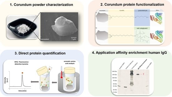
3. Results and Discussion

3.1. Corundum Particle Characterization

The corundum raw material used in this work is an industrial product used for abrasive purposes. TEM, ESEM, BET, DLS, EDS, and zeta potential measurements were performed to characterize the corundum particles and give insight into the shape, size, and surface properties of this material. Before functionalization, the particles were purified, as shown in the Supplementary Materials. In a BET measurement, a surface area of 4.5 m2/g was determined. DLS measurements showed an average particle size of 0.78 µm ± 0.14 (Supplementary Materials). However, the TEM and ESEM micrographs revealed that a broad range of particle sizes and shapes are present due to the milling process. Figure 1 shows a macroscopic view of the raw material (a), a TEM micrograph for a corundum particle (b) as well as an ESEM micrograph (c). The surface of the particle appears to be nonporous and the particles have sharp edges. These images are consistent with the mean particle sizes and surface area determined by DLS and BET.

3.2. Surface Functionalization with Amino Phosphonic Acids, Activated with Glutaraldehyde or Oxidized Hyperbranched Polyglycerol

In order to immobilize proteins or peptides on the corundum surface covalently, 12-amino dodecyl phosphonic acid (12-APA) was used as an interface to provide a firmly bound functional group that can be modified in further steps. TEM-EDS measurements after the functionalization with 12-APA showed an increase in phosphorus and carbon, supporting the attachment of 12-APA to the corundum surface (Supplementary Materials, Figure S7). Once the phosphonic acid is attached to the surface, a further reaction with a crosslinker is necessary to link the desired protein to the corundum surface. This was achieved by using the well-established bifunctional crosslinker glutaraldehyde, which will react in a nucleophilic addition reaction with the amine group of the phosphonic acid, and in the same manner with the amino groups of protein mainly provided by lysine residues. A hyperbranched polyglycerol (PG) was explored in this work to find an alternative to the toxic and poorly defined glutaraldehyde. After some diols are cleaved to aldehyde groups, the PG is allowed to react in a similar manner as the glutaraldehyde does, with the exception that the PG is highly branched in structure. Aside from the expected lower toxicity, it was expected to lead to a higher protein-binding capacity due to the existence of more functional groups that can be linked to the protein. The PG was oxidized for 16 h with a molar ratio of 1.4:1 (hydroxyl groups/ periodate) in PBS. Our results suggest that the oxidation overnight led to higher protein-binding capacities than oxidation for one hour (data not shown). Figure 2 shows the oxidation reaction of the diol cleavage by NaIO4. In this reaction, the diols are cleaved, resulting in aldehyde groups that can be modified later on.
In a 1H NMR experiment, a newly formed signal caused by terminal aldehyde groups (about 9.5 ppm) confirmed the formation of aldehyde groups after oxidation with NaIO4. Another sharp singlet at about 8.3 ppm indicated the presence of formic acid in the oxidation solution, which is a byproduct of the diol cleavage (Supplementary Materials, Figure S8). The oxidized PG was then incubated with the amino phosphonic acid functionalized corundum in the same way as glutaraldehyde. Zeta potential measurements were performed to confirm a change in the surface chemistry (Table 1). A reduction of the imines by NaCNBH3 was not performed for these samples.

3.3. Protein Immobilization and Direct Quantification with Aromatic Amino Acid Analysis (AAAA)

Bovine serum albumin (BSA) was used as a model protein to determine which tested conjugation strategy offers the highest protein-binding capacity. Figure 3 shows the simplified functionalization schemes for corundum.
Direct protein quantification on particles is an elegant way to circumvent the challenges that are faced when trying to quantify the unbound amount of protein in a supernatant. In a recent work by Tchipilov et al., the disadvantages of such an indirect approach to quantification were discussed in detail [46]. Aromatic amino acid analysis (AAAA), in contrast, is a useful and very precise approach for direct protein quantification in liquids or on solid surfaces. The particles coated with proteins were dispersed in 37% hydrochloric acid containing DL-methionine as a reductant to protect sensitive amino acids from oxidative degradation. The mixture was heated in a copper block for 1 h at 150 °C. After centrifugation to remove the corundum particles, the hydrolyzed protein solution is diluted and injected into an HPLC system for quantification. The amino acid sequence of the protein BSA is known, and therefore the number of the respective aromatic amino acids, too. The total protein content of the sample can be calculated with the use of an external aromatic amino acid calibration. In this work, the fluorescence detection of tyrosine was used to calculate the amount of immobilized protein on the corundum particles. Figure 4 illustrates the described workflow.
The amount of immobilized BSA was calculated with a calibration based on a standard amino acid solution containing L-tyrosine. Quantification of BSA on 12-APA-modified corundum showed a value of 2.54 mg ± 0.02 when functionalized with oxidized polyglycerol and 1.96 mg ± 0.02 with glutaraldehyde. This data suggests that using 12-APA in combination with oxidized polyglycerol can bind more protein than glutaraldehyde.

3.4. Affinity Enrichment of IgG from Human Plasma with Protein-A-Functionalized Corundum

Prior to the affinity enrichment experiment, the non-specific binding properties of the functionalized corundum were investigated to ensure that plasma proteins do not interact non-specifically with the surface. Corundum particles were functionalized with 12-APA, subsequently with glutaraldehyde or polyglycerol, and finally with BSA. Human plasma was then incubated with the particles for 1 h. The material was washed three times with PBS/Tween before the elution of adsorbed proteins with 2% SDS was performed. Such a harsh elution is expected to elute all proteins that are bound in a non-covalent manner [47]. SDS-PAGE showed low NSB for the polyglycerol-functionalized particles, whereas, for glutaraldehyde, slightly more proteins were eluted after human plasma incubation (Supplementary Materials, Figure S10). Polyglycerol was chosen for the proof-of-concept experiment, where the IgG should be isolated from human plasma with 12-APA-modified corundum particles that were functionalized with oxidized polyglycerol (PG) and subsequently conjugated to protein A. Protein A selectively binds the Fc part of many immunoglobulins; therefore, it can be used as an affinity binder to isolate IgG from a complex matrix such as plasma, which is estimated to contain up to 10,000 different proteins [48]. The amount of immobilized protein A was quantified using the previously described method. An amount of 0.5 ± 0.04 mg protein A per 1 g corundum was determined. Human plasma was diluted at 1:10 and incubated with the functionalized particles. Several washing steps and centrifugations were performed to remove all loosely bound material. The particles were then treated with a sodium dodecyl sulfate (SDS) solution (2%, tris base, pH 6.8) and incubated for 15 min to elute the bound IgG. The solution was separated from the corundum via centrifugation and then heated for complete protein denaturation before it was loaded onto the SDS gel. Figure 5 shows the described workflow and the SDS gel obtained in this experiment.
The silver-stained SDS-PAGE separation indicates the successful isolation of IgG from human plasma. In addition, no protein A leakage (“leaching”) was observed, confirming the covalent attachment of protein A to the oxidized polyglycerol.
To determine the IgG-binding capacity, protein A functionalized corundum was incubated with 1:10 diluted human plasma for 1 h and was subsequently washed three times with PBST and once with lab water before it was treated with glycine/HCl buffer of pH 2.3 for 15 min. After neutralization, a capacity of 1.8 mg IgG per 1 g corundum was determined with a commercial BCA assay.

4. Conclusions

In this work, corundum powder, intended to be used in industrial applications, was purified, characterized, and functionalized to create a new platform for the isolation and enrichment of targeted proteins from complex matrixes like human plasma. This approach leads to an affinity method that can be easily performed in any lab possessing a standard centrifuge. This makes protein isolation independent of access to complex column-based instruments or magnetic separation systems.
The raw corundum material showed some impurities in the TEM micrographs; therefore, a brief purification protocol was established. The functionalization with a long-chain amino phosphonic acid was used to generate a surface with primary amine groups. In a subsequent step, glutaraldehyde was compared with oxidized polyglycerol (PG) as linkers. Our results showed that in direct comparison, the oxidized PG could bind more BSA than glutaraldehyde, making it a viable alternative. In the next experiment, protein A was conjugated to corundum modified with 12-APA and oxidized PG to isolate IgG from human plasma. The results showed the successful affinity enrichment of IgG with high purity. No leaching of protein A was detected even under harsh conditions, which confirms the covalent attachment of protein A to the surface. Furthermore, the non-specific binding (NSB) of matrix proteins (human plasma) was found to be very low, which shows that the oxidized PG also acts as an excellent surface coating. These proof-of-concept experiments demonstrate the performance and flexibility of these novel materials based on corundum as a platform for affinity separations.

Supplementary Materials

The following supporting information can be downloaded at: https://www.mdpi.com/article/10.3390/separations9090252/s1, Figure S1: Dynamic light scattering (DLS) measurements of corundum F1200 in 0.1 M PBS indicated an average particle size of 0.78 µm; Figure S2: TEM micrograph of unpurified corundum: (a) Corundum surface covered with needle-shaped crystals, (b) elemental mapping showing sodium- and chloride-containing structures; Figure S3: Purification protocol for corundum powder; Figure S4: TEM-EDS analysis of corundum before (black) and after (red) purification; Figure S5: BET measurements: Isotherm linear plot and BET surface area plot; Figure S6: Calibration line of tyrosine (amino acid standard solution). Fluorescence detection was performed at 272 nm (Ex.) and 303 nm (Em.); Figure S7: TEM-EDS analysis of corundum before (red) and after (black) 12-APA functionalization; Figure S8: NMR of untreated PG (red) and oxidized PG (blue). The NMR signal at a chemical shift of 9.5 ppm indicates terminal aldehydes of the polyglycerol that were formed due to oxidation with NaIO4. The very sharp singlet at a chemical shift of 8.3 ppm corresponds to formic acid, also a product of the cleavage; Figure S9: ESEM micrographs of functionalized corundum. A surface layer was not visible; Figure S10: SDS-PAGE of corundum functionalized with bovine serum albumin (BSA), conjugated with glutaraldehyde or polyglycerol. The functionalized corundum was incubated with 1:10 diluted human plasma (HP). After the elution with 2% of SDS, almost no protein elution was detected for the polyglycerol/BSA-functionalized corundum, whereas for the glutaraldehyde/BSA, at least one weak protein band (arrow) is visible. Furthermore, no elution (leaching) of the covalently bound BSA was observed.

Author Contributions

Conceptualization, J.L.V. and M.G.W.; methodology, J.L.V. and M.G.W.; validation, J.L.V., P.H.S. and M.G.W.; formal analysis, J.L.V. and P.H.S.; investigation J.L.V., P.H.S., I.F., K.G., K.M. and A.Z.; resources, J.L.V., P.H.S., I.F., K.G., K.M. and A.Z.; data curation, J.L.V., P.H.S., I.F., K.G., K.M. and A.Z.; writing—original draft preparation, J.L.V. and M.G.W.; writing—review and editing, J.L.V. and M.G.W.; visualization, J.L.V.; supervision, M.G.W.; project administration, M.G.W.; funding acquisition, M.G.W. All authors have read and agreed to the published version of the manuscript.

Funding

This research received no external funding.

Institutional Review Board Statement

Not applicable.

Informed Consent Statement

Not applicable.

Data Availability Statement

Not applicable.

Acknowledgments

Special thanks to DendroPharm GmbH, Berlin, for their technical support concerning the hyperbranched polyglycerol.

Conflicts of Interest

The authors declare no conflict of interest.

References

  1. Schneck, N.A.; Phinney, K.W.; Lee, S.B.; Lowenthal, M.S. Quantification of cardiac troponin I in human plasma by immunoaffinity enrichment and targeted mass spectrometry. Anal. Bioanal. Chem. 2018, 410, 2805–2813. [Google Scholar] [CrossRef] [PubMed]
  2. Makawita, S.; Diamandis, E.P. The bottleneck in the cancer biomarker pipeline and protein quantification through mass spectrometry–based approaches: Current strategies for candidate verification. Clin. Chem. 2010, 56, 212–222. [Google Scholar] [CrossRef]
  3. Sauer, P.W.; Burky, J.E.; Wesson, M.C.; Sternard, H.D.; Qu, L. A high-yielding, generic fed-batch cell culture process for production of recombinant antibodies. Biotechnol. Bioeng. 2000, 67, 585–597. [Google Scholar] [CrossRef]
  4. Aires da Silva, F.; Corte-Real, S.; Goncalves, J. Recombinant antibodies as therapeutic agents. Biodrugs 2008, 22, 301–314. [Google Scholar] [CrossRef] [PubMed]
  5. Łącki, K.M.; Riske, F.J. Affinity Chromatography: An Enabling Technology for Large-Scale Bioprocessing. Biotechnol. J. 2020, 15, 1800397. [Google Scholar] [CrossRef] [PubMed]
  6. Groman, E.V.; Wilchek, M. Recent developments in affinity chromatography supports. Trends Biotechnol. 1987, 5, 220–224. [Google Scholar] [CrossRef]
  7. Cuatrecasas, P.; Wilchek, M.; Anfinsen, C.B. Selective enzyme purification by affinity chromatography. Proc. Natl. Acad. Sci. USA 1968, 61, 636–643. [Google Scholar] [CrossRef]
  8. Göke, B.; Keim, V. HPLC and FPLC. Int. J. Pancreatol. 1992, 11, 109–116. [Google Scholar] [CrossRef] [PubMed]
  9. Whiteaker, J.R.; Zhao, L.; Zhang, H.Y.; Feng, L.-C.; Piening, B.D.; Anderson, L.; Paulovich, A.G. Antibody-based enrichment of peptides on magnetic beads for mass-spectrometry-based quantification of serum biomarkers. Anal. Biochem. 2007, 362, 44–54. [Google Scholar] [CrossRef] [PubMed]
  10. Huang, B.; Changhe, L.; Zhang, Y.; Wenfeng, D.; Min, Y.; Yuying, Y.; Han, Z.; Xuefeng, X.; Dazhong, W.; Debnath, S. Advances in fabrication of ceramic corundum abrasives based on sol–gel process. Chin. J. Aeronaut. 2021, 34, 1–17. [Google Scholar] [CrossRef]
  11. Selvakumaran, D.; Arunachalam, N.; Vijayaraghavan, L.; Balan, A. Performance comparison of sol-gel with white alumina abrasives for grinding of super duplex stainless steel (SDSS). Procedia Manuf. 2018, 26, 1448–1458. [Google Scholar] [CrossRef]
  12. Krell, A.; Blank, P.; Wagner, E.; Bartels, G. Advances in the grinding efficiency of sintered alumina abrasives. J. Am. Ceram. Soc. 1996, 79, 763–769. [Google Scholar] [CrossRef]
  13. Nadolny, K. State of the art in production, properties and applications of the microcrystalline sintered corundum abrasive grains. Int. J. Adv. Manuf. Technol. 2014, 74, 1445–1457. [Google Scholar] [CrossRef]
  14. Pujari, S.P.; Scheres, L.; Marcelis, A.T.; Zuilhof, H. Covalent surface modification of oxide surfaces. Angew. Chem. Int. Ed. 2014, 53, 6322–6356. [Google Scholar] [CrossRef]
  15. Luschtinetz, R.; Oliveira, A.F.; Duarte, H.A.; Seifert, G. Self-assembled Monolayers of Alkylphosphonic Acids on Aluminum Oxide Surfaces–A Theoretical Study. Z. Anorg. Allg. Chem. 2010, 636, 1506–1512. [Google Scholar] [CrossRef]
  16. Messerschmidt, C.; Schwartz, D.K. Growth mechanisms of octadecylphosphonic acid self-assembled monolayers on sapphire (corundum): Evidence for a quasi-equilibrium triple point. Langmuir 2001, 17, 462–467. [Google Scholar] [CrossRef]
  17. Bauer, T.; Schmaltz, T.; Lenz, T.; Halik, M.; Meyer, B.; Clark, T. Phosphonate-and carboxylate-based self-assembled monolayers for organic devices: A theoretical study of surface binding on aluminum oxide with experimental support. ACS Appl. Mater. Interfaces 2013, 5, 6073–6080. [Google Scholar] [CrossRef]
  18. Thissen, P.; Valtiner, M.; Grundmeier, G. Stability of phosphonic acid self-assembled monolayers on amorphous and single-crystalline aluminum oxide surfaces in aqueous solution. Langmuir 2010, 26, 156–164. [Google Scholar] [CrossRef]
  19. Frasconi, M.; Mazzei, F.; Ferri, T. Protein immobilization at gold–thiol surfaces and potential for biosensing. Anal. Bioanal. Chem. 2010, 398, 1545–1564. [Google Scholar] [CrossRef]
  20. Silin, V.; Weetall, H.; Vanderah, D.J. SPR studies of the nonspecific adsorption kinetics of human IgG and BSA on gold surfaces modified by self-assembled monolayers (SAMs). J. Colloid Interface Sci. 1997, 185, 94–103. [Google Scholar] [CrossRef] [PubMed]
  21. Bain, C.D.; Whitesides, G.M. Modeling organic surfaces with self-assembled monolayers. Angew. Chem. 1989, 101, 522–528. [Google Scholar] [CrossRef]
  22. Klykov, O.; Weller, M.G. Quantification of N-hydroxysuccinimide and N-hydroxysulfosuccinimide by hydrophilic interaction chromatography (HILIC). Anal. Methods 2015, 7, 6443–6448. [Google Scholar] [CrossRef]
  23. Barbosa, O.; Ortiz, C.; Berenguer-Murcia, Á.; Torres, R.; Rodrigues, R.C.; Fernandez-Lafuente, R. Glutaraldehyde in bio-catalysts design: A useful crosslinker and a versatile tool in enzyme immobilization. RSC Adv. 2014, 4, 1583–1600. [Google Scholar] [CrossRef]
  24. Migneault, I.; Dartiguenave, C.; Bertrand, M.J.; Waldron, K.C. Glutaraldehyde: Behavior in aqueous solution, reaction with proteins, and application to enzyme crosslinking. BioTechniques 2004, 37, 790–802. [Google Scholar] [CrossRef] [PubMed]
  25. Clinton, F. Sodium cyanoborohydride—A highly selective reducing agent for organic functional groups. Synthesis 1975, 3, 135–146. [Google Scholar] [CrossRef]
  26. Zeiger, E.; Gollapudi, B.; Spencer, P. Genetic toxicity and carcinogenicity studies of glutaraldehyde—A review. Mutat. Res./Rev. Mutat. Res. 2005, 589, 136–151. [Google Scholar] [CrossRef]
  27. Beauchamp, R.O.; St Clair, M.B.G.; Fennell, T.R.; Clarke, D.O.; Morgan, K.T.; Karl, F.W. A critical review of the toxicology of glutaraldehyde. Crit. Rev. Toxicol. 1992, 22, 143–174. [Google Scholar] [CrossRef]
  28. Calderón, M.; Quadir, M.A.; Sharma, S.K.; Haag, R. Dendritic polyglycerols for biomedical applications. Adv. Mater. 2010, 22, 190–218. [Google Scholar] [CrossRef]
  29. Unbehauen, M.L.; Fleige, E.; Paulus, F.; Schemmer, B.; Mecking, S.; Moré, S.D.; Haag, R. Biodegradable core–multishell nanocarriers: Influence of inner shell structure on the encapsulation behavior of dexamethasone and tacrolimus. Polymers 2017, 9, 316. [Google Scholar] [CrossRef]
  30. Pant, K.; Groger, D.; Bergmann, R.; Pietzsch, J.; Steinbach, J.; Graham, B.; Spiccia, L.; Berthon, F.; Czarny, B.; Devel, L. Synthesis and biodistribution studies of 3H-and 64Cu-labeled dendritic polyglycerol and dendritic polyglycerol sulfate. Bioconjug. Chem. 2015, 26, 906–918. [Google Scholar] [CrossRef] [PubMed]
  31. Frey, H.; Haag, R. Dendritic polyglycerol: A new versatile biocompatible material. Rev. Mol. Biotechnol. 2002, 90, 257–267. [Google Scholar] [CrossRef]
  32. Höger, K.; Becherer, T.; Qiang, W.; Haag, R.; Frieß, W.; Küchler, S. Polyglycerol coatings of glass vials for protein resistance. Eur. J. Pharm. Biopharm. 2013, 85, 756–764. [Google Scholar] [CrossRef] [PubMed]
  33. Yu, Y.; Frey, H. Controllable nonspecific protein adsorption by charged hyperbranched polyglycerol thin films. Langmuir 2015, 31, 13101–13106. [Google Scholar] [CrossRef]
  34. Moore, E.; Delalat, B.; Vasani, R.; Thissen, H.; Voelcker, N.H. Patterning and biofunctionalization of antifouling hyperbranched polyglycerol coatings. Biomacromolecules 2014, 15, 2735–2743. [Google Scholar] [CrossRef] [PubMed]
  35. Hao, M.; Wu, T.; Chen, Q.; Lian, X.; Wu, H.; Shi, B. Hyperbranched polyglycerols as robust up-conversion nanoparticle coating layer for feasible cell imaging. Polymers 2020, 12, 2592. [Google Scholar] [CrossRef]
  36. Jones, G. Polymerization of glutaraldehyde at fixative pH. J. Histochem. Cytochem. 1974, 22, 911–913. [Google Scholar] [CrossRef] [PubMed]
  37. Rembaum, A.; Margel, S.; Levy, J. Polyglutaraldehyde: A new reagent for coupling proteins to microspheres and for labeling cell-surface receptors. J. Immunol. Methods 1978, 24, 239–250. [Google Scholar] [CrossRef] [PubMed]
  38. Moks, T.; Abrahmsén, L.; Nilsson, B.; Hellman, U.; Sjöquist, J.; Uhlén, M. Staphylococcal protein A consists of five IgG-binding domains. Eur. J. Biochem. 1986, 156, 637–643. [Google Scholar] [CrossRef] [PubMed]
  39. Pecher, H.S.; Zimathies, A.; Weller, M.G. Oligoepoxide-Based Monoliths: Synthesis and Application as Affinity Capillary Column for Enrichment of Immunoglobulin G. Macromol. Chem. Phys. 2012, 213, 2398–2403. [Google Scholar] [CrossRef]
  40. Wilke, M.; Röder, B.; Paul, M.; Weller, M.G. Sintered glass monoliths as supports for affinity columns. Separations 2021, 8, 56. [Google Scholar] [CrossRef]
  41. Nayak, N.; Mazzei, R.; Giorno, L.; Crespo, J.G.; Portugal, C.A.; Poerio, T. Protein Attachment Mechanism for Improved Functionalization of Affinity Monolith Chromatography (AMC). Molecules 2022, 27, 4496. [Google Scholar] [CrossRef]
  42. Schneider, C.A.; Rasband, W.S.; Eliceiri, K.W. NIH Image to ImageJ: 25 years of image analysis. Nat. Methods 2012, 9, 671–675. [Google Scholar] [CrossRef]
  43. Hesse, A.; Weller, M.G. Protein quantification by derivatization-free high-performance liquid chromatography of aromatic amino acids. J. Amino Acids 2016, 2016, 7374316. [Google Scholar] [CrossRef] [PubMed]
  44. Reinmuth-Selzle, K.; Tchipilov, T.; Backes, A.T.; Tscheuschner, G.; Tang, K.; Ziegler, K.; Lucas, K.; Pöschl, U.; Fröhlich-Nowoisky, J.; Weller, M.G. Determination of the protein content of complex samples by aromatic amino acid analysis, liquid chromatography-UV absorbance, and colorimetry. Anal. Bioanal. Chem. 2022, 414, 4457–4470. [Google Scholar] [CrossRef] [PubMed]
  45. Image from the Protein Data Bank (rcsb.org) PDB ID 4F5S. Bujacz, A.; Bujacz, G. Crystal Structure of Bovine Serum Albumin. 2012. Available online: https://www.wwpdb.org/pdb?id=pdb_00004f5s (accessed on 30 August 2022). [CrossRef]
  46. Tchipilov, T.; Raysyan, A.; Weller, M.G. Methods for the quantification of particle-bound protein—Application to reagents for lateral-flow immunoassays (LFIA). Preprints 2022, 2022030332. [Google Scholar] [CrossRef]
  47. Sousa, M.M.; Steen, K.W.; Hagen, L.; Slupphaug, G. Antibody cross-linking and target elution protocols used for immunoprecipitation significantly modulate signal-to noise ratio in downstream 2D-PAGE analysis. Proteome Sci. 2011, 9, 1–8. [Google Scholar] [CrossRef]
  48. Pernemalm, M.; Sandberg, A.; Zhu, Y.; Boekel, J.; Tamburro, D.; Schwenk, J.M.; Björk, A.; Wahren-Herlenius, M.; Åmark, H.; Östenson, C.-G. In-depth human plasma proteome analysis captures tissue proteins and transfer of protein variants across the placenta. Elife 2019, 8, e41608. [Google Scholar] [CrossRef] [PubMed]
Figure 1. (a) Macroscopic view of the raw corundum powder, (b) TEM micrograph, and (c) ESEM micrograph showing purified particles with an irregular shape but a smooth, nonporous surface.
Separations 09 00252 g001
Figure 2. Oxidation of diols in polyglycerol (PG) with sodium periodate. The formation of aldehyde groups could be shown in a 1H NMR experiment (Supplementary Materials, Figure S8).
Separations 09 00252 g002
Figure 3. A simplified scheme of corundum particles modified with 12-aminododecylphosphonic acid (12-APA), the (a) crosslinkers glutaraldehyde, or (b) oxidized polyglycerol, respectively, and finally conjugated to bovine serum albumin (BSA); Structures are not to scale; BSA structure from [45].
Separations 09 00252 g003
Figure 4. Workflow of aromatic amino acid analysis (AAAA) of protein-functionalized corundum. Red dots symbolize aromatic amino acids in the sequence, green dots all other amino acids: (a) Protein bound to corundum particles, (b) hydrolysis in HCl at 150 °C for 1 h, (c) dilution of hydrolysis supernatant, and (d) HPLC analysis of tyrosine with fluorescence detection (excitation 272 nm/emission 303 nm).
Separations 09 00252 g004
Figure 5. Affinity enrichment of IgG (Y symbol) from human plasma. Corundum was coated with 12-APA and oxidized polyglycerol: (a) Human plasma containing a high number of different proteins, (b) incubation with protein-A-functionalized corundum, (c) binding of IgG to protein A (u symbol), (d) centrifugation, and removal of the supernatant and washing, and (e) SDS-PAGE after the elution of selectively bound IgG by a 2% SDS solution.
Separations 09 00252 g005
Table 1. Zeta potential measurements of unfunctionalized and functionalized corundum. Measurements were performed in 1 mM KCl at a pH of 7.
SampleZeta Potential [mV]
Corundum4.28 ± 0.01
Corundum-12-APA−2.94 ± 0.37
Corundum-12-APA-glutaraldehyde−15.00 ± 0.52
Corundum-12-APA-oxidized polyglycerol−15.10 ± 3.70
Publisher’s Note: MDPI stays neutral with regard to jurisdictional claims in published maps and institutional affiliations.

