Article

Curative Effects of Dianthus orientalis against Paracetamol Triggered Oxidative Stress, Hepatic and Renal Injuries in Rabbit as an Experimental Model

1
Department of Zoology, University of Malakand, Chakdara 18800, Khyber Pakhtunkhwa, Pakistan
2
Department of Zoology, Islamia College Peshawar, Peshawar 25120, Khyber Pakhtunkhwa, Pakistan
3
Department of Botany, Islamia College Peshawar, Peshawar 25120, Khyber Pakhtunkhwa, Pakistan
4
Department of Biotechnology, University of Malakand, Chakdara 18800, Khyber Pakhtunkhwa, Pakistan
5
Laboratory of Animal Health, Food Hygiene and Quality, Department of Agriculture, University of Ioannina, 47132 Arta, Greece
6
Department of Pharmacology and Toxicology, College of Pharmacy, King Saud University, P.O. Box 2455, Riyadh 11451, Saudi Arabia
*
Author to whom correspondence should be addressed.
Separations 2023, 10(3), 182; https://doi.org/10.3390/separations10030182
Submission received: 12 February 2023 / Revised: 2 March 2023 / Accepted: 3 March 2023 / Published: 7 March 2023
(This article belongs to the Section Analysis of Natural Products and Pharmaceuticals)

Abstract

:
The aim of the present study investigates the hepatoprotective, nephroprotective and hematopoietic and antioxidant effects of Dianthus orientalis leaves aqueous extract (DO.AQ) in rabbits intoxicated with paracetamol. Different experimental groups were formed, i.e., group N, group T, group ELD, group EMD, group EHD and group SM. The groups with leaves aqueous extract of Dianthus orientalis of 200 and 400 mg/kg body weight, i.e., group EMD and group EHD, showed remedial effects; however, a high dose extract significantly (p < 0.05) reduced the elevated serum levels of alanine transaminase ALT, aspartate transaminase AST and alkaline phosphatase ALP and renal related indices such as serum creatinine, urea and uric acid, and serum electrolytes such as Ca, Mg, P, Na and K, as well as the total count of RBC, WBC, platelets and hemoglobin Hb concentration, mean corpuscular hemoglobin MCH concentration and hematocrit HCT values. Additionally, the extract showed positive effects on the lipid profile, i.e., decreasing levels of cholesterol, triglycerides and LDL and increasing levels of HDL. The levels of thiobarbituric acid reactive substances TBARS, glutathione GSH and radical scavenging activity were also evaluated in liver and kidney homogenates. Paracetamol fed animals had high levels of thiobarbituric acid reactive substances and low levels of glutathione GSH and radical scavenging activity (RSA). Extract ingestion caused a significant increase in glutathione and radical scavenging activity RSA levels, while reducing the (TBARS) levels, showing that the extracts have antioxidant potentials. The antioxidant capacity of the Dianthus orientalis leaves aqueous extract at various dosages demonstrated an increased inhibition of DPPH, i.e., 2, 2-diphenyl-1-picrylehydrazyle free radical. The histological study of the liver and kidney supports the protective activity of Dianthus orientalis leaves aqueous extract against paracetamol intoxication with optimistic effects regarding oxidative stress condition and serum electrolytes balance.

1. Introduction

The liver is an essential organ in the body, playing a significant role in controlling a number of biological activities such as metabolism, secretion, and storage. Therefore, it protects the body from toxic substances through detoxification and the elimination of xenobiotics from the body [1,2]. This exposes the liver to a variety of endogenous and external harmful substances that can cause hepatotoxicity [3].
Many regularly used chemicals and drugs cause cellular and metabolic damage to the liver [4]. Acetaminophen, usually referred to as paracetamol, is a well-known painkiller and fever reducing agent that causes liver and kidney toxicity. When given in a normal amount, paracetamol has no harmful effects, but a high dose can harm the liver [5]. Acetaminophen-induced liver injury is caused by the toxic metabolite N-acetyl-p-benzoquinoneimine (NABQI), manufactured by cytochrome P-450 enzymes [6,7]. This substance is normally altered by conjugation with glutathione, but in a high dose this element is generated in such excessive concentrations that it overpowers the detoxification process, causing hepatocyte damage and cell death [8]. According to [9], different high dosages of acetaminophen showed a noteworthy increase in liver enzyme activities.
The kidneys are also important homeostatic organs [10]. Toxic overdose of drugs leads to renal toxicity, often associated with numerous metabolic disorders such as imbalances in serum electrolytes, urea, uric acid, and creatinine [11,12]; these are crucial biomarkers for detecting kidney function [13]. Reactive oxygen species (ROS) are produced in both hepatotoxicity and nephrotoxicity, and they damage the integrity of cell membranes and release cellular enzymes such as hepatic transaminases, such as (ALT), (AST), (ALP) and lactate dehydrogenase (LDH), into the bloodstream and increase levels of thiobarbituric acid reactive substances (TBARS). Other biomarkers, such as glutathione (GSH), superoxide dismutase (SOD), and malondialdehyde (MDA), as well as haematological indices including PCV, Hb, TLC, MCH, MCHC, and MCV, are also affected by hepatocellular injury [14,15,16,17,18]. Thus, ROS are involved in programmed cell death, which induces oxidative stress and damages biomolecules such as lipids, nucleic acids, proteins and carbohydrates [19]. Chemo-inhibition is a method for reducing the risk of hepatotoxicity that uses both natural and synthetic substances [20]. By restoring free radical effects to normal, antioxidant drugs contribute significantly to the prevention of oxidative damage [21]. Numerous studies have demonstrated the powerful antioxidant capacity of medicinal plants which are rich in chemicals that scavenge free radicals [22,23], such as phenolic compounds, triterpenoids, flavonoids, alkaloids, and tannin [24].
The genus Dianthus L. belongs to the family Caryophyllaceae and comprises about 300 species distributed in Asia, Europe, North America, and Africa (Reeve, 1967). The Mediterranean region is the primary hub of genus diversity [25,26]. They are frequently referred to as carnations and pinks. The plants are annual, biennial or perennial. Dianthus orientalis Adams is a herbaceous perennial plant widely distributed in southern and eastern Turkey and Asia [27]. The plant is traditionally used as a natural cure, for example using its leaves as a diuretic and pollen as a desiccant, vulnerary, astringent, haemostatic, or diuretic [28].
Most of world’s population relies on herbal medicine for their primary care. Herbal drugs or plant derived agents are used in combination with allopathic medicine to cure all types of diseases, including liver illness; therefore, this study aimed to investigate the curative role of the selected plant extract [29]. As a component of Pakistani medicinal flora, aqueous extracts of Dianthus orientalis leaves have been investigated for hepatoprotective, nephroprotective, and other serum biochemical activities. It is therefore hypothesized that this plant will give the best results regarding the healing of liver and kidney injuries induced in the laboratory and, further, should be explored based on scientific principles and should also be approved as a chief medicine in other related illnesses.

2. Materials and Methods

2.1. Collection and Extraction of Plant Materials

The plant Dianthus orientalis voucher name (UOM/V.No.179) was placed in the herbarium in the Department of Botany, University of Malakand. The plant was collected from the Swat district of Pakistan and identified by Prof. Dr. Muhammad Nisar, University of Malakand, for further processing. A mechanical grinder was used to slice and grind the fresh Dianthus orientalis leaves. The crushed material was added to 1000 m/L of deionized water and shaken for 45 h at normal temperature. After filtration of the mixture, the filtrate was converted to a thick paste extract by shifting into a rotary evaporator under low pressure and high temperature (40 °C), and finally the aqueous extract of Dianthus orientalis (DO.AQ) was prepared [30].

2.2. Acute Toxicity Test

The rabbits (average weight 800–1300 mg) were fasted for 14 h and were placed into four groups of five rabbits each. Using a steel bulbed needle, rabbits were given an intraperitoneal injection of 100, 200, 400 or 800 mg/kg. After that, the rabbits were given unrestricted access to water and food for 48 h while being monitored for signs of acute poisoning following the method as described by Sana et al. [31].

2.3. Experimental Animals, Chemicals, Treatment Regime and Ethical Approval

The experimental animals (rabbits weighing on average 800–1300 mg) were purchased from a nearby market. These animals were kept in cages for acclimatization prior to the experimentation, as described by Chattopadhyay et al. [32], in accordance with the “Guide for the Care and Use of Laboratory Animals,” and with the approval of the Ethical Committee of the Department of Zoology, University of Malakand reviewed aims objectives of the study vide reference No UOM/Zoo/21-191. The experimental animals were categorized into six (6) groups, each with five animals [33]. All animals received extract and chemicals continuously during the course of 21 days (0–7, 8–14, and 15–21 days) by oral administration.
The chemicals, such as silymarin and paracetamol, were manufactured by BEIJING INFOARK Co., Ltd. (Beijing Infoark Technology Development Co., Ltd., Beijing, China) Haidian District, Beijing, China and Johnson Matthey (Shanghai) Catalyst Co., Ltd. China.
The treatment plan is listed underneath.
Group N: This group animals kept as normal with no treatment.
Group T: Ingest paracetamol (200 mg/kg body weight (BW) one time daily for 21 days regularly.
Group ELD: Received paracetamol (200 mg/kg BW) and DO.AQ extract (100 mg/kg BW, daily once for 21 days regularly.
Group EMD: Administered paracetamol (200 mg/kg BW) and (DO.AQ) extract (200 mg/kg BW) daily for 21 days regularly.
Group EHD: Treated with (DO.AQ) extract (400 mg/kg BW) and paracetamol (200 mg/kg BW) once daily for 21 days continuously.
Group SM: Received silymarin (50 mg/kg and paracetamol (200 mg/kg BW) once daily for 21 days continually.

2.4. Analysis of Hematological, Liver and Kidney Function

2.4.1. Blood Collection

Blood was collected on day 7, first week (W1), day 14, second week (W2) and ultimately on day 21, third week (W3) from all groups of rabbits by carotid hemorrhage into centrifuge tubes. The blood samples were centrifuged at 3000 rev/h. for 10 min in a bench centrifuge to acquire clear serum, which was employed for the evaluation of biochemical, liver and kidney function tests [34].

2.4.2. Hematological Parameters

Erythrocytes, hemoglobin concentration, hematocrit, mean corpuscular volume, mean corpuscular hemoglobin, mean corpuscular hemoglobin concentration and platelets were examined by using the technique of [35,36].

2.4.3. Liver and Kidney Function Test via Biochemical Analysis

Serum was utilized for the assessment of liver and kidney functions via assessing the levels of ALT, AST, and ALP using an auto-analyzer (Olympus AU 600, Olympus, Nagoya, Japan) according to [37]. Glucose level was assessed by the process of [38]. Serum urea, uric acid and creatinine were estimated following [39]’s procedure, and total serum cholesterol, HDL, LDL and triglyceride were determined by the method of [40].

2.5. Histopathological Analysis

Histopathology of the kidney and liver was performed according to the procedure in [41]. All of the animals were dissected at the end of the experiment, and the livers and kidneys from the animals of the respective group were removed and promptly preserved in a solution containing 10% formalin and 0.9% sodium chloride. The tissues were then placed in paraffin, thinly partitioned by means of a microtome, stained with haematoxylin and eosin (H&E) for typical morphological evaluation, and then examined under a light microscope (BX50; Olympus, Tokyo). The photographs were produced using a digital camera system (Pixcera Co., Osaka, Japan) mounted on the microscope.

2.6. Statistical Analysis

The outcomes of the current study were scrutinized using ANOVA (one-way) and Tukey test, utilizing the latest version of graph pad prism.

3. Results

3.1. Acute Toxicity

During this experiment, all of the rabbits appeared to be healthy, with no signs of toxicity, illness or death observed in any of the groups. The acute oral administration of DO.AQ extract to rabbits has demonstrated that the plant is safe and can be used medicinally in this species, even at a dose of 800 mg/kg BW.

3.2. Blood Hematological Analysis

Acetaminophen (paracetamol) consumption significantly (p < 0.05) reduced the levels of hemoglobine, red blood cell number, HCT value, MCV value, MCH and MCHC levels, whereas an increase was seen in the concentration of WBC, platelets, lymphocytes, neutrophils and monocytes. The consumption of DO.AQ extract on day 21 (third week, W3) of the study at doses of 200 mg to 400 mg/kg body weight significantly (p < 0.05) stabilized the blood parameters. However, as shown in Table 1 and Table 2 and Figure 1 and Figure 2, no significant changes were noticed at all doses of DO.AQ extract throughout the first and second weeks of treatment (W1, W2).

3.3. Hepatic and Renal Function Test

3.3.1. Liver and Kidney Serum Markers

When the animals were given paracetamol (200 mg/kg BW), a statistically significant (p < 0.05) increase in the AST level, ALT, and ALP values were seen in all experimental animal groups, i.e., T, ELD, EMD, EHD and SM, in comparison to group N. Additionally, serum creatinine, urea and uric acid concentrations were elevated in the same way. The DO.AQ extract showed a therapeutic effect on the serum biochemical markers levels. At all dosages of DO.AQ extract, the result was not significant throughout the first and second weeks of treatment (W1, W2). Nevertheless, during the third week (W3) of the experiment, a significant (p < 0.05) normalizing effect was seen in comparison to the control and silymarin groups. It was revealed that increasing the dosage of DO.AQ extract from 200 to 400 mg/kg BW had tremendous therapeutic effects on serum biochemistry, as demonstrated in Table 3 and Figure 3, respectively.

3.3.2. Lipid Profile

Significant (p < 0.05) effects have been shown by the administration of paracetamol on the lipid profile of experimental animal groups, i.e., T, ELD, EMD, EHD and SM in comparison to group N, respectively. A decrease in the HDL and increase in the levels of cholesterol, LDL, triglyceride and glucose were observed with the ingestion of paracetamol. No changes occurred with the feeding of DO.AQ extracts at different doses in the first week of experimentation at all. In the second and third week, the extract at a low dose of DO.AQ extract (100 mg/kg BW) and medium dose of DO.AQ extract (200 mg/kg BW) demonstrated a non-significant (p < 0.05) effect. Nevertheless, a highly significant (p < 0.05) effect was recorded with the intake of 400 mg/kg BW of DO.AQ extract, which brought the serum lipid profile and glucose levels to the normal level when compared to control and silymarin groups, respectively. It was also observed that the administration of extract at a high dose of 400 mg/kg BW had a regulatory effect at the end-of-experiment last week at day 21, as shown in Figure 4 and Table 4.

3.3.3. Serum Electrolytes

Paracetamol disrupt the levels of serum electrolytes, for instance Ca, Mg, Cl, Na and K. No changes were observed with the treatment of DO.AQ extracts at different doses in the first week of experimentation at all. In the second and third week, the DO.AQ extract at a low dose (100 mg/kg BW) and a medium dose (200 mg/kg BW) showed a non-significant (p < 0.05) effect, though significant (p < 0.05) results were recorded with the intake of DO.AQ extract at a dose rate of 400 mg/kg BW for serum electrolytes at day 21 of the experiment. Additionally, the serum electrolytes were carried to the normal level when compared to control and silymarin groups, respectively, in Table 5.

3.4. Analysis of Liver and Kidney Antioxidants

3.4.1. Liver and Kidney GSH Levels

Paracetamol caused hepatotoxicity and nephrotoxicity, as a significant (p < 0.05) decrease in hepatic and renal glutathione (GSH) was seen in group-T rabbits compared to group-N (control animals). Similar outcomes were seen in group-ELD animals similar to group-T animals; however, group-EMD animals displayed a slight but not significant recovery after DO.AQ extract treatment. This shows that the DO.AQ extract at low dose (100 mg/kg BW) has no therapeutic effect on GSH levels in the hepatic and renal tissues, whereas the DO.AQ extract at dose (200 mg/kg BW) had a minor but not significant effect (group-EMD). In contrast, when animals in group-EHD were given high doses of DO.AQ extract (400 mg/kg BW), the GSH levels in the hepatic and renal tissues were significantly (p < 0.05) better than those in group-N and group-T. Results from rabbits receiving 400 mg/kg of extract were similar to those of group-SM, which ingested silymarin (50 mg/kg) and had a considerable increase in GSH levels, as shown in Table 6.

3.4.2. RSA Level in Liver and Kidney

After the comparison of group-N and group-T, the effects of the increasing dosages (100, 200, and 400 mg/kg) of DO.AQ extracts caused dose-dependent, substantial (p < 0.05) increases in the percent %RSA levels of hepatotoxic rabbits after ingestion. In comparison to all other treatment groups, paracetamol control groups had considerably lower %RSA levels. The administration of the extract DO.AQ at a dose of 100 mg/kg BW to group-ELD animals showed no impact on the reduction of %RSA levels. After treatment with the extract at a dose of 200 mg/kg BW, a modest increase in the %RSA level was seen in the rabbits of group-EMD. When compared to paracetamol control and normal control animals, a statistically significant (p < 0.05) increase in %RSA level was seen after 400 mg/kg BW of extract was given to group-EHD animals. A dose of 50 mg/kg BW of silymarin considerably raises %RSA levels when given to group-SM rabbits after intoxication with paracetamol, as shown in Table 6.

3.4.3. TBRAS Level in Liver and Kidney

Table 6 shows the level of thiobarbituric acid reactive substances (TBARS) in various experimental animal groups. The amount of TBARS in the liver and kidney increased significantly (p < 0.05) in animals exposed to paracetamol (200 mg/kg BW) compared to control animals (group-N versus group-T). Animals in group-ELD treated with paracetamol, followed by 100 mg/kg BW of DO.AQ extract, did not exhibit any reductions in the amount of TBARS in the liver and kidney, producing outcomes identical to those of group-T. In group-EMD animals, the level of TBARS was decreased by the DO.AQ extract at a dose of 200 mg/kg BW; however, when compared to normal control animals, this reduction was not statistically significant. In comparison to the normal control and toxic control groups (group-N and group-T), the administration of 400 mg/kg BW of DO.AQ extract to group-EHD led to a significant (p < 0.05) reduction in the amount of TBARS in the liver and kidney. The TBRAS level significantly (p < 0.05) decreased following paracetamol intoxication in the experimental group-SM, which was fed silymarin at a dose of 50 mg/kg BW.

3.5. The Antioxidant Activity of DO.AQ Extract Based on DPPH Free Radical

The findings of antioxidant activity against DPPH are presented in Table 7. The percent inhibition at the lowest concentration, 100 ppm, was 46.50%, followed by 47.38%, 55.60%, 67.89% and 71.6% at 150 ppm, 250 ppm, 300 ppm and 400 ppm, respectively. The percentage inhibition increased as the extract concentration increased. This increase in percentage inhibition is indicative of the antioxidant capacity of Dianthus orientalis (DO.AQ) extract.

3.6. Histopathology

The liver and kidney histological structures of different groups, i.e., N, T, ELD, EMD, EHD and SM, are shown in Figure 5:
In group N of both liver and kidney parenchyma, the linings of endothelia of central veins were found to be normal with normal shape.
Group T, where paracetamol at dose rate 200 mg/kg BW was administered, damage of liver and kidney cells showed swelling, necrosis of hepatocytes, deterioration and minor steatosin, as are clearly seen in the image.
Furthermore, group ELD treated with 100 mg/kg BW of DO.AQ extract and 200 mg/kg BW of paracetamol showed no remedial outcome on liver and kidney histology and necrosis, as can be seen.
Group EMD received DO.AQ extract at dose 200 mg/kg BW, and it had no important effect on liver and kidney histological structures; inflammation, necrosis, swelling, degeneration and steatosis were observed.
In group EHD, which ingested DO.AQ extract at dose rate (400 mg/kg BW+ Paracetamol 200 mg/kg BW), normal liver and kidney parenchyma and hepatocytes with the no necrosis was shown. Group SM fed with silymarin 100 mg/kg BW + Paracetamol 200 mg/kg BW had a significant curative effect on liver and kidney histology, as shown in the figure of group SM.

4. Discussion

Dianthus is reported to have potent antioxidant effects [42] and anti-inflammatory and wound healing activities [43]. However, the plant has not been well explored, which is why the present study was established for investigation. Dianthus orientalis leaves aqueous extract (DO.AQ) was tested on paracetamol intoxicated rabbits for its antioxidant potential as well as hepatocurative and nephroprotective activities. The consumption of excessive amounts of acetaminophen (paracetamol) can damage the liver and kidneys of experimental animals, resulting in the generation of reactive oxygen species (ROS) that decimate the integrity of cell membranes, leading to cell death and imposing oxidative stress [
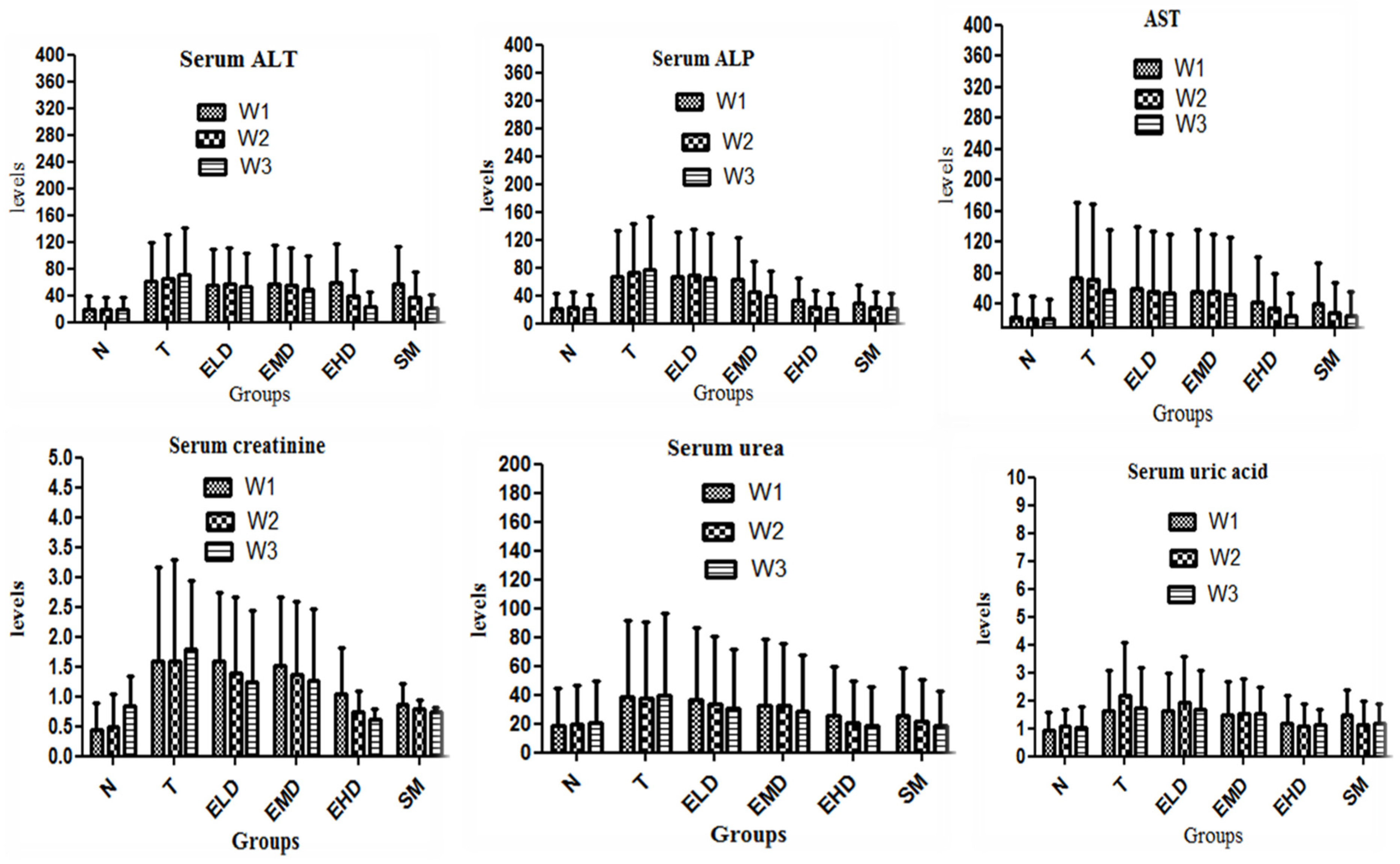
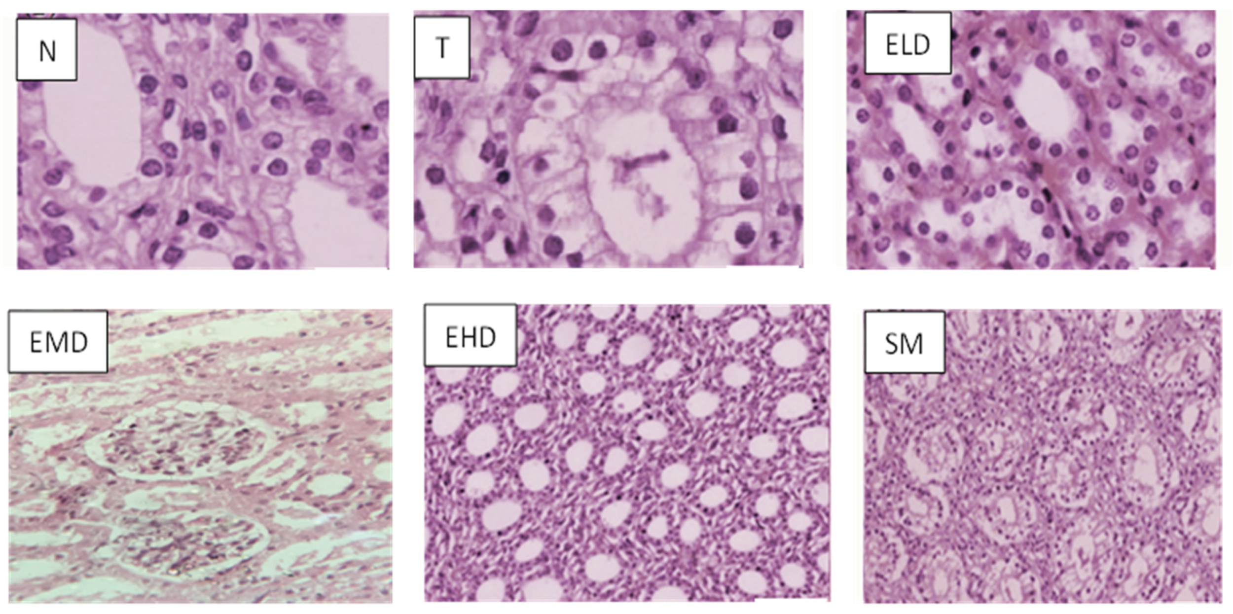
44]. The current study demonstrated that the consumption of paracetamol disrupted blood haematology, liver enzymes such as ALT, AST, and ALP, serum biomarkers such as urea, creatinine, and uric acid, as well as lipid profile, and also altered the histomorphological architecture, as described by [45,46,47]. A similar study has been conducted by Amang et al., 2020, demonstrating that Opilia celtidifolia is a plant used in Cameroonian ethnomedicine to cure jaundice. By reducing the levels of ALT, AST, ALP, and other serum indicators including urea, creatinine, and uric acid into the normal range, it was discovered that the (DO.AQ) extract had hepato- and nephroprotective capabilities.
Another parallel study showed that P. divaricata treatment restored altered biochemical and histopathological findings associated with PCM induced hepatotoxicity Singh, et al., 2016.
Various extract doses (100 mg/kg BW and 200 mg/kg BW) administered during the first and second treatment weeks (W1 and W2) had no noticeable impact. The high dose (400 mg/kg BW) extract, meanwhile, significantly decreased the serum enzymes by the end of the third week. Serum biochemical and histopathological status were observed to be regulated in silymarin-treated animals during the last week (W3) of study. These curative properties of (DO.AQ) extract are a result of the antioxidant activity and stabilization of the lysosomal membrane. These effects were equivalent to those of a group treated with silymarin, a standard hepatoprotective agent [48]. A similar study was also conducted by [49]; he studied the protective effect of Parthenium hysterophorus against CCL4 and paracetamol-induced toxicity. Our outcomes were in line with earlier research, where Ascorbic Acid was utilized as a nephroprotective drug and paracetamol was used to cause kidney injury in animals [
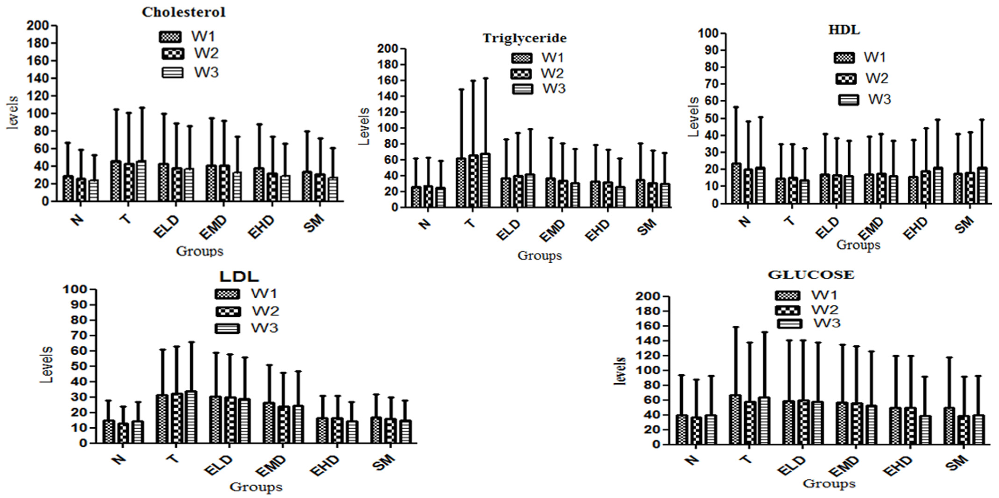
50]. Serum lipids have been linked to the development of numerous heart problems. Blood contains a variety of lipids, although LDL and HDL are more important than others in terms of heart-related ailments and are more common in evaluating heart problems [51].
According to the findings of the current study, liver damage brought on by paracetamol overdose is associated with a considerable increase in serum glucose and lipid levels, including cholesterol, TG, LDL, and a decrease in HDL [52]. Treatment with 400 mg/kg of Dianthus orientalis. aqueous extract has shown excellent reductions in serum glucose and lipid levels, including cholesterol, LDL, and triglyceride levels, while also increasing HDL levels. High levels of HDL cholesterol are considered to be good cholesterol as it is responsible for eradicating extra cholesterol by transporting it to the liver, thus helping prevent heart problems. For the digestion of lipids, glucose is essential. It produces pyruvic acid by glycolysis or to be converted into fatty acids, TC and TG.
The impact of paracetamol consumption is also noticeable in the animals’ haematology, where significant (p < 0.05) reductions in the values of RBC, HB, MCV, MCH, and MCHC and an increase in WBC, PLT, lymphocyte, neutrophil, and monocyte levels were seen. Similar investigations have also been conducted, showing that hepatotoxicity might alter haematological parameters such as packed cell volume, HB, total leucocyte count (TLC), MCH, MCHC, and MCV, indicating involvement with the haematological profile [53]. The range of harmful effects of any exogenous material, such as plant extract, on the blood composition of an animal can be ascertained through the analysis of haematological parameters [54,55].
Low levels of RBC, HB, MCV, MCH, and MCHC are noticeably increased by high dosages of DO.AQ extract, while WBC, PLT, lymphocyte, neutrophil, and monocyte concentrations are lowered to normal range. The normalization of these hematological parameters was observed from the third week of treatment. The high dose (400 mg/kg BW) of DO.AQ extract had a significant therapeutic effect, although the low and medium doses (100 mg/kg and 200 mg/kg BW) of the extract demonstrated no significant effect. Dianthus orientalis leaves aqueous extract (DO.AQ) significantly enhanced haematological parameters, which may indicate that it contains phytochemicals that could stimulate the formation of erythropoietin in experimental animals’ stem cells. Erythropoietin is a glycoprotein hormone that encourages bone marrow stem cells to produce red blood cells [56]. The results of the current investigation are consistent with those of [57], which described how Caulis bambusae (Bamboo) stem extract affected haematological and biochemical markers in chinchilla rabbits.
Exogenous antioxidants and the host’s own endogenous antioxidant defense system, which includes enzymatic and non-enzymatic antioxidants such as SOD, CAT, and GSH, can both prevent tissue damage brought on by oxidative stress. Reduced glutathione (GSH) is a potent nucleophilic, 3-peptide (L-γ-glutamyl cysteinyl glycine) antioxidant that is crucial for cellular defense and detoxifies reactive oxygen species by interacting with and removing harmful compounds, taking care of the inflammatory cytokine chain reactions [58]. Reduced GSH levels in different tissues leave the body’s defenses vulnerable to reactive oxygen species (ROS), which increases the risk of peroxidative injury. According to the findings of the current investigation, paracetamol treatment decreased GSH levels in the liver and kidneys through the process of lipid peroxidation, which may have led to the production of free radicals [59]. When compared to the control rabbits, GSH and %RSA levels in the liver and kidneys of paracetamol hepatotoxic rabbits were significantly decreased, indicating weak antioxidative defense and damage to the liver and kidneys.
Various dosages of DO.AQ extracts were administered to rabbits receiving paracetamol treatment. The levels of GSH and %RSA in the liver and kidneys were increased by a high dose extract (400 mg/kg BW), demonstrating that the plant extract’s antioxidant effect is dose dependent. A similar study was conducted by [60], using carbon tetrachloride; they discovered a substantial decrease in liver GSH content on carbon tetrachloride ingestion. The observed reduced level of GSH in paracetamol-toxic rabbits may be related to high levels of reactive substance scavenging activity, which were triggered because of liver and renal cell necrosis and apoptosis or a potential decrease in hepatic and renal GSH synthesis. The findings of the current investigation are similar to those of [61], which examined the protective impact of Aerva jevanica against ethanol-induced hepatic injury in rats; he came to the conclusion that Aerva jevanica (AJME) contained antioxidants and hepatoprotective properties. Silymarin increased the quantity of glutathione and the activity of antioxidant enzymes, whereas a dose-dependent decrease in lipid peroxidation was observed with silymarin and extracts [62].
High amounts of the reactive oxygen species (ROS) can cause the destruction of biological molecules including lipids, proteins, and nucleic acids. Antioxidant defenses have evolved to eliminate most of these oxidant mediators in order to prevent this condition. Oxidative stress develops when the delicate equilibrium between oxidative damage and defense systems is disturbed [63,64], and it has been shown that high lipid peroxide concentrations increase TBARS levels in the liver and kidneys of paracetamol-fed rabbits, as TBARS is an index of lipid peroxidation. Our findings demonstrate that TBARS levels were high in the liver and kidney of paracetamol control animals because of high lipid peroxidation. The liver and kidney TBARS levels were low in animals given a high dose of Dianthus orientalis aqueous extract. This reduction in TBARS levels may be caused by DO.AQ extracts’ active components, which may be involved in scavenging ROS and enhancing the antioxidant capacities of tissues. Our study agrees with [65], which analyzed the oxidative stress variables in streptozotocin induced diabetic rats and treated them with piper leaf [66,67].
Dianthus orientalis leaves aqueous extract (DO.AQ) was tested in the current investigation for its in vitro antioxidant activity against the DPPH free radical scavenging system, since it showed some variation in the level of various pharmacological activities. The % inhibition increased as the concentration of the extract increased. This rise in % inhibition shows the antioxidant capacity of Dianthus orientalis. The exact process by which plant extracts reduce lipid peroxidation is unknown, although it is possible that their antioxidant qualities are a key factor. Alkaloids, carotenoids, and phenolic compounds are the three main chemical components of plants. Due to their antioxidant properties, these chemicals are particularly effective against acute or chronic liver and kidney disorders, comprising cancer and heart ailments.

5. Conclusions

It has been concluded that leaves’ aqueous extract of Dianthus orientalis (DO.AQ) is very effective against liver and kidney injury caused by the overdose of paracetamol. The DO.AQ recovers various serum related markers and restores the lipid profile, and also regulates various serum antioxidants parameters, and thus has powerful antioxidant capacity. The DO.AQ may be a great source for new medications, and needs advanced assessment and investigation.

Author Contributions

Conceptualization, B.A., A.M.Y., H.M., A.A.K. and T.A.; methodology, B.A., A.M.Y., H.M., A.A.K. and T.A.; software, T.A.; validation, A.A.; formal analysis, T.A.; investigation, B.A., A.M.Y., H.M., A.A.K. and T.A.; resources, M.A. and A.A.; data curation, T.A.; writing—original draft preparation, T.A. and M.A; writing—review and editing, T.A. and A.F.A.; visualization, A.A.; supervision, T.A. project administration, A.A. and M.A.; funding acquisition, T.A. All authors have read and agreed to the published version of the manuscript.

Funding

This research received no external funding.

Institutional Review Board Statement

The study was conducted in accordance with the “Guide for the Care and Use of Laboratory Animals”, and approved by the Ethical Committee of the Department of Zoology, University of Malakand (protocol code No UOM/Zoo/21-191 and 1 March 2023).

Data Availability Statement

Not applicable.

Acknowledgments

The authors greatly acknowledge and express their gratitude to the Researchers Supporting Project number (RSP2023R462), King Saud University, Riyadh, Saudi Arabia.

Conflicts of Interest

The authors declare no conflict of interest.

References

  1. Hall, J.E.; Hall, M.E. Guyton and Hall Textbook of Medical Physiology e-Book; Elsevier Health Sciences: London, UK, 2020; ISBN 9780323640039. [Google Scholar]
  2. Ozougwu, J.C. Physiology of the liver. Int. J. Res. Pharm. Biosci. 2017, 4, 13–24. [Google Scholar]
  3. Corsini, A.; Bortolini, M. Drug-induced liver injury: The role of drug metabolism and transport. J. Clin. Pharmacol. 2013, 53, 463–474. [Google Scholar] [CrossRef]
  4. Coleman, M.D. Human Drug Metabolism; John Wiley & Sons: Hoboken, NJ, USA, 2020. [Google Scholar]
  5. Lee, W.M. Acetaminophen (APAP) hepatotoxicity—Isn’t it time for APAP to go away? J. Hepatol. 2017, 67, 1324–1331. [Google Scholar] [CrossRef] [Green Version]
  6. Hashemi, S.A.; Allameh, A.; Aleagha, M.S.E.; Daraeib, B. Stem Cell Factor Attenuates Formation of Acetaminophen–Glutathione Conjugate in Kidney of Mice Treated With a Toxic Dose of Acetaminophen. Bull. Environ. Pharmacol. Life Sci. 2014, 3, 20–26. [Google Scholar]
  7. Pingili, R.B.; Pawar, A.K.; Challa, S.R. Effect of chrysin on the formation of N-acetyl-p-benzoquinoneimine, a toxic metabolite of paracetamol in rats and isolated rat hepatocytes. Chem.-Biol. Interact. 2019, 302, 123–134. [Google Scholar] [CrossRef] [PubMed]
  8. Blondet, N.M.; Messner, D.J.; Kowdley, K.V.; Murray, K.F. Mechanisms of hepatocyte detoxification. In Physiology of the Gastrointestinal Tract; Elsevier: Amsterdam, The Netherlands, 2018; pp. 981–1001. [Google Scholar]
  9. Choi, E.; Alsop, D.; Wilson, J.Y. The effects of chronic acetaminophen exposure on the kidney, gill and liver in rainbow trout (Oncorhynchus mykiss). Aquat. Toxicol. 2018, 198, 20–29. [Google Scholar] [CrossRef] [PubMed]
  10. Sümer, E.; Senturk, G.E.; Demirel, Ö.U.; Yesilada, E. Comparative biochemical and histopathological evaluations proved that receptacle is the most effective part of Cynara scolymus against liver and kidney damages. J. Ethnopharmacol. 2020, 249, 112458. [Google Scholar] [CrossRef] [PubMed]
  11. Heidari, R.; Ahmadi, A.; Mohammadi, H.; Ommati, M.M.; Azarpira, N.; Niknahad, H. Mitochondrial dysfunction and oxidative stress are involved in the mechanism of methotrexate-induced renal injury and electrolytes imbalance. Biomed. Pharmacother. 2018, 107, 834–840. [Google Scholar] [CrossRef]
  12. Chakraborty, R.; Sen, S. Nephroprotective activity of Pisonia aculeata L. leaf extract against cisplatin induced nephrotoxicity and renal dysfunction in experimental rodents. Indian J. Exp. Biol. (IJEB) 2020, 58, 770–776. [Google Scholar]
  13. Medina, M.F.; Gonzalez, M.E.; Klyver, S.M.R.; Odstrcl, I.M.A. Histopathological and biochemical changes in the liver, kidney, and bloodof amphibians intoxicated with cadmium. Turk. J. Biol. 2016, 40, 229–238. [Google Scholar] [CrossRef]
  14. Bakheet, M.S.; Haredy, H.H.; Abdesalam, A.; Abd, H.K. Hepatotoxicity implies chemical-driven liver damage induced by certain medicinal and other chemical agents. Int. Inv. J. Med. Med. Sci. 2015, 2, 144–164. [Google Scholar]
  15. Prieto, I.; Monsalve, M. ROS homeostasis, a key determinant in liver ischemic-preconditioning. Redox Biol. 2017, 12, 1020–1025. [Google Scholar] [CrossRef] [Green Version]
  16. Ma, P.; Yan, B.; Zeng, Q.; Liu, X.; Wu, Y.; Jiao, M.; Liu, C.; Wu, J.; Yang, X. Oral exposure of Kunming mice to diisononyl phthalate induces hepatic and renal tissue injury through the accumulation of ROS. Protective effect of melatonin. Food Chem. Toxicol. 2014, 68, 247–256. [Google Scholar] [CrossRef] [PubMed]
  17. Kumar, S.; Raman, R.P.; Prasad, K.P.; Srivastava, P.; Kumar, S.; Rajendran, K. Effects on haematological and serum biochemical parameters of Pangasianodon hypophthalmus to an experimental infection of Thaparocleidus sp.(Monogenea: Dactylogyridae). Exp. Parasitol. 2018, 188, 1–7. [Google Scholar] [CrossRef] [PubMed]
  18. Deshpande, N.; Kandi, S.; Muddeshwar, M.; Das, R.; Ramana, K. A study of biochemical and hematological markers in alcoholic liver cirrhosis. World J. Nutr. Health 2014, 2, 24–27. [Google Scholar]
  19. Rani, V.; Deep, G.; Singh, R.K.; Palle, K.; Yadav, U.C. Oxidative stress and metabolic disorders: Pathogenesis and therapeutic strategies. Life Sci. 2016, 148, 183–193. [Google Scholar] [CrossRef]
  20. Singh, M.; Kaur, M.; Silakari, O. Flavones: An important scaffold for medicinal chemistry. Eur. J. Med. Chem. 2014, 84, 206–239. [Google Scholar] [CrossRef]
  21. Yaribeygi, H.; Butler, A.E.; Barreto, G.E.; Sahebkar, A. Antioxidative potential of antidiabetic agents: A possible protective mechanism against vascular complications in diabetic patients. J. Cell. Physiol. 2019, 234, 2436–2446. [Google Scholar] [CrossRef]
  22. Ozkan, G.; Kamiloglu, S.; Ozdal, T.; Boyacioglu, D.; Capanoglu, E. Potential use of Turkish medicinal plants in the treatment of various diseases. Molecules 2016, 21, 257. [Google Scholar] [CrossRef]
  23. Hassan, W.; Noreen, H.; Rehman, S.; Gul, S.; Amjad Kamal, M.; Paul Kamdem, J.; Zaman, B.; BT da Rocha, J. Oxidative stress and antioxidant potential of one hundred medicinal plants. Curr. Top. Med. Chem. 2017, 17, 1336–1370. [Google Scholar] [CrossRef]
  24. Nguyen, V.; Le, V.; Vo, T.; Bui, L.; Anh, H.; Danh, V. Preliminary phytochemical screening and determination of total polyphenols and flavonoids content in the leaves of Houttuynia cordata Thunb. Proc. IOP Conf. Ser. Mater. Sci. Eng. 2020, 736, 062013. [Google Scholar] [CrossRef]
  25. Kadereit, G.; Borsch, T.; Weising, K.; Freitag, H. Phylogeny of Amaranthaceae and Chenopodiaceae and the evolution of C4 photosynthesis. Int. J. Plant Sci. 2003, 164, 959–986. [Google Scholar] [CrossRef]
  26. Safikhani, K.; Mahmoodi, M. New record of dianthus pendulus (Caryophyllaceae) from Iran. Iran. J. Bot. 2020, 26, 19–21. [Google Scholar]
  27. HAZAR, D.; BAKTIR, İ. Identification and Evaluation of Propagation Techniques of Dianthus orientalis Adams. ANADOLU Ege Tarımsal Araştırma Enstitüsü Derg. 2018, 28, 37–44. [Google Scholar]
  28. Khaledi, M.; Asadi-Samani, M.; Mahmoodi-Kouhi, A.; Gholipour, A. Antibacterial effect of the hydroalcoholic extracts of four Iranian medicinal plants on Staphylococcus aureus and Acinetobacter baumanii. Int. J. Pharm. Phytopharm. Res. 2017, 7, 10–14. [Google Scholar]
  29. Sadat-Hosseini, M.; Farajpour, M.; Boroomand, N.; Solaimani-Sardou, F. Ethnopharmacological studies of indigenous medicinal plants in the south of Kerman, Iran. J. Ethnopharmacol. 2017, 199, 194–204. [Google Scholar] [CrossRef]
  30. d’Acampora, A.J.; Rossi, L.F.; Ely, J.B.; Vasconcellos, Z.A.d. Is animal experimentation fundamental? Acta Cir. Bras. 2009, 24, 423–425. [Google Scholar] [CrossRef]
  31. Sana; Ur Rahman, S.; Zahid, M.; Khan, A.A.; Aziz, T.; Iqbal, Z.; Ali, W.; Khan, F.F.; Jamil, S.; Shahzad, M.; et al. Hepatoprotective effects of walnut oil and Caralluma tuberculata against paracetamol in experimentally induced liver toxicity in mice. Acta Biochim. Pol. 2022, 69, 871–878. [Google Scholar]
  32. Chattopadhyay, J.; Sarkar, R. Chaos to order: Preliminary experiments with a population dynamics models of three trophic levels. Ecol. Model. 2003, 163, 45–50. [Google Scholar] [CrossRef]
  33. Albus, U. Guide for the Care and Use of Laboratory Animals, 8th ed; National Academies Press: Washington, DC, USA, 2011. Available online: https://olaw.nih.gov/sites/default/files/Guide-for-the-Care-and-Use-of-Laboratory-Animals.pdf (accessed on 1 January 2023).
  34. Donovan, J.; Brown, P. Blood collection. Curr. Protoc. Immunol. 2006, 73, 1–7. [Google Scholar] [CrossRef]
  35. Huang, Y.-L.; Hu, Z.-D. Lower mean corpuscular hemoglobin concentration is associated with poorer outcomes in intensive care unit admitted patients with acute myocardial infarction. Ann. Transl. Med. 2016, 4, 190. [Google Scholar] [CrossRef] [PubMed] [Green Version]
  36. Özyürek, E.; Cetintaş, S.; Ceylan, T.; ÖğÜş, E.; Haberal, A.; Gürakan, B.; Özbek, N. Complete blood count parameters for healthy, small-for-gestational-age, full-term newborns. Clin. Lab. Haematol. 2006, 28, 97–104. [Google Scholar] [CrossRef]
  37. Forouzandeh, H.; Azemi, M.E.; Rashidi, I.; Goudarzi, M.; Kalantari, H. Study of the protective effect of Teucrium polium L. extract on acetaminophen-induced hepatotoxicity in mice. Iran. J. Pharm. Res. IJPR 2013, 12, 123. [Google Scholar] [PubMed]
  38. Sathya, A.; Siddhuraju, P. Protective effect of bark and empty pod extracts from Acacia auriculiformis against paracetamol intoxicated liver injury and alloxan induced type II diabetes. Food Chem. Toxicol. 2013, 56, 162–170. [Google Scholar] [CrossRef]
  39. Lu, H.-Y.; Ning, X.-Y.; Chen, Y.-Q.; Han, S.-J.; Chi, P.; Zhu, S.-N.; Yue, Y. Predictive value of serum creatinine, blood urea nitrogen, uric acid, and β2-microglobulin in the evaluation of acute kidney injury after orthotopic liver transplantation. Chin. Med. J. 2018, 131, 1059. [Google Scholar] [CrossRef]
  40. Penumarthy, S.; Penmetsa, G.S.; Mannem, S. Assessment of serum levels of triglycerides, total cholesterol, high-density lipoprotein cholesterol, and low-density lipoprotein cholesterol in periodontitis patients. J. Indian Soc. Periodontol. 2013, 17, 30. [Google Scholar]
  41. Saito, H.; Kameda, Y.; Masui, K.; Murakami, S.; Kondo, T.; Ito, H.; Oshita, F.; Tsuboi, M.; Yokose, T.; Noda, K. Correlations between thin-section CT findings, histopathological and clinical findings of small pulmonary adenocarcinomas. Lung Cancer 2011, 71, 137–143. [Google Scholar] [CrossRef]
  42. Li, S.; Li, S.-K.; Gan, R.-Y.; Song, F.-L.; Kuang, L.; Li, H.-B. Antioxidant capacities and total phenolic contents of infusions from 223 medicinal plants. Ind. Crops Prod. 2013, 51, 289–298. [Google Scholar] [CrossRef]
  43. Turan, I.; Demir, S.; Aliyazicioglu, R.; Kilinc, K.; Ozer Yaman, S.; Akbulut Cakiroglu, K.; Kanbolat, S.; Ayazoglu Demir, E.; Mentese, A.; Aliyazicioglu, Y. Dimethyl sulfoxide extract of Dianthus carmelitarum induces S phase arrest and apoptosis in human colon cancer cells. Nutr. Cancer 2019, 71, 1181–1188. [Google Scholar] [CrossRef]
  44. El-Maddawy, Z.K.; El-Sayed, Y.S. Comparative analysis of the protective effects of curcumin and N-acetyl cysteine against paracetamol-induced hepatic, renal, and testicular toxicity in Wistar rats. Environ. Sci. Pollut. Res. 2018, 25, 3468–3479. [Google Scholar] [CrossRef]
  45. Mossa, A.-T.H.; Swelam, E.S.; Mohafrash, S.M. Sub-chronic exposure to fipronil induced oxidative stress, biochemical and histopathological changes in the liver and kidney of male albino rats. Toxicol. Rep. 2015, 2, 775–784. [Google Scholar] [CrossRef] [PubMed] [Green Version]
  46. Dawood, M.A.; Moustafa, E.M.; Gewaily, M.S.; Abdo, S.E.; AbdEl-Kader, M.F.; SaadAllah, M.S.; Hamouda, A.H. Ameliorative effects of Lactobacillus plantarum L-137 on Nile tilapia (Oreochromis niloticus) exposed to deltamethrin toxicity in rearing water. Aquat. Toxicol. 2020, 219, 105377. [Google Scholar] [CrossRef]
  47. Sanjeev, S.; Bidanchi, R.M.; Murthy, M.K.; Gurusubramanian, G.; Roy, V.K. Influence of ferulic acid consumption in ameliorating the cadmium-induced liver and renal oxidative damage in rats. Environ. Sci. Pollut. Res. 2019, 26, 20631–20653. [Google Scholar] [CrossRef] [PubMed]
  48. Pithayanukul, P.; Nithitanakool, S.; Bavovada, R. Hepatoprotective potential of extracts from seeds of Areca catechu and nutgalls of Quercus infectoria. Molecules 2009, 14, 4987–5000. [Google Scholar] [CrossRef] [Green Version]
  49. Amang, A.P.; Kodji, E.; Mezui, C.; Baane, M.P.; Siwe, G.T.; Kuissu, T.M.; Emakoua, J.; Tan, P.V. Hepatoprotective Effects of Aqueous Extract of Opilia celtidifolia (Opiliaceae) Leaves against Ethanol-Induced Liver Damage in Rats. Evid.-Based Complement. Altern. Med. 2020, 2020, 6297475. [Google Scholar] [CrossRef] [PubMed]
  50. Aziz, T.; Fawad, I.; Ali Khan, A.; Ur Rahman, S.; Zamani, G.Y.; Alharbi, M.; Alshammari, A.; Alasmari, A.F. Assessing the pharmacological and biochemical effects of Salvia hispanica (Chia seed) against oxidized Helianthus annuus (sunflower) oil in selected animals. Acta Biochim. Pol. 2023, 6621, 1–8. [Google Scholar] [CrossRef]
  51. Saleem, M.; Asif, A.; Akhtar, M.F.; Saleem, A. Hepatoprotective potential and chemical characterization of Artocarpus lakoocha fruit extract. Bangladesh J. Pharmacol. 2018, 13, 90–97. [Google Scholar] [CrossRef] [Green Version]
  52. Adeneye, A.; Olagunju, J. Protective effect of oral ascorbic acid (Vitamin C) on acetaminophen-induced renal injury in rats. Afr. J. Biomed. Res. 2009, 12, 55–61. [Google Scholar]
  53. Jain, H.R.; Shetty, V.; Singh, G.; Shetty, S. A study of lipid profile in diabetes mellitus. Int. J. Sci. Study 2016, 4, 55–60. [Google Scholar]
  54. El-Gindy, Y.; Zeweil, H. Effects of parsley supplementation on the seminal quality, blood lipid profile and oxidant status of young and old male rabbits. World Rabbit Sci. 2017, 25, 215–223. [Google Scholar] [CrossRef] [Green Version]
  55. Iweala, E.E.; Osundiya, A.O. Biochemical, haematological and histological effects of dietary supplementation with leaves of Gnetum africanum Welw. on paracetamol-induced hepatotoxicity in rats. Int. J. Pharmacol. 2010, 6, 872–879. [Google Scholar] [CrossRef]
  56. Ahmad, B.; Yousafzai, A.M.; Zeb, A.; Ali, W.; Khan, N.Z.; Aasim, M.; Ahmad, S.; Ullah, S.; Khan, A.A.; Naz, F. Therapeutic role of Typha elephantina leaves aqueous extract in paracetamol intoxicated rabbits. Pak. J. Pharm. Sci. 2021, 34, 737–745. [Google Scholar] [PubMed]
  57. Faggio, C.; Fazio, F.; Marafioti, S.; Arfuso, F.; Piccione, G. Oral administration of Gum Arabic: Effects on haematological parameters and oxidative stress markers in Mugil cephalus. Iran. J. Fish. Sci. 2015, 14, 60–72. [Google Scholar]
  58. Fibach, E. Erythropoiesis in vitro—A research and therapeutic tool in thalassemia. J. Clin. Med. 2019, 8, 2124. [Google Scholar] [CrossRef] [PubMed] [Green Version]
  59. Khan, M.I.R.; Saha, R.K.; Saha, H. Muli bamboo (Melocanna baccifera) leaves ethanolic extract a non-toxic phyto-prophylactic against low pH stress and saprolegniasis in Labeo rohita fingerlings. Fish Shellfish Immunol. 2018, 74, 609–619. [Google Scholar] [CrossRef]
  60. Ashtiani, H.R.A.; Bakhshandi, A.K.; Rahbar, M.; Mirzaei, A.; Malekpour, A.; Rastegar, H. Glutathione, cell proliferation and differentiation. Afr. J. Biotechnol. 2011, 10, 6348–6363. [Google Scholar]
  61. Ramos, A.; Correia, A.; Antunes, S.; Gonçalves, F.; Nunes, B. Effect of acetaminophen exposure in Oncorhynchus mykiss gills and liver: Detoxification mechanisms, oxidative defence system and peroxidative damage. Environ. Toxicol. Pharmacol. 2014, 37, 1221–1228. [Google Scholar] [CrossRef]
  62. Schmitt, B.; Vicenzi, M.; Garrel, C.; Denis, F.M. Effects of N-acetylcysteine, oral glutathione (GSH) and a novel sublingual form of GSH on oxidative stress markers: A comparative crossover study. Redox Biol. 2015, 6, 198–205. [Google Scholar] [CrossRef] [Green Version]
  63. Khan, R.A. Protective Effect of Aerva jevanica Against Ethanol Induced Hepatic Stress in Rats: A Randomized Control Report. Indian J. Pharm Edu. Res. 2017, 51, S110–S114. [Google Scholar] [CrossRef] [Green Version]
  64. Khan, M.R.; Siddique, F. Antioxidant effects of Citharexylum spinosum in CCl4 induced nephrotoxicity in rat. Exp. Toxicol. Pathol. 2012, 64, 349–355. [Google Scholar] [CrossRef]
  65. Weinberg, F.; Ramnath, N.; Nagrath, D. Reactive oxygen species in the tumor microenvironment: An overview. Cancers 2019, 11, 1191. [Google Scholar] [CrossRef] [PubMed] [Green Version]
  66. Latha, R. Anti-Hyperglycemic and Anti-Oxidant Activities of Ethanolic Extract of Lantana Camara Leaves. Ph.D. Dissertation, JKK Nattraja College of Pharmacy, Komarapalayam, India, 2016. [Google Scholar]
  67. Rama Devi, K.; Srinivasan, R.; Kannappan, A.; Santhakumari, S.; Bhuvaneswari, M.; Rajasekar, P.; Prabhu, N.M.; Veera Ravi, A. In vitro and in vivo efficacy of rosmarinic acid on quorum sensing mediated biofilm formation and virulence factor production in Aeromonas hydrophila. Biofouling 2016, 32, 1171–1183. [Google Scholar] [CrossRef] [PubMed]
Figure 1. Graphical representation of hematological indices at week 1, week 2 and week 3 of different experimental groups (Table 1).
Separations 10 00182 g001
Figure 2. Graphical representation of blood cells at week 1, week 2 and week 3 of various experimental groups (Table 2).
Separations 10 00182 g002
Figure 3. Graphical representation of liver related and other serum parameters at week 1, week 2 and week 3 of different experimental groups (Table 3).
Separations 10 00182 g003
Figure 4. Graphical representation of serum markers at week 1, week 2 and week 3 of various experimental animal groups (Table 4).
Separations 10 00182 g004
Figure 5. Kidney and liver micrographs of various experimental animal groups, i.e., N = normal, T = toxic, ELD = Extract low dose (100 mg/kg BW), EMD = Extract medium dose (200 mg/kg BW), EHD = Extract high dose (400 mg/kg BW), SM = silymarin (100 mg/kg BW).
Separations 10 00182 g005aSeparations 10 00182 g005b
Table 1. The effects of DO.AQ extracts on hematological indices of various experimental groups.
GroupsRBC × 103/µLHB G/dLMCV G/dLMCH G/dLMCHC G/dLHCT%
W1W2W3W1W2W3W1W2W3W1W2W3W1W2W3W1W2W3
N7.3 ± 0.39 a6.9 ±0.34 a7.0 ± 0.37 a11 ± 0.79 a12 ± 0.25 a13 ± 0.78 a19 ± 1.3 b19 ± 1.6 b70 ± 1.9 a35 ± 3.1 a31 ± 1.8 a34 ± 2.4 a36 ± 1.6 a35 ± 2.2 a38 ± 1.1 a41 ± 3.0 a42 ± 1.1 a40 ± 1.3 a
T5.21 ± 0.2 b3.2 ± 0.33 b3.2 ± 0.42 b8.7 ± 0.52 b8.5 ± 0.21 b8.2 ± 0.8 b22 ± 1.2 b24 ± 1.7 b36 ± 2.6 b19 ± 0.84 b18 ± 2.0 b17 ± 1.2 b22 ± 0.11 b23 ± 1.1 b20 ± 1.1 b21 ± 1.3 b18 ± 0.3 b20 ± 0.6 b
ELD5.7 ± 1.01 b3.4 ± 0.22 b4.5 ± 0.23 c8.6 ± 0.82 b8.1 ± 0.71 b8.3 ± 0.7 b26 ± 0.84 b29 ± 3.6 c41 ± 3.1 c19 ± 1.55 b19 ± 1.35 b22 ± 0.03 c22 ± 1.2 b24 ± 0.65 b21 ± 0.83 b22 ± 0.3 b21 ± 00 b25 ± 0.3 b
EMD5.9 ± 0.3 b3.8 ± 0.21 b4.8 ± 0.15 c9.9 ± 0.73 b9.2 ± 0.61 b9.3 ± 0.8 b34 ± 2.3 c41 ± 1.5 a51 ± 2.5 d19 ± 2.1 b22 ± 0.84 c212 ± 1.2 c23 ± 1.2 b23 ± 0.4 b28 ± 1.1 c21 ± 01 b25 ± 0.42 b30 ± 1.6 c
EHD6.4 ± 0.26 c5.8 ± 0.20 c6.8 ± 0.21 a11 ± 0.00 b11 ± 1.52 c40 ± 1.7 a39 ± 4.2 a43 ± 1.8 a67 ± 3.7 a23 ± 0.2 c24 ± 0.24 c31 ± 1.6 a27 ± 0.1 c25 ± 1.3 c31 ± 1.7 a27 ± 1.2 c35 ± 2.1 c42 ± 1.2 a
SM6.0 ± 0.092 c5.6 ± 0.04 c6.8 ± 0.58 a9.9 ± 0.0 b9.1 ± 0.10 b10 ± 1.92 a51 ± 0.0 c64 ± 1.7 a69 ± 3.5 a25 ± 1.9 c28 ± 2.0 a32 ± 1.7 a27 ± 1.7 c30 ±1.1 a35 ± 0.5 a27 ± 0.74 c3 6± 3.0 a41 ± 0.1 a
N = Normal control without any treatment, T = only paracetamol only at dose rate 200 BW, ELD = paracetamol (200 mg) + (DO.AQ) extract (100 mg/kg BW), EMD = paracetamol (200 mg) + (DO.AQ) extract (200 mg/kg BW), EHD = paracetamol (200 mg) + (DO.AQ) extract (400 mg/kg BW), SM = paracetamol (200 mg) + Silymarin (50 mg/kg BW). The same alphabet in the same row shows no significant difference (p < 0.05). Different alphabets in the same row show significant difference (p < 0.05).
Table 2. The effects of DO.AQ extracts on leucocytes and other associated parameters of various experimental groups.
GroupsWBC × 103/µLPLT G/dLNeutrophils G/dLLymphocytes %Monocytes %
W1W2W3W1W2W3W1W2W3W1W2W3W1W2W3
N5.9 ± 0.3 a6.9 ± 2.1 a7.8 ± 0.2 a134 ± 4.5 a142 ± 1.3 a141 ± 2.a34 ± 1.3 a37 ± 2.2 a36 ± 2.3 a31 ± 2.9 a34 ± 3.6 a35 ± 1.6 a33 ± 1.6 a32 ± 1.3 a33 ± 0.2 a
T13 ± 2.1 b14 ± 1.1 b15 ± 1.1 b255 ± 3.2 b264 ± 5.1 b270 ± 1.8 b64 ± 4.2 b63 ± 1.6 b67 ± 3.6 b70 ± 1.9 b68 ± 2.1 b71 ± 0.75 b71 ± 2.3 b25 ± 1.4 b25 ± 0.2 b
ELD12 ± 0.1 b13 ±0.51 c13 ± 0.1 c252 ± 1.5 b2451 ± 2.3 c231 ± 2.2 c58 ± 1.1 c52 ± 3.3 c45 ± 2.5 c66 ± 1.6 c67 ± 1.3 c63 ± 2.6 c 65 ± 3.2 c23 ± 0.2 c22 ± 0.1 c
EMD11 ± 0.1 b13 ± 0.5 c12 ± 0.1 c223 ± 5.6 c225 ± 3.6 d221 ± 3.6 d42 ± 2.6 d44 ± 2.4 d46 ± 3.7 c63 ± 2.7 c57 ± 2.1 d44 ± 1.4 d62 ± 3.1 c22 ± 0.4 c21 ± 0.4 c
EHD11 ± 0.39 c11 ±0.71 d7.7 ±0.1 a221 ± 2.3 d191 ± 3.5 e151 ± 2.3 a47 ± 3.8 d42 ± 1.4 d37 ± 2.7 a44 ± 1.89 d44 ± 2.832 ± 1.8 a46 ± 1.2 d18 ± 1.4 d 16 ± 0.2 a
SM10 ± 0.1 c7.7 ± 0.14 a7.8 ± 0.1 a176 ± 4.2 e168 ± 5.6 e149 ± 3.5 a42 ± 2.5 d33 ± 3.2 a39 ± 1.8 a43 ± 3.2 d33 ± 3.8 a39 ± 0.7 a47 ± 12.2 d17 ± 2.4 d17 ± 0.3 a
N = control animals no treatment, T = paracetamol only (200 BW), ELD = paracetamol (200 mg) + (DO.AQ) extract (100 mg/kg BW), EMD = paracetamol (200 mg) + (DO.AQ) extract (200 mg/kg BW), EHD = paracetamol (200 mg) + (DO.AQ) extract (400 mg/kg BW), SM = paracetamol (200 mg) + Silymarin (50 mg/kg BW). The same alphabet in the same row shows no significant difference (p < 0.05). Different alphabets in the same row show significant difference (p < 0.05).
Table 3. The effects of (DO.AQ) extracts on serum biochemical markers of various experimental groups.
GroupsSerum ALTSerum ASTSerum ALPSerum CreatinineSerum UreaSerum Uric Acid
W1W2W3W1W2W3W1W2W3W1W2W3W1W2W3W1W2W3
N40 ± 2.2 a38 ± 3.1 a37 ± 3.1 a43 ± 2.1 a41 ± 2.2 a39 ± 2.1 a43 ± 1.1 a41 ± 1.0 a45 ± 1.8 a0.88 ± 00.2 a0.87 ± 1.7 a1.3 ± 0.4 a41 ± 1.8 a37 ± 1.4 a35 ± 0.3 a1.6 ± 0.6 a1.3 ± 0.3 a1.9 ± 0.4 a
T141 ± 1.2 b146 ± 4.2 b153 ± 3.1 b145 ± 4.3 b151 ± 2.3 b135 ± 3.1 b156 ± 2.2 b162 ± 3.4 b172 ± 3.2 b2.4 ± 0.8 b2.4 ± 0.0 b2.5 ± 0.2 b76 ± 3.2 b82 ± 1.2 b81 ± 2.2 b4.4 ± 0.2 b4.3 ± 0.7 b4.6 ± 0.29 b
ELD121 ± 1.3 c132 ± 4.2 c121 ± 2.4 c154 ± 3.3 c122 ± 1.2 c121 ± 2.1 c159 ± 3.0 b124 ± 1.8 c91 ± 2.3 c2.2 ± 0.7 b2.6 ± 0.0 b2.5 ± 0.03 c73 ± 2.9 c62 ± 2.1 c64 ± 1.4 c4.32 ± 0.6 b3.1 ± 0.4 c3.4 ± 0.4 c
EMD131 ± 3.1 d131 ± 2.2 d94 ± 2.2 d84 ± 1.1 d81 ± 2.2 d69 ± 3.2 d126 ± 3.1 c82 ± 2.0 d85 ± 1.3 c2.7 ± 0.5 b2.2 ± 0.02 b1.4± 0.4 d 66 ± 0.4 c57 ± 0.64 d56 ± 1.3 d3.3 ± 0.32 c3.5 ± 0.52.9 ± 0.21 d
EHD121 ± 2.1 e63 ± 1.4 e42 ± 2.3 a82 ± 2.3 e70 ± 3.0 e48 ± 1.2 a98 ± 3.1 d6 3 ± 2.3 e57 ± 4.3 a2.3 ± 0.3 b1.6 ± 0.0 1 c1.5 ± 0.3 a62 ± 1.3 d 47 ± 1.21 e37 ± 0.75 a2.5 ± 0.03 d2.2 ± 0.1 d1.4 ± 0.3 a
SM80 ± 2.3 e62 ± 2.2 e41 ± 2.5 a75 ± 2.1 e40 ± 1.9 a40 ± 2.1 a81 ± 2.2 e67 ± 1.2 e53 ± 1.0 a1.7 ± 0.3 c1.7 ± 0.1 c1.4 ± 0.3 a55 ± 2.7 d43 ± 3.2 e40 ± 1.1 a2.4 ± 1.31 a2.2 ± 0.02 d1.7 ± 0.3 a
N = control animals, no treatment, T = paracetamol only (200 BW), ELD = paracetamol (200 mg) + (DO.AQ) extract (100 mg/kg BW), EMD = paracetamol (200 mg) + (DO.AQ) extract (200 mg/kg BW), EHD = paracetamol (200 mg) + (DO.AQ) extract (400 mg/kg BW), SM = paracetamol (200 mg) + Silymarin (50 mg/kg BW). The same alphabet in the same row shows no significant difference (p < 0.05). Different alphabets in the same row show significant difference (p < 0.05).
Table 4. The effects of (DO.AQ) extracts on the lipid profile of various experimental groups.
GroupsCholesterolTriglyceridesHDLLDLGlucose mg/dL
W1W2W3W1W2W3W1W2W3W1W2W3W1W2W3
N56 ± 2.2 a49 ± 3.2 a44 ± 3.2 a51 ± 3.1 a51 ± 3.1 a49 ± 3.0 a49 ± 2.3 a47 ± 2.2 a40 ± 3.4 a26 ± 2.1 a27 ± 3.2 a25 ± 1.7 a84 ± 2.21 a79 ± 2.4 a82 ± 1.4 a
T87 ± 3.4 b84 ± 2.2 b89 ± 3.1 b132 ± 2.1 b142 ± 3.2 b154 ± 4.2 b29 ± 4.1 b28 ± 3.1 b27 ± 1.5 b61 ± 3.4 b61 ± 1.3 b66 ± 2.5 b117 ± 3.00 b119 ± 2.12 b121 ± 3.01 b
ELD3 ± 2.1 b74 ± 2.1 b71 ± 2.5 b81 ± 3.1 c81 ± 5.175 ± 3.1 c34 ± 2.2 b32 ± 2.3 c31 ± 2.1 c51 ± 5.1 b54 ± 2.3 c47 ± 2.4 c114 ± 4.33 c113 ± 3.23 c113 ± 4.26 c
EMD79 ± 1.4 b77 ± 4.2 b62 ± 4.1 c89 ± 2.2 c75 ± 2.3 c61 ± 2.2 d36 ± 4.2 b33 ± 3.4 c34 ± 1.3 d57 ± 3.1 b40 ± 2.6 d41 ± 3.1 c107 ± 2.11 d105 ± 3.31 d101 ± 2.31 d
EHD73.25 c61 ± 2.1 c55 ± 3.1 a69 ± 1.7 d64 ± 2.1 d53 ± 3.2 a33 ± 3.1 b32 ± 3.4 d37 ± 2.1 a41 ± 5.2 c32 ± 2.3 e26 ± 2.4 a96 ± 3.21 d91 ± 2.22 ac96 ± 3.12 a
SM66 ± 2.3 c62 ± 3.2 c51 ± 3.2 a70 ± 4.1 d52 ± 3.1 a51 ± 2.3 a42 ± 3.3 c39 ± 2.5 d36 ± 4.3 a37 ± 2.3 d33 ± 3.1 e30 ± 2.3 a107 ± 2.1 ac105 ± 3.1 ac95 ± 2.5 a
N = control animals, no treatment, T = paracetamol only (200 BW), ELD = paracetamol (200 mg) + (DO.AQ) extract (100 mg/kg. BW), EMD = paracetamol (200 mg) + (DO.AQ) extract (200 mg/kg BW), EHD = paracetamol (200 mg) + (DO.AQ) extract (400 mg/kg BW), SM = paracetamol (200 mg) + Silymarin (50 mg/kg BW). The same alphabet in the same row shows no significant difference (p < 0.05). Different alphabets in the same row show significant difference (p < 0.05).
Table 5. The effects of (DO.AQ) extracts on serum albumen, glucose and serum electrolytes of various experimental groups.
GroupsC (mmol/L)Mg (mmol/dL)Cl (mmol/dL)Na (mmol/dL)K (mmol/dL)P (mmol/dL)
W3W1W2W3W1W2W3W1W2W3W1W2W3W1W2W3W1W2W3
N82 ± 1.4 a3.98 ± 0.31 a3.91 ± 1.3 a3.42 ± 2.1 a0.61 ± 1.00 a0.62 ± 31.0 a0.64 ± 11.0 a89.1 ± 1.12 a78.0 ± 2.1 a88.0 ± 1.1 a124.4 ± 2.2 a123 ± 2.31 a127 ± 3.2 a4.91 ± 1.11 a4.1 ± 2.1 a4.25 ± 1.0 a2.72 ± 0.00 a3.00 ± 0.00 a2.91 ± 1.00 a
T121 ± 3.01 b6.01 ± 0.23 b5.03 ± 1.12 b6.171 ± 2.17 b0.86 ± 0.02 b0.89 ± 0.62 b0.90 ± 1.51 b110.6 ± 0.4 b116 ± 0.8 b119 ± 2.51 b143.4 ± 1.3 b149.4 ± 3.7 b144 ± 2.86 b6.82 ± 0.34 b7.02 ± 0.52 b6.90 ± 0. 2 b3.69 ± 0.04 b4.12 ± 0.0 b4.76 ± 0.12 b
ELD113 ± 4.26 c6.15 ± 0.14 c5.95 ± 2.14 c5.99 ± 1.32 c0.67 ± 0.01 c0.715 ± 0.03 c0.66 ± 0.04 c104.1 ± 3.1 c103 ± 2.1 c102 ± 3.1 c148.3 ± 2.26 c145 ± 3.14 c145 ± 3.14 c6.22 ± 0.31 c6.34 ± 0.461 c6.31 ± 1.01 c3.36 ± 0.1 c3.46 ± 0.4 c3.35 ± 0.6 c
EMD101 ± 2.31 d5.90 ± 0.13 c5.89 ± 0.32 c5.12 ± 0.12 d0.66 ± 0.21 c0.67 ± 0.01 c0.65 ± 0.01 d103.1 ± 1.4 c102 ± 2.1 c99 ± 2.4 d139.1 ± 0.41 c138 ± 3.11 c133 ± 2.41 d5.96 ± 0.13 c5.76 ± 1.11 c5.52 ± 0.31 d3.47 ± 0.02 c3.43 ± 0.01 c3.83 ± 0.031 d
EHD83± 3.12 a4.52 ± 1.20 c4.22 ± 0.10 c34.22 ± 0.20 a0.78 ± 1.02 c0.71 ± 0.03 d0.58 ± 2.02 a96.4 ± 2.6 c93.5 ± 1.3 d83.5 ± 2.1 a138.4 ± 2.43 c136.4 ± 1.63 ac126.4 ± 2.23 aa4.99 ± 0.21 c4.79 ± 0.34 d4.29 ± 0.31 a1.56 ± 0.00 c1.99 ± 0.10 cd2.26 ± 1.10 a
SM95 ± 2.5 a5.26 ± 0.1 c4.32 ± 0.23 d3.82 ± 0.41 a0.69 ± 0.21 c0.68 ± 0.01 d0.62 ± 0.02 a103.2 ± 2.2 b99.12 ± 3.2 d89.12 ± 2.5 a143.7 ± 0.31 bc133.2 ± 0.34 ad125 ± 0.13 a5.23 ± 0.38 b4.63 ± 0.21 c4.13 ± 0.30 a3.83 ± 0.23 c3.12 ± 0.00 ac2.12 ± 0.00 a
N = control animals, no treatment, T = only paracetamol (200 BW), ELD = paracetamol (200 mg) + (DO.AQ) extract (100 mg/kg BW), EMD = paracetamol (200 mg) + (DO.AQ) extract (200 mg/kg BW), EHD = paracetamol (200 mg) + (DO.AQ) extract (400 mg/kg BW), SM = paracetamol (200 mg) + Silymarin (50 mg/kg BW). The same alphabet in the same row shows no significant difference (p < 0.05). Different alphabets in the same row show significant difference (p < 0.05).
Table 6. Liver and kidney antioxidant markers of various experimental groups.
GroupsLiver GSHKidney GSHLiver RSAKidney RSALiver TBARSKidney TBARS
N37.35 ± 1.14 a27.12 ± 1.76 a54.39± 2.031 a29 ± 0.705 a13 ± 1.3 a13 ± 1.75 a
T15.65 ± 3.5 b14.12 ± 1.73 b27.73 ± 1.179 b18.4 ± 2.37 b33 ± 1.2 b24 ± 0.52 b
ELD15.41 ± 1.3 b15.18 ± 1.41 b38.13 ± 1.741 c21.8 ± 1.44 b34 ± 0.8 b22 ± 2.2 b
EMD23.20 ± 2.34 c17.3 ± 2.84 c42.41 ± 2.21 d22.23 ± 1.905 b26 ± 1.4 c18 ± 1.85 c
EHD38.31 ± 0.23 a26.16 ± 1.391 a56.36 ± 1.32 a26.7 ± 2.51 a13 ± 0.63 a14 ± 1.1 a
SM36.12 ± 1.51 a27.29 ± 2.04 a57.32 ± 1.21 a25.66 ± 1.43 b9.0 ± 0.75 a13 ± 0.32 a
N = control group, no treatment, T = only paracetamol (200 BW), ELD = paracetamol (200 mg) + (DO.AQ) extract (100 mg/kg BW), EMD = paracetamol (200 mg) + (DO.AQ) extract (200 mg/kg BW), EHD = paracetamol (200 mg) + (DO.AQ) extract (400 mg/kg BW), SM = paracetamol (200 mg) + Silymarin (50 mg/kg BW). The same alphabet in the same row shows no significant difference (p < 0.05). Different alphabets in the same row show significant difference (p < 0.05).
Table 7. DPPH free radical scavenging activity of the extract at different concentrations.
DO.AQ Extract
Concentration
NoAbsorption% InhibitionMean ± SEM
100 ppm10.5648.74%46.50 ± 2.46
20.100
30.85
150 ppm10.18849.88%47.90 ± 3.89
20.191
30.149
250 ppm10.16056.53%55.60 ± 3.10
20.135
30.172
300 ppm10.12069.90%67.89 ± 2.31
20.112
30.121
400 ppm10.04173.33%71.9 ± 1.65
20.072
30.104
Disclaimer/Publisher’s Note: The statements, opinions and data contained in all publications are solely those of the individual author(s) and contributor(s) and not of MDPI and/or the editor(s). MDPI and/or the editor(s) disclaim responsibility for any injury to people or property resulting from any ideas, methods, instructions or products referred to in the content.

