Review

Onco-Receptors Targeting in Lung Cancer via Application of Surface-Modified and Hybrid Nanoparticles: A Cross-Disciplinary Review

1
Faculty of Pharmacy, Institute of Pharmaceutical Technology and Regulatory Affairs, University of Szeged, 6720 Szeged, Hungary
2
Hamdard Institute of Pharmaceutical Sciences, Hamdard University Islamabad Campus, Islamabad 76400, Pakistan
3
Department of Pharmacy, Faculty of Biological Sciences, Quaid-i-Azam University, Islamabad 45320, Pakistan
4
Department of Pharmacology and Clinical Pharmacy, School of Pharmacy, Bandung Institute of Technology, Bandung 40132, Indonesia
5
Department of Physics, University of Zabol, Zabol 98613-35856, Iran
6
Department of Chemistry, Shahid Bahonar University of Kerman, Kerman 6169-14111, Iran
7
POLYMAT and Kimika Aplikatua Saila, Kimika Fakultatea, University of the Basque Country UPV/EHU, Joxe Mari Korta Zentroa, Tolosa Hiribidea 72, 20018 Donostia, San Sebastián, Spain
8
Centro de Física de Materiales, CSIC-UPV/EHU, Paseo Manuel Lardizábal 5, 20018 Donostia, San Sebastián, Spain
9
Donostia International Physics Center (DIPC), Paseo Manuel Lardizábal 4, 20018 Donostia, San Sebastián, Spain
*
Authors to whom correspondence should be addressed.
Processes 2021, 9(4), 621; https://doi.org/10.3390/pr9040621
Submission received: 1 March 2021 / Revised: 27 March 2021 / Accepted: 29 March 2021 / Published: 1 April 2021

Abstract

:
Lung cancer is among the most prevalent and leading causes of death worldwide. The major reason for high mortality is the late diagnosis of the disease, and in most cases, lung cancer is diagnosed at fourth stage in which the cancer has metastasized to almost all vital organs. The other reason for higher mortality is the uptake of the chemotherapeutic agents by the healthy cells, which in turn increases the chances of cytotoxicity to the healthy body cells. The complex pathophysiology of lung cancer provides various pathways to target the cancerous cells. In this regard, upregulated onco-receptors on the cell surface of tumor including epidermal growth factor receptor (EGFR), integrins, transferrin receptor (TFR), folate receptor (FR), cluster of differentiation 44 (CD44) receptor, etc. could be exploited for the inhibition of pathways and tumor-specific drug targeting. Further, cancer borne immunological targets like T-lymphocytes, myeloid-derived suppressor cells (MDSCs), tumor-associated macrophages (TAMs), and dendritic cells could serve as a target site to modulate tumor activity through targeting various surface-expressed receptors or interfering with immune cell-specific pathways. Hence, novel approaches are required for both the diagnosis and treatment of lung cancers. In this context, several researchers have employed various targeted delivery approaches to overcome the problems allied with the conventional diagnosis of and therapy methods used against lung cancer. Nanoparticles are cell nonspecific in biological systems, and may cause unwanted deleterious effects in the body. Therefore, nanodrug delivery systems (NDDSs) need further advancement to overcome the problem of toxicity in the treatment of lung cancer. Moreover, the route of nanomedicines’ delivery to lungs plays a vital role in localizing the drug concentration to target the lung cancer. Surface-modified nanoparticles and hybrid nanoparticles have a wide range of applications in the field of theranostics. This cross-disciplinary review summarizes the current knowledge of the pathways implicated in the different classes of lung cancer with an emphasis on the clinical implications of the increasing number of actionable molecular targets. Furthermore, it focuses specifically on the significance and emerging role of surface functionalized and hybrid nanomaterials as drug delivery systems through citing recent examples targeted at lung cancer treatment.

1. Introduction

Lung cancer is one of the most prevalent diseases and the leading causes of death worldwide [1]. It is more common in males than in females and based on an estimation, this type of cancer caused 154,050 deaths in 2018 [2]. One of the most common causes of this devastating disease is chronic tobacco usage. The major reason for its high mortality is the late diagnosis of the disease, and in most cases, lung cancer is diagnosed at the fourth stage when the cancer has already metastasized to the nearby organs [3]. Among lung cancer patients, 85% exhibit nonsmall cell lung cancer (NSCLC) while the rest (15%) of the patients have small cell lung cancer (SCLC). The survival of the patients suffering from lung cancer mainly depends upon the early diagnosis and efficient surgical removal of the tumor tissues. Among the different treatments, chemotherapy is the most recommended therapy to treat lung cancer. However, the major limitation of conventional chemotherapy is related to the presence of inefficient drugs at the target site, which ultimately compromises the therapeutic efficacy [4]. To reduce this problem, repeated administration of systemic chemotherapy at higher concentrations is required, which is allied with dose-related systemic toxicities. Moreover, in conventional therapies the uptake of the cytotoxic agents by the healthy cells can increase the chances of cytotoxicity in these normal cells. Hence, novel approaches are required for both the diagnosis and treatment of lung cancers [5].
Due to numerous limitations associated with these conventional methods, several researchers have exploited nanotechnology-based approaches for the efficient diagnosis and delivery of therapeutic agents [6]. Among various nanoparticle-mediated drug delivery systems, the most frequently used ones for lung cancer treatment include polymeric nanoparticles [7,8], liposomes [9,10], bionanoparticles [11,12] and metallic nanoparticles [13,14]. These nanoparticles have been very effective due to their small size, large surface area, high biocompatibility and reduced renal clearance. Although the use of nanoparticles has shown several advantages [15], their site-specific delivery is still a problem for which passive and active-targeting approaches are necessary [16,17].
The passive targeting approach utilizes the exploitation of the enhanced permeability and retention (EPR) effect. In many disease conditions, including lung cancers, the endothelial lining of the blood vessels exhibits higher permeability than in normal conditions [17,18]. The presence of this leaky vasculature allows the higher permeation of the nanoparticles into the target site [19]. Moreover, the lack of a normal lymphatic drainage system in the tumor site contributes to higher levels of retention of the nanoparticles. However, this idiosyncratic property cannot be applied to low molecular weight drugs which have a small residence time and rapid excretion from the tumorous cells. Low molecular weight drugs can be encapsulated in unionized drug carriers to improve their pharmacokinetics (elongated systematic circulation), increasing tumor selectivity and lowering side effects. This phenomenon of tumor-targeting is called “passive” and depends upon the properties of the carrier molecule (its molecular weight and residence time) and the tumor anatomy (vascularity, porosity, etc.), but does not have any ligands for specific cells’ binding sites. The EPR effect provides a 20–30% higher concentration of the drug targeted delivery of the tumorous site compared to normal body tissues [20,21].
EPR effect is extremely dependent on the intrinsic pathways of tumor cells’ growth and it is controlled specifically by the rate of angiogenesis and lymphangiogenesis, the rate of perivascular tumor development, stromal thickness response and the intratumor pressure. All these elements, along with the physicochemical properties of nanoparticles, can influence the efficiency of the drug’s targeted delivery [22]. However, the extrusion properties of the newly formed tumor’s blood vessels have an impact on the nanomedicine impregnation; it causes an increase in the interstitial pressure, which may hinder the retention of the drug carriers in the tumor tissues. Furthermore, due to the imbalance between the pro- and antiangiogenetic signaling in different points of the tumorous tissues, the blood vessels are deviant with enlarged, curvy and saccular pathways, unorganized interconnection processes and branching. This miscellaneous blood circulation causes an irregular growth of the tumor cells and those cells surrounding the blood vessel grow rapidly compared to those that are far away, because of low oxygen and nutrition supply. This explains why the outer sites of large tumorous tissues have less blood supply (i.e., 1–2 cm in diameter in mice) and why is most often difficult for nanomedicines to reach the cores of tumor cells. Although the interstitial pressure is high in the inner portion of the tumor, the extrusion rate is unexpectedly small. This pattern was observed in some different types of murine and human tumor cells. The increased interstitial pressure does not only hinder the drug supply to the core tumorous tissues but also retards the growth of new blood vessels. This causes a higher blood supply to flow towards the tumor cells’ periphery, indicating that there is the possibility of modifying the EPR effect chemically or mechanically to improve the growth of the blood vessels for the retention of the drug-loaded nanocarriers. It is worth mentioning here that some types of EPR enhancers like bradykinin (kinin), nitric acid, peroxynitrite, prostaglandins, etc. may cause hypertension that could enhance tumor extrusion [23].
To further improve the targeted delivery of the imaging modalities and therapeutic agents against lung cancer, many researchers have also exploited the receptor-mediated delivery of theranostics [24]. Several receptors are overexpressed in lung cancer, like oxytocin, vasopressin, chemokine, epidermal growth factor, bradykinins, bombsein, folate and tyrosine receptors. The majority of the lung cancer receptors are categorized as G-protein coupled receptors. These receptors have a potential role in the formation, progression and metastasis of lung cancer and are involved in angiogenesis process during tumor development and also during the progression of the cancer to the nearby organs [25]. The overexpression of several kinds of receptors in lung cancer has been exploited by researchers for the site-specific delivery of theranostics. As compared with the passive targeted approach, a higher amount of the drug can be made to reach the target site through active targeted delivery of the imaging modalities and therapeutic agents.
Active targeting is compulsory for the proper distribution of drugs, genes and theranostics to the action site so the therapeutic effect on normal body tissue can be avoided. By using active targeting, a sufficient amount of drug is placed at the tumor site increasing the drug efficiency by many folds. Thus, active targeting nanosystems are more efficient than passive targeting ones. Active targeting is possible exclusively when the nanocarriers are enriched with ligands that are specific for the overexpressed receptors in lung cancers [26]. This phenomenon enhances the binding capacity of the drug and imaging modalities to the tumor tissues and thus increases the drug entrapment capacity at the tumor site. Hundreds of ligands and antibodies have been discovered against the abovementioned receptors and are exploited for targeted delivery of the drug cargoes to the target site. A strong ligand/receptor binding affinity serves as role model to promote active binding technology. This can improve the targeted delivery of theranostics and therapeutic agents on the one hand and overcome the problems allied with conventional approaches on the other hand [26]. The visual illustration of various nanotechnology-based theranostic delivery approaches are shown in Figure 1.
This review first summarizes the current knowledge of the pathways implicated in the different types of lung cancer with an emphasis on the clinical implications of the increasing number of actionable molecular targets. The utilization of different targeting approaches to combat the toxicity of the chemotherapeutic agents is discussed here. The mechanism through which this targeted delivery is attained is also described. In this context, this review could attract the interest of medical scientists who are involved in biological systems. The second specific focus of this review is on the role of the surface-modified and hybrid nanomaterials as drug delivery systems in combating lung cancer. This spotlight was achieved through citing most the recent and representative examples targeted at lung cancer treatment. From this perspective, it could be highly interesting for material scientists.
The fields of biology and material science are traditionally rather separated, as much as they naturally rely on the very same basic principles. Through the novel cross-disciplinary focus of this review, we attempt to overcome this gap and create a more synergetic perspective on both areas, which will be highly beneficial for the scientific community given the plethora of discussions and discoveries that can be envisaged.

2. Pathways for Targeting Lung Cancer

Lung cancer is histologically classified into NSCLC and SCLC. The complex interplay between pathological changes and oncogenic mutations alters the signaling of multiple pathways and the expression of chemokines and various receptors. In turn, a modified tumor microenvironment facilitates the growth, proliferation, angiogenesis, metastasis and survival of the cancer cells. Traditional treatment strategies for the lung cancer include chemotherapy, radiotherapy and surgical excision. However, conventional chemotherapeutic agents have compromised therapeutic efficacy owing to pharmacokinetic issues, solubility problems and nonspecific action in normal cells with resultant toxicities. Moreover, high drug doses, tumor-associated alteration of pathways and subsequent treatment with multiple therapies will contribute to the occurrence of tumor resistance against chemotherapeutic agents [27] Therefore, the focus is now laid on the suppression of upregulated pathways including EGFR, RAS-RAF-MEK-ERK/MAPK, JAK-STAT, PI3K/AKT/mTOR through newly designed, specifically targeted small molecule inhibitors and antibodies (Figure 2). For instance, specific EGFR inhibitor (erlotinib) and PI3K/AKT/mTOR inhibitor (everolimius) replaced the first-line chemotherapy [28]. The most common genetic mutations in the lung cancer, along with their mode of aberration and the associated small molecule inhibitors to target specific pathways, are mentioned in Table 1. Nevertheless, the small-molecule-mediated targeted therapy is relatively successful and increases survival rates but is prone to therapeutic failure because of cancer relapse, and increased drug resistances due to targeting site mutations [29].
Hence, developing a highly targeted drug delivery system for specific action into the tumorous cells at an optimal dose is of great necessity. Broadly, lung cancer can be targeted through either passive or active targeting mechanisms or both. Passive drug delivery follows a certain principle to be deposited into the lung tissues under the EPR effect. EPR is attributed to leaky vasculature and deteriorative epithelial integrity that allows residence and accumulation of small sized particles into the lung tumorous tissue [30], which act either as a carrier to deliver the drug or act directly as a therapeutic moiety. In passive targeting, particle size is the main determinant for distribution and deposition in the lungs. For instance, large particles around >5 μm have fewer chances to concentrate and are mostly exhaled out of the lungs. Particles in the range of 1–5 μm are phagocytosed by the alveolar macrophages and particles with size <1 μm could be deposited in the alveolar cells with minimal clearance by the immune cells [31].
To achieve improved tumor-specific targeting and to avoid possible threats with dislocation and clearance of passively targeted delivery carriers, active targeting of overly expressed onco-receptors with specific ligands brings better outcomes [30]. The inhibition of overexpressed receptor functions through specifically targeting moieties modulates the expression of cancer projectors and improves drug action in the tumor-specific lung tissues. Various overexpressed receptors in the tumor microenvironment include EGFR, TFR, FR and CD44 receptor [32]. Tumor receptors and tumor-associated immune cells have a role in cancer growth, proliferation, metastasis and angiogenesis. Therefore, receptor-mediated targeting and immune cell targeting alter the onco-proteins’ expression and inhibit oncogenic pathways to stop cancer growth and progression.

3. Onco-receptor Targets in Lung Tumors and Vasculature

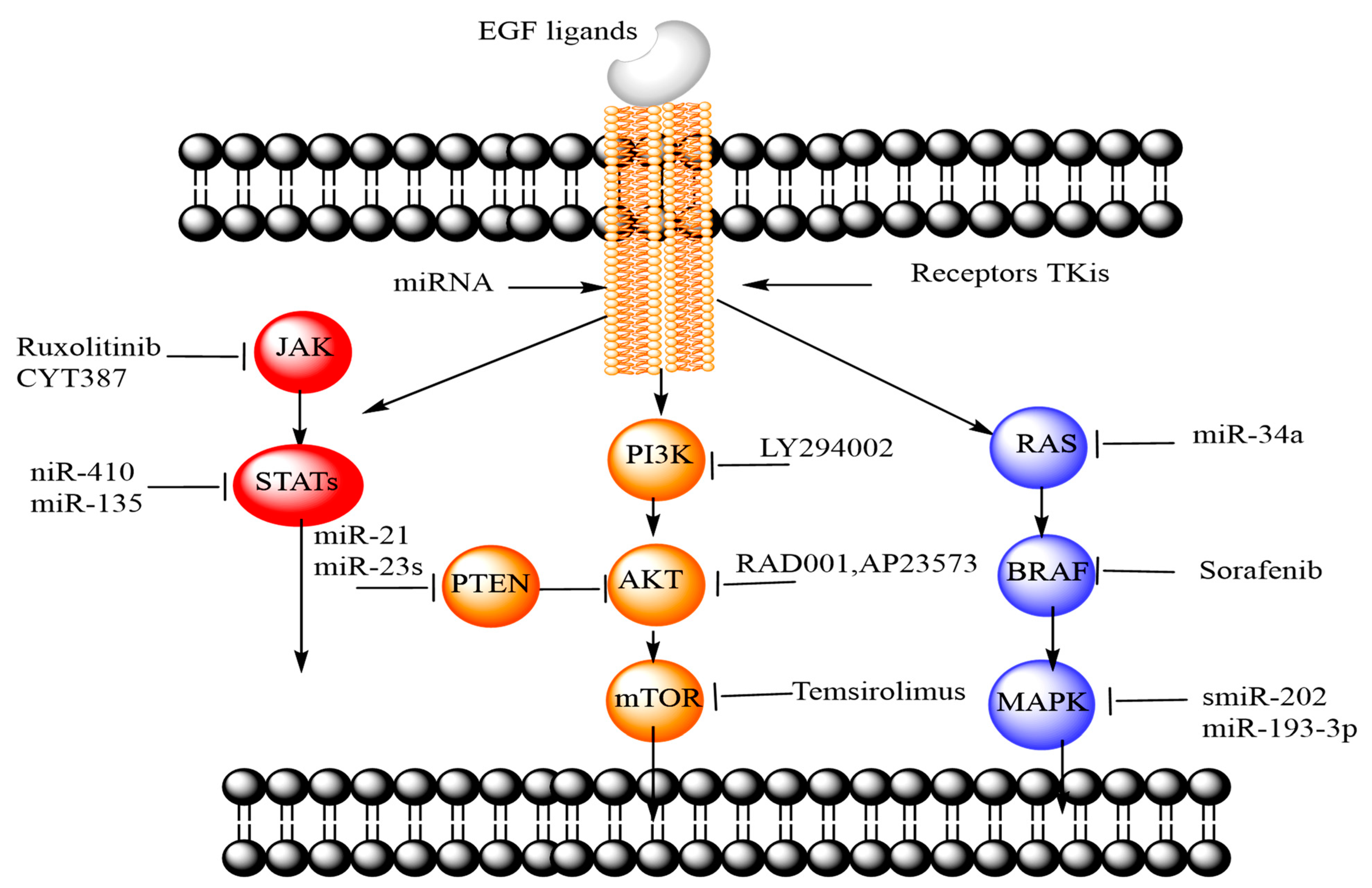
3.1. Epidermal Growth Factor Receptor (EGFR)

The EGFR is a cell surface peptide receptor from the ErbB family of tyrosine kinase. It consists of the extracellular region with two homologous ligand-binding domains and two cysteine-rich domains, a single slanging transmembrane domain and an intracellular region comprising juxtamembrane, a tyrosine kinase domain and a regulatory region [53]. EGFR regulates growth, differentiation and migration of the alveolar and bronchial epithelial cells under normal conditions, while overfunctioning in cancer facilitates the proliferation, metastasis, and invasion of lung cancer cells [54]. EGFR is among the highly expressed onco-receptors in 85% of NSCLC, with negligible involvement in SCLC [55]. Various monoclonal antibodies (panitumumab, cetuximab) and tyrosine kinase inhibitors (erlotinib, gefitinib, lapatinib) are used to target EGFR to treat lung cancer [55]. Furthermore, antisense oligonucleotides, affibodies, peptides, and nanobodies worked to inhibit EGFR [56]. Recently, it has been observed that ligand anchored nanocarriers specifically bind to extracellular domains of EGFR to release the drugs intracellularly for the tumor-specific inhibition of the signaling pathway. Under this approach, biotinylated-EGF ligand-bound gelatin nanocarriers have delivered increased concentrations of cisplatin to the lung cancer cells and significantly reduced tumor volume via inhalation route [57]. Similarly, DNA aptamer conjugated chitosan-liposome complexes have delivered erlotinib specifically to the lung cancer cells via EGFR [58]. Additionally, monoclonal antibody linked polymeric nanoparticles have shown promising results against acquired EGFR-kinase resistance in cancer cell lines and could be designed to suppress EGFR resistant pathways in the lung tumor [59]. Ligand-bound nanocarriers favor site-specific tyrosine kinase inhibitors or monoclonal antibodies’ delivery to the lung cancer cells, reduce off-site toxicities and endosomal clearance, and improve therapeutic efficacy with sustained drug release rate.

3.2. Transferrin Receptor (TFR)

Transferrin (TF) is a nonheme glycoprotein (~180 kDa), mainly responsible for iron (ferric ions) transport in the body [60]. Therefore, TFR (CD71) is expressed by the normal epithelial and immune cells. In tumors, the overexpression of TFR facilitates fast iron transport to accomplish the nutritional demand of the cancer cells. The expression of TFR in cancer cells is 10-fold higher than the expression in normal cells [54]. Overexpressed TFR can be targeted by the ligands including TF, ferritin and anti-TFR antibody, thus improving the tumor targeting efficiency of the carrier system. TFR is highly upregulated in lung cancer; about 88% of NSCLC cases have elevated TFR-1 levels [61]. In one study, the blocking of TFR through the anti-TFR antibody significantly retarded the cell proliferation of the lung adenocarcinoma cell lines [62]. Furthermore, TF conjugated doxorubicin (DOX) liposomes increased cellular internalization in A549 lung cancer cells compared to alveolar type I (ATI) and alveolar type II (ATII) cells [63]. Similarly, antibodies and peptides targeted TFR and inhibited tumor growth or induced apoptosis of the tumor cells [64].

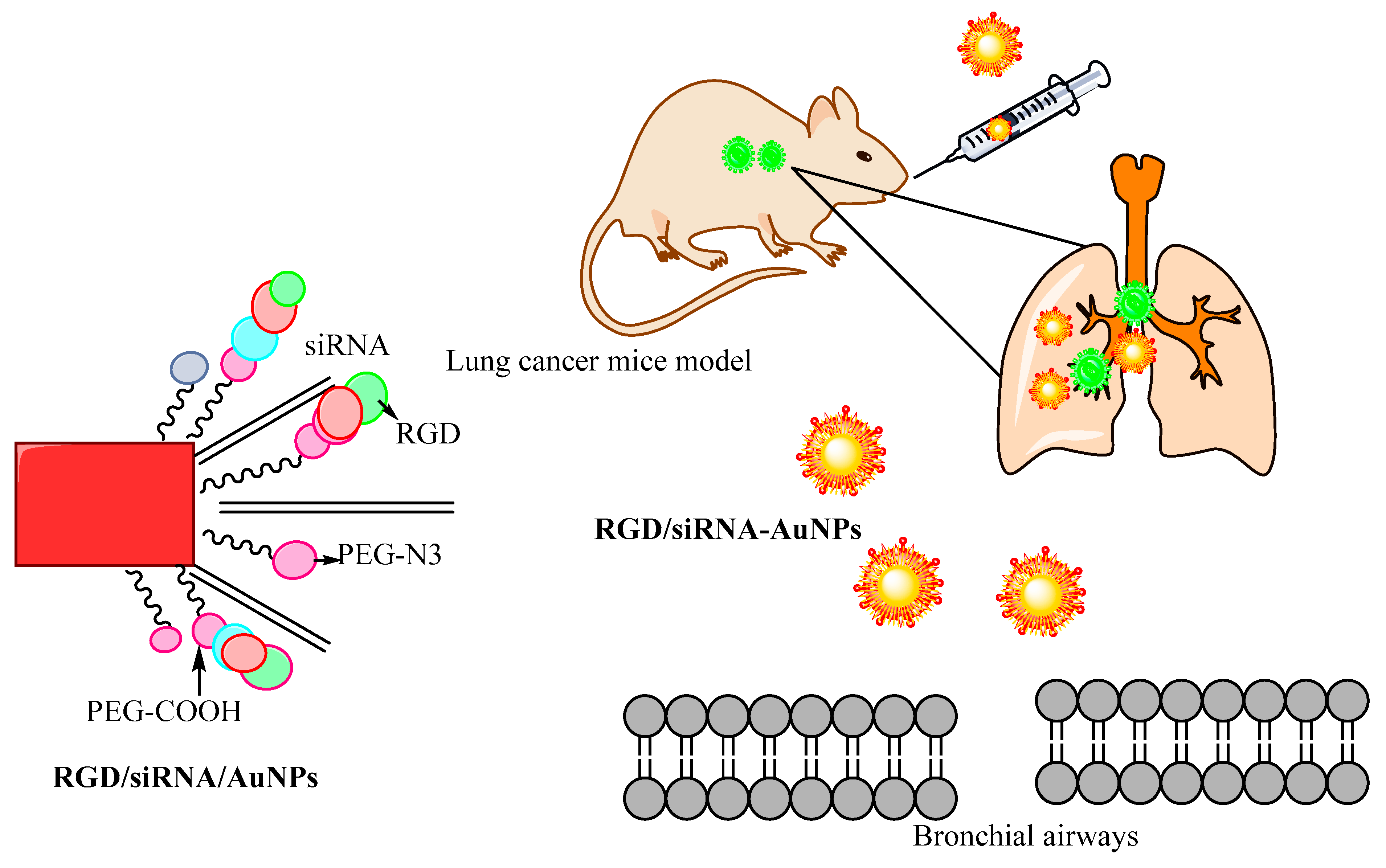
3.3. α. vβ3 Integrin Receptor

Integrins belong to the transmembrane heterodimeric glycoproteins family, consisting of the 18 α and 8 β subunits [65]. Integrins are expressed in multiple forms in many tumor-associated cell types. In lung cancer, the integrins αv, α5, β1, β3 and β5 have been demonstrated to develop the survival and metastasis of cancer cells [66]. The role of integrins encompasses cell–matrix adhesion, the maintenance of cellular morphology, differentiation and proliferation [54]. About 82% of NSCLC cases have higher integrin expression, while only 13% of SCLC expressed integrins [67]. The widespread functions of integrins in lung cancer suggest that their inhibition could be beneficial in tumor targeting and therapy. It was demonstrated that the inhibition of the αvβ3 and αvβ5 integrins with targeted ligands can block the endothelial cell angiogenesis and tumor metastasis [66]. In this context, arginylglycylaspartic acid (RGD) peptide has the potential to target αvβ3 integrin, thus facilitating drug delivery to the lung cancer. In one study, RGD anchored poly(lactide-co-glycolide) (PLGA)-chitosan nanocarriers successfully delivered paclitaxel (PTX) specifically to lung cancer, while normal human bronchial epithelial cells with poor integrin expression had negligible cytotoxic effects of PTX [68]. Furthermore, cyclic peptide anchored formulation elevated the localized drug concentration and suppressed the tumor cells in the subcutaneous and orthotopic A549 xenograft mice models as compared to the free drug controls [69].

3.4. Folate Receptors (FRs)

FRs are from a family of glycoproteins (35–40 kDa) having a strong binding affinity for folic acid (FA). FRs are differentiated into four isoforms including FRα, FRβ, FRγ and FRδ [70]. Normal human cells have a very low content of FRs, whereas FRs are overexpressed in a variety of tumor cells—the first two isoforms (FRα, FRβ) are the most common [70,71]. In NSCLC, FRα is overly expressed especially in adenocarcinoma [54]. Therefore, FA or FR monoclonal antibodies could serve as a ligand to target lung cancer. Folate can be conjugated to chemotherapeutic agents, microparticles, nanocarriers, lipidic systems and oligonucleotides to directly target FR-positive tumor cells. Folate-PEG-modified cytochrome c nanomicelles have demonstrated selective targeting and internalization by FR expressed on the HeLa cells compared to FR negative cell lines [72]. Similarly, DOX and small interfering RNA (siRNA) were loaded into folate-biotin conjugated starch nanoparticles for codelivery into human lung cancer cells (A549). Folate-mediated codelivery has shown enhanced cytotoxicity and reduced proliferation of the A549 cells. The cytotoxicity was competitively inhibited in the presence of free folate; further, the expression of insulinlike growth factor 1 receptor (IGF1R) proteins was decreased through the treatment [73].

3.5. Cluster of Differentiation 44 (CD44)

CD44 is a cell-surface based glycoprotein receptor with a specific affinity for hyaluronic acid (HA). The binding of HA to the receptor regulates cell adhesion and the differentiation and migration of the normal cells [74]. In tumors, CD44 has the important functions of cell adhesion, growth, proliferation, metastasis and induction of the cancer cell resistance [74,75]. CD44 is highly upregulated in squamous cell metaplasia and NSCLC [76] and is involved in metastasis of NSCLC to the lymph node [77]. HA, as an anionic glycosaminoglycan and a polymeric ligand, can be anchored to the surface of the particles or itself is able to self-assemble to target the lung cancer [78,79]. For instance, HA anchored polyethyleneimine-PEG nanoparticles specifically delivered siRNA to lung cancer cells [78]. Furthermore, enzyme hyaluronidase-1 expressed heavily in the malignant tumors degraded HA, thus facilitating drug release from HA in the target cancer cells [80].

3.6. Other Onco-Receptors

Several other receptors are heavily expressed in the lung tumor microenvironment including luteinizing hormone-releasing hormone (LHRH) receptors [81], chemotactic chemokines receptor 4 (CXCR4) [82], fibroblast growth factor receptor [83], tyrosine kinase AXL receptor [84], vascular endothelial growth factor receptor (VEGFR) [85], death receptor/TNF-related apoptosis-inducing ligand-receptor (DR4/TRAIL-R1) [86], β2-adrenergic receptors (β2-AR) [87] and lectin receptors [88]. Targeting these receptors through specific ligands can inhibit lung cancer survival, growth and metastasis.

4. Extracellular Nanovesicles in Targeting Lung Cancer

The concept of applying nanoparticles for lung cancer targeting shares a lot of similarities with the function of extracellular vesicles (EVs). In this regard, we briefly review these particles in this section. EVs are cell-derived, membrane-bound particles known to mediate intercellular signaling and are sensitive in organ-specific metastasis. Depending on the biogenesis pathways or their subcellular origin and size, EVs are also referred to as apoptotic bodies, microvesicles or exosomes [89,90,91]. EVs confined from distinct body fluids transport immune response-related and immune-modulatory molecules. These molecules include proteins, lipids, and nucleic acids. Recent studies considered EVs as one of the main components in the tumor microenvironment. In the tumor microenvironment, the EVs are able to transport the biomolecules to the less malignant cells. As the result, the less malignant cells receiving the EVs may continue to show increased metastatic and migratory behavior [92].
Integrin receptors are enriched in small EVs and are major players in mediating EV functions. For example, αvβ3 integrin is upregulated during cancer progression and is known to account for the migration of cancer cells. These nanovesicles’ signaling is capable of modifying the tumor cell’s structure, characteristics and functionality, such as overcoming drug resistance [93,94]. In their study, Hoshino et al. demonstrated that the tumor-derived lung-tropic EVs carry integrins α6β1 and α6β4, which are favorably taken up by lung fibroblasts and surfactant protein C-positive epithelial cells. The authors demonstrated that the incorporation of EVs by lung resident cells enhanced the expression of the proinflammatory gene S100 and promoted the lung metastasis [95].
From the therapeutic perspective, EVs are novel drug delivery systems and have more biosafety and biocompatibility characteristics than other synthetic surface functionalized or hybrid nanoparticles. In this context, EVs can be divided into unmodified and modified EVs [96]. Similar to nonfunctionalized nanoparticles, unmodified EVs have shown less efficacy in various performed studies. Therefore, scientists are now developing modified EVs through the introduction of therapeutic molecules into EVs or modifying the surface components of EVs to enhance their efficacies in terms of tissue targeting and site specificity [97]. For example, Nakase et al. modified EVs with octaarginine peptide, which resulted in enhanced cellular EV uptake via the active induction of macropinocytosis without cytotoxicity. Additionally, the increased accumulation of EVs at the targeting site showed greater therapeutic effect [98]. Recent studies on EV-mediated lung cancer targeting at a specific site highlighted that there are many limitations involved in the modification of these nanovesicles, as the strategies adopted for modification may damage the EV membrane and consequently compromise the therapeutic efficacy of the EVs [99,100,101,102,103]. To overcome these limitations, surface modification of the synthetic nanoparticles has demonstrated more promising results. For instance in a recent report, α3β1 integrins were targeted in NSCLC through cyclic peptide linked polymersome containing docetaxel. The results demonstrated better cellular uptake of cyclic peptide anchored formulation by A549 human lung cancer cells than by free docetaxel (DTX) and nontargeted polymersome.

5. Immunological Targets in Lung Cancer

Since the tumor is associated with pathophysiological, cellular and biochemical alterations, several immune cells like T-lymphocytes, macrophages, natural killer cells, B cells, MDSCs and dendritic cells infiltrate the lung tumor microenvironment. Hence, TAMs, MDSCs and regulator T-cells can be targeted through different ligands and strategies to modulate the tumor activity and reduce tumor progression [104].

5.1. Tumor-Associated Macrophages (TAMs)

Traditionally, activated macrophages of different phenotypes have commonly been categorized as M1 and M2 macrophages. M1 macrophages are activated through the classical pathway and are involved in proinflammatory response, while M2 macrophages are alternatively activated and associated with anti-inflammatory action. At first, macrophages polarize to M1 to assist the host immune response against an antigen, then they attain M2 phenotype to repair the damaged tissues. Macrophages linked with tumors are known as TAMs and are classified into two phenotypes—M1 and M2 (M1 type TAMs suppress cancer progression, while M2 type TAMs promote it). TAMs are characterized by increased M2/M1 ratio and play a crucial role in tumor progression, metastasis, matrix remodeling, angiogenesis and tumor resistance [105,106]. TAMs produce cytokines, growth factors like epithelial growth factor, matrix metalloproteinase-9, angiopoietin, etc. to assist tumor development. Therefore, TAM targeting can bring benefits to treat lung cancer. TAMs can be targeted through different ways including the repolarization of M2 into M1 cells, the prevention of macrophage recruitment into the tumor or the direct termination of M2 cells [104]. Moreover, several receptors such as C-type lectin, CD44, FRs have been expressed on the surface of TAMs, which can be specifically targeted for tumor eradication [107,108,109]. C-type lectin receptors are Ca2+ dependent carbohydrate recognition proteins and have multiple types including mannose receptor, macrophage galactose-type lectin-C and dectin receptor. Hence, different carbohydrate moieties are used to target C-type lectin receptors like mannose, glucose, D-galactose, N-acetyl-D-glucosamine (NAG) and maltose [110]. Recently, various mannose receptor targeting strategies involving mannose anchored liposomes, solid lipid nanoparticles, polymeric nanocarriers, niosomes, dendrimers and quantum dots have been fabricated to modulate macrophage function in the tumor [110]. In this quest, biotin and mannose conjugated lipid-coated calcium zoledronate nanocarriers have shown higher internalization in both TAMs and cancer cells, restraining tumor growth, progression and angiogenesis [111].

5.2. Myeloid-Derived Suppressor Cells (MDSCs)

MDSCs are the heterogeneous immature population of cells, comprised of myeloid progenitor cells, immature macrophages, immature dendritic cells and immature granulocytes [112]. MDSCs release immunosuppressive cytokines to retard immune system action against the tumor and thereby facilitating the tumor progression [113]. Thus, MDSCs are the major target cells in cancer immunotherapy. Several strategies have been designed to suppress MDSCs function in lung cancer including:
(a)
Differentiation of MDSCs into mature myeloid cells [113];
(b)
Suppression of MDSCs amplification through inhibition of stem cell function [114], VEGF [115] or STAT3 pathway [116];
(c)
Direct elimination of MDSCs by antibodies or chemotherapeutic drugs like gemcitabine [117];
(d)
Attenuating MDSCs functioning [118];
(e)
Inhibition of the immune checkpoint to restore the antitumor immune response [119].
Nowadays, nanocarriers employing different options to target MDSCs, promote MDSCs maturation and modulate their function to regress tumor progression and angiogenesis [120]. Furthermore, MDSCs cell membrane coated iron oxide magnetic nanoparticles successfully evaded the immune system, actively targeted cancer cells along with magnetic and photothermal-induced ablation of the cancer cells [121].

5.3. Regulators T-Cells

Infiltrated regulatory T-cells in the tumors have downregulated the activation and response of cytotoxic T-cells against lung cancer [122,123]. Regulatory T-cells play an integral role in tumor development; thus, they could be targeted to suppress the tumor. For instance, glucocorticoid-induced tumor necrosis factor receptor-related protein (GITR) ligand was linked to PLGA nanoparticles for active targeting of regulator T-cells. The complex nanosystem, together with photothermal and photodynamic therapy, has remarkably reduced the tumor growth and recurrence [124,125].

6. Delivery Routes of Nanoparticles in Targeting Lung Cancer

The delivery of nanomedicines to the lungs increases the sustained local drug concentration to treat lung cancer [32]. Most chemotherapeutics act on normal tissues due to their nontargeting nature, leading to adverse effects [126]. Therefore, targeted drug delivery requires a low dose, which results in fewer systemic side effects. Although drugs can be administered through oral, intravenous or inhalational routes, the research on oral drug delivery to lungs has not shown promising results, as only a limited amount of the drug molecules are delivered to the lung tumors [127]. Additionally, the majority of the anticancer formulations are used as intravenous dosage forms. However, as most of the chemotherapeutic agents for lung cancer treatment are hydrophobic in nature, high doses and/or surface modification are needed to improve their systemic bioavailability [128]. On the other hand, the inhalational route is the most attractive option due to lower side effects and high biodistribution [129]. A safe and effective mean of lung cancer theranostics are the chemotherapeutic agents formulated by nanotechnology-based carriers, which are a novel targeted drug delivery “inhalational nanomedicine” that can be administered through the inhalational route [130]. In this context, the easiest way of drug delivery is inhalation by aerosols to target the cancerous tissues of the lung. The differential accumulation of drug particles or aerosol droplets in different regions of the lungs depends on their sizes. Drugs can be formulated as solutes or particles in aerosol droplets of appropriate size and used in drug delivery [131].
Okamoto et al. formulated gene powders with chitosan as a nonviral vector and mannitol as a dry powder carrier to compare their gene expression and therapeutic adequacy to intravenous or intratracheal gene solutions in mice having pulmonary metastasis prepared by injecting CT26 cells. In both normal and tumorous tissues, the genes expressed by intratracheal powder were higher than the one expressed by intravenous or intratracheal solutions, indicating that therapeutic gene powders are efficient for lung cancer treatment [132]. In another study, Dames et al. revealed that the targeted delivery of aerosols to the affected lung tissue might improve therapeutic efficiency and reduce undesired side effects. The authors showed theoretically that targeted aerosol delivery with superparamagnetic iron oxide nanoparticles along with a target-directed magnetic gradient field can be achieved to treat localized lung disease [133]. Ngwa et al. examine the potential of nanoparticle drones (smart nanomaterials) in targeting lung cancer. They compared and assessed inhalation (air) versus the traditional intravenous routes of navigating physiological barriers using such drones. They concluded that the inhalation route might be more promising for targeting tumor cells with radiosensitizers and cannabinoids in terms of maximizing the damage to lung tumor cells while minimizing any collateral damage or side effects [134].

7. Surface Modification of Nanoparticles to Combat Toxicity in Lungs Cancer

One of the challenging factors in the delivery of drugs to the lungs is to understand the interactions of the nanoparticles with the biological systems. The chemotherapeutic agents in the form of NDDSs are cell nonspecific, resulting in the undesired attack of healthy cells (an important factor in the failure of conventional nanotechnology cancer therapy). This is the reason why still further advancements need to be carried out in the field of NDDSs. The fast clearance of nanoparticles decreases the efficiency of sustained drug delivery and their translocation might bring nanoparticles to undesired areas of the body causing toxicity. Due to the complex nature of nanoparticles, research studies have led to different views of the nanomaterials’ safety [135,136]. The physical properties of nanoparticles, such as morphology, geometry, dimensions and surface charge, have been found to change their therapeutic effect. Rod-shaped particles are more toxic than spherical particles. Long fibers cause inflammation because they are less likely to be engulfed by macrophages, thus minimizing their elimination from the system [136]. Nanoparticles produce pulmonary toxicity by oxidative stress because of the production of reactive oxygen species within the biological system [137]. It is evident from a research study that cytotoxicity occurs due to the production of free radicals after exposure to 3.5 to 23.3 µg/ml cerium oxide (CeO2) nanoparticles. It causes oxidative stress in the cells by reducing glutathione and α-tocopherol levels and elevating the production of malondialdehyde and lactate dehydrogenase, which are indicators of lipid peroxidation and cell membrane damage, respectively [138]. The accumulation of nanoparticles in the tissue due to slow clearance produces potential free radicals as well as the prevalence of numerous phagocytic cells in the organs of the reticuloendothelial system (RES) making the lungs the main targets of oxidative stress [139].
According to a research study, 15 nm and 46 nm silicon dioxide (SiO2) nanoparticles significantly reduced cell viability in a dose-dependent and time-dependent manner in bronchoalveolar carcinoma-derived cells at 10–100 μg/mL dosage. Both types of SiO2 nanoparticles have higher cytotoxicity than the positive control material (Min-U-Sil 5). The reactive oxygen species (ROS) generated by exposure to 15 nm SiO2 nanoparticles produces oxidative stress in these cells as reflected by reduced glutathione levels and the elevated production of malondialdehyde and lactate dehydrogenase, indicative of lipid peroxidation and membrane damage [140].
Surface functionalized nanoparticles have received tremendous importance as drug carriers. The physicochemical or biological properties of the nanoparticles can be altered by modifying their surfaces with different functional groups through covalent or noncovalent bonding, such as the adsorption of biologically active molecules (i.e., proteins, surfactants, enzymes, antibodies or nucleic acids) [141]. Nanoparticles functionalized with biodegradable polymers could be evaluated as the best chemotherapeutic delivery system. The surface chemistry of these nanoparticles must be carefully controlled as it is the shell of the nanoparticles that is in contact with body organs and fluids. As an example, nanoparticles have been coated with hydrophilic polymers or functionalized with ligands or proteins to enhance their circulation time or to achieve site specific delivery, respectively [142]. In another example, it was shown that the coating of nanoparticles with polymers could reduce their toxicity by changing their half-life distribution, disposition, stimuli reactivity and therapeutic application [30]. The oily nature of the nanocapsule’s core can accommodate high loadings of lipophilic anticancer drugs [143]. Moreover, magnetic nanoparticles functionalized with polymers, monoclonal antibodies, peptides, heparin, hormones or other biologics are very effective and highly specific for cell biology and cancer therapeutic applications [144]. Surface modification of the NDDSs allows the targeted delivery of therapeutic agent such as antibodies and ligands (i.e., TF, FA, lactoferrins, lectins and mannose derivatives) into the tumors [135]. Additionally, surface PEGylation (the process by which polyethylene glycol chains are attached to biological molecules) does not only enhance the colloidal stability of nanoparticles but also increases their accumulation at the tumor site and decreases opsonization [145]. It was shown that surface-modified nanoparticles with 1, 2 dipalmitoylphosphatidylcholine (DPPC) are less prone to phagocytosis. The presence of phospholipids inhibits the adsorption of opsonic proteins on the inhaled nanoparticles, allowing them to escape phagocytosis [146]. In one research study, multiwalled carbon nanotubes (MWCNTs) were functionalized with amine-terminated poly (amidoamine) (PAMAM) dendrimers modified with fluorescein isothiocyanate (FI) and FA. This modified system acted as both a drug targeted system and a pH-responsive system for delivering DOX into cancerous cells [147]. Meenach et al. used an advanced organic spray-drying method to manufacture inhalable lung surfactant-based carriers comprising synthetic phospholipids, DPPC and dipalmitoylphosphatidylglycerol (DPPG), loaded with PTX, for targeted pulmonary delivery as high-performing nanoparticulate dry powder inhalers [148]. Li et al. suggested that a tumor-targeted PEGylated LPD formulation (liposome-polycation-DNA complex) enhanced cellular uptake by specific receptor-mediated pathways. They showed that the targeted drug delivery system caused a strong gene-silencing mediated by RNAi through delivering siRNA to the tumor cells after intravenous administration [149,150]. Grabowski et al. described the cytotoxicity and inflammatory action of nanoparticles made of PLGA through in vitro analysis on A549 human lung epithelial cells. Three different neutral, positively or negatively charged PLGA nanoparticles (230 nm) were obtained by using different types of stabilizers (polyvinyl alcohol, chitosan, or Pluronic® F68). For comparison, polystyrene nanoparticles were used as nonbiodegradable polymeric nanoparticles and titanium dioxide (anatase and rutile) as inorganic nanoparticles. As the result, the PLGA-based and polystyrene nanoparticles were less toxic than or equally toxic to titanium dioxide nanoparticles. On the contrary, the inflammatory response measured by the release of interleukin 6 (IL-6), IL-8, monocyte chemoattractant protein-1 (MCP-1) and tumor necrosis factor α (TNF-α) cytokines was low for all nanoparticles [151]. The PLGA-based nanoparticles led to a higher inflammatory response, which was correlated with a higher uptake of these nanoparticles. The authors claimed that both the coating of the PLGA nanoparticles and the nature of the core play a key role in the cell response.

8. Comparison between Surface-Modified Nanoparticles with Other Targeting Methods in Lung Cancer Treatment

There are many ongoing contributions of nanoparticles in the field of targeting lung cancer. However, there are a few limitations that inhibit amplifying their applications, including low stability, greater immunogenicity, nonuniform distribution, increased rate of clearance, poor ability in encapsulating imaging and targeting agents and unspecified internalization (via passive delivery method) at the malignant site. The morphology of the lung itself is a big barrier for the optimal transformation of agents into it. Therefore, it is important to design nanoparticulate systems that could reduce these complications. The functionalized nanoparticles can be used as powerful theranostic tools to enhance the delivery of drugs to the malignant site [34]. Many studies have been carried out comparing the performance of functionalized and nonfunctionalized nanoparticles in lung cancer targeting. For example, Chung et al. developed and compared the PLGA nanoparticles ligated with heparin, chitosan and pluronic with nonconjugated PLGA nanoparticles. The viability tests for both normal and tumor cells showed the less cytotoxic effect of the nanoparticles. The in vitro cellular uptake of the nanoparticles for both chitosan and heparin functionalization showed the desired effects. The in vivo tumor model study exhibited that there was a positive but insufficient effect of chitosan decorated nanoparticles, although it showed enhanced accumulation that was almost 2.4-fold higher than that of the control nanoparticles. The results concluded that the surface functionalization of the PLGA nanoparticles with chitosan and heparin may be an efficient strategy for the enhanced tumor theranostics [152]. Patil et al. performed a study with the aim of achieving targeted delivery through the single-step surface functionalization of nanoparticles with a tissue recognition ligand. They used biotin and a folic acid ligand to functionalize the PLA-PEG nanoparticles. The surface modification was confirmed through NMR, transmission electron microscopy and tumor cell uptake study. In comparison to the bare nanoparticles, the functionalized nanoparticles showed more precise and efficient results with greater binding affinity at the delivery site. The in vivo study result of the surface-modified PTX-loaded PLA-PEG nanoparticles showed an enhanced efficacy in comparison to the nonmodified nanoparticles [153]. The same authors in another study developed biotin functionalized PLGA nanoparticles encapsulating a combination of PTX and P-glycoprotein (P-gp) inhibitor tariquidar to overcome tumor drug resistance. The dual agent nanoparticles showed higher cell inhibition in the cell line study in comparison to only PTX-loaded ones. Additionally, performing in vivo studies in a mouse model, these nanoparticles demonstrated considerably enhanced inhibition of tumor growth. The authors concluded that these dual agent nanoparticles could be applied as an efficient system to overcome tumor drug resistance [154]. In another report, Xia et al. developed DOX-loaded selenium (Se) nanoparticles and functionalized them with cyclic peptide (Arg–Gly–Asp–D-Phe–Cys [RGDfC]) to fabricate tumor targeting delivery. The aim of the study was to improve the antitumor efficacy of DOX in NSCLC. This nanodrug carrier displayed an efficient cellular uptake in A549 cells and entered the A549 cells mainly by clathrin-mediated endocytosis. Interestingly, comparing active targeting with the passive targeting delivery system, the authors concluded that the RGDfC functionalized DOX-loaded Se nanoparticles provide a promising approach for lung carcinoma therapy [155]. Perepelyuk et al. studied the therapeutic efficacy and in vivo efficacy of mucin1-aptamer-modified miRNA-29-loaded hybrid nanoparticles in a lung tumor model. The results displayed that the presence of MUC1-aptamer conjugates increase the delivery of miRNA-29b to the tumor cells. Moreover, the downregulation of DNMT3B by MAFMILHNs resulted in the inhibition of tumor growth in a mouse model [156]. Table 2 presents a summary of the recent studies on comparison between surface-modified and unmodified nanoparticles in lung cancer treatment.

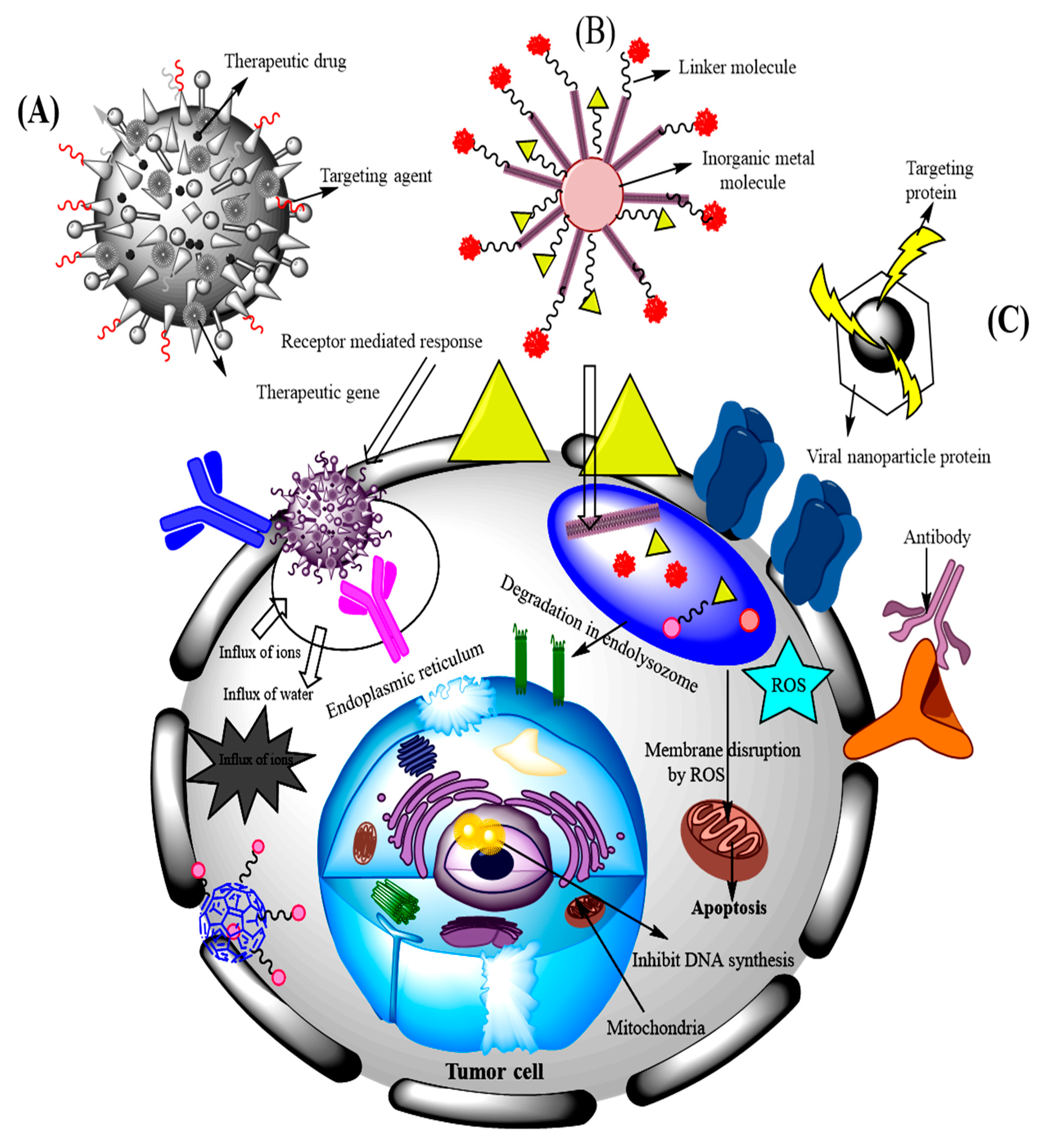
9. Different Types and Applications of Surface-Modified and Hybrid Nanoparticles for Targeting Lung Cancer

Nanoparticles are included in the drug delivery systems to overcome certain issues such as low solubility and permeability related to tumor targeting. The most significant advantages of nanoparticles are their excellent loading capacity and high surface to volume ratios. Various organic and inorganic nanomaterials have emerged as novel tools for cancer diagnosis and therapy due to their unique characteristics. In this review, based on the main structural moiety of nanoparticle we broadly divide them into three types: organic nanoparticles, inorganic nanoparticles and hybrid nanoparticles. The combinatorial therapeutic approach via hybrid nanoparticles is discussed in a separate subsection too.

9.1. Organic Nanoparticles

Organic nanoparticles can be defined as solid particles composed of organic compounds (mainly polymers, lipids or proteins). They have been widely studied for decades, presenting a large variety of materials and exciting applications in cancer therapies. There are many biopolymeric nanoparticles that are utilized in the drug delivery systems. For example, PLGA is a biodegradable copolymer approved by the US Food and Drug Administration (FDA) for use in distinct biological products. PLGA nanoparticles can be used to obtain extended and sustained delivery of therapeutic agents including protein, peptide, RNA, DNA and small molecules to their particular target sites [170,171]. As an example, Karra et al. developed cetuximab functionalized PLGA nanoparticles and loaded them with PTX. The results confirmed the in vitro targeting performance and enhanced the cellular internalization along with cytotoxicity of this targeted delivery system in lung cancer cells overexpressing EGFR. The intravenous administration of the nanoparticles to mice results in the considerable inhibition of tumor growth and the reduction of mortality rates. Pharmacokinetics studies results showed no increase in the aggregation of nanoparticles at the tumor tissue site. The authors concluded the promising potential of this system for enhanced efficacy against lung cancer [172]. In another report, Patil et al. compared YSA peptide functionalized and nonfunctionalized PLGA nanoparticles to improve delivery to bleomycin treated cultured endothelial cells in a bleomycin induced lung injury mouse model. When human umbilical vein endothelial cells (HUVEC) were treated with bleomycin, the 3 h uptake of both types of nanoparticles was increased up to 2-fold. The results showed that in mice the bleomycin injury led to 2.3 and 4.7 times increases in the lung concentrations of the nonfunctionalized and YSA-functionalized nanoparticles, respectively. The authors stated that PLGA nanoparticle delivery to cultured vascular endothelial cells and mouse lungs in vivo was higher directly after bleomycin treatment, with the delivery likely to be higher for YSA-functionalized nanoparticles [173].
Single chain technology is a new term developed in nanotechnology in order to broaden the functions of soft nano-objects through chain compaction. Using single chain technology, individual copolymer chains of different natures, compositions and molar masses have been folded intramolecularly to develop single chain nanoparticles (SCNPs). This leads to very small size polymer nanoparticles in the sub-20 nm size [174,175]. The folding is achieved by the self-assembly or crosslinking of functional groups on the precursor polymer, or rather moderated by the external cross-linker. There are several ways to develop SCNPs including dynamic and irreversible covalent crosslinking reactions such as cycloaddition. Moreover, there are huge number of SCNPs that have been introduced, from single and multiblock to star particles, hairpins and tadpole molecules. There are only a few examples present where a functionalized group has been incorporated into SCPNs [176]. However, these functionalized SCNPs still have not been used for lung cancer targeting. In this context, an insight was given by Benito et al. who evaluated the use of SCPNs based on poly(methacrylic acid) in targeting pancreatic adenocarcinoma. They functionalized SCPNs with somatostatin analogue PTR86 as a targeting moiety since these somatostatin receptors are overexpressed in pancreatic cancer. The imaging results showed a higher accumulation of targeted SCPNs in the tumor compared to the nontargeted nanoparticles, which was due to the enhanced retention in the tissues [177]. Later, Kröger et al. also reported the greater potential of these types of nanoparticles for cellular targeting [178,179].
Dendrimers are another class of polymers that are constructed by the stepwise addition of layers (generations) of molecules around a central core. This unique physicochemical properties of dendrimers enable a facile utilization of them as templates to funcionalize nanoparticles [180]. In this regard, a group of researchers reported greater penetration and higher stability of siRNA by implementation of surface-modified poly(propyleneimine) dendrimers. The siRNA nanoparticles were coated by a dithiol bearing cross linker that followed by a layer of PEG. In addition, a synthetic derivative of LHRH was linked at the end of the PEG polymer to conduct siRNA nanoparticles to the cancer cell. The developed system showed time- and concentration-dependent cellular uptake under in vitro conditions. It was proposed by the authors that this approach could be used for the in vivo systemic delivery of siRNA for efficient cancer therapy [181].
Solid lipid nanoparticles or lipid nanoparticles are nanoparticles composed of lipids as a matrix which are exceptionally biodegradable and biocompatible. They possess superior properties such as high drug payload, increased drug stability, large scale production and sterilization [182]. For instance, in one study Pooja et al. developed and evaluated TF conjugated and etoposide loaded solid lipid nanoparticles. The tissue distribution and pharmacokinetics were studied in Balb/c mice. The nanoparticles showed great anticancer activity of etoposide via antiproliferative assay and induced apoptosis in A549 cells. It was concluded that over expressed TF-receptors showed enhanced efficacy in NSCLC [183]. Liposomes are similar in design to lipid nanoparticles, but slightly different in composition and function. Riaz et al. developed the TF-7 surface functionalized liposomes loaded with quercetin (QR) for lung cancer therapy. These liposomes were evaluated for cellular uptake and in vitro cytotoxicity study and they exhibited higher cytotoxicity and S-phase cell cycle arrest. The in vivo study showed enhanced liposomes accumulation in the lungs and sustained release up to 96 h [184].
Considering that albumin has remarkable roles in human body, it can be used in the area of medicine and disease treatment. As an example, Yang et al. used hematoporphyrin (HP) functionalized albumin nanoparticles for cancer therapy. These nanoparticles further modified with gamma emitting nuclides (99mTc). HP-albumin nanoparticles showed improved accumulation in A549 and CT-26 cancer cell lines. The evaluation of the pharmacokinetics of 99mTc chelated HP-albumin nanoparticles via the scintigraphic imaging of rabbits resulted in acceptable imaging properties in the rabbit with a longer biological half-life compared to 99mTc-HP. The authors concluded these modified albumin nanoparticles could be applied as a diagnostic tool for cancer as well as the obvious application for photodynamic therapy [185].

9.2. Inorganic Nanoparticles

Inorganic nanoparticles including gold, silver, iron oxide and silica nanoparticles have been widely studied as therapeutic agents for cancer treatments in biomedical fields [186]. Among them, gold nanoparticles are attractive constituents for nanoparticle polymer hybrid materials as they support localized surface plasmon resonances, and the wavelength region of the surface plasmon resonance peak can be adjusted finely through the geometric parameters of the particles [187,188]. In one study, Heo et al. developed the gold nanoparticles surface-functionalized with PEG, biotin and rhodamine B and linked beta-cyclodextrin (β-CD). The specific interactions of these nanoparticles with cancer cells such as HeLa, A549 and MG63, as well as normal NIH3T3 cells, were evaluated. The authors observed that the modified nanoparticles were more effectively involved with the cancer cells. Confocal laser scanning microscopy (CLSM), fluorescence-activated cell-sorting (FACS) and cell viability analyses showed that the surface functionalized nanoparticles played a significant role in the diagnosis and treatment of the cancer cells, and could be used in theranostic agents [189]. Guo et al. developed a multifunctional nanocarrier encapsulated with methotrexate via electrostatic interaction between gold nanocluster conjugate chitosan and nucleolin targeting aptamer (AS1411). The in vivo study demonstrated that intravenous administration of nanodrug carrier systems into BALB/c mice caused the accumulation of methotrexate at the tumor site. The results suggested that the developed functionalized system can be applied for an effective delivery for anticancer agents and shows enhanced potential in clinical applications [190]. João Conde et al. fabricated the gold nanoparticles conjugated with siRNA/RGD and studied in a lung cancer murine model. The RGD treatment showed a significant downregulation followed by tumor growth inhibition and the increased survival of the tumor bearing transgenic mice. The results demonstrated that RGD gold nanoparticles stimulate the delivery by intratracheal application in mice that leads to the suppression of tumor cell proliferation. The enhanced targeted delivery of gold nanoparticles encapsulated with siRNA to cancer cells works towards effective silencing of the oncogene. The study showed gold nanoparticles stimulated the inflammatory and immune responses that can promote the therapeutic effect of the siRNA to reduce the tumor size at very low doses [191]. The schematic illustration of this study is described in Figure 3, which shows the enhanced efficacy of siRNA loaded into functionalized nanoparticles.
Applications of silica or silicon dioxide (SiO2) as another inorganic nanoparticle are broadly investigated in drug delivery. For example, Munaweera et al. prepared cisplatin and cisplatin/nitricoxide-loaded amine functionalized mesoporous silica nanoparticles for the treatment of lung cancer. The results demonstrated that for nonsmall lung cancer cell lines (i.e., H596 and A549), the toxicity of cisplatin/nitric oxide-loaded silica nanoparticles was higher than that of silica nanoparticles loaded with only cisplatin. The nitric oxide-activated sensitization of the tumor cell death, which showed that nitric oxide is a potential enhancer of platinum-based lung cancer therapy [192]. Another type of inorganic nanoparticles with biomedical applications is zirconium oxide (ZrO2). In one study, ZrO2 nanoparticles were coated with aminopropilsilane, tetraoxidecanoic acid or acrylic acid. The studied results showed dose-dependent signs of effectiveness. It was concluded that surface modifications of the ZrO2 nanoparticles had very small effects on the inflammatory lungs of rats and mice but it had very clear efficacy in the allergic mouse model used. The results stated that the allergic mice are more responsive to exposure to surface-modified nanoparticles [193]. The unique properties of molybdenum disulfide (MoS2) make it an attractive candidate for drug delivery applications [194]. In their study, Wei Zhang et al. developed the riboflavin 5′-monophosphate sodium salt functionalized 2D MoS2 nanosheets prepared by the simple ultrasonication method, then they applied this nanocomposite having fine electrochemical redox activity as a platform to immobilize DNA probe. The results showed that the signal detection platform showed greater sensitivity with the limit of detection of 1.2 × 10⁻¹⁷ mol L−1 for PIK3CA gene from lung malignancy. The constructed biosensor was easy to achieve and could detect different pathogenic DNA without an intricate label process [195].
Magnetic nanoparticles can produce heat under the magnetic field and can also deliver drugs to the lung cancer site [196,197]. Among them, iron oxide nanoparticles are widely studied systems for biomedical applications [198,199]. In their study, Huang et al. reported the synergy effect of superparamagnetic iron oxide nanoparticles along with an anticancer drug (β-lapachone) for improved cancer therapy. The authors suggested that combination of superparamagnetic iron oxide nanoparticles with reactive oxygen species-producing drugs could conceivably enhance drug efficiency, thus presenting a synergistic strategy to integrate imaging and therapeutic functions in the discovery of theranostic nanomedicine [200]. In another study, dextran coated iron oxide nanoparticles were modified with the TAT peptide and they were used to improve the efficiency of the radiation. After performing the internalization study, it was revealed that TAT functionalized nanoparticles enhanced the generation of the reactive oxygen species in comparison to the nanoparticles without any surface modifications. These modified nanoparticles also affected the mitochondrial integrity of A549 cells in combination with the radiation, which resulted in a synergistic decrease in cell viability [201,202].

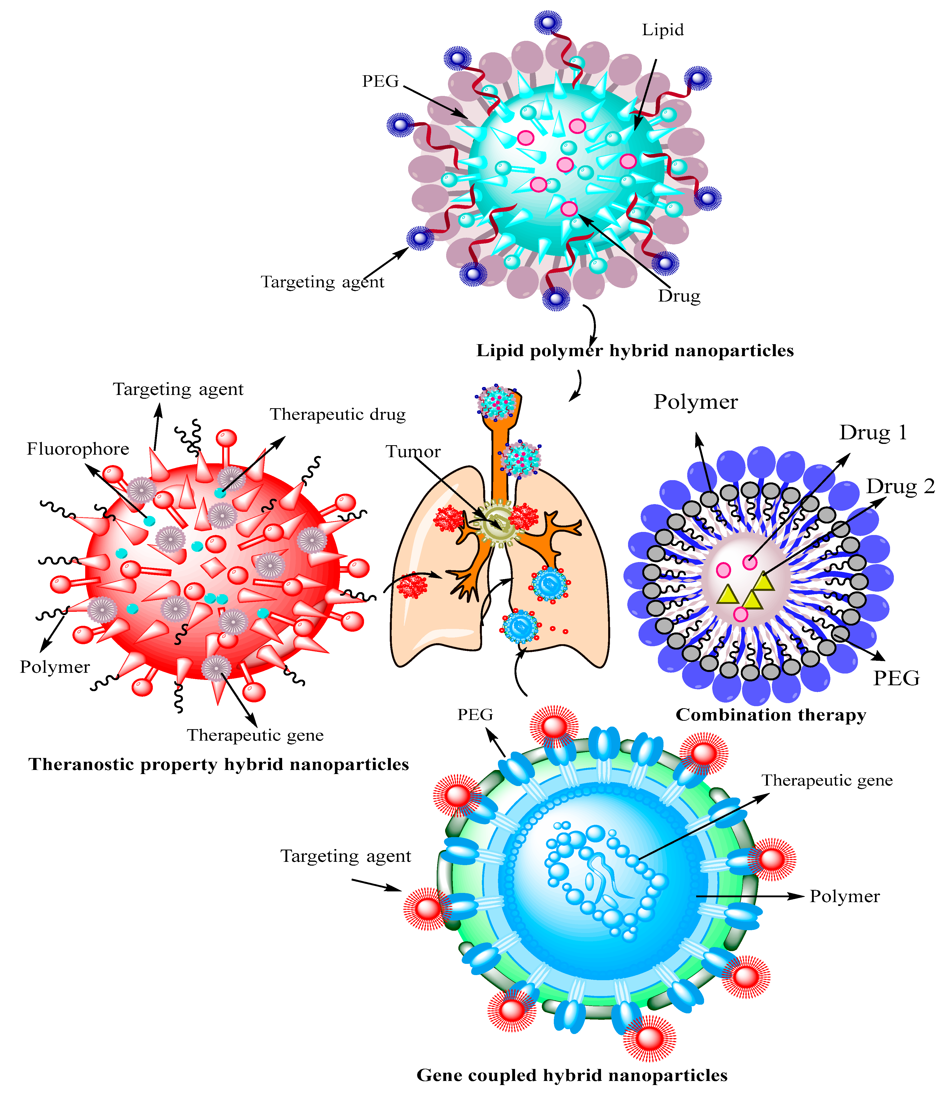
9.3. Hybrid Nanoparticles

In order to enhance the efficacy of the therapeutic regimen in lung cancer, it is necessary to develop new systems that can increase the survival rates. The development of hybrid nanoparticles (that could comprise both inorganic and organic structural moieties) in conjugation with other genes, biomolecules and other drugs are promising therapeutics systems for efficient targeting [203]. These types of nanoparticles are important for targeting the tumor site, for its early diagnosis and to measure the risk of malignancy in neighboring cells. These hybrid types are classified into diagnostics, therapeutic and theranostic nanoparticles. Figure 4 graphically shows the different types of hybrid nanoparticles that have been applied for lung cancer targeting.
In this regard, Sacko et al. studied anticancer effect of a combination therapy of miRNA-29b and genistein loaded in mucin-1 (MUC 1)-aptamer functionalized hybrid nanoparticles in NSCLC A549 cell line. This nanodrug carrier displayed a superior antiproliferative effect compared to individual genistein and miRNA-29b-loaded nanoparticles, thus, it can be considered a potential treatment modality for A549 cell line [204]. The same research group studied the pharmacokinetic response of novel antineurotensin receptor 1 monoclonal antibody (anti-NTSR1-mAb)-functionalized antimutant K-ras siRNA-loaded hybrid nanoparticles and compared it with that of naked siRNA formulation. As with the main findings, the plasma terminal half-life of the siRNA-loaded nanoparticle-delivered was 11 times higher than that of the naked siRNA formulation. In addition, high performance liquid chromatography (HPLC) analysis showed that the hybrid carrier system could protect the encapsulated siRNA against degradation in systemic circulation. The authors concluded that these hybrid nanoparticles can function as an effective nonviral vector for siRNA delivery for both experimental and clinical uses [205].
In another study, EGFR-targeted superaparamagnetic iron oxide nanoparticles (SPIONs) were conjugated to carboxy-terminated pluronic F127. The authors investigated the inhalation delivery of these nanoparticles as a potential approach for lung cancer treatment. As in the main findings, EGFR targeting enhanced tumor retention of SPIONs while minimizing systemic exposure. Additionally, magnetic hyperthermia using these nanoparticles resulted in a significant inhibition of in vivo tumor growth [206].
Another type of hybrid delivery system are lipid polymer nanoparticles or core shell lipid polymer nanoparticles, which combine the good biodegradability of polymeric nanoparticles with the excellent biomimetic characteristics of liposomes, and they are effective carrier systems for the delivery of anticancer drugs into the tumor site [207]. In this context, Bivash Mandal et al. showed that a hybrid system containing biodegradable polycaprolactone (as the core) and phospholipid-shell was able to deliver erlotinib into the lung cancer cells. Performing cell viability studies by this erlotinib-loaded hybrid system, a significant decrease in proliferation of A549 cells was observed, which affords this system a potential application to deliver erlotinib into lung cancer cells [208]. A similar study was carried out by Song et al. in which they showed the enhanced properties of EGFR-targeted lipid polymer hybrid nanoparticles in the codelivery of resveratrol and DTX. They developed this nanocarrier system by the conjugation of EGF and target the EGFR on the surface of the lung cancer cells in order to increase the endocytosis. Resveratrol (as an antioxidant) improved the production of reactive oxygen species through inducing cytotoxicity. The results exhibited the enhanced antitumor efficacy of resveratrol and DTX in both in vitro and in vivo studies [209]. Another related study reports the synthesis and characterization of lipid-coated poly D,L-lactic-co-glycolic acid nanoparticles that were modified with TF to deliver the DOX into A549 cells. These DOX-loaded hybrid nanoparticles exhibited higher cytotoxicity against lung cancer cells and showed an improved therapeutic effect in the lung cancer-bearing nude mice in comparison to their nontargeted counterparts. This finding marks this approach as an efficient targeted drug‑delivery system for lung cancer therapy [210]. In another study, naturally occurring chitosan and hyaluronic acid were deposited on negatively charged hybrid solid lipid nanoparticles through layer-by-layer (LbL) assembly. Next, this hybrid system was loaded with DOX/dextran sulfate complex with the aim of tumor specific targeting. Employing this approach under in vivo studies, the DOX half-life was increased and its elimination rate was decreased compared to those measured for the uncoated solid lipid nanoparticles [211].
Another interesting type of nanocarriers in cancer therapy are hydrazine-based pH-sensitive nanoparticles. For example, Li et al. designed a dual-ligand lipid based nanoparticle system, in which TF conjugated PEG hydrazone nanoparticles were used for the codelivery of DTX and baicalein into A549 cells. Decorating the lipid nanoparticle with TF could internalize them into the cancer cells. Moreover, this hybrid nanocarrier achieved significant synergistic effects, the best tumor inhibition ability and the lowest systemic toxicity [212]. A recent progression in lung cancer therapy is gene therapy of lung cancer by applying siRNA hybrid nanoparticles [213]. Applying hybrid nanoparticles was useful to carefully transport the siRNA into the cytoplasm and cross the limitations of the traditional gene therapy [214]. For instance, the encapsulation of siRNA in calcium phosphate nanoparticles coated with DOPA (dioleoylphosphatydic acid) could target H460 lung cancer cells [215].
An overview of some other recent studies (that were not mentioned in this section) on developing nanoparticle-based delivery systems as a therapy against lung cancer is presented in Table 3.

9.4. Combinatorial Therapeutic Approach via Hybrid Nanoparticles

The encapsulation of different drugs with multiple sites of action is considered to be an efficient method for targeting malignant cells. The application of combinatorial therapeutic approaches will reduce the dose and resistance of the applied anticancer drugs. However, each anticancer drug has a specific biochemical activity; therefore, combined administration would be inappropriate and ineffective in targeting lung cancers. Moreover, combining more drugs can cause harmful effects on healthy organs [229]. Various hybrid combination therapies have been designed for lung cancer targeting. Multifunctional hybrid nanoparticles have gained more recognition as a combinatorial therapeutic approach. For example, in one study an amphiphilic triblock copolymer functionalized with deoxycholate was synthesized and was used as a nanocarrier to codeliver DOX and PTX into lung cancer cells. Each residue of the copolymeric system comprises a unique property for the complex construction. The codelivery of DOX and PTX using hybrid nanovesicles exhibited an enhanced antitumor effect by reducing the growth of the A549 cells in lung cancer [230]. Another study demonstrated the enhanced efficacy of dual drug delivery in lung cancer therapy, in which PLGA/methacrylic acid copolymer nanoparticles were developed for the codelivery of DOX and chrysin. This nanoformulation significantly reduced the proliferation of A549 cells. The loaded agents showed higher antitumor activity under the in vitro cell line study [231]. Based on several studies, it is concluded that the combination therapies using two or more drugs in a single nanoparticle might be more useful for tumor targeting. Hybrid nanoparticle-based combination therapy, including chemotherapy with hyperthermia, can help to reduce the mortality rate in lung cancer patients. Through combining hyperthermia therapy with chemotherapy, upon increasing the temperature of tumor environment up to 40–45 °C, the malignant cells are killed but the healthy cells are not affected [232]. The hybrid magnetic nanoparticles can be used for combination therapy (chemo and hyperthermia) to achieve better antitumor efficacy. In one study, hydroxyapatite nanoparticles encapsulated with cisplatin were developed to target lung cancer by combining chemotherapy with hyperthermia. The results of this study showed a greater uptake of nanoparticles in A549 cells by activation of the (ERK) signaling pathway [233]. Figure 5 presents a schematic graphic design of combination therapy approaches via different types of nanoparticles.

10. Some Clinical Studies and Marketed Formulation of Nanoparticles for Lung Cancer Treatment

The application of nanoparticle-based drug delivery systems for lung cancer treatments is still in the development phase. Nevertheless, there are already some nanoparticle-based drugs in the market and various nanobased therapeutics are being used in clinical studies. Abraxane is the first nanotechnology-based drug for lung cancer therapy that has passed regulatory scrutiny and is already on the market and can be used to treat breast and pancreatic cancer as well. In this formulation, PTX is bonded to albumin nanoparticles as a delivery vehicle [234]. Genexol-PM is another PTX-loaded formulation, which is based on polymeric (PEG-PLA) nanoparticle micelles and has been approved in Europe and South Korea for the treatment of breast cancer and NSCLC [235,236].
In recent years, the US FDA has approbated numerous investigational new drug (IND) applications for nanoformulations, enabling clinical trials for lung cancer. Considering these developments, it seems that nanotechnology could improve the drugs’ effects, overcoming their inherent conventional limits. In Table 4, a list of the FDA approved nanoparticle-based drug delivery systems and a list of INDs being tested in clinical trials are shown [237,238].

11. Conclusions and Future Perspectives

In spite of the achieved advances in the drug delivery systems for lung cancer targeting, this cancer type is still the main cause of many deaths in the world. The major problem with the present treatment strategies is a lack of tools and smarter carrier systems for the drug targeting of malignant cells. It is evident from numerous research studies that the surface modification strategy reduces toxicity by changing the half-life, distribution, disposition, stimuli reactivity and therapeutic application. All the applications of surface-modified and hybrid nanoparticles paved the way for the clinical therapeutics through engineered and fine-tuned delivery to the lung tumor while reducing its side effects. These nanomaterials for lung cancer targeting are categorized into diagnostic, therapeutic, and theranostic multifunctional systems. The alteration of the onco-receptor function and modulating their pathways through specific strategies inhibit tumor growth and development through enhanced tumor-specific action. Surface-modified and hybrid nanoparticles can also help to advance the diagnosis process of lung cancer by stepping ahead from anatomical to molecular imaging for more precise diagnosis. However, the chemo-physiological aspects of these carriers should be carefully evaluated for optimal diagnosis. There are several conjugated nanodrugs recently filed in the list of the FDA’s new drug applications. Camptothecin, a potent antineaoplastic agent being studied alone and in combination with other drugs, has been included in various phase 1 and phase 2 clinical trials for targeting lung cancer (SCLN and NSCLC) and other solid tumors. For evaluating the surface-modified and hybrid nanoparticles it is mandatory to evaluate the in vivo toxicity and biodistribution of the nanohybrid carriers. By addressing the challenges in the development of optimized modified/functionalized nanoparticles, it will be possible to translocate these systems for clinical applications. The current review described why and how hybrid or surface-modified nanocarrier systems have the potential to be the most insolent delivery system for targeting lung cancer. We anticipate that this cross-disciplinary review could inspire further research and discovery on the design and performance of surface-modified and hybrid nanoparticles for targeted drug delivery and lung cancer therapy.

Author Contributions

Conceptualization, A.R.; writing—original draft preparation, F.S., M.B., M.Z., M.Q., Q.U.A. and E.G.; writing—review and editing, A.R. and M.A.A.; supervision, A.R. and M.A.A. All authors have read and agreed to the published version of the manuscript.

Funding

The APC was funded through PHOTO-EMULSION project. Financing entity: European Union H2020-MSCA-ITN-2017.

Acknowledgments

M.A.A. would like to thank Radmila Tomovska for her great help in providing APC funding.

Conflicts of Interest

The authors declare no conflict of interest.

Abbreviations

AgNPssilver nanoparticles
anti-NTSR1-mAbantineurotensin receptor 1 monoclonal antibody
ATIalveolar type I
ATIIalveolar type II
AuNPsgold nanoparticles
β2-ARβ2-adrenergic receptors
β-CDbeta-cyclodextrin
CCL2chemokine (C-C motif) ligand 2
CD44cluster of differentiation 44
CeO2cerium oxide
CLNscationic lipid nano-systems
CLSMconfocal laser scanning microscopy
CNTcarbon nanotubes
DNAdeoxyribonucleic acid
DPPC1, 2 dipalmitoylphosphatidylcholine
DPPGdipalmitoylphosphatidylglycerol
DOPAdioleoylphosphatydic acid
DOXdoxorubicin
DR4/TRAIL-Rdeath receptor/TNF-related apoptosis inducing ligand-receptor
DTXdocetaxel
EGFRepidermal growth factor receptor
EGR-1early growth response-1
EML4echinoderm microtubule-associated protein-like 4
EPRenhanced permeability and retention
ERKextracellular signal-regulated kinase
EVsextracellular vesicles
FAfolic acid
FACSfluorescence-activated cell-sorting
FDAfood and drug administration
FIfluorescein isothiocyanate
FRfolate receptor
GDPguanine diphosphate
GITRglucocorticoid-induced tumor necrosis factor receptor-related
GTPguanine triphosphate
HAhyaluronic acid
HIF-1hypoxia-inducible factor-1
HPhemamiddleorphyrin
HPLChigh performance liquid chromatography
HUVEChuman umbilical vein endothelial cells
IL-6interleukin 6
INDinvestigational new drug
JAK-STATjanus kinase-signal transducer and activator of transcription
KLF2Krüppel-like Factor 2
KLF4Krüppel-like Factor 4
LbLlayer-by-layer
LHRHluteinizing hormone-releasing hormone
LPDliposome-polycation-DNA complex
MAAmethacrylic acid
MAPKmitogen-activated protein kinase
MCP-1monocyte chemoattractant protein-1
MDSCsmyeloid-derived suppressor cells
MoS2molybdenum disulfide
MWCNTsmulti-walled carbon nanotubes
NAGN-acetyl-D-glucosamine
NDDSnanodrug delivery systems
NPsNanoparticles
NSCLSnon-small cell lung cancer
OCAq9,10-anthraquinone-2-carboxylic acid
PAMAMpoly(amidoamine)
PEGpolyethylene glycol
PI3K/AKT/mTORphosphatidylinositol 3-kinase/protein kinase B/mammalian target of rapamycin mTOR
PIP2phosphatidylinositol 4,5-bisphosphate
PIP3phosphatidylinositol 3,4,5-bisphosphate
PLApolylactide
PLGApoly(lactide-co-glycolide)
PNApeptide nucleic acid
PNIPAAmpoly-(N-isopropylacrylamide)
PTXpaclitaxel
QDquantum dot
(QRquercetin
RAFrapidly accelerated fibrosarcoma
RBrose bengal
RESreticuloendothelial system
RGDarginylglycylaspartic acid
RNAribonucleic acid
RNAiRNA interference
ROSreactive oxygen species
SCNPsingle chain nanoparticle
SCLCsmall cell lung cancer
SiO2silicon dioxide
siRNAsmall interfering RNA
SLNPssolid lipid nanoparticles
SPIONssuperaparamagnetic iron oxide nanoparticles
TAMstumor-associated macrophages
TCL-SPIONthermally crosslinked supermagnetic iron oxide
TFtransferrin
TFRtransferrin receptor
TLR3toll-like receptor 3
TNFαtumour necrosis factor α
VEGFRvascular endothelial growth factor receptor
ZO1zonula occludens 1
ZrO2zirconium oxide

References

  1. Lim, R.B.L. End-of-life care in patients with advanced lung cancer. Ther. Adv. Respir. Dis. 2016, 10, 455–467. [Google Scholar] [CrossRef] [Green Version]
  2. Howlader, N.; Forjaz, G.; Mooradian, M.J.; Meza, R.; Kong, C.Y.; Cronin, K.A.; Mariotto, A.B.; Lowy, D.R.; Feuer, E.J. The effect of advances in lung-cancer treatment on population mortality. New Engl. J. Med. 2020, 383, 640–649. [Google Scholar] [CrossRef]
  3. Bade, B.C.; Cruz, C.S.D. Lung cancer 2020: Epidemiology, etiology, and prevention. Clin. Chest Med. 2020, 41, 1–24. [Google Scholar] [CrossRef]
  4. Rizvi, N.A.; Cho, B.C.; Reinmuth, N.; Lee, K.H.; Luft, A.; Ahn, M.-J.; van den Heuvel, M.M.; Cobo, M.; Vicente, D.; Smolin, A. Durvalumab with or without tremelimumab vs. standard chemotherapy in first-line treatment of metastatic non–small cell lung cancer: The MYSTIC phase 3 randomized clinical trial. JAMA Oncol. 2020, 6, 661–674. [Google Scholar] [CrossRef] [Green Version]
  5. Subbiah, S.; Nam, A.; Garg, N.; Behal, A.; Kulkarni, P.; Salgia, R. Small cell lung cancer from traditional to innovative therapeutics: Building a comprehensive network to optimize clinical and translational research. J. Clin. Med. 2020, 9, 2433. [Google Scholar] [CrossRef]
  6. Sukumar, U.K.; Bhushan, B.; Dubey, P.; Matai, I.; Sachdev, A.; Packirisamy, G. Emerging applications of nanoparticles for lung cancer diagnosis and therapy. Int. Nano Lett. 2013, 3, 45. [Google Scholar] [CrossRef] [Green Version]
  7. Wang, X.; Chen, H.; Zeng, X.; Guo, W.; Jin, Y.; Wang, S.; Tian, R.; Han, Y.; Guo, L.; Han, J.; et al. Efficient lung cancer-targeted drug delivery via a nanoparticle/MSC system. Acta Pharm. Sin. B 2019, 9, 167–176. [Google Scholar] [CrossRef]
  8. Hu, J.; Fu, S.; Peng, Q.; Han, Y.; Xie, J.; Zan, N.; Chen, Y.; Fan, J. Paclitaxel-loaded polymeric nanoparticles combined with chronomodulated chemotherapy on lung cancer: In vitro and in vivo evaluation. Int J. Pharm. 2017, 516, 313–322. [Google Scholar] [CrossRef]
  9. Lin, C.; Zhang, X.; Chen, H.; Bian, Z.; Zhang, G.; Riaz, M.K.; Tyagi, D.; Lin, G.; Zhang, Y.; Wang, J.; et al. Dual-ligand modified liposomes provide effective local targeted delivery of lung-cancer drug by antibody and tumor lineage-homing cell-penetrating peptide. Drug Deliv. 2018, 25, 256–266. [Google Scholar] [CrossRef]
  10. Song, X.L.; Ju, R.J.; Xiao, Y.; Wang, X.; Liu, S.; Fu, M.; Liu, J.J.; Gu, L.Y.; Li, X.T.; Cheng, L. Application of multifunctional targeting epirubicin liposomes in the treatment of non-small-cell lung cancer. Int. J. Nanomed. 2017, 12, 7433–7451. [Google Scholar] [CrossRef] [Green Version]
  11. Perepelyuk, M.; Sacko, K.; Thangavel, K.; Shoyele, S.A. Evaluation of MUC1-Aptamer Functionalized Hybrid Nanoparticles for Targeted Delivery of miRNA-29b to Nonsmall Cell Lung Cancer. Mol. Pharm. 2018, 15, 985–993. [Google Scholar] [CrossRef]
  12. Dostalova, S.; Vasickova, K.; Hynek, D.; Krizkova, S.; Richtera, L.; Vaculovicova, M.; Eckschlager, T.; Stiborova, M.; Heger, Z.; Adam, V. Apoferritin as an ubiquitous nanocarrier with excellent shelf life. Int. J. Nanomed. 2017, 12, 2265–2278. [Google Scholar] [CrossRef] [Green Version]
  13. Ramalingam, V.; Varunkumar, K.; Ravikumar, V.; Rajaram, R. Target delivery of doxorubicin tethered with PVP stabilized gold nanoparticles for effective treatment of lung cancer. Sci. Rep. 2018, 8, 3815. [Google Scholar] [CrossRef] [PubMed]
  14. Wang, Z.; Qiao, R.; Tang, N.; Lu, Z.; Wang, H.; Zhang, Z.; Xue, X.; Huang, Z.; Zhang, S.; Zhang, G.; et al. Active targeting theranostic iron oxide nanoparticles for MRI and magnetic resonance-guided focused ultrasound ablation of lung cancer. Biomaterials 2017, 127, 25–35. [Google Scholar] [CrossRef] [Green Version]
  15. Chaturvedi, V.K.; Singh, A.; Singh, V.K.; Singh, M.P. Cancer nanotechnology: A new revolution for cancer diagnosis and therapy. Curr. Drug Metab. 2019, 20, 416–429. [Google Scholar] [CrossRef]
  16. Iyer, S.; Prajapati, R.; Ramesh, A.; Basavalingegowda, M.; Todur, S.; Kavishvar, S.; Vijaykumar, R.; Naik, R.; Kulkarni, P.; Bhatt, A.D. The future of lung cancer therapy: Striding beyond conventional EGFR and ALK treatments. Mol. Clin. Oncol. 2019, 10, 469–475. [Google Scholar] [CrossRef]
  17. Bertrand, N.; Wu, J.; Xu, X.; Kamaly, N.; Farokhzad, O.C. Cancer nanotechnology: The impact of passive and active targeting in the era of modern cancer biology. Adv. Drug Deliv. Rev. 2014, 66, 2–25. [Google Scholar] [CrossRef] [Green Version]
  18. Kumar, M.; Jha, A.; Dr, M.; Mishra, B. Targeted drug nanocrystals for pulmonary delivery: A potential strategy for lung cancer therapy. Expert Opin. Drug Deliv. 2020, 17, 1459–1472. [Google Scholar] [CrossRef]
  19. Vanza, J.D.; Patel, R.B.; Patel, M.R. Nanocarrier centered therapeutic approaches: Recent developments with insight towards the future in the management of lung cancer. J. Drug Deliv. Sci. Technol. 2020, 60, 102070. [Google Scholar] [CrossRef]
  20. Patel, K.; Patel, K. Challenges and Recent Progress of Nano Sized Drug Delivery Systems for Lung Cancer Therapy: A Review. Himal. J. Health Sci. 2020, 5, 58–62. [Google Scholar]
  21. Iyer, A.K.; Khaled, G.; Fang, J.; Maeda, H. Exploiting the enhanced permeability and retention effect for tumor targeting. Drug Discov. Today 2006, 11, 812–818. [Google Scholar] [CrossRef] [PubMed]
  22. Haider, N.; Fatima, S.; Taha, M.; Rizwanullah, M.; Firdous, J.; Ahmad, R.; Mazhar, F.; Khan, M.A. Nanomedicines in diagnosis and treatment of cancer: An update. Curr. Pharm. Des. 2020, 26, 1216–1231. [Google Scholar] [CrossRef] [PubMed]
  23. A Razak, S.A.; A Wahab, H.; Fisol, F.A.; Abdulbaqi, I.M.; Parumasivam, T.; Mohtar, N.; Mohd Gazzali, A. Advances in Nanocarriers for Effective Delivery of Docetaxel in the Treatment of Lung Cancer: An Overview. Cancers 2021, 13, 400. [Google Scholar] [CrossRef]
  24. Behera, A.; Padhi, S. Passive and active targeting strategies for the delivery of the camptothecin anticancer drug: A review. Environ. Chem. Lett. 2020, 18, 1–11. [Google Scholar] [CrossRef]
  25. Gorain, B.; Bhattamishra, S.K.; Choudhury, H.; Nandi, U.; Pandey, M.; Kesharwani, P. Chapter 3—Overexpressed Receptors and Proteins in Lung Cancer. In Nanotechnology-Based Targeted Drug Delivery Systems for Lung Cancer; Kesharwani, P., Ed.; Academic Press: Cambridge, MA, USA, 2019; pp. 39–75. [Google Scholar] [CrossRef]
  26. Karpuz, M.; Silindir-Gunay, M.; Ozer, A.Y.; Ozturk, S.C.; Yanik, H.; Tuncel, M.; Aydin, C.; Esendagli, G. Diagnostic and therapeutic evaluation of folate-targeted paclitaxel and vinorelbine encapsulating theranostic liposomes for non-small cell lung cancer. Eur. J. Pharm. Sci. 2020, 156, 105576. [Google Scholar] [CrossRef]
  27. Yuan, M.; Huang, L.-L.; Chen, J.-H.; Wu, J.; Xu, Q. The emerging treatment landscape of targeted therapy in non-small-cell lung cancer. Signal. Transduct. Target. Ther. 2019, 4, 1–14. [Google Scholar] [CrossRef] [Green Version]
  28. Neel, D.S.; Bivona, T.G. Resistance is futile: Overcoming resistance to targeted therapies in lung adenocarcinoma. NPJ Precis. Oncol. 2017, 1, 1–6. [Google Scholar] [CrossRef]
  29. Pathak, A.; Tanwar, S.; Kumar, V.; Banarjee, B.D. Present and future prospect of small molecule & related targeted therapy against human cancer. Vivechan Int. J. Res. 2018, 9, 36. [Google Scholar]
  30. Nguyen, H.X. Targeted Delivery of Surface-Modified Nanoparticles: Modulation of Inflammation for Acute Lung Injury. In Surface Modification of Nanoparticles for Targeted Drug Delivery; Springer: Berlin/Heidelberg, Germany, 2019; pp. 331–353. [Google Scholar]
  31. Carvalho, T.C.; Peters, J.I.; Williams, R.O., 3rd. Influence of particle size on regional lung deposition—What evidence is there? Int. J. Pharm. 2011, 406, 1–10. [Google Scholar] [CrossRef]
  32. Ray, L. Polymeric Nanoparticle-Based Drug/Gene Delivery for Lung Cancer. In Nanotechnology-Based Targeted Drug Delivery Systems for Lung Cancer; Elsevier: Amsterdam, The Netherlands, 2019; pp. 77–93. [Google Scholar]
  33. Palmer, J.D.; Zaorsky, N.G.; Witek, M.; Lu, B. Molecular markers to predict clinical outcome and radiation induced toxicity in lung cancer. J. Thorac. Dis. 2014, 6, 387. [Google Scholar]
  34. Mottaghitalab, F.; Farokhi, M.; Fatahi, Y.; Atyabi, F.; Dinarvand, R. New insights into designing hybrid nanoparticles for lung cancer: Diagnosis and treatment. J. Control. Release 2019, 295, 250–267. [Google Scholar] [CrossRef] [PubMed]
  35. Bartholomew, C.; Eastlake, L.; Dunn, P.; Yiannakis, D. EGFR targeted therapy in lung cancer; an evolving story. Respir. Med. Case Rep. 2017, 20, 137–140. [Google Scholar] [CrossRef] [PubMed]
  36. Koivunen, J.P.; Mermel, C.; Zejnullahu, K.; Murphy, C.; Lifshits, E.; Holmes, A.J.; Choi, H.G.; Kim, J.; Chiang, D.; Thomas, R. EML4-ALK fusion gene and efficacy of an ALK kinase inhibitor in lung cancer. Clin. Cancer Res. 2008, 14, 4275–4283. [Google Scholar] [CrossRef] [PubMed] [Green Version]
  37. Selinger, C.I.; Rogers, T.-M.; Russell, P.A.; O’toole, S.; Yip, P.; Wright, G.M.; Wainer, Z.; Horvath, L.G.; Boyer, M.; McCaughan, B. Testing for ALK rearrangement in lung adenocarcinoma: A multicenter comparison of immunohistochemistry and fluorescent in situ hybridization. Mod. Pathol. 2013, 26, 1545–1553. [Google Scholar] [CrossRef] [Green Version]
  38. Cope, N.; Candelora, C.; Wong, K.; Kumar, S.; Nan, H.; Grasso, M.; Novak, B.; Li, Y.; Marmorstein, R.; Wang, Z. Mechanism of BRAF activation through biochemical characterization of the recombinant full-length protein. Chembiochem A Eur. J. Chem. Biol. 2018, 19, 1988. [Google Scholar] [CrossRef]
  39. Papadimitrakopoulou, V. Development of PI3K/AKT/mTOR pathway inhibitors and their application in personalized therapy for non–small-cell lung cancer. J. Thorac. Oncol. 2012, 7, 1315–1326. [Google Scholar] [CrossRef] [Green Version]
  40. Ji, M.; Guan, H.; Gao, C.; Shi, B.; Hou, P. Highly frequent promoter methylation and PIK3CA amplification in non-small cell lung cancer (NSCLC). BMC Cancer 2011, 11, 147. [Google Scholar] [CrossRef] [Green Version]
  41. Tan, A.C. Targeting the PI3K/Akt/mTOR pathway in non-small cell lung cancer (NSCLC). Thorac. Cancer 2020, 11, 511–518. [Google Scholar] [CrossRef] [PubMed] [Green Version]
  42. Pylayeva-Gupta, Y.; Grabocka, E.; Bar-Sagi, D. RAS oncogenes: Weaving a tumorigenic web. Nat. Rev. Cancer 2011, 11, 761–774. [Google Scholar] [CrossRef] [Green Version]
  43. Gibbons, D.L.; Byers, L.A.; Kurie, J.M. Smoking, p53 mutation, and lung cancer. Mol. Cancer Res. 2014, 12, 3–13. [Google Scholar] [CrossRef] [Green Version]
  44. Campling, B.G.; el-Deiry, W.S. Clinical implications of p53 mutations in lung cancer. Methods Mol. Med. 2003, 75, 53–77. [Google Scholar] [CrossRef]
  45. Marks, J.L.; Gong, Y.; Chitale, D.; Golas, B.; McLellan, M.D.; Kasai, Y.; Ding, L.; Mardis, E.R.; Wilson, R.K.; Solit, D. Novel MEK1 mutation identified by mutational analysis of epidermal growth factor receptor signaling pathway genes in lung adenocarcinoma. Cancer Res. 2008, 68, 5524–5528. [Google Scholar] [CrossRef] [PubMed] [Green Version]
  46. Heigener, D.F.; Gandara, D.R.; Reck, M. Targeting of MEK in lung cancer therapeutics. Lancet Respir. Med. 2015, 3, 319–327. [Google Scholar] [CrossRef]
  47. Nakata, Y.; Kimura, A.; Katoh, O.; Kawaishi, K.; Hyodo, H.; Abe, K.; Kuramoto, A.; Satow, Y. c-kit point mutation of extracellular domain in patients with myeloproliferative disorders. Br. J. Haematol. 1995, 91, 661–663. [Google Scholar] [CrossRef]
  48. Naeem, M.; Dahiya, M.; Clark, J.I.; Creech, S.D.; Alkan, S. Analysis of c-kit protein expression in small-cell lung carcinoma and its implication for prognosis. Hum. Pathol. 2002, 33, 1182–1187. [Google Scholar] [CrossRef]
  49. Lai, Y.; Wang, X.; Zeng, T.; Xing, S.; Dai, S.; Wang, J.; Chen, S.; Li, X.; Xie, Y.; Zhu, Y.; et al. Serum VEGF levels in the early diagnosis and severity assessment of non-small cell lung cancer. J. Cancer 2018, 9, 1538–1547. [Google Scholar] [CrossRef] [Green Version]
  50. Frezzetti, D.; Gallo, M.; Maiello, M.R.; D’Alessio, A.; Esposito, C.; Chicchinelli, N.; Normanno, N.; de Luca, A. VEGF as a potential target in lung cancer. Expert Opin. Ther. Targets 2017, 21, 959–966. [Google Scholar] [CrossRef]
  51. Griffin, R.; Ramirez, R.A. Molecular targets in non–small cell lung cancer. Ochsner J. 2017, 17, 388–392. [Google Scholar]
  52. Bergethon, K.; Shaw, A.T.; Ou, S.-H.I.; Katayama, R.; Lovly, C.M.; McDonald, N.T.; Massion, P.P.; Siwak-Tapp, C.; Gonzalez, A.; Fang, R. ROS1 rearrangements define a unique molecular class of lung cancers. J. Clin. Oncol. 2012, 30, 863. [Google Scholar] [CrossRef] [Green Version]
  53. Ferguson, K.M. Structure-based view of epidermal growth factor receptor regulation. Annu. Rev. Biophys. 2008, 37, 353–373. [Google Scholar] [CrossRef] [Green Version]
  54. Alhajj, N.; Chee, C.F.; Wong, T.W.; Rahman, N.A.; Abu Kasim, N.H.; Colombo, P. Lung cancer: Active therapeutic targeting and inhalational nanoproduct design. Expert Opin. Drug Deliv. 2018, 15, 1223–1247. [Google Scholar] [CrossRef]
  55. Cadranel, J.; Ruppert, A.-M.; Beau-Faller, M.; Wislez, M. Therapeutic strategy for advanced EGFR mutant non-small-cell lung carcinoma. Crit. Rev. Oncol. Hematol. 2013, 88, 477–493. [Google Scholar] [CrossRef]
  56. Yewale, C.; Baradia, D.; Vhora, I.; Patil, S.; Misra, A. Epidermal growth factor receptor targeting in cancer: A review of trends and strategies. Biomaterials 2013, 34, 8690–8707. [Google Scholar] [CrossRef]
  57. Tseng, C.-L.; Su, W.-Y.; Yen, K.-C.; Yang, K.-C.; Lin, F.-H. The use of biotinylated-EGF-modified gelatin nanoparticle carrier to enhance cisplatin accumulation in cancerous lungs via inhalation. Biomaterials 2009, 30, 3476–3485. [Google Scholar] [CrossRef]
  58. Li, F.; Mei, H.; Xie, X.; Zhang, H.; Liu, J.; Lv, T.; Nie, H.; Gao, Y.; Jia, L. Aptamer-conjugated chitosan-anchored liposomal complexes for targeted delivery of erlotinib to EGFR-mutated lung cancer cells. AAPS J. 2017, 19, 814–826. [Google Scholar] [CrossRef]
  59. Zheng, Y.; Su, C.; Zhao, L.; Shi, Y. mAb MDR1-modified chitosan nanoparticles overcome acquired EGFR-TKI resistance through two potential therapeutic targets modulation of MDR1 and autophagy. J. Nanobiotechnol. 2017, 15, 66. [Google Scholar] [CrossRef]
  60. Schieber, C.; Bestetti, A.; Lim, J.P.; Ryan, A.D.; Nguyen, T.L.; Eldridge, R.; White, A.R.; Gleeson, P.A.; Donnelly, P.S.; Williams, S.J. Conjugation of Transferrin to Azide-Modified CdSe/ZnS Core–Shell Quantum Dots using Cyclooctyne Click Chemistry. Angew. Chem. Int. Ed. 2012, 51, 10523–10527. [Google Scholar] [CrossRef]
  61. Kukulj, S.; Jaganjac, M.; Boranic, M.; Krizanac, S.; Santic, Z.; Poljak-Blazi, M. Altered iron metabolism, inflammation, transferrin receptors, and ferritin expression in non-small-cell lung cancer. Med. Oncol. 2010, 27, 268–277. [Google Scholar] [CrossRef]
  62. Wu, Y.; Xu, J.; Chen, J.; Zou, M.; Rusidanmu, A.; Yang, R. Blocking transferrin receptor inhibits the growth of lung adenocarcinoma cells in vitro. Thorac. Cancer 2018, 9, 253–261. [Google Scholar] [CrossRef] [Green Version]
  63. Anabousi, S.; Bakowsky, U.; Schneider, M.; Huwer, H.; Lehr, C.-M.; Ehrhardt, C. In vitro assessment of transferrin-conjugated liposomes as drug delivery systems for inhalation therapy of lung cancer. Eur. J. Pharm. Sci. 2006, 29, 367–374. [Google Scholar] [CrossRef]
  64. Daniels, T.R.; Bernabeu, E.; Rodríguez, J.A.; Patel, S.; Kozman, M.; Chiappetta, D.A.; Holler, E.; Ljubimova, J.Y.; Helguera, G.; Penichet, M.L. The transferrin receptor and the targeted delivery of therapeutic agents against cancer. Biochim. Biophys. Acta BBA Gen. Subj. 2012, 1820, 291–317. [Google Scholar] [CrossRef] [Green Version]
  65. Hynes, R.O. Integrins: Bidirectional, allosteric signaling machines. Cell 2002, 110, 673–687. [Google Scholar] [CrossRef] [Green Version]
  66. Aksorn, N.; Chanvorachote, P. Integrin as a molecular target for anti-cancer approaches in lung cancer. Anticancer Res. 2019, 39, 541–548. [Google Scholar] [CrossRef] [Green Version]
  67. Bartolazzi, A.; Cerboni, C.; Flamini, G.; Bigotti, A.; Lauriola, L.; Natali, P.G. Expression of α3β1 integrin receptor and its ligands in human lung tumors. Int. J. Cancer 1995, 64, 248–252. [Google Scholar] [CrossRef]
  68. Babu, A.; Amreddy, N.; Muralidharan, R.; Pathuri, G.; Gali, H.; Chen, A.; Zhao, Y.D.; Munshi, A.; Ramesh, R. Chemodrug delivery using integrin-targeted PLGA-Chitosan nanoparticle for lung cancer therapy. Sci. Rep. 2017, 7, 1–17. [Google Scholar] [CrossRef]
  69. Zou, Y.; Sun, Y.; Guo, B.; Wei, Y.; Xia, Y.; Huangfu, Z.; Meng, F.; van Hest, J.C.; Yuan, J.; Zhong, Z. α3β1 Integrin-Targeting Polymersomal Docetaxel as an Advanced Nanotherapeutic for Nonsmall Cell Lung Cancer Treatment. ACS Appl. Mater. Interfaces 2020, 12, 14905–14913. [Google Scholar] [CrossRef]
  70. Morales-Cruz, M.; Delgado, Y.; Castillo, B.; Figueroa, C.M.; Molina, A.M.; Torres, A.; Milián, M.; Griebenow, K. Smart Targeting to Improve Cancer Therapeutics. Drug Des. Dev. Ther. 2019, 13, 3753. [Google Scholar] [CrossRef] [Green Version]
  71. Shen, J.; Hu, Y.; Putt, K.S.; Singhal, S.; Han, H.; Visscher, D.W.; Murphy, L.M.; Low, P.S. Assessment of folate receptor alpha and beta expression in selection of lung and pancreatic cancer patients for receptor targeted therapies. Oncotarget 2018, 9, 4485. [Google Scholar] [CrossRef] [Green Version]
  72. Morales-Cruz, M.; Cruz-Montañez, A.; Figueroa, C.M.; González-Robles, T.; Davila, J.; Inyushin, M.; Loza-Rosas, S.A.; Molina, A.M.; Muñoz-Perez, L.; Kucheryavykh, L.Y. Combining stimulus-triggered release and active targeting strategies improves cytotoxicity of cytochrome c nanoparticles in tumor cells. Mol. Pharm. 2016, 13, 2844–2854. [Google Scholar] [CrossRef]
  73. Li, L.; He, S.; Yu, L.; Elshazly, E.H.; Wang, H.; Chen, K.; Zhang, S.; Ke, L.; Gong, R. Codelivery of DOX and siRNA by folate-biotin-quaternized starch nanoparticles for promoting synergistic suppression of human lung cancer cells. Drug Deliv. 2019, 26, 499–508. [Google Scholar] [CrossRef]
  74. Templeton, A.K.; Miyamoto, S.; Babu, A.; Munshi, A.; Ramesh, R. Cancer stem cells: Progress and challenges in lung cancer. Stem Cell Investig. 2014, 1, 9. [Google Scholar]
  75. Misra, S.; Hascall, V.C.; Markwald, R.R.; Ghatak, S. Interactions between hyaluronan and its receptors (CD44, RHAMM) regulate the activities of inflammation and cancer. Front. Immunol. 2015, 6, 201. [Google Scholar] [CrossRef] [Green Version]
  76. Penno, M.B.; August, J.T.; Baylin, S.B.; Mabry, M.; Linnoila, R.I.; Lee, V.S.; Croteau, D.; Yang, X.L.; Rosada, C. Expression of CD44 in human lung tumors. Cancer Res. 1994, 54, 1381–1387. [Google Scholar]
  77. Zhao, S.; He, J.-L.; Qiu, Z.-X.; Chen, N.-Y.; Luo, Z.; Chen, B.-J.; Li, W.-M. Prognostic value of CD44 variant exon 6 expression in non-small cell lung cancer: A meta-analysis. Asian Pac. J. Cancer Prev. 2014, 15, 6761–6766. [Google Scholar] [CrossRef] [PubMed] [Green Version]
  78. Ganesh, S.; Iyer, A.K.; Morrissey, D.V.; Amiji, M.M. Hyaluronic acid based self-assembling nanosystems for CD44 target mediated siRNA delivery to solid tumors. Biomaterials 2013, 34, 3489–3502. [Google Scholar] [CrossRef] [PubMed] [Green Version]
  79. Cadete, A.; Olivera, A.; Besev, M.; Dhal, P.K.; Gonçalves, L.; Almeida, A.J.; Bastiat, G.; Benoit, J.-P.; de la Fuente, M.; Garcia-Fuentes, M. Self-assembled hyaluronan nanocapsules for the intracellular delivery of anticancer drugs. Sci. Rep. 2019, 9, 1–11. [Google Scholar] [CrossRef]
  80. Yoo, J.; Park, C.; Yi, G.; Lee, D.; Koo, H. Active targeting strategies using biological ligands for nanoparticle drug delivery systems. Cancers 2019, 11, 640. [Google Scholar] [CrossRef] [Green Version]
  81. Li, X.; Taratula, O.; Taratula, O.; Schumann, C.; Minko, T. LHRH-targeted drug delivery systems for cancer therapy. Mini Rev. Med. Chem. 2017, 17, 258–267. [Google Scholar] [CrossRef]
  82. Meng, W.; Xue, S.; Chen, Y. The role of CXCL12 in tumor microenvironment. Gene 2018, 641, 105–110. [Google Scholar] [CrossRef]
  83. Hallinan, N.; Finn, S.; Cuffe, S.; Rafee, S.; O’Byrne, K.; Gately, K. Targeting the fibroblast growth factor receptor family in cancer. Cancer Treat. Rev. 2016, 46, 51–62. [Google Scholar] [CrossRef] [Green Version]
  84. Rankin, E.B.; Giaccia, A.J. The receptor tyrosine kinase AXL in cancer progression. Cancers 2016, 8, 103. [Google Scholar] [CrossRef] [PubMed]
  85. Das, M.; Wakelee, H. Targeting VEGF in lung cancer. Expert Opin. Ther. Targets 2012, 16, 395–406. [Google Scholar] [CrossRef]
  86. Kim, I.; Byeon, H.J.; Kim, T.H.; Lee, E.S.; Oh, K.T.; Shin, B.S.; Lee, K.C.; Youn, Y.S. Doxorubicin-loaded porous PLGA microparticles with surface attached TRAIL for the inhalation treatment of metastatic lung cancer. Biomaterials 2013, 34, 6444–6453. [Google Scholar] [CrossRef] [PubMed]
  87. Nilsson, M.B.; Le, X.; Heymach, J.V. β-Adrenergic Signaling in Lung Cancer: A Potential Role for Beta-Blockers. J. Neuroimmune Pharmacol. 2020, 15, 27–36. [Google Scholar] [CrossRef] [PubMed]
  88. Thom, I.; Schult-Kronefeld, O.; Burkholder, I.; Goern, M.; Andritzky, B.; Blonski, K.; Kugler, C.; Edler, L.; Bokemeyer, C.; Schumacher, U.; et al. Lectin histochemistry of metastatic adenocarcinomas of the lung. Lung Cancer 2007, 56, 391–397. [Google Scholar] [CrossRef]
  89. Kalluri, R.; LeBleu, V.S. The biology, function, and biomedical applications of exosomes. Science 2020, 367, eaau6977. [Google Scholar] [CrossRef]
  90. Xu, R.; Rai, A.; Chen, M.; Suwakulsiri, W.; Greening, D.W.; Simpson, R.J. Extracellular vesicles in cancer—Implications for future improvements in cancer care. Nat. Rev. Clin. Oncol. 2018, 15, 617–638. [Google Scholar] [CrossRef]
  91. Veerman, R.E.; Akpinar, G.G.; Eldh, M.; Gabrielsson, S. Immune cell-derived extracellular vesicles—Functions and therapeutic applications. Trends Mol. Med. 2019, 25, 382–394. [Google Scholar] [CrossRef]
  92. Mo, Z.; Cheong, J.Y.A.; Xiang, L.; Le, M.T.; Grimson, A.; Zhang, D.X. Extracellular vesicle-associated organotropic metastasis. Cell Prolif. 2021, 54, e12948. [Google Scholar] [CrossRef]
  93. Altei, W.F.; Pachane, B.C.; dos Santos, P.K.; Ribeiro, L.N.M.; Sung, B.H.; Weaver, A.M.; Selistre-de-Araujo, H.S. Inhibition of alphavbeta3 integrin impairs adhesion and uptake of tumor-derived small extracellular vesicles. Cell Commun. Signal. 2020, 18, 158. [Google Scholar] [CrossRef]
  94. Singh, A.; Fedele, C.; Lu, H.; Nevalainen, M.T.; Keen, J.H.; Languino, L.R. Exosome-mediated transfer of αvβ3 integrin from tumorigenic to nontumorigenic cells promotes a migratory phenotype. Mol. Cancer Res. 2016, 14, 1136–1146. [Google Scholar] [CrossRef] [Green Version]
  95. Hoshino, A.; Costa-Silva, B.; Shen, T.-L.; Rodrigues, G.; Hashimoto, A.; Mark, M.T.; Molina, H.; Kohsaka, S.; di Giannatale, A.; Ceder, S. Tumour exosome integrins determine organotropic metastasis. Nature 2015, 527, 329–335. [Google Scholar] [CrossRef] [Green Version]
  96. Wiklander, O.P.; Brennan, M.Á.; Lötvall, J.; Breakefield, X.O.; Andaloussi, S.E. Advances in therapeutic applications of extracellular vesicles. Sci. Transl. Med. 2019, 11, eaav8521. [Google Scholar] [CrossRef]
  97. Murphy, D.E.; de Jong, O.G.; Brouwer, M.; Wood, M.J.; Lavieu, G.; Schiffelers, R.M.; Vader, P. Extracellular vesicle-based therapeutics: Natural versus engineered targeting and trafficking. Exp. Mol. Med. 2019, 51, 1–12. [Google Scholar] [CrossRef]
  98. Nakase, I.; Noguchi, K.; Aoki, A.; Takatani-Nakase, T.; Fujii, I.; Futaki, S. Arginine-rich cell-penetrating peptide-modified extracellular vesicles for active macropinocytosis induction and efficient intracellular delivery. Sci. Rep. 2017, 7, 1–11. [Google Scholar] [CrossRef]
  99. Keklikoglou, I.; Cianciaruso, C.; Güç, E.; Squadrito, M.L.; Spring, L.M.; Tazzyman, S.; Lambein, L.; Poissonnier, A.; Ferraro, G.B.; Baer, C. Chemotherapy elicits pro-metastatic extracellular vesicles in breast cancer models. Nat. Cell Biol. 2019, 21, 190–202. [Google Scholar] [CrossRef] [Green Version]
  100. Liu, Y.; Gu, Y.; Han, Y.; Zhang, Q.; Jiang, Z.; Zhang, X.; Huang, B.; Xu, X.; Zheng, J.; Cao, X. Tumor exosomal RNAs promote lung pre-metastatic niche formation by activating alveolar epithelial TLR3 to recruit neutrophils. Cancer Cell 2016, 30, 243–256. [Google Scholar] [CrossRef] [PubMed] [Green Version]
  101. Vu, L.T.; Peng, B.; Zhang, D.X.; Ma, V.; Mathey-Andrews, C.A.; Lam, C.K.; Kiomourtzis, T.; Jin, J.; McReynolds, L.; Huang, L. Tumor-secreted extracellular vesicles promote the activation of cancer-associated fibroblasts via the transfer of microRNA-125b. J. Extracell. Vesicles 2019, 8, 1599680. [Google Scholar] [CrossRef] [Green Version]
  102. Zhou, W.; Fong, M.Y.; Min, Y.; Somlo, G.; Liu, L.; Palomares, M.R.; Yu, Y.; Chow, A.; O’Connor, S.T.F.; Chin, A.R. Cancer-secreted miR-105 destroys vascular endothelial barriers to promote metastasis. Cancer Cell 2014, 25, 501–515. [Google Scholar] [CrossRef] [Green Version]
  103. Zeng, Z.; Li, Y.; Pan, Y.; Lan, X.; Song, F.; Sun, J.; Zhou, K.; Liu, X.; Ren, X.; Wang, F.; et al. Cancer-derived exosomal miR-25-3p promotes pre-metastatic niche formation by inducing vascular permeability and angiogenesis. Nat. Commun. 2018, 9, 5395. [Google Scholar] [CrossRef] [Green Version]
  104. Huai, Y.; Hossen, M.N.; Wilhelm, S.; Bhattacharya, R.; Mukherjee, P. Nanoparticle interactions with the tumor microenvironment. Bioconjugate Chem. 2019, 30, 2247–2263. [Google Scholar] [CrossRef] [PubMed]
  105. Mukhtar, M.; Ali, H.; Ahmed, N.; Munir, R.; Talib, S.; Khan, A.S.; Ambrus, R. Drug delivery to macrophages: A review of nano-therapeutics targeted approach for inflammatory disorders and cancer. Expert Opin. Drug Deliv. 2020, 17, 1239–1257. [Google Scholar] [CrossRef] [PubMed]
  106. Zhang, M.; He, Y.; Sun, X.; Li, Q.; Wang, W.; Zhao, A.; Di, W. A high M1/M2 ratio of tumor-associated macrophages is associated with extended survival in ovarian cancer patients. J. Ovarian Res. 2014, 7, 19. [Google Scholar] [CrossRef] [PubMed] [Green Version]
  107. O’Shannessy, D.J.; Somers, E.B.; Wang, L.C.; Wang, H.; Hsu, R. Expression of folate receptors alpha and beta in normal and cancerous gynecologic tissues: Correlation of expression of the beta isoform with macrophage markers. J. Ovarian Res. 2015, 8, 29. [Google Scholar] [CrossRef] [Green Version]
  108. Hou, Y.C.; Chao, Y.J.; Tung, H.L.; Wang, H.C.; Shan, Y.S. Coexpression of CD44-positive/CD133-positive cancer stem cells and CD204-positive tumor-associated macrophages is a predictor of survival in pancreatic ductal adenocarcinoma. Cancer 2014, 120, 2766–2777. [Google Scholar] [CrossRef] [Green Version]
  109. Yan, H.; Kamiya, T.; Suabjakyong, P.; Tsuji, N.M. Targeting C-Type Lectin Receptors for Cancer Immunity. Front. Immunol. 2015, 6, 408. [Google Scholar] [CrossRef] [Green Version]
  110. Patil, T.S.; Deshpande, A.S. Mannosylated nanocarriers mediated site-specific drug delivery for the treatment of cancer and other infectious diseases: A state of the art review. J. Control. Release 2020, 320, 239–252. [Google Scholar] [CrossRef]
  111. Zang, X.; Zhou, J.; Zhang, X.; Chen, D.; Han, Y.; Chen, X. Dual-targeting Tumor Cells and Tumor Associated Macrophages with Lipid Coated Calcium Zoledronate for Enhanced Lung Cancer Chemoimmunotherapy. Int. J. Pharm. 2020, 594, 120174. [Google Scholar] [CrossRef]
  112. Gabrilovich, D.I.; Nagaraj, S. Myeloid-derived suppressor cells as regulators of the immune system. Nat. Rev. Immunol. 2009, 9, 162–174. [Google Scholar] [CrossRef]
  113. Iclozan, C.; Antonia, S.; Chiappori, A.; Chen, D.-T.; Gabrilovich, D. Therapeutic regulation of myeloid-derived suppressor cells and immune response to cancer vaccine in patients with extensive stage small cell lung cancer. Cancer Immunol. Immunother. 2013, 62, 909–918. [Google Scholar] [CrossRef] [Green Version]
  114. Pan, P.-Y.; Wang, G.X.; Yin, B.; Ozao, J.; Ku, T.; Divino, C.M.; Chen, S.-H. Reversion of immune tolerance in advanced malignancy: Modulation of myeloid-derived suppressor cell development by blockade of stem-cell factor function. Blood 2008, 111, 219–228. [Google Scholar] [CrossRef] [PubMed]
  115. Ma, J.; Xu, H.; Wang, S. Immunosuppressive role of myeloid-derived suppressor cells and therapeutic targeting in lung cancer. J. Immunol. Res. 2018, 2018, 6319649. [Google Scholar] [CrossRef] [PubMed]
  116. Zhou, J.; Qu, Z.; Sun, F.; Han, L.; Li, L.; Yan, S.; Stabile, L.P.; Chen, L.-F.; Siegfried, J.M.; Xiao, G. Myeloid STAT3 promotes lung tumorigenesis by transforming tumor immunosurveillance into tumor-promoting inflammation. Cancer Immunol. Res. 2017, 5, 257–268. [Google Scholar] [CrossRef] [Green Version]
  117. Sawant, A.; Schafer, C.C.; Jin, T.H.; Zmijewski, J.; Hubert, M.T.; Roth, J.; Sun, Z.; Siegal, G.P.; Thannickal, V.J.; Grant, S.C. Enhancement of antitumor immunity in lung cancer by targeting myeloid-derived suppressor cell pathways. Cancer Res. 2013, 73, 6609–6620. [Google Scholar] [CrossRef] [Green Version]
  118. Srivastava, M.K.; Dubinett, S.; Sharma, S. Targeting MDSCs enhance therapeutic vaccination responses against lung cancer. Oncoimmunology 2012, 1, 1650–1651. [Google Scholar] [CrossRef] [Green Version]
  119. Ajona, D.; Ortiz-Espinosa, S.; Moreno, H.; Lozano, T.; Pajares, M.J.; Agorreta, J.; Bértolo, C.; Lasarte, J.J.; Vicent, S.; Hoehlig, K. A combined PD-1/C5a blockade synergistically protects against lung cancer growth and metastasis. Cancer Discov. 2017, 7, 694–703. [Google Scholar] [CrossRef] [Green Version]
  120. Domvri, K.; Petanidis, S.; Anestakis, D.; Porpodis, K.; Bai, C.; Zarogoulidis, P.; Freitag, L.; Hohenforst-Schmidt, W.; Katopodi, T. Dual photothermal MDSCs-targeted immunotherapy inhibits lung immunosuppressive metastasis by enhancing T-cell recruitment. Nanoscale 2020, 12, 7051–7062. [Google Scholar] [CrossRef]
  121. Yu, G.T.; Rao, L.; Wu, H.; Yang, L.L.; Bu, L.L.; Deng, W.W.; Wu, L.; Nan, X.; Zhang, W.F.; Zhao, X.Z. Myeloid-Derived Suppressor Cell Membrane-Coated Magnetic Nanoparticles for Cancer Theranostics by Inducing Macrophage Polarization and Synergizing Immunogenic Cell Death. Adv. Funct. Mater. 2018, 28, 1801389. [Google Scholar] [CrossRef]
  122. Erfani, N.; Mehrabadi, S.M.; Ghayumi, M.A.; Haghshenas, M.R.; Mojtahedi, Z.; Ghaderi, A.; Amani, D. Increase of regulatory T cells in metastatic stage and CTLA-4 over expression in lymphocytes of patients with non-small cell lung cancer (NSCLC). Lung Cancer 2012, 77, 306–311. [Google Scholar] [CrossRef]
  123. Ganesan, A.P.; Johansson, M.; Ruffell, B.; Yagui-Beltran, A.; Lau, J.; Jablons, D.M.; Coussens, L.M. Tumor-infiltrating regulatory T cells inhibit endogenous cytotoxic T cell responses to lung adenocarcinoma. J. Immunol. 2013, 191, 2009–2017. [Google Scholar] [CrossRef] [Green Version]
  124. Ou, W.; Jiang, L.; Thapa, R.K.; Soe, Z.C.; Poudel, K.; Chang, J.-H.; Ku, S.K.; Choi, H.-G.; Yong, C.S.; Kim, J.O. Combination of NIR therapy and regulatory T cell modulation using layer-by-layer hybrid nanoparticles for effective cancer photoimmunotherapy. Theranostics 2018, 8, 4574. [Google Scholar] [CrossRef]
  125. Beissert, S.; Schwarz, A.; Schwarz, T. Regulatory T cells. J. Investig. Dermatol. 2006, 126, 15–24. [Google Scholar] [CrossRef] [Green Version]
  126. Tseng, C.-L.; Wu, S.Y.-H.; Wang, W.-H.; Peng, C.-L.; Lin, F.-H.; Lin, C.-C.; Young, T.-H.; Shieh, M.-J. Targeting efficiency and biodistribution of biotinylated-EGF-conjugated gelatin nanoparticles administered via aerosol delivery in nude mice with lung cancer. Biomaterials 2008, 29, 3014–3022. [Google Scholar] [CrossRef]
  127. Azarmi, S.; Roa, W.H.; Löbenberg, R. Targeted delivery of nanoparticles for the treatment of lung diseases. Adv. Drug Deliv. Rev. 2008, 60, 863–875. [Google Scholar] [CrossRef]
  128. Lu, J.; Liong, M.; Zink, J.I.; Tamanoi, F. Mesoporous silica nanoparticles as a delivery system for hydrophobic anticancer drugs. Small 2007, 3, 1341–1346. [Google Scholar] [CrossRef] [PubMed]
  129. Gasselhuber, A.; Dreher, M.R.; Rattay, F.; Wood, B.J.; Haemmerich, D. Comparison of conventional chemotherapy, stealth liposomes and temperature-sensitive liposomes in a mathematical model. PLoS ONE 2012, 7, e47453. [Google Scholar] [CrossRef] [PubMed] [Green Version]
  130. Ahmad, J.; Akhter, S.; Rizwanullah, M.; Amin, S.; Rahman, M.; Ahmad, M.Z.; Rizvi, M.A.; Kamal, M.A.; Ahmad, F.J. Nanotechnology-based inhalation treatments for lung cancer: State of the art. Nanotechnol. Sci. Appl. 2015, 8, 55. [Google Scholar] [PubMed] [Green Version]
  131. Patton, J.S.; Fishburn, C.S.; Weers, J.G. The lungs as a portal of entry for systemic drug delivery. Proc. Am. Thorac. Soc. 2004, 1, 338–344. [Google Scholar] [CrossRef]
  132. Okamoto, H.; Shiraki, K.; Yasuda, R.; Danjo, K.; Watanabe, Y. Chitosan–interferon-β gene complex powder for inhalation treatment of lung metastasis in mice. J. Control. Release 2011, 150, 187–195. [Google Scholar] [CrossRef]
  133. Dames, P.; Gleich, B.; Flemmer, A.; Hajek, K.; Seidl, N.; Wiekhorst, F.; Eberbeck, D.; Bittmann, I.; Bergemann, C.; Weyh, T.; et al. Targeted delivery of magnetic aerosol droplets to the lung. Nat. Nanotechnol. 2007, 2, 495–499. [Google Scholar] [CrossRef]
  134. Ngwa, W.; Kumar, R.; Moreau, M.; Dabney, R.; Herman, A. Nanoparticle drones to target lung cancer with radiosensitizers and cannabinoids. Front. Oncol. 2017, 7, 208. [Google Scholar] [CrossRef] [Green Version]
  135. Chivere, V.T.; Kondiah, P.P.; Choonara, Y.E.; Pillay, V. Nanotechnology-based biopolymeric oral delivery platforms for advanced cancer treatment. Cancers 2020, 12, 522. [Google Scholar] [CrossRef] [Green Version]
  136. Sung, J.C.; Pulliam, B.L.; Edwards, D.A. Nanoparticles for drug delivery to the lungs. Trends Biotechnol. 2007, 25, 563–570. [Google Scholar] [CrossRef] [PubMed]
  137. Gill, S.; Löbenberg, R.; Ku, T.; Azarmi, S.; Roa, W.; Prenner, E.J. Nanoparticles: Characteristics, mechanisms of action, and toxicity in pulmonary drug delivery—A review. J. Biomed. Nanotechnol. 2007, 3, 107–119. [Google Scholar] [CrossRef]
  138. Lin, W.; Huang, Y.W.; Zhou, X.D.; Ma, Y. Toxicity of cerium oxide nanoparticles in human lung cancer cells. Int. J. Toxicol. 2006, 25, 451–457. [Google Scholar] [CrossRef] [PubMed]
  139. Aillon, K.L.; Xie, Y.; El-Gendy, N.; Berkland, C.J.; Forrest, M.L. Effects of nanomaterial physicochemical properties on in vivo toxicity. Adv. Drug Deliv. Rev. 2009, 61, 457–466. [Google Scholar] [CrossRef] [PubMed] [Green Version]
  140. Lin, W.; Huang, Y.-W.; Zhou, X.-D.; Ma, Y. In vitro toxicity of silica nanoparticles in human lung cancer cells. Toxicol. Appl. Pharmacol. 2006, 217, 252–259. [Google Scholar] [CrossRef]
  141. Rybak-Smith, M. Effect of surface modification on toxicity of nanoparticles. Encycl. Nanotechnol. 2012, 2012, 645–652. [Google Scholar]
  142. Patra, J.K.; Das, G.; Fraceto, L.F.; Campos, E.V.R.; Rodriguez-Torres, M.D.P.; Acosta-Torres, L.S.; Diaz-Torres, L.A.; Grillo, R.; Swamy, M.K.; Sharma, S.; et al. Nano based drug delivery systems: Recent developments and future prospects. J. Nanobiotechnol. 2018, 16, 71. [Google Scholar] [CrossRef] [Green Version]
  143. Oyarzun-Ampuero, F.; Kogan, M.J.; Neira-Carrillo, A.; Morales, J.O. Surface-modified nanoparticles to improve drug delivery. Amino Acids Polyglutamic Acid Polyasparagine 2014, 16, 18. [Google Scholar]
  144. Gautam, M.; Poudel, K.; Yong, C.S.; Kim, J.O. Prussian blue nanoparticles: Synthesis, surface modification, and application in cancer treatment. Int. J. Pharm. 2018, 549, 31–49. [Google Scholar] [CrossRef]
  145. Yoo, J.-W.; Chambers, E.; Mitragotri, S. Factors that control the circulation time of nanoparticles in blood: Challenges, solutions and future prospects. Curr. Pharm. Des. 2010, 16, 2298–2307. [Google Scholar] [CrossRef]
  146. Mangal, S.; Gao, W.; Li, T.; Zhou, Q.T. Pulmonary delivery of nanoparticle chemotherapy for the treatment of lung cancers: Challenges and opportunities. Acta Pharmacol. Sin. 2017, 38, 782–797. [Google Scholar] [CrossRef] [PubMed]
  147. Wen, S.; Liu, H.; Cai, H.; Shen, M.; Shi, X. Targeted and pH-responsive delivery of doxorubicin to cancer cells using multifunctional dendrimer-modified multi-walled carbon nanotubes. Adv. Healthc. Mater. 2013, 2, 1267–1276. [Google Scholar] [CrossRef] [PubMed]
  148. Meenach, S.A.; Anderson, K.W.; Hilt, J.Z.; McGarry, R.C.; Mansour, H.M. High-performing dry powder inhalers of paclitaxel DPPC/DPPG lung surfactant-mimic multifunctional particles in lung cancer: Physicochemical characterization, in vitro aerosol dispersion, and cellular studies. AAPS PharmSciTech 2014, 15, 1574–1587. [Google Scholar] [CrossRef] [PubMed] [Green Version]
  149. Li, S.D.; Huang, L. Surface-modified lpd nanoparticles for tumor targeting. Ann. New York Acad. Sci. 2006, 1082, 1–8. [Google Scholar] [CrossRef]
  150. Gao, J.; Liu, W.; Xia, Y.; Li, W.; Sun, J.; Chen, H.; Li, B.; Zhang, D.; Qian, W.; Meng, Y. The promotion of siRNA delivery to breast cancer overexpressing epidermal growth factor receptor through anti-EGFR antibody conjugation by immunoliposomes. Biomaterials 2011, 32, 3459–3470. [Google Scholar] [CrossRef]
  151. Grabowski, N.; Hillaireau, H.; Vergnaud, J.; Santiago, L.A.; Kerdine-Romer, S.; Pallardy, M.; Tsapis, N.; Fattal, E. Toxicity of surface-modified PLGA nanoparticles toward lung alveolar epithelial cells. Int. J. Pharm. 2013, 454, 686–694. [Google Scholar] [CrossRef]
  152. Chung, Y.I.; Kim, J.C.; Kim, Y.H.; Tae, G.; Lee, S.Y.; Kim, K.; Kwon, I.C. The effect of surface functionalization of PLGA nanoparticles by heparin- or chitosan-conjugated Pluronic on tumor targeting. J. Control. Release 2010, 143, 374–382. [Google Scholar] [CrossRef]
  153. Patil, Y.B.; Toti, U.S.; Khdair, A.; Ma, L.; Panyam, J. Single-step surface functionalization of polymeric nanoparticles for targeted drug delivery. Biomaterials 2009, 30, 859–866. [Google Scholar] [CrossRef] [Green Version]
  154. Patil, Y.; Sadhukha, T.; Ma, L.; Panyam, J. Nanoparticle-mediated simultaneous and targeted delivery of paclitaxel and tariquidar overcomes tumor drug resistance. J. Control. Release 2009, 136, 21–29. [Google Scholar] [CrossRef]
  155. Xia, Y.; Chen, Y.; Hua, L.; Zhao, M.; Xu, T.; Wang, C.; Li, Y.; Zhu, B. Functionalized selenium nanoparticles for targeted delivery of doxorubicin to improve non-small-cell lung cancer therapy. Int. J. Nanomed. 2018, 13, 6929. [Google Scholar] [CrossRef] [Green Version]
  156. Perepelyuk, M.; Maher, C.; Lakshmikuttyamma, A.; Shoyele, S.A. Aptamer-hybrid nanoparticle bioconjugate efficiently delivers miRNA-29b to non-small-cell lung cancer cells and inhibits growth by downregulating essential oncoproteins. Int. J. Nanomed. 2016, 11, 3533. [Google Scholar]
  157. Li, S.; Fang, C.; Zhang, J.; Liu, B.; Wei, Z.; Fan, X.; Sui, Z.; Tan, Q. Catanionic lipid nanosystems improve pharmacokinetics and anti-lung cancer activity of curcumin. Nanomedicine 2016, 12, 1567–1579. [Google Scholar] [CrossRef] [PubMed]
  158. Soni, N.; Soni, N.; Pandey, H.; Maheshwari, R.; Kesharwani, P.; Tekade, R.K. Augmented delivery of gemcitabine in lung cancer cells exploring mannose anchored solid lipid nanoparticles. J. Colloid Interface Sci. 2016, 481, 107–116. [Google Scholar] [CrossRef] [PubMed]
  159. Li, X.-T.; He, M.-L.; Zhou, Z.-Y.; Jiang, Y.; Cheng, L. The antitumor activity of PNA modified vinblastine cationic liposomes on Lewis lung tumor cells: In vitro and in vivo evaluation. Int. J. Pharm. 2015, 487, 223–233. [Google Scholar] [CrossRef] [PubMed]
  160. Wang, X.-B.; Zhou, H.-Y. Molecularly targeted gemcitabine-loaded nanoparticulate system towards the treatment of EGFR overexpressing lung cancer. Biomed. Pharmacother. 2015, 70, 123–128. [Google Scholar] [CrossRef] [PubMed]
  161. Li, F.; Mei, H.; Gao, Y.; Xie, X.; Nie, H.; Li, T.; Zhang, H.; Jia, L. Co-delivery of oxygen and erlotinib by aptamer-modified liposomal complexes to reverse hypoxia-induced drug resistance in lung cancer. Biomaterials 2017, 145, 56–71. [Google Scholar] [CrossRef] [PubMed]
  162. Choi, S.H.; Byeon, H.J.; Choi, J.S.; Thao, L.; Kim, I.; Lee, E.S.; Shin, B.S.; Lee, K.C.; Youn, Y.S. Inhalable self-assembled albumin nanoparticles for treating drug-resistant lung cancer. J. Control. Release 2015, 197, 199–207. [Google Scholar] [CrossRef]
  163. Singh, R.P.; Sharma, G.; Singh, S.; Patne, S.C.; Pandey, B.L.; Koch, B.; Muthu, M.S. Effects of transferrin conjugated multi-walled carbon nanotubes in lung cancer delivery. Mater. Sci. Eng. C 2016, 67, 313–325. [Google Scholar] [CrossRef]
  164. Almurshedi, A.S.; Radwan, M.; Omar, S.; Alaiya, A.A.; Badran, M.M.; Elsaghire, H.; Saleem, I.Y.; Hutcheon, G.A. A novel pH-sensitive liposome to trigger delivery of afatinib to cancer cells: Impact on lung cancer therapy. J. Mol. Liq. 2018, 259, 154–166. [Google Scholar] [CrossRef]
  165. Mottaghitalab, F.; Kiani, M.; Farokhi, M.; Kundu, S.C.; Reis, R.L.; Gholami, M.; Bardania, H.; Dinarvand, R.; Geramifar, P.; Beiki, D. Targeted delivery system based on gemcitabine-loaded silk fibroin nanoparticles for lung cancer therapy. ACS Appl. Mater. Interfaces 2017, 9, 31600–31611. [Google Scholar] [CrossRef]
  166. De Souza Oliveira, R.C.; Corrêa, R.J.; Teixeira, R.S.P.; Queiroz, D.D.; da Silva Souza, R.; Garden, S.J.; de Lucas, N.C.; Pereira, M.D.; Forero, J.S.B.; Romani, E.C. Silica nanoparticles doped with anthraquinone for lung cancer phototherapy. J. Photochem. Photobiol. B Biol. 2016, 165, 1–9. [Google Scholar] [CrossRef] [PubMed]
  167. Yu, M.K.; Jeong, Y.Y.; Park, J.; Park, S.; Kim, J.W.; Min, J.J.; Kim, K.; Jon, S. Drug-loaded superparamagnetic iron oxide nanoparticles for combined cancer imaging and therapy in vivo. Angew. Chem. Int. Ed. 2008, 47, 5362–5365. [Google Scholar] [CrossRef] [PubMed]
  168. Peng, G.; Tisch, U.; Adams, O.; Hakim, M.; Shehada, N.; Broza, Y.Y.; Billan, S.; Abdah-Bortnyak, R.; Kuten, A.; Haick, H. Diagnosing lung cancer in exhaled breath using gold nanoparticles. Nat. Nanotechnol. 2009, 4, 669–673. [Google Scholar] [CrossRef]
  169. Amreddy, N.; Babu, A.; Panneerselvam, J.; Srivastava, A.; Muralidharan, R.; Chen, A.; Zhao, Y.D.; Munshi, A.; Ramesh, R. Chemo-biologic combinatorial drug delivery using folate receptor-targeted dendrimer nanoparticles for lung cancer treatment. Nanomed. Nanotechnol. Biol. Med. 2018, 14, 373–384. [Google Scholar] [CrossRef]
  170. Ghitman, J.; Biru, E.I.; Stan, R.; Iovu, H. Review of hybrid PLGA nanoparticles: Future of smart drug delivery and theranostics medicine. Mater. Des. 2020, 193. [Google Scholar] [CrossRef]
  171. Li, J.; Zhang, C.; Li, J.; Fan, L.; Jiang, X.; Chen, J.; Pang, Z.; Zhang, Q. Brain delivery of NAP with PEG-PLGA nanoparticles modified with phage display peptides. Pharm. Res. 2013, 30, 1813–1823. [Google Scholar] [CrossRef]
  172. Karra, N.; Nassar, T.; Ripin, A.N.; Schwob, O.; Borlak, J.; Benita, S. Antibody conjugated PLGA nanoparticles for targeted delivery of paclitaxel palmitate: Efficacy and biofate in a lung cancer mouse model. Small 2013, 9, 4221–4236. [Google Scholar] [CrossRef]
  173. Patil, M.A.; Upadhyay, A.K.; Hernandez-Lagunas, L.; Good, R.; Carpenter, T.C.; Sucharov, C.C.; Nozik-Grayck, E.; Kompella, U.B. Targeted delivery of YSA-functionalized and non-functionalized polymeric nanoparticles to injured pulmonary vasculature. Artif. Cells Nanomed. Biotechnol. 2018, 46, S1059–S1066. [Google Scholar] [CrossRef] [Green Version]
  174. Maiz, J.; Verde-Sesto, E.; Asenjo-Sanz, I.; Fouquet, P.; Porcar, L.; Pomposo, J.A.; de Molina, P.M.; Arbe, A.; Colmenero, J. Collective Motions and Mechanical Response of a Bulk of Single-Chain Nano-Particles Synthesized by Click-Chemistry. Polymers 2021, 13, 50. [Google Scholar] [CrossRef] [PubMed]
  175. De-La-Cuesta, J.; González, E.; Pomposo, J.A. Advances in fluorescent single-chain nanoparticles. Molecules 2017, 22, 1819. [Google Scholar] [CrossRef] [Green Version]
  176. Verde-Sesto, E.; Arbe, A.; Moreno, A.J.; Cangialosi, D.; Alegría, A.; Colmenero, J.; Pomposo, J.A. Single-chain nanoparticles: Opportunities provided by internal and external confinement. Mater. Horiz. 2020, 7, 2292–2313. [Google Scholar] [CrossRef]
  177. Benito, A.B.; Aiertza, M.K.; Marradi, M.; Gil-Iceta, L.; Shekhter Zahavi, T.; Szczupak, B.; Jimenez-Gonzalez, M.; Reese, T.; Scanziani, E.; Passoni, L.; et al. Functional Single-Chain Polymer Nanoparticles: Targeting and Imaging Pancreatic Tumors in Vivo. Biomacromolecules 2016, 17, 3213–3221. [Google Scholar] [CrossRef]
  178. Kroger, A.P.P.; Paulusse, J.M.J. Single-chain polymer nanoparticles in controlled drug delivery and targeted imaging. J. Control. Release 2018, 286, 326–347. [Google Scholar] [CrossRef]
  179. Kroger, A.P.P.; Komil, M.I.; Hamelmann, N.M.; Juan, A.; Stenzel, M.H.; Paulusse, J.M.J. Glucose Single-Chain Polymer Nanoparticles for Cellular Targeting. ACS Macro Lett. 2019, 8, 95–101. [Google Scholar] [CrossRef] [PubMed]
  180. Sandoval-Yanez, C.; Castro Rodriguez, C. Dendrimers: Amazing Platforms for Bioactive Molecule Delivery Systems. Materials 2020, 13, 570. [Google Scholar] [CrossRef] [Green Version]
  181. Taratula, O.; Garbuzenko, O.B.; Kirkpatrick, P.; Pandya, I.; Savla, R.; Pozharov, V.P.; He, H.; Minko, T. Surface-engineered targeted PPI dendrimer for efficient intracellular and intratumoral siRNA delivery. J. Control. Release 2009, 140, 284–293. [Google Scholar] [CrossRef] [PubMed] [Green Version]
  182. Mishra, V.; Bansal, K.K.; Verma, A.; Yadav, N.; Thakur, S.; Sudhakar, K.; Rosenholm, J.M. Solid Lipid Nanoparticles: Emerging Colloidal Nano Drug Delivery Systems. Pharmaceutics 2018, 10, 191. [Google Scholar] [CrossRef] [Green Version]
  183. Pooja, D.; Kulhari, H.; Tunki, L.; Chinde, S.; Kuncha, M.; Grover, P.; Rachamalla, S.S.; Sistla, R. Nanomedicines for targeted delivery of etoposide to non-small cell lung cancer using transferrin functionalized nanoparticles. RSC Adv. 2015, 5, 49122–49131. [Google Scholar] [CrossRef]
  184. Riaz, M.K.; Zhang, X.; Wong, K.H.; Chen, H.; Liu, Q.; Chen, X.; Zhang, G.; Lu, A.; Yang, Z. Pulmonary delivery of transferrin receptors targeting peptide surface-functionalized liposomes augments the chemotherapeutic effect of quercetin in lung cancer therapy. Int. J. Nanomed. 2019, 14, 2879. [Google Scholar] [CrossRef] [Green Version]
  185. Yang, S.-G.; Chang, J.-E.; Shin, B.; Park, S.; Na, K.; Shim, C.-K. 99mTc-hematoporphyrin linked albumin nanoparticles for lung cancer targeted photodynamic therapy and imaging. J. Mater. Chem. 2010, 20, 9042–9046. [Google Scholar] [CrossRef]
  186. Wang, F.; Li, C.; Cheng, J.; Yuan, Z. Recent Advances on Inorganic Nanoparticle-Based Cancer Therapeutic Agents. Int. J. Env. Res. Public Health 2016, 13, 1182. [Google Scholar] [CrossRef]
  187. Tagliazucchi, M.; Blaber, M.G.; Schatz, G.C.; Weiss, E.A.; Szleifer, I. Optical properties of responsive hybrid au@polymer nanoparticles. ACS Nano 2012, 6, 8397–8406. [Google Scholar] [CrossRef] [PubMed]
  188. Slaughter, L.S.; Willingham, B.A.; Chang, W.S.; Chester, M.H.; Ogden, N.; Link, S. Toward plasmonic polymers. Nano Lett. 2012, 12, 3967–3972. [Google Scholar] [CrossRef]
  189. Heo, D.N.; Yang, D.H.; Moon, H.-J.; Lee, J.B.; Bae, M.S.; Lee, S.C.; Lee, W.J.; Sun, I.-C.; Kwon, I.K. Gold nanoparticles surface-functionalized with paclitaxel drug and biotin receptor as theranostic agents for cancer therapy. Biomaterials 2012, 33, 856–866. [Google Scholar] [CrossRef] [PubMed]
  190. Guo, X.; Zhuang, Q.; Ji, T.; Zhang, Y.; Li, C.; Wang, Y.; Li, H.; Jia, H.; Liu, Y.; Du, L. Multi-functionalized chitosan nanoparticles for enhanced chemotherapy in lung cancer. Carbohydr. Polym. 2018, 195, 311–320. [Google Scholar] [CrossRef]
  191. Conde, J.; Ambrosone, A.; Sanz, V.; Hernandez, Y.; Marchesano, V.; Tian, F.; Child, H.; Berry, C.C.; Ibarra, M.R.; Baptista, P.V.; et al. Design of multifunctional gold nanoparticles for in vitro and in vivo gene silencing. ACS Nano 2012, 6, 8316–8324. [Google Scholar] [CrossRef]
  192. Munaweera, I.; Shi, Y.; Koneru, B.; Patel, A.; Dang, M.H.; di Pasqua, A.J.; Balkus, K.J. Nitric oxide- and cisplatin-releasing silica nanoparticles for use against non-small cell lung cancer. J. Inorg. Biochem. 2015, 153, 23–31. [Google Scholar] [CrossRef]
  193. Vennemann, A.; Alessandrini, F.; Wiemann, M. Differential effects of surface-functionalized zirconium oxide nanoparticles on alveolar macrophages, rat lung, and a mouse allergy model. Nanomaterials 2017, 7, 280. [Google Scholar] [CrossRef] [Green Version]
  194. Liu, T.; Wang, C.; Gu, X.; Gong, H.; Cheng, L.; Shi, X.; Feng, L.; Sun, B.; Liu, Z. Drug delivery with PEGylated MoS2 nano-sheets for combined photothermal and chemotherapy of cancer. Adv. Mater. 2014, 26, 3433–3440. [Google Scholar] [CrossRef]
  195. Zhang, W.; Yang, J.; Wu, D. Surface-functionalized MoS2 nanosheets sensor for direct electrochemical detection of PIK3CA gene related to lung cancer. J. Electrochem. Soc. 2020, 167, 027501. [Google Scholar] [CrossRef]
  196. Saadat, M.; Manshadi, M.K.D.; Mohammadi, M.; Zare, M.J.; Zarei, M.; Kamali, R.; Sanati-Nezhad, A. Magnetic particle targeting for diagnosis and therapy of lung cancers. J. Control. Release 2020, 328, 776–791. [Google Scholar] [CrossRef] [PubMed]
  197. Mukherjee, S.; Liang, L.; Veiseh, O. Recent Advancements of Magnetic Nanomaterials in Cancer Therapy. Pharmaceutics 2020, 12, 147. [Google Scholar] [CrossRef] [PubMed] [Green Version]
  198. Bloemen, M.; van Stappen, T.; Willot, P.; Lammertyn, J.; Koeckelberghs, G.; Geukens, N.; Gils, A.; Verbiest, T. Heterobifunctional PEG ligands for bioconjugation reactions on iron oxide nanoparticles. PLoS ONE 2014, 9, e109475. [Google Scholar] [CrossRef] [PubMed] [Green Version]
  199. Maleki, H.; Simchi, A.; Imani, M.; Costa, B. Size-controlled synthesis of superparamagnetic iron oxide nanoparticles and their surface coating by gold for biomedical applications. J. Magn. Magn. Mater. 2012, 324, 3997–4005. [Google Scholar] [CrossRef]
  200. Huang, G.; Chen, H.; Dong, Y.; Luo, X.; Yu, H.; Moore, Z.; Bey, E.A.; Boothman, D.A.; Gao, J. Superparamagnetic iron oxide nanoparticles: Amplifying ROS stress to improve anticancer drug efficacy. Theranostics 2013, 3, 116. [Google Scholar] [CrossRef]
  201. Hauser, A.K.; Mitov, M.I.; Daley, E.F.; McGarry, R.C.; Anderson, K.W.; Hilt, J.Z. Targeted iron oxide nanoparticles for the enhancement of radiation therapy. Biomaterials 2016, 105, 127–135. [Google Scholar] [CrossRef] [Green Version]
  202. Hauser, A.K.; Mathias, R.; Anderson, K.W.; Hilt, J.Z. The effects of synthesis method on the physical and chemical properties of dextran coated iron oxide nanoparticles. Mater. Chem. Phys. 2015, 160, 177–186. [Google Scholar] [CrossRef] [Green Version]
  203. Sailor, M.J.; Park, J.H. Hybrid nanoparticles for detection and treatment of cancer. Adv. Mater. 2012, 24, 3779–3802. [Google Scholar] [CrossRef]
  204. Sacko, K.; Thangavel, K.; Sunday, A. Shoyele. Codelivery of genistein and miRNA-29b to A549 cells using aptamer-hybrid nanoparticle bioconjugates. Nanomaterials 2019, 9, 1052. [Google Scholar] [CrossRef] [Green Version]
  205. Perepelyuk, M.; Thangavel, C.; Liu, Y.; Den, R.B.; Lu, B.; Snook, A.E.; Shoyele, S.A. Biodistribution and pharmacokinetics study of siRNA-loaded anti-NTSR1-mAb-functionalized novel hybrid nanoparticles in a metastatic orthotopic murine lung cancer model. Mol. Ther. Nucleic Acids 2016, 5, e282. [Google Scholar] [CrossRef] [PubMed]
  206. Sadhukha, T.; Wiedmann, T.S.; Panyam, J. Inhalable magnetic nanoparticles for targeted hyperthermia in lung cancer therapy. Biomaterials 2013, 34, 5163–5171. [Google Scholar] [CrossRef] [PubMed] [Green Version]
  207. Mandal, B.; Bhattacharjee, H.; Mittal, N.; Sah, H.; Balabathula, P.; Thoma, L.A.; Wood, G.C. Core–shell-type lipid–polymer hybrid nanoparticles as a drug delivery platform. Nanomed. Nanotechnol. Biol. Med. 2013, 9, 474–491. [Google Scholar] [CrossRef] [PubMed]
  208. Mandal, B.; Mittal, N.K.; Balabathula, P.; Thoma, L.A.; Wood, G.C. Development and in vitro evaluation of core-shell type lipid-polymer hybrid nanoparticles for the delivery of erlotinib in non-small cell lung cancer. Eur. J. Pharm. Sci. 2016, 81, 162–171. [Google Scholar] [CrossRef] [PubMed]
  209. Song, Z.; Shi, Y.; Han, Q.; Dai, G. Endothelial growth factor receptor-targeted and reactive oxygen species-responsive lung cancer therapy by docetaxel and resveratrol encapsulated lipid-polymer hybrid nanoparticles. Biomed. Pharm. 2018, 105, 18–26. [Google Scholar] [CrossRef] [PubMed]
  210. Guo, Y.; Wang, L.; Lv, P.; Zhang, P. Transferrin-conjugated doxorubicin-loaded lipid-coated nanoparticles for the targeting and therapy of lung cancer. Oncol. Lett. 2015, 9, 1065–1072. [Google Scholar] [CrossRef] [PubMed] [Green Version]
  211. Ramasamy, T.; Tran, T.H.; Choi, J.Y.; Cho, H.J.; Kim, J.H.; Yong, C.S.; Choi, H.G.; Kim, J.O. Layer-by-layer coated lipid-polymer hybrid nanoparticles designed for use in anticancer drug delivery. Carbohydr. Polym. 2014, 102, 653–661. [Google Scholar] [CrossRef] [PubMed]
  212. Li, S.; Wang, L.; Li, N.; Liu, Y.; Su, H. Combination lung cancer chemotherapy: Design of a pH-sensitive transferrin-PEG-Hz-lipid conjugate for the co-delivery of docetaxel and baicalin. Biomed. Pharm. 2017, 95, 548–555. [Google Scholar] [CrossRef]
  213. Itani, R.; Al Faraj, A. siRNA Conjugated Nanoparticles-A Next Generation Strategy to Treat Lung Cancer. Int. J. Mol. Sci. 2019, 20, 6088. [Google Scholar] [CrossRef] [Green Version]
  214. Yang, X.Z.; Dou, S.; Wang, Y.C.; Long, H.Y.; Xiong, M.H.; Mao, C.Q.; Yao, Y.D.; Wang, J. Single-step assembly of cationic lipid-polymer hybrid nanoparticles for systemic delivery of siRNA. ACS Nano 2012, 6, 4955–4965. [Google Scholar] [CrossRef] [PubMed]
  215. Li, J.; Yang, Y.; Huang, L. Calcium phosphate nanoparticles with an asymmetric lipid bilayer coating for siRNA delivery to the tumor. J. Control. Release 2012, 158, 108–114. [Google Scholar] [CrossRef] [PubMed] [Green Version]
  216. Foldbjerg, R.; Dang, D.A.; Autrup, H. Cytotoxicity and genotoxicity of silver nanoparticles in the human lung cancer cell line, A549. Arch. Toxicol. 2011, 85, 743–750. [Google Scholar] [CrossRef]
  217. Wang, C.; Li, X.; Wang, Y.; Liu, Z.; Fu, L.; Hu, L. Enhancement of radiation effect and increase of apoptosis in lung cancer cells by thio-glucose-bound gold nanoparticles at megavoltage radiation energies. J. Nanopart. Res. 2013, 15, 1–12. [Google Scholar] [CrossRef]
  218. Liu, F.; Sun, J.; Yu, W.; Jiang, Q.; Pan, M.; Xu, Z.; Mo, F.; Liu, X. Quantum dot-pulsed dendritic cell vaccines plus macrophage polarization for amplified cancer immunotherapy. Biomaterials 2020, 242, 119928. [Google Scholar] [CrossRef] [PubMed]
  219. Zhang, T.; Chen, Y.; Ge, Y.; Hu, Y.; Li, M.; Jin, Y. Inhalation treatment of primary lung cancer using liposomal curcumin dry powder inhalers. Acta Pharm. Sin. B 2018, 8, 440–448. [Google Scholar] [CrossRef] [PubMed]
  220. Samadi, S.; Moradkhani, M.; Beheshti, H.; Irani, M.; Aliabadi, M. Fabrication of chitosan/poly (lactic acid)/graphene oxide/TiO2 composite nanofibrous scaffolds for sustained delivery of doxorubicin and treatment of lung cancer. Int. J. Biol. Macromol. 2018, 110, 416–424. [Google Scholar] [CrossRef] [PubMed]
  221. Kim, S.-W.; Lee, Y.K.; Lee, J.Y.; Hong, J.H.; Khang, D. PEGylated anticancer-carbon nanotubes complex targeting mitochondria of lung cancer cells. Nanotechnology 2017, 28, 465102. [Google Scholar] [CrossRef] [PubMed]
  222. Mohamad Saimi, N.I.; Salim, N.; Ahmad, N.; Abdulmalek, E.; Abdul Rahman, M.B. Aerosolized Niosome Formulation Containing Gemcitabine and Cisplatin for Lung Cancer Treatment: Optimization, Characterization and In Vitro Evaluation. Pharmaceutics 2021, 13, 59. [Google Scholar] [CrossRef] [PubMed]
  223. Hamishehkar, H.; Bahadori, M.B.; Vandghanooni, S.; Eskandani, M.; Nakhlband, A.; Eskandani, M. Preparation, characterization and anti-proliferative effects of sclareol-loaded solid lipid nanoparticles on A549 human lung epithelial cancer cells. J. Drug Deliv. Sci. Technol. 2018, 45, 272–280. [Google Scholar] [CrossRef]
  224. Rossi, S.M.; Ryan, B.K.; Kelly, H.M. Evaluation of the activity of a chemo-ablative, thermoresponsive hydrogel in a murine xenograft model of lung cancer. Br. J. Cancer 2020, 123, 369–377. [Google Scholar] [CrossRef]
  225. Zhang, Q.; Liu, Q.; Du, M.; Vermorken, A.; Cui, Y.; Zhang, L.; Guo, L.; Ma, L.; Chen, M. Cetuximab and Doxorubicin loaded dextran-coated Fe3O4 magnetic nanoparticles as novel targeted nanocarriers for non-small cell lung cancer. J. Magn. Magn. Mater. 2019, 481, 122–128. [Google Scholar] [CrossRef]
  226. Md, S.; Alhakamy, N.A.; Aldawsari, H.M.; Husain, M.; Kotta, S.; Abdullah, S.T.; Fahmy, U.A.; Alfaleh, M.A.; Asfour, H.Z. Formulation Design, Statistical Optimization, and In Vitro Evaluation of a Naringenin Nanoemulsion to Enhance Apoptotic Activity in A549 Lung Cancer Cells. Pharmaceuticals 2020, 13, 152. [Google Scholar] [CrossRef]
  227. Wang, M.; Wang, K.; Deng, G.; Liu, X.; Wu, X.; Hu, H.; Zhang, Y.; Gao, W.; Li, Q. Mitochondria-Modulating Porous Se@ SiO2 Nanoparticles Provide Resistance to Oxidative Injury in Airway Epithelial Cells: Implications for Acute Lung Injury. Int. J. Nanomed. 2020, 15, 2287. [Google Scholar] [CrossRef] [PubMed] [Green Version]
  228. Wei, D.; Xin, Y.; Rong, Y.; Li, Y.; Zhang, C.; Chen, Q.; Qin, S.; Wang, W.; Hao, Y. A Mesoporous Gd-MOF with Lewis Basic Sites for 5-Fu Delivery and Inhibition of Human Lung Cancer Cells In Vivo and In Vitro. J. Inorg. Organomet. Polym. Mater. 2019, 30, 1–11. [Google Scholar] [CrossRef]
  229. Hoffner, B.; Leighl, N.B.; Davies, M. Toxicity management with combination chemotherapy and programmed death 1/programmed death ligand 1 inhibitor therapy in advanced lung cancer. Cancer Treat. Rev. 2020, 85, 101979. [Google Scholar] [CrossRef] [PubMed]
  230. Lv, S.; Tang, Z.; Li, M.; Lin, J.; Song, W.; Liu, H.; Huang, Y.; Zhang, Y.; Chen, X. Co-delivery of doxorubicin and paclitaxel by PEG-polypeptide nanovehicle for the treatment of non-small cell lung cancer. Biomaterials 2014, 35, 6118–6129. [Google Scholar] [CrossRef] [PubMed]
  231. Jabbari, S.; Ghamkhari, A.; Javadzadeh, Y.; Salehi, R.; Davaran, S. Doxorubicin and chrysin combination chemotherapy with novel pH-responsive poly [(lactide-co-glycolic acid)-block-methacrylic acid] nanoparticle. J. Drug Deliv. Sci. Technol. 2018, 46, 129–137. [Google Scholar] [CrossRef]
  232. Chatterjee, D.K.; Diagaradjane, P.; Krishnan, S. Nanoparticle-mediated hyperthermia in cancer therapy. Ther. Deliv. 2011, 2, 1001–1014. [Google Scholar] [CrossRef] [Green Version]
  233. Huang, J.Y.; Chen, M.H.; Kuo, W.T.; Sun, Y.J.; Lin, F.H. The characterization and evaluation of cisplatin-loaded magnetite–hydroxyapatite nanoparticles (mHAp/CDDP) as dual treatment of hyperthermia and chemotherapy for lung cancer therapy. Ceram. Int. 2015, 41, 2399–2410. [Google Scholar] [CrossRef]
  234. Desai, N.; Trieu, V.; Yao, Z.; Louie, L.; Ci, S.; Yang, A.; Tao, C.; De, T.; Beals, B.; Dykes, D.; et al. Increased antitumor activity, intratumor paclitaxel concentrations, and endothelial cell transport of cremophor-free, albumin-bound paclitaxel, ABI-007, compared with cremophor-based paclitaxel. Clin. Cancer Res. 2006, 12, 1317–1324. [Google Scholar] [CrossRef] [PubMed] [Green Version]
  235. Kim, D.W.; Kim, S.Y.; Kim, H.K.; Kim, S.W.; Shin, S.W.; Kim, J.S.; Park, K.; Lee, M.Y.; Heo, D.S. Multicenter phase II trial of Genexol-PM, a novel Cremophor-free, polymeric micelle formulation of paclitaxel, with cisplatin in patients with advanced non-small-cell lung cancer. Ann. Oncol. 2007, 18, 2009–2014. [Google Scholar] [CrossRef]
  236. Lee, K.S.; Chung, H.C.; Im, S.A.; Park, Y.H.; Kim, C.S.; Kim, S.B.; Rha, S.Y.; Lee, M.Y.; Ro, J. Multicenter phase II trial of Genexol-PM, a Cremophor-free, polymeric micelle formulation of paclitaxel, in patients with metastatic breast cancer. Breast Cancer Res. Treat. 2008, 108, 241–250. [Google Scholar] [CrossRef] [PubMed]
  237. Ventola, C.L. Progress in Nanomedicine: Approved and Investigational Nanodrugs. Pharm. Ther. 2017, 42, 742–755. [Google Scholar]
  238. Havel, H.; Finch, G.; Strode, P.; Wolfgang, M.; Zale, S.; Bobe, I.; Youssoufian, H.; Peterson, M.; Liu, M. Nanomedicines: From Bench to Bedside and Beyond. AAPS J. 2016, 18, 1373–1378. [Google Scholar] [CrossRef] [PubMed]
  239. Bobo, D.; Robinson, K.J.; Islam, J.; Thurecht, K.J.; Corrie, S.R. Nanoparticle-Based Medicines: A Review of FDA-Approved Materials and Clinical Trials to Date. Pharm. Res. 2016, 33, 2373–2387. [Google Scholar] [CrossRef]
  240. Caster, J.M.; Patel, A.N.; Zhang, T.; Wang, A. Investigational nanomedicines in 2016: A review of nanotherapeutics currently undergoing clinical trials. Wiley Interdiscip. Rev. Nanomed. Nanobiotechnol. 2017, 9. [Google Scholar] [CrossRef]
  241. Sainz, V.; Conniot, J.; Matos, A.I.; Peres, C.; Zupancic, E.; Moura, L.; Silva, L.C.; Florindo, H.F.; Gaspar, R.S. Regulatory aspects on nanomedicines. Biochem. Biophys. Res. Commun. 2015, 468, 504–510. [Google Scholar] [CrossRef]
  242. U.S. Food & Drug Administration. Novel Drug Approvals for 2017. FDA 2018. Available online: https://www.fda.gov/drugs/new-drugs-fda-cders-new-molecular-entities-and-new-therapeutic-biological-products/novel-drug-approvals-2018 (accessed on 31 March 2021).
  243. U.S. Food & Drug Administration. Novel Drug Approvals for 2016; U.S. Food & Drug Administration: Washington, DC, USA, 2016.
  244. Tse, T.; Fain, K.M.; Zarin, D.A. How to avoid common problems when using ClinicalTrials.gov in research: 10 issues to consider. BMJ 2018, 361, k1452. [Google Scholar] [CrossRef] [Green Version]
Figure 1. Schematic presentation of various methods for delivery of therapeutic agents against lung cancer including (A) polymeric nanoparticle-based approach, (B) metallic nanoparticle-based approach and (C) bioparticle-based approach.
Processes 09 00621 g001
Figure 2. Oncogenic signaling pathways and drugs targeting abnormal signaling of EGFR, VEGFR, PI3/AKT/mTOR, RAS/BRAF/MAK, JAK/STAT pathways. Reproduced from the reference [27].
Processes 09 00621 g002
Figure 3. Inflammatory response and therapeutic siRNA silencing via RGD nanoparticles in a lung cancer mouse model.
Processes 09 00621 g003
Figure 4. Schematic representations of different types of hybrid nanoparticles used for lung cancer targeting (i.e., theranostic hybrid nanoparticles, gene hybrid nanoparticles, combination therapy of hybrid nanoparticles and lipid polymer hybrid nanoparticles).
Processes 09 00621 g004
Figure 5. Schematic representation of combination therapy approaches in targeting lung cancer. (A) Combination lipid nanoparticles with imaging and therapeutic moiety (B) Polymer based nanocapsule hybrid therapy with imaging and targeting moiety along with therapeutic agent (C) Inorganic-polymer combination nanoparticle coencapsulated with drug molecule, targeting agent and imaging moiety.
Processes 09 00621 g005
Table 1. Most common mutations in lung cancers and their relevant mechanisms.
OncogeneAberrationActivation MechanismType of Lung TumorTargeted Drug InhibitorsReferences
EGFRGatekeeper or oncogene mutation/
Amplification
Ligand binding → Activation of tyrosine kinase → phosphorylation of EGFRNSCLC, ADCErlotinib, Gefitinib, Cetuximab,[33,34,35]
EML/ALKFusionFusion of amino terminal of EML4 to intracellular kinase → ALK tyrosine kinase receptor rearrangement leads to activationNSCLC, ADCLorlatinib, ensartinib, crizotinib, alectinib[34,36,37]
BRAFMutation/fusion/kinase duplicationAutophosphorylation of kinase loop and MEK protein bindingNSCLC, ADCDabrafenib, Vemurafenib[34,38]
PI3KModified/ActivatedPIP2 and PIP3 phosphorylation → placement of serine threonine kinase AKT into membrane → PI3K phosphorylationNSCLC, SCLCLY294002, wortmannin[39,40,41]
mTORActivatedPIP2 and PIP3 phosphorylation → placement of serine threonine kinase AKT into membrane → mTOR phosphorylationNSCLC, SCLCRidaforolimus, Rapamycin, sirolimus[39,40]
RASMutationConversion of GDP to GTP to activate G-protein (RAS) receptorNSCLS, ADCTipifarinib, Lonafarinib, salirasib, sorafenib[34,42]
p53Mutation/DeletionInactivating of missense gene mutationsADC, SCLCAdvexin (adenoviral vector)[33,43,44]
MEKActivatedRAS activationNSCLS, ADCSelumetinib, sorafenib, trametinib[45,46]
c-KITOverexpressionRegulatory and functional c-KIT mutations → activation of protein kinaseSCLCImatinib, STI-571 (Gleevec)[47,48]
VEGFOverexpressionHIF-1 or EGR-1 upregulation → VEGF expressionSCLC, NSCLSBevacizumab[33,49,50]
ROS1RearrangementAutophosphorylationNSCLSCrizotinib[51,52]
Epidermal growth factor receptor (EGFR); small cell lung cancer (SCLC); nonsmall cell lung cancer (NSCLC); adenocarcinoma (ADC); phosphatidylinositide-3 kinase (PI3K); hypoxia-inducible factor-1 (HIF-1); early growth response-1 (EGR-1); guanine diphosphate (GDP); guanine triphosphate (GTP); vascular endothelial growth factor (VEGF); echinoderm microtubule associated proteinlike-4 (EML4); phosphatidylinositol 4,5-bisphosphate (PIP2); phosphatidylinositol 3,4,5-bisphosphate (PIP3).
Table 2. Summary of the recent studies on comparison between surface-modified and unmodified nanoparticles in lung cancer treatment.
Nanodrug Carrier TypeEncapsulated DrugLigand/Targeting MoietyOutcomesReference
Cationic lipid nanosystems (CLNs)CurcuminSurface charged particleGreater bioavailability pharmacokinetics inhibitory effect on cell growth and invasion, enhanced apoptosis in LL/2 cells, increased antitumor effect of curcumin loaded, CLNs in C57BL/6 J mice compared with control, reduced tumor volume and growth[157]
Solid lipid nanoparticles (SLNPs)GemcitabineMannoseReduced hemolysis due to the presence of cationic ammonium on the surface of SLNs, significant toxicity on A549 cells in vitro, greater uptake into A549 cells by receptor mediated endocytosis, enhanced concentration in lungs in in vivo studies[158]
Cationic liposomesVinblastinePeptide nucleic acid (PNA)Greater internalization of targeted liposome into LL/2 cells in vitro, inducing apoptosis in LL/2 cells, greater antitumor efficacy of PNA-modified vinblastine cationic liposome in tumor-bearing mice, increased survival rate of animals treated with PNA-modified liposomes[159]
Polylactic acid (PLA)GemcitabineCetuximabGreater uptake into A549 cells via EGFR mediated endocytosis, enhanced antiproliferative activity of targeted nanoparticles against lung cancer cells compared with nonmodified nanoparticles[160]
Cationic liposomeErlotinib/oxygenAnti-EFGR aptamerGreater cellular uptake, greater erlotinib resistance in vitro, inhibiting the tumor growth in xenograft model, accumulation of targeted liposomes at the site of tumor compared with other organs[161]
Albumin self-assemblyDoxorubicin/TRAIL proteinNot applicable (N/A)Enhanced antiproliferative activity of doxorubicin and TRAIL protein on lung cancer (H226) cells, significant antitumor efficacy in BALB/c nu/nu mice having H226 cell induced tumor.[162]
Multiwalled carbon nanotube (MWCNT)Docetaxel/curcumine-6TransferrinGreater uptake of targeted MWNT into A549 cells, cell cycle arrest in phase (sub-G1),significantly reduced lung toxicity of targeted MWNT.[163]
pH sensitive liposomesAfatinibN/AEnhanced stability of CL and PSL, induction of apoptosis in H-1975 cells[164]
Silk fibroinGemcitabineSP5–52 peptideIncreased potential in LL/2 cells targeting in both in vitro and in vivo studies, enhanced reduction in proliferation of tumor cells, greater accumulation of targeted nanoparticles at the site[165]
Silica10-Anthraquinone-2-carboxylic Acid (OCAq)/rose bengal (RB)N/AEnhanced efficacy of silica nanoparticles conjugated with dyes for photodynamic therapy, two folds phototoxicity on A549 cells by generating oxygen radicals[166]
Thermally crosslinked supermagnetic iron oxide (TCL-SPION)Cyanine/DoxurubicinN/AGreater fluorescent intensity of TCL-SPION at tumor site compared to other tissues, greater accumulation of DOX encapsulated TCL-SPION at the tumor site.[167]
Gold nanoparticlesN/AN/AFor diagnosis of lung cancer by analyzing the volatile organic compounds in cancer patients[168]
Polyamidoamine dendrimerCis-diamine platinumFolate/HuR siRNAGreater antiproliferative effect on H1299 cells by codelivery of anticancer drug and siRNA, enhanced toxicity of targeted formulation in comparison to nontargeted at tumor site[169]
Table 3. Summary of some recent studies on design of nanoparticle-based delivery approaches for therapy against lung cancer.
Nanoparticles TypeSystem Description and the Main FindingReference
Silver NPs 1
(AgNPs)
-
Poly vinyl pyrrolidone coated AgNPs were used in this study.
-
After exposure to AgNPs, DNA damage induced by ROS was detected as an increase in bulky DNA adducts by 32P postlabeling and these NPs were suggested as a mediator of ROS-induced genotoxicity.
[216]
Gold NPs
(AuNPs)
-
Glucose-bound AuNPs combined with radiation, can increase cytotoxicity on A549 cells not only by arresting the G2/M phase, but also by increasing apoptosis.
[217]
Quantum dot
(QD)
-
The QD-pulsed dendritic cell vaccine was introduced as a new combination therapy to amplify antitumor immunity.
-
This combination boosts antigen-specific T-cell immunity and actively inhibits local tumor growth and tumor metastasis in vivo.
[218]
Liposome
-
A liposomal curcumin dry powder inhaler for inhalation treatment of primary lung cancer was developed.
-
This liposomal system showed higher anticancer effects than the other medications regarding pathology and the expression of many cancers.
[219]
Graphene
-
Graphene oxide/TiO2/DOX loaded polymer composites were developed in the forms of nanofibers.
-
In the presence of magnetic field, these nanofibers showed higher proliferation inhibition effect on target lung cancer cells.
[220]
Carbon nanotubes
(CNT)
-
PEG-coated CNT nanodrugs were designed that improves the mitochondrial targeting of lung cancer cells.
-
This system increased the anticancer efficacy by increasing mitochondria accumulation rate of cytosol released anticancer nanodrugs.
[221]
Niosome
-
A noisome-based formulation containing gemcitabine and cisplatin was presented for lung cancer treatment.
-
This system reduced cytotoxicity effects against both MRC5 and A549 comparing to with control (gemcitabine and cisplatin alone) after 72 h of treatment.
[222]
Solid lipid NPs
(SLNPs)
-
Sclareol-loaded SLNPs was formulated and tested for potential geno-cytotoxicity upon A549 lung cancer cells.
-
Flow cytometry analyses determined early and late apoptosis in sclareol and sclareol-loaded SLNPs treated cells.
[223]
Hydrogel
-
A poloxamer-based thermoresponsive hydrogel was developed to exert local tumor control.
-
This hydrogel demonstrated a dose-dependent cancer cell-specific toxicity in vitro and was retained in situ for at least 14 days in the xenograft model.
[224]
Iron oxide magnetic NPs
-
DOX and cetuximab were co-conjugated to dextran-coated Fe3O4 magnetic nanoparticles.
-
These NPs significantly suppress cell proliferation of A549 cells as compared with A549 cells treated with NPs only conjugated with DOX.
[225]
Nanoemulsion system
-
Naringenin nanoemulsions for oral delivery were developed using employing a Box–Behnken design.
-
These nanoemulsion were more effective than the naringenin solution in reducing Bcl2 expression, while increasing proapoptotic Bax and caspase-3 activity.
[226]
Porous Se@SiO2 NPs
-
Porous Se@SiO2 NPs were fabricated with antioxidant properties.
-
These NPs significantly increased the resistance of airway epithelial cells under oxidative injury and shifted lipopolysaccharide-induced gene expression profile closer to the untreated controls.
[227]
Metal–organic frameworks (MOFs)
-
A hydrolytically stable mesoporous gadolinium -MOF was prepared.
-
This nanostructure provided lewis basic sites for 5-Fu delivery and inhibition of human lung cancer cells in vivo and in vitro.
[228]
1 NPs: Nanoparticles.
Table 4. List of FDA approved and under investigation nanoparticles encapsulated with anticancer drugs.
List of FDA Approved Anticancer Drug Loaded Nanoparticles
Trade nameGeneric nameBenefits of encapsulation via nanoparticlesReference
Doxil (Janssen)Doxorubicin HCl liposome
injection
Increased delivery to disease site, decreased systemic toxicity of free drug[239,240]
Marqibo (Spectrum Pharmaceuticals)Liposomal vincristineIncreased delivery to tumor site, decreased systemic toxicity[241]
Onivyde (Ipsen Biopharmaceuticals)Liposomal irinotecanIncreased delivery to tumor site, decreased systemic toxicity[241]
Vyxeos (Jazz Pharmaceuticals)Liposomal daunorubicin
and cytarabine
Increased efficacy through synergistic delivery of coencapsulated agents[242]
List of INDs encapsulated by modified and unmodified nanoparticles under clinical trials
Nanoparticle type (Investigation ID)Encapsulated DrugClinical trial phaseReference
Pegylated Liposomal MM-302DOXPhase 1[243]
HER2-Targeted LiposomesDOXPhase 1[244]
Thermo sensitive Liposome Thermodox (Celsion Corp.)DOXPhase 3[244]
Conjugate
Cyclodextran-PEG polymeric nanoparticle CRLX101
Camptothecinphase1 and 2
clinical trials
[243]
Polymeric nanoparticle Conjugated
CRLX301
Docetaxelphase ½a[243]
Polyglutamic acid-conjugated nanoparticle (poliglumex) OpaxioPaclitaxelPhase3[239]
Publisher’s Note: MDPI stays neutral with regard to jurisdictional claims in published maps and institutional affiliations.

