Article

A Fructan Sucrase Secreted Extracellular and Purified in One-Step by Gram-Positive Enhancer Matrix Particles

School of Chemical Engineering and Technology, Tianjin University, Tianjin 300350, China
*
Author to whom correspondence should be addressed.
Processes 2021, 9(1), 95; https://doi.org/10.3390/pr9010095
Submission received: 26 November 2020 / Revised: 31 December 2020 / Accepted: 31 December 2020 / Published: 5 January 2021
(This article belongs to the Special Issue Application of Proteomics and Enzyme Technologies in Foods)

Abstract

:
Fructan sucrase is a kind of biological enzyme that catalyzes the synthesis of fructan, and fructan is a polysaccharide product with important industrial application value. In this study, the Fructan sucrase gene of Bacillus subtilis was cloned to plasmid PET-28A-ACMA-Z, and three clones were obtained after the transformation of Escherichia coli BL21, namely BS-FF, BSO, and BS. The clones BS-FF and BSO secreted the recombinant enzymes outside the cells, while the clone BS expressed them inside the cells. The induction experiment results showed that the optimum IPTG concentration in the medium was 0.5 mM and 1.0 mM for clones BS-FF and BSO, respectively, while the incubation conditions were at 28 °C for 8 h. The recombinant fructan sucrase was purified one step using a material called GEM particles. The results indicated that 95.25% of fructan sucrase expressed by the clone BS-FF could be secreted into the extracellular area, and even 98.78% by the clone BSO. With the above purification system, the receiving rate of the recombinant enzyme for clones BS-FF and BSO was 97.70% and 84.99%, respectively. As for the bioactivity of recombinant fructan sucrase, the optimum temperature and pH were 50 °C and 5.6, respectively. The Km and Vmax of it were 33.96 g/L and 0.63 g/(L·min), respectively. The engineered strains with the high extracellular secretion of fructan sucrase were constructed, and a one-step method for the purification of the recombinant enzyme was established. The results might provide a novel selection for the enzymatic production of fructan on a large scale.

1. Introduction

Recently, lots of health-promoting properties have been reported for fructans, including antioxidant activity, enhancement of the intestinal immune response, promotion of the growth/activity of beneficial colonic lactic acid bacteria, low caloric value, anticancerous, hypercholesteremic, and enhanced calcium absorption properties [1,2,3,4]. Meanwhile, fructans are widely used in pharmaceutical, food, textile, detergent, and other industries, such as anti-tumor agents [5]. Fructans can be used as non-digestible sweeteners, dietary fiber, and prebiotics in the food industry [6,7]. However, fructan is a functional oligosaccharide that is rare in nature and difficult to synthesize chemically. Microbial biosynthesis is mainly applied for the industrial production of fructans in recent years. Microbial enzymes in industrial applications have many advantages over plant enzymes and animal enzymes because they can be efficiently mass-produced. Meanwhile, higher product yield can be obtained at a relatively low cost [8]. The production of fructan mainly involves biosynthesis by microorganisms with sucrose as the raw material or by fructan sucrase enzymatic synthesis. Studies have shown that enzyme catalytic technology has great potential in the food field. [9]. However, only a few of the microorganisms that have been found to be able to produce fructan produce the enzyme at a high capacity, severely limiting the large-scale industrial production and application of fructan. Therefore, the production of fructan by using biological enzyme engineering technology has become the main production method, and the mass production, purification of high-efficiency fructan sucrase has become the focus of these studies. Fructan sucrase performs three different catalytic functions, polymerization, hydrolysis, and transfructosylation, and these functions depend on the kind of acceptor molecule used by the enzyme [10]. Many studies have indicated that fructan sucrase is present in Clostridium acetobutylicum [11], Lactobacillus reuteri [12], Bacillus licheniformis ANT 179 [13], and Leuconstoc mesenteroides Lm 17 [14]. Ishida Ryuichi et al. [15] cloned and expressed fructan sucrase from L. mesenteroides NTM048, a probiotic strain with immunomodulatory activity, and purified the enzyme. Liu, Qian et al. [16] identified a new fructan sucrase capable of producing fructan from Brenneria goodwinii. In the above studies, the fructan sucrase was expressed and purified with a Ni2+-charged HisTrap HP column (GE Healthcare), which bound the His6-tagged LvnS protein, to obtain pure fructan sucrase. This purification method has many disadvantages, such as high cost, long time, and low efficiency. Moreover, extracellular proteins that strongly facilitate downstream processing in industrial production and thus reduce production costs are preferred. Whether in theory or industrial production, the extracellular secretion of protein has enormous advantages over traditional protein production. Secretion of proteins is highly needed to produce large amounts of protein, as it avoids the need for cytoplasmic proteases, facilitates proper folding, minimizes purification steps, and reduces the cost [17]. Besides, the folding of expressed protein in the periplasm or medium can effectively improve the quality, including stability, solubility, and biological activity of the product [18]. Furthermore, to obtain protein secreted into the periplasm, the outer membrane must be lysed; therefore, protein secretion into the periplasm is less advantageous than protein secretion into the culture medium. Many proteins tend to form inclusion bodies without activity if they cannot be transported out of the cell promptly. Fructan sucrase may be considered such a protein, and facilitating the secretion of fructan might be the best and most efficient way to obtain fructan at a high yield. Extracellular secretion of cytosolic protein into the medium without the requirement of significant cell lysis may be considered an outstanding, efficient, and convenient approach [19]. It has been reported that recombinant maltogenic amylase with codon-optimized versions of native E. coli signal peptides could be effectively transported across inner membranes [20]. Moreover, recombinant α-amylase encoded on plasmids was also reported to be expressed extracellularly in E. coli [21]. So far, many raise the E. coli protein secreted extracellular strategies have been reported. So far, a lot of strategies have been reported to improve the secretion of extracellular proteins in Escherichia coli [19]. These studies focused on the application of distinct extracellular secretion pathways [22], optimization of induction conditions [23], co-expression of major exocrine components [24], and structure of leaky strains [25]. Studies have theorized ad shown that some B. subtilis strains possess the fructan sucrase gene and produce the corresponding fructan sucrase. However, little has been known about fructan sucrase until now because the production of fructan sucrase in wild strains is so low that it is difficult to isolate and purify the protein. Therefore, the expression and extracellular secretion of fructan sucrase by genetic engineering was attempted.
In this study, the fructan sucrase was expressed from B. subtilis ZW019 in E. coli by generating different expression constructs with various secretory signal peptides. Meanwhile, the secretion of fructan sucrase was investigated to improve the yield of the enzyme under distinct induction conditions. The recombinant fructan sucrase was purified one step by GEM particles. This study aims to simplify the purification steps and reduce the cost, which provides a possibility for the industrial production of fructan.

2. Materials and Methods

2.1. Bacterial Strains, Culture Conditions, and Plasmids

B. subtilis ZW019 which was isolated from fermented tofu was used as a source of the fructan sucrase gene in this study. E. coli DH5α was used for universal gene cloning, and E. coli BL21(DE3) was used for gene expression vectors. All beakers and glass Petri dishes were sterilized at 121 °C, 1 bar per 20 min [26]. All three strains were grown in LB medium (1% tryptone, 0.5% yeast extract, and 1% NaCl) at 37 °C, and glycerol stocks were stored at –80 °C. Lactococcus lactis NZ9000 grown in an M17 medium containing 0.5% glucose at 30 °C without shaking was used to prepare GEM particles. The plasmid pET-28a-AcmA-Z maintained in our lab was used as the backbone for the two signals peptide-fructan sucrase fusions.

2.2. Cloning of the Fructan Sucrase Gene

Plasmid DNA preparation and deoxyribonucleic acid (DNA) extraction were carried out according to Molecular Cloning [27]. In this study, genomic DNA was extracted using a genomic DNA purification kit. (Tiangen Biotech, Tianjin, China). The coding sequence was amplified by polymerase chain reaction (PCR) using the specific primers shown in Table 1. The amplified PCR fragment was sequenced by GENEWIZ Co., Ltd. (Tianjin, China). The PCR product was purified by agarose gel electrophoresis, digested with restriction enzymes, and inserted into the corresponding sites of the plasmid pET-28a-AcmA-Z to produce the recombinant plasmid pET-28a-AcmA-fructan sucrase-Z. The transformants carrying the desired gene were screened on solid medium containing kanamycin [28]. The inserted gene was detected by PCR and double enzyme (BamHI and PstI) digestion. The full-length fructan sucrase-encoding gene, 1332 bp (GenBank accession no. MT038999), was synthesized by GENEWIZ Co., Ltd. (Tianjin, China). The recombinant expression vector was transformed into E. coli BL21(DE3) for further expression.

2.3. Determination of Inducement Conditions

The secretion of fructan sucrase under distinct culture time, temperature, and IPTG concentration was measured, to promote the production of the enzyme. The enzyme was inducted at 37 °C, 28 °C, and 18 °C [29] with 0.5 mM IPTG. The activity of fructan sucrase was tested at 2, 4, 6, 8, 12, and 16 h. The IPTG induction concentrations were set to 0.1, 0.2, 0.5, 1.0 mM, and the fixed induction temperature was 28 °C. The activity of fructan sucrase was detected only from 0–8 h and measured every 2 h.

2.4. Expression and Purification of the Recombinant Enzyme

E. coli BL21 (DE3) containing gene recombinant expression vector was cultured overnight at 37 °C in LB medium containing kanamycin (50 µg/mL). Then, the culture solution was diluted (2%) to sterilized LB medium (1 L) containing kanamycin (50 µg/mL), and cultured until the OD600 reached 0.6. Heterologous expression of fructan sucrase was induced by IPTG. The culture was collected by centrifugation at 13,000× g for 20 min at 4 °C. The culture supernatant was directly labeled the fraction secreted extracellularly. The sediment was washed with 12 mL of 50 mM Tris-HCl (pH 7.2) buffer solution and then ultrasonically disrupted by sonication on ice (2-s pulses with a 4-s rest between pulses, 35 °C, 45% power, mode 02 probe 06, 20 min total). After sonication, the solution was centrifuged at 12,800× g for 17 min; the supernatants were labeled the soluble intracellular fraction, and the cell debris was marked as the insoluble intracellular fraction. The cell debris was resuspended in 10 mL of 50 mM Tris-HCl buffer solution. Total enzyme activity was equal to the sum of extracellular and intracellular enzyme activity. Twenty microliters of protein sample were mixed with 20 μL of loading buffer and then incubated in a water bath at 100 °C for 10 min to denature proteins. Forty microliters of each sample were subjected to sodium dodecyl sulfate-polyacrylamide gel electrophoresis (SDS-PAGE) analysis. SDS-PAGE was performed by 12% separating gel and 5% stacking gel on a mini-vertical electrophoresis unit SE 250 (GE Healthcare, PITT, PA, USA). The protein bands were visualized by staining with Coomassie Brilliant Blue R-250 and discolored with distilled water. To facilitate separation and purification, the AcmA tag was assembled to the C-terminus of fructan sucrase as a purification tag [30]. The GEM particles by the L. lactis NZ9000 boiled in 0.1 mM HCl for 30 min to obtain, and then washed with PBS. According to boiling grown L. lactis NZ9000 in 0.1 mM HCl for 30 min, the GEM particles were gained, followed by extensive washing with PBS [31,32]. The GEM method was used to purify the secreted components, which were labeled as “purified secretions”. The recombinant fructan sucrase-AcmA fusion protein was mixed with the GEM particles for 30 min [30]. The efficiency of the above purification method was determined by SDS-PAGE electrophoresis.

2.5. Determination of the Enzyme Activity

The activity of fructan sucrase was estimated by testing the release of reducing sugar in a solution (2 mL), which contains 10% sucrose solution, fructan sucrase, and 200 mM sodium phosphate buffer (pH 5.6), after incubation in a 50 °C water bath for 30 min. One milliliter of 3,5-dinitrosalicylic acid (DNS) solution was added to the reaction mixture to quench the reaction, and then the mixture was cultured in boiling water for 10 min or until the color was observed. After the solution was cooled to room temperature, the light absorption value was detected at 540 nm. One unit of fructan sucrase activity was defined as the amount of enzyme needed to release 1 μmol of reducing sugar per minute under the assay conditions [19].

2.6. Effect of pH, Temperature, and Ions on the Activity of the BSO Recombinant Fructan Sucrase

Under the most suitable conditions, the secretion of protein expressed from the recombinant expression vector in E. coli was almost the same as that in B. subtilis, so the BSO recombinant expression vectors were selected to study the properties of secreted fructan sucrase. The effect of pH on the activity of fructan sucrase was determined by changing the pH of the substrate between 4.0 and 6.6. In a 50 mM Tris-HCl (pH 7.2) buffer solution, the enzyme activity was measured at different reaction temperatures from 25 °C to 65 °C (tested at 5 °C intervals), and the effect of temperature on the BSO fructan sucrase activity was analyzed. To determine the effect of different ions on the activity of the BSO recombinant fructan sucrase, K+, Mg2+, Ca2+, Fe2+, Ba2+, Ni2+, Mn2+, and Zn2+ were added to the reaction, and other variables were controlled to be consistent. The concentration of metal ions was 5 mM and 50 mM, respectively.

2.7. Determining the Kinetic Parameters of BSO Recombinant Fructan Sucrase

The kinetic parameters were determined at 50 °C with sucrose concentrations ranging from 15.6 to 200 mM (sodium acetate-acetate buffer, pH = 5.6). A plot was constructed with the inverse of the substrate concentration and the inverse of the reaction rate, and a linear fit of the plot was used to obtain the value of the Michaelis–Menten constant Km.

2.8. Data Analysis

All measurements and assays were performed more than three times, and the data were analyzed and the figures were produced by GraphPad Prism 5.0 (GraphPad Software, San Diego, CA, USA). The values are presented as the means ± standard deviations. A p-value < 0.01 indicated significant differences in the analysis of multiple comparison tests and variance (ANOVA).

3. Results and Discussion

3.1. Cloning of the Fructan Sucrase Gene

The gene encoding fructan sucrase from B. subtilis was cloned with the native signal sequence, named BS-FF, or without the native signal sequence, named BS, and expressed in E. coli BL21. Clones containing the secretory signal peptide of E. coli fused with the fructan sucrase gene of B. subtilis were named BSO. To avoid false-positive clones, the commonly used primers AcmA-Term and T7 were used to identify positive clones by PCR. Lots of exogenous proteins have been expressed in E. coli successfully [33].

3.2. Effect of Temperature, Time, IPTG Concentration on Induced Expression

3.2.1. Effect of Temperature on Induction Expression

The effect of the cultivation temperature on the expression of BSO and BS-FF, which contain secretion signal peptides, was detected at three different temperatures (18, 28, and 37 °C). As shown in Figure 1A, at 18 °C or 37 °C, the extracellular activity of fructan sucrase was very low because the temperature had a strong influence on the metabolic rate of E. coli. At 18 °C, the expression level of the foreign gene was insufficient; at 37 °C the expressed fructan sucrase could not be transported out of the cell on time, and inactive inclusion bodies were formed in the cell, so secretory expression was low at this temperature. However, at a culture temperature of 28 °C, recombinant fructan sucrase was induced with 0.5 mM IPTG, and the recombinant enzymes were successfully produced in high amounts. However, the BS clones, which contained no signal peptide, did not exhibit secretory expression (Figure 1A).

3.2.2. Effect of Time on Induced Expression

The secreted expression of IPTG-induced fructan sucrase in E. coli gradually increased over time and showed a downward trend after the highest enzyme activity was reached; the enzyme activity reached the highest value 8 h after IPTG was added (Figure 1A).

3.2.3. Effect of IPTG Concentration on Induced Expression

As depicted in Figure 1B, the optimal IPTG concentration to induce the expression of the BSO recombinant enzyme was approximately 1.0 mM, and the optimal IPTG concentration to induce the expression of the BS-FF recombinant enzyme was approximately 0.5 mM. Enzyme expressed from the BS recombinant expression vector was still not secreted at either IPTG concentration.
The BS-FF clones with the secretory signal peptide of B. subtilis secreted 95.25% of the total fructan sucrase outside the cell, while the BSO clones with the secretory signal peptide of E. coli secreted 98.78% of the fructan sucrase outside the cell. BS cells expressing fructan sucrose without a secreted signaling peptide secreted 6.48% of the total fructan sucrase. The small amount of extracellular activity detected might be due to the lysis of a few host cells. The intracellular fructan sucrase activity of the intracellular soluble fraction of BS cells was 66.78%. No enzyme activity was detected in the insoluble intracellular fraction of BS cells. However, as shown in Figure 2, the target protein was present in the insoluble part of the cell. The extracellular enzyme activity of BS was very low because BS does not contain secreted signal peptides, intracellular fructans sucrase cannot be transferred to the extracellular space and intracellular inclusion bodies were formed [34,35]. It is well known that inclusion body formation limits protein overexpression in E. coli [36]. The inclusion bodies were pellets that lacked both an advanced structure and biological activity. By reducing the induction temperature and IPTG concentration, the formation of inclusion bodies could be avoided as much as possible [37]. Only exceedingly low fructan sucrase activity (0.1 U/mL) was determined in the culture medium when the expression of fructan sucrase was induced with 0.5 mM IPTG at 18 or 37 °C, and this result was similar to previous reports [38,39]. In contrast, the activity of the expressed recombinant enzyme was highest at the optimal induction temperature of 28 °C.

3.3. Expression and Purification of Recombinant Fructan Sucrase

The secretion expression of BSO was induced at 28 °C with 1.0 mM IPTG. BS-FF expression was induced at 28 °C with 0.5 mM IPTG. The cells were cultured for 8 h after induction to maximize extracellular secretion. The supernatant was collected, centrifuged at 4 °C, 12,000× g for 30 min, and the protein distribution was detected. SDS-PAGE analysis showed that a distinct protein band appeared (approximately 58 kDa) (Figure 2). A DNS assay was performed; the amount of fructan sucrase secreted to the outside of the cell reached 95.25% for the clones with the secretory signal peptide of B. subtilis, while for the clones with the signal peptide sequence of E. coli, the amount of fructan sucrase secreted to the outside of the cell reached 98.78%. GEM particles were mixed with the supernatant after centrifugation at room temperature for more than 30 min. The supernatant after centrifugation was mixed with GEM particles at room temperature for more than 30 min to achieve the purification of the enzyme. As shown in Figure 2, GEM particles did not contain any protein, so no impurities were introduced during the purification process. GEM particles could bind to the AcmA tag with most of the extracellular target protein, and the target bands were thicker and more visible. At the same time, there were no bands other than the target bands (Figure 2), indicating that the purification with GEM particles was sufficient. In this experiment, the GEM purification method we choose saves the purification time and reduces the purification steps compared with the traditional Ni column purification method [40]. The traditional Ni column purification method requires the use of a large amount of imidazole to elute the enzyme from the Ni column [41]. However, the GEM particles used in this study did not need any strong chemical reagents in the purification process. On the other hand, the soluble fraction of the cell was analyzed by SDS-PAGE. As shown in Figure 2, the soluble fraction did not contain the target band, indicating that the cells did not contain fructan sucrase. There were many nontarget bands in the intracellular soluble components, indicating that there were many nontarget proteins in the intracellular components, which made it difficult to isolate and purify fructan sucrase. The signal peptides from B. subtilis and E. coli share common features. The secretion efficiency of these microbes depends not only on the signal peptide but also on the sequence of the mature protein, especially the sequence at the N-terminus [42]. The E. coli expression system has been studied more than the B. subtilis secretion system. Subtilisin, mannanase, chitinase, α-amylase, and xylanase have been expressed in E. coli. It seems possible that signal peptides from Bacillus species could be applied for the extracellular secretion of other recombinant proteins produced by the E. coli system. Similarly, the secretory signal peptide of E. coli is also very suitable for secreting the fructan sucrase gene of B. subtilis.

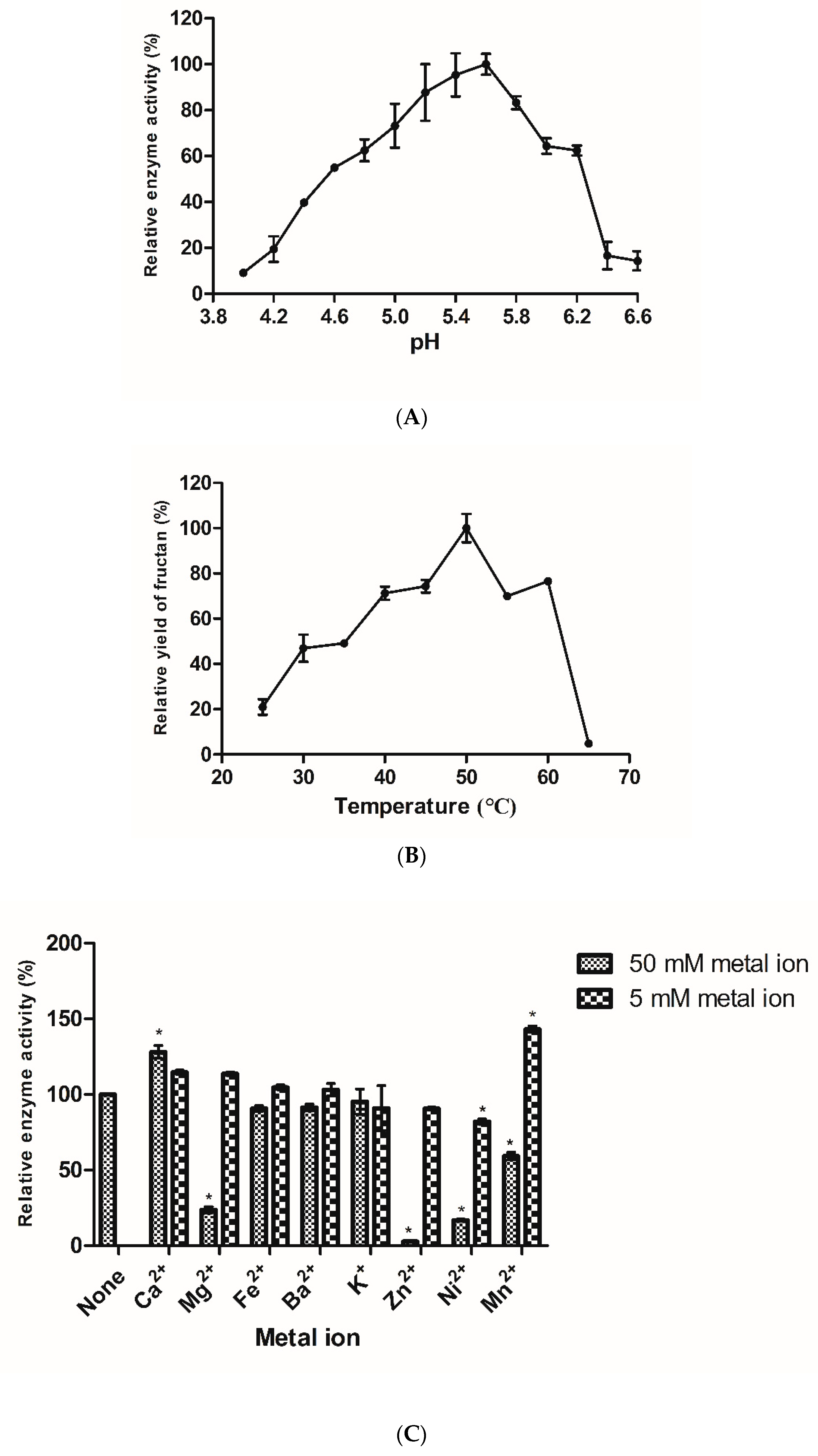
3.4. Effect of pH, Temperature, and Ions on the Activity of Recombinant Fructan Sucrase

3.4.1. Effect of pH on the Activity of Fructan Sucrase

Under the optimal induction conditions, the secreting effect of the clone with the signal peptide gene sequence of E. coli was almost the same as that of the clone with the signal peptide gene sequence of B. subtilis, so the BSO recombinant expression vectors were selected to study the properties of secreted fructan sucrase. As shown in Figure 3A, maximal activity was achieved at pH 5.6. From pH 4.0–5.6, the activity of fructan sucrase increased with the increase in pH, and the highest value was observed at pH 5.6. When the pH was lower than 4.6, the activity of fructan sucrase was low (<50% of the maximum). The fructan sucrase of B. subtilis ZW019 was adapted to the slightly acidic pH, which was similar to the conclusion of a fructan sucrase produced by other strains [43].

3.4.2. Effect of Temperature on the Activity of Fructan Sucrase

As shown in Figure 3B, from 25–50 °C, fructan sucrase activity increased with increasing temperature, while activity decreased sharply when the temperature was increased from 50–65 °C. The optimum temperature was 50 °C, and the enzyme activity was almost completely lost at 65 °C, indicating that fructan sucrase was not resistant to high temperatures and was sensitive to heat.

3.4.3. Effect of Ions on the Activity of Recombinant Fructan Sucrase

Generally, metal ions have a great influence on protein folding and catalytic processes [44]. As shown in Figure 3C, the following metal ions only slightly inhibited enzyme activity at low concentrations: K+, Zn2+, and Ni2+. On the other hand, Ca2+, Mg2+, and Mn2+ strongly promoted fructan sucrase activity, especially Mn2+, which increased the fructan sucrase activity by approximately 143%. Ba2+ and K+ at low concentrations had little influence on the activity of fructan sucrase. At higher concentrations, Mg2+, Zn2+, Ni2+, and Mn2+ significantly inhibited the enzyme activity, Fe2+, Ba2+, and K+ slightly inhibited the activity of fructan sucrase, and Ca2+ promoted the enzyme activity. The divalent metal cations play a key role in the extracellular expression of recombinant protein in E. coli [23,45].

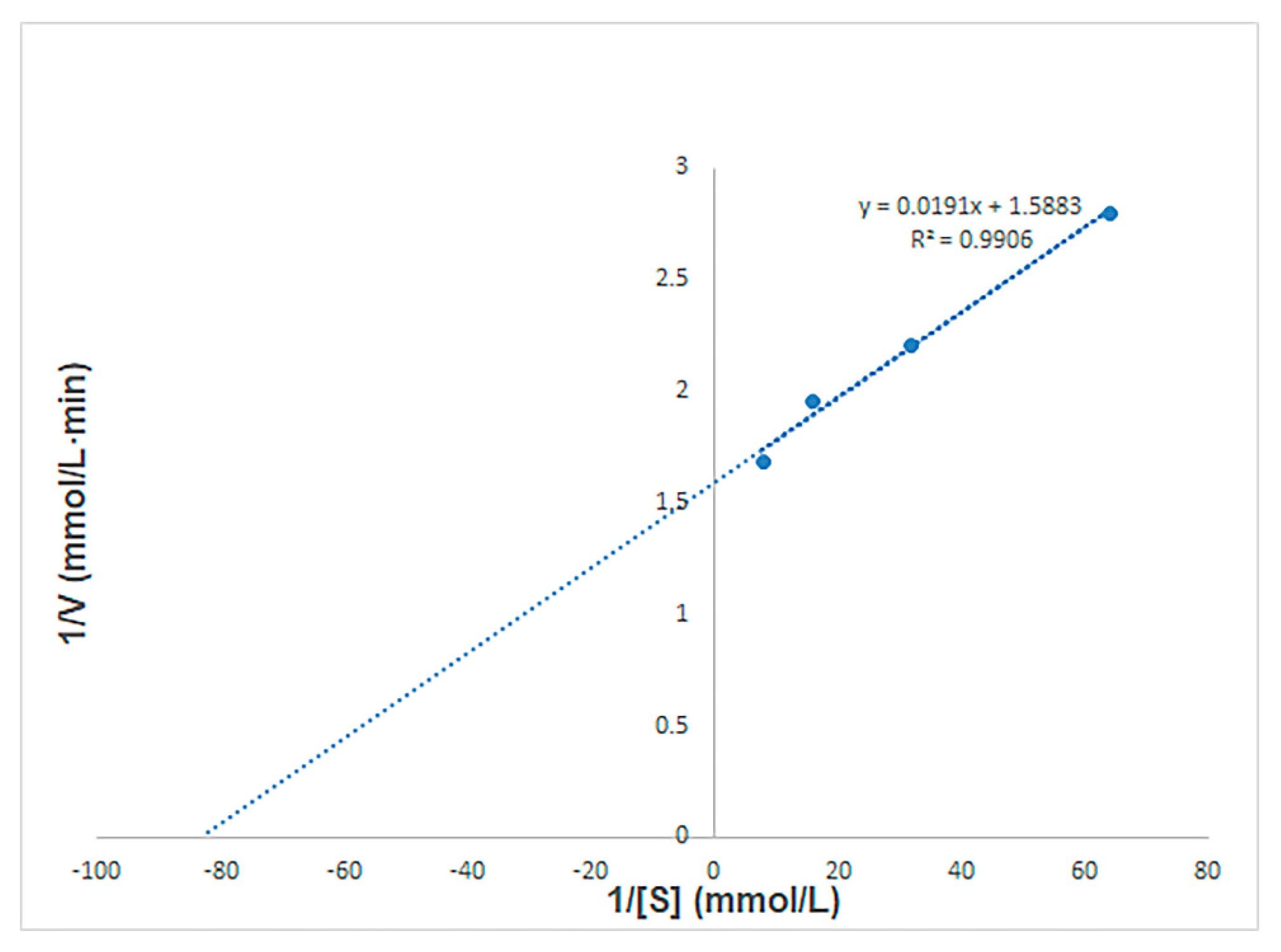
3.5. The Kinetic Parameters of Recombinant Fructan Sucrase

The Km and Vmax values were detected by nonlinear fit analysis based on Eadie-Hofstee plots [46]. As shown in Figure 4, the initial reaction rate was determined with a sucrose concentration of 15.6–200 mM. Recombinant fructan sucrase hydrolyzed sucrose at 50 °C, the Michaelis–Menten constant (Km) was 33.96 g/L, and the maximum reaction rate (Vmax) was 0.63 g/(L·min). According to a recently published review of fructan sucrase [47,48], it still suggests that fructan sucrase in this study has a special catalytic ability to transform sucrose as a substrate.

4. Conclusions

In the study, a difficult to isolate and purity fructan sucrase produced wild B. subtilis strain was firstly expressed extracellularly in E. coli. Signal peptides suitable for the secretion of fructan sucrase in E. coli were found. The signal peptides from E. coli and B. subtilis both enabled the secretion of recombinant proteins into the extracellular environment. Extracellular fructan sucrase was purified and immobilized by GEM particles in one step. The effect of pH, temperature, and ions on the activity of recombinant fructan sucrase were determined. The work presented herein illustrates a useful method to easily and efficiently get fructan sucrase, which will provide a basis for the enzymology of bacillus fructan sucrase and its industrial application.

Author Contributions

J.W., conceptualization, methodology, investigation, and writing—original draft; H.X., data curation, investigation, formal analysis, and writing—review and editing. F.Z., investigation, writing—review and editing, and formal analysis; B.Z., software, validation, and supervision; M.X., validation, supervision, and software; Z.Z., supervision and software; Y.H., project administration, resources, supervision, and funding acquisition. All authors have read and agreed to the published version of the manuscript.

Funding

This research was funded by the Key Technology R & B Program of Tianjin, China, grant number 19YFZCSN00100.

Institutional Review Board Statement

Not applicable.

Informed Consent Statement

Not applicable.

Data Availability Statement

Data is in agreement with the MDPI Research Data Policies. Most of the data are available in this manuscript. More specific data can be requested from the corresponding author.

Acknowledgments

This work was financially supported by the Key Technology R & B Program of Tianjin, China (19YFZCSN00100).

Conflicts of Interest

The authors declare no conflict of interest. The funders had no role in the design of the study; in the collection, analyses, or interpretation of data; in the writing of the manuscript, or in the decision to publish the results.

References

  1. Belghith, K.S.; Dahech, I.; Hamden, K.; Feki, A.; Mejdoub, H.; Belghith, H. Hypolipidemic effect of diet supplementation with bacterial levan in cholesterol-fed rats. Int. J. Biol. Macromol. 2012, 50, 1070–1074. [Google Scholar] [CrossRef]
  2. Byun, B.Y.; Lee, S.J.; Mah, J.H. Antipathogenic activity and preservative effect of levan (β-2, 6-fructan), a multifunctional polysaccharide. Int. J. Food Sci. Technol. 2014, 49, 238–245. [Google Scholar] [CrossRef]
  3. Dahech, I.; Harrabi, B.; Hamden, K.; Feki, A.; Mejdoub, H.; Belghith, H.; Belghith, K.S. Antioxidant effect of nondigestible levan and its impact on cardiovascular disease and atherosclerosis. Int. J. Biol. Macromol. 2013, 58, 281–286. [Google Scholar] [CrossRef]
  4. Srikanth, R.; Siddartha, G.; Reddy, C.H.; Harish, B.S.; Ramaiah, M.J.; Uppuluri, K.B. Antioxidant and anti-inflammatory levan produced from Acetobacter xylinum NCIM2526 and its statistical optimization. Carbohydr. Polym. 2015, 123, 8–16. [Google Scholar] [CrossRef] [PubMed]
  5. Methus, K.; Rath, P.; Thanapon, C.; Karan, W.; Surasak, C. Computational design of oligosaccharide producing levansucrase from Bacillus licheniformis RN-01 to improve its thermostability for production of levan-type fructooligosaccharides from sucrose. Int. J. Biol. Macromol. 2020, 160, 252–263. [Google Scholar] [CrossRef]
  6. Srikanth, R.; Reddy, C.H.; Siddartha, G.; Ramaiah, M.J.; Uppuluri, K.B. Uppuluri review on production, characterization and applications of microbial levan. Carbohydr. Polym. 2015, 120, 102–114. [Google Scholar] [CrossRef] [PubMed]
  7. Ragab, T.I.M.; Shalaby, A.S.G.; Awdan, S.A.E.; El-Bassyouni, G.T.; Salama, B.M.; Helmy, W.A.; Esawy, M.A. Role of levan extracted from bacterial honey isolates in curing peptic ulcer: In vivo. Int. J. Biol. Macromol 2020, 142, 64–573. [Google Scholar] [CrossRef] [PubMed]
  8. Raveendran, S.; Parameswaran, B.; Ummalyma, S.B.; Abraham, A.; Mathew, A.K.; Madhavan, A.; Rebello, S.A. Pandey Applications of microbial enzymes in food industry. Food Technol. Biotechnol. 2018, 56, 16–30. [Google Scholar] [CrossRef] [PubMed]
  9. Lalitagauri, R.; Sunita, P.; Debabrata, B. Enzymes—An existing and promising tool of food processing industry. Recent Pat. Biotechnol. 2016, 10, 58–71. [Google Scholar] [CrossRef]
  10. Trujilloa, L.E.; Arrietaa, J.G.; Dafhnisa, F.; Garcíab, J.; Valdésb, J.; Tambarac, Y.; Pérezb, M.; Hernándeza, L. Fructo-oligosaccharides production by the Gluconacetobacter diazotrophicus levansucrase expressed in the methylotrophic yeast Pichia pastoris. Enzym. Microb Technol. 2001, 28, 139–144. [Google Scholar] [CrossRef]
  11. Gao, S.; Qi, X.; Hart, D.J.; Gao, H.; An, Y. Expression and characterization of levansucrase from Clostridium acetobutylicum. J. Agric. Food Chem. 2017, 65, 867–871. [Google Scholar] [CrossRef] [PubMed]
  12. Ni, D.; Xu, W.; Bai, Y.; Zhang, W.; Zhang, T.; Mu, W. Biosynthesis of levan from sucrose using a thermostable levansucrase from Lactobacillus reuteri LTH5448. Int. J. Biol. Macromol. 2018, 113, 29–37. [Google Scholar] [CrossRef] [PubMed]
  13. Xavier, J.R.; Ramana, K.V. Optimization of levan production by cold-active Bacillus licheniformis ANT 179 and fructooligosaccharide synthesis by its levansucrase. Appl. Biochem. Biotechnol. 2017, 181, 986–1006. [Google Scholar] [CrossRef] [PubMed]
  14. Iliev, I.; Vasileva, T.; Bivolarski, V.; Salim, A.; Morel, S.; Rabier, P.; Gabriel, V. Optimization of the expression of levansucrase L17 in recombinant E. coli. Biotechnol. Biotechnol. Equip. 2018, 32, 477–486. [Google Scholar] [CrossRef] [Green Version]
  15. Ishida, R.; Sakaguchi, K.; Matsuzaki, C.; Katoh, T.; Ishida, N.; Yamamoto, K.; Hisa, K. Levansucrase from Leuconostoc mesenteroides NTM048 produces a levan exopolysaccharide with immunomodulating activity. Biotechnol. Lett. 2016, 38, 681–687. [Google Scholar] [CrossRef]
  16. Liu, Q.; Yu, S.H.; Zhang, T.; Jing, B.; Mu, W.M. Efficient biosynthesis of levan from sucrose by a novel levansucrase from Brenneria goodwinii. Carbohydr. Polym. 2017, 157, 1732–1740. [Google Scholar] [CrossRef]
  17. Jong, W.S.; Saurí, A.; Luirink, J. Extracellular production of recombinant proteins using bacterial autotransporters. Curr. Opin. Biotechnol. 2010, 21, 646–652. [Google Scholar] [CrossRef]
  18. Choi, J.H.; Lee, S.Y. Secretory and extracellular production of recombinant proteins using Escherichia coli. Appl. Microbiol. Biotechnol. 2004, 64, 625–635. [Google Scholar] [CrossRef]
  19. Su, L.; Xu, C.; Woodard, R.W.; Chen, J.; Wu, J. A novel strategy for enhancing extracellular secretion of recombinant proteins in Escherichia coli. Appl. Microbiol. Biotechnol. 2013, 97, 6705–6713. [Google Scholar] [CrossRef]
  20. Sambasivam, G.; Nair, S.; Karthikeyan, G. Effect of codon-optimized E-coli signal peptides on recombinant Bacillus stearothermophilus maltogenic amylase periplasmic localization, yield and activity. J. Ind. Microbiol. Biotechnol. 2014, 41, 1435–1442. [Google Scholar] [CrossRef]
  21. Chen, J.; Gai, Y.; Fu, G.; Zhou, W.; Zhang, D.; Wen, J. Enhanced extracellular production of α-amylase in Bacillus subtilisby optimization of regulatory elements and over-expression of PrsA lipoprotein. Biotechnol. Lett. 2015, 37, 899–906. [Google Scholar] [CrossRef] [PubMed]
  22. Angkawidjaja, C.; Kuwahara, K.; Omori, K.; Koga, Y.; Takano, K.; Kanaya, S. Extracellular secretion of Escherichia coli alkaline phosphatase with a C-terminal tag by type I secretion system: Purification and biochemical characterization. Protein. Eng. Des. Sel. 2006, 19, 337–343. [Google Scholar] [CrossRef] [PubMed]
  23. Li, Z.F.; Li, B.; Liu, Z.G.; Wang, M.; Gu, Z.B.; Du, G.C.; Wu, J.; Chen, J. Calcium leads to further increase in glycine-enhanced extracellular secretion of recombinant alpha-cyclodextrin glycosyltransferase in Escherichia coli. J. Agric. Food Chem. 2009, 57, 6231–6237. [Google Scholar] [CrossRef] [PubMed]
  24. Matos, C.F.R.O.; Branston, S.D.; Albiniak, A.; Dhanoya, A.; Robinson, C. High-yield export of a native heterologous protein to the periplasm by the Tat translocation pathway in Escherichia coli. Biotechnol. Bioeng. 2012, 109, 2533–2542. [Google Scholar] [CrossRef] [PubMed]
  25. Kujau, M.J.; Hoischen, C.; Riesenberg, D.; Gumpert, J. Expression and secretion of functional miniantibodies McPC603scFvDhlx in cell-wall-less L-form strains of Proteus mirabilisand Escherichia coli: A comparison of the synthesis capacities of L-form strains with an E. coli producer strain. Appl. Microbiol. Biotechnol. 1998, 49, 51–58. [Google Scholar] [CrossRef]
  26. Lech, M. Optimisation of protein-free waste whey supplementation used for the industrial microbiological production of lactic acid. Biochem. Eng. J. 2020, 157, 107531. [Google Scholar] [CrossRef]
  27. Wood, E.J. Molecular cloning, a laboratory manual. Biochem. Educ. 1983, 11, 182–183. [Google Scholar] [CrossRef]
  28. Liu, S.L.; Du, K.; Chen, W.Z.; Liu, G.; Xing, M. Effective approach to greatly enhancing selective secretion and expression of three cytoplasmic enzymes in Escherichia coli through synergistic effect of EDTA and lysozyme. J. Ind. Microbiol. Biotechnol. 2012, 39, 1301–1307. [Google Scholar] [CrossRef]
  29. Guo, Y.; Bian, W.; Zhang, Y. Expression in Escherichia coli, purification and characterization of LRSAM1, a LRR and RING domain E3 ubiquitin ligase. Protein Expres Purif. 2017, 129, 158–161. [Google Scholar] [CrossRef]
  30. Zhao, F.K.; Song, Q.Z.; Wang, B.B.; Du, R.P.; Han, Y.; Zhou, Z.J. Secretion of the recombination α-amylase in Escherichia coli and purification by the gram-positive enhancer matrix (GEM) particles. Int. J. Biol. Macromol. 2019, 123, 91–96. [Google Scholar] [CrossRef]
  31. Li, P.C.; Qiao, X.W.; Zheng, Q.S.; Hou, J.B. Immunogenicity and immunoprotection of porcine circovirus type 2 (PCV2) Cap protein displayed by Lactococcus lactis. Vaccine 2016, 34, 696–702. [Google Scholar] [CrossRef] [PubMed]
  32. Saluja, V.; Visser, M.R.; Veer, W.T.; Roosmalen, M.L.V.; Leenhouts, K.; Hinrichs, W.L.J.; Huckriede, A.; Frijlink, H.W. Influenza antigen-sparing by immune stimulation with Gram-positive enhancer matrix (GEM) particles. Vaccine 2010, 28, 7963–7969. [Google Scholar] [CrossRef] [PubMed]
  33. Wang, P.L.; Qin, W.T.; Xu, J.T.; Yan, Y.R.; Tian, J.; Wu, N.F.; Yao, B. Enhancing the soluble expression of an amylase in Escherichia coli by the mutations related to its domain interactions. Protein Expr. Purif. 2016, 120, 35–41. [Google Scholar] [CrossRef] [PubMed]
  34. Sumitomo, N.; Ozaki, K.; Kawai, S.; Ito, S. Nucleotide sequence of the gene for an alkaline endoglucanase from an Alkalophilic Bacillus and its expression in Escherichia coli and Bacillus subtilis. Biosci. Biotechnol. Biochem. 1992, 6, 872–877. [Google Scholar] [CrossRef]
  35. Solingen, P.; Meijer, D.; Kleij, W.A.; Barnett, C.; Bolle, R.; Power, S.D.; Jones, B.E. Cloning and expression of an endocellulase gene from a novel Streptomycete isolated from an East African soda lake. Extremophiles 2001, 5, 333–341. [Google Scholar] [CrossRef] [PubMed]
  36. Srensen, H.P.; Mortensen, K.K. Advanced genetic strategies for recombinant protein expression in Escherichia coli. J. Biotechnol. 2005, 115, 113–128. [Google Scholar] [CrossRef]
  37. Meng, F.Q.; Zhao, H.Z.; Zhang, C.; Lu, F.X.; Bie, X.M. Expression of a novel bacteriocin—The plantaricin Pln1—In Escherichia coli and its functional analysis. Protein Expr. Purif. 2016, 119, 85–93. [Google Scholar] [CrossRef]
  38. Laymon, R.A.; Adney, W.S.; Mohagheghi, A.; Himmel, M.E.; Thomas, S.R. Cloning and expression of full-length Trichoderma reesi cellobiohydrolase I cDNAs in Escherichia coli. Appl. Biochem. Biotechnol. 1996, 57–58, 389–397. [Google Scholar] [CrossRef]
  39. Tang, B.; Pan, H.; Zhang, Q.; Ding, L. Cloning and expression of cellulase gene EG1 from Rhizopus stolonifer var. reflexus TP-02 in Escherichia coli. Bioresour. Technol. 2009, 100, 6129–6132. [Google Scholar] [CrossRef]
  40. Zhao, B.; Du, R.; Wang, J.Y.; Xu, M.; Han, Y.; Zhou, Z.J. Purification and biochemical characterization of a novel glucansucrase from Leuconostoc citreum B-2. Biotechnol. Lett. 2020, 42, 1535–1545. [Google Scholar] [CrossRef]
  41. Liu, Y.; Huang, L.; Guo, W.; Jia, L.B.; Fu, Y.; Gui, S.; Lu, F.P. Cloning, expression, and characterization of a thermostable and pH-stable laccase from Klebsiella pneumoniae and its application to dye decolorization. Process Biochem. 2016, 53, 125–134. [Google Scholar] [CrossRef]
  42. Malik, B.; Rashid, N.; Ahmad, N.; Akhtar, M. Escherichia coli signal peptidase recognizes and cleaves the signal sequence of α-amylase originating from Bacillus licheniformis. Biochemistry (Moscow) 2013, 78, 958–962. [Google Scholar] [CrossRef] [PubMed]
  43. Hövels, M.; Kosciow, K.; Kniewel, J.; Jakob, F.; Deppenmeier, U. High yield production of levan-type fructans by Gluconobacter japonicus LMG 1417. Int. J. Biol. Macromol. 2020, 164, 295–303. [Google Scholar] [CrossRef] [PubMed]
  44. Oliveira, A.N.D.; Oliveira, L.A.D.; Andrade, J.S. Partial characterization of amylases of two Indigenous Central Amazonian Rhizobia Strains. Braz. Arch. Biol. Techn. 2010, 53, 35–45. [Google Scholar] [CrossRef] [Green Version]
  45. Ding, R.; Li, Z.; Chen, S.; Wu, D.; Wu, J.; Chen, J. Enhanced secretion of recombinant α-cyclodextrin glucosyltransferase from E. coli by medium additives. Process. Biochem. 2010, 45, 880–886. [Google Scholar] [CrossRef]
  46. Fuhrmann, G.F.; Völker, B. Misuse of graphical analysis in nonlinear sugar transport kinetics by Eadie-Hofstee plots. Biochim. Biophys. Acta 1993, 1145, 180–182. [Google Scholar] [CrossRef]
  47. Xu, W.; Ni, D.; Zhang, W.; Guang, C.; Zhang, T.; Mu, W. Recent advances in Levansucrase and Inulosucrase: Evolution, characteristics, and application. Crit. Rev. Food Sci. Nutr. 2019, 59, 3630–3647. [Google Scholar] [CrossRef]
  48. Hill, A.; Chen, L.; Mariage, A.; Petit, J.L.; Berardinis, V.D.; Karboune, S. Discovery of new levansucrase enzymes with interesting properties and improved catalytic activity to produce levan and fructooligosaccharides. Catal. Sci. Technol. 2019, 9, 2931–2944. [Google Scholar] [CrossRef]
Figure 1. Effect of temperature, time, and IPTG concentration on the expression of BS, BSO, and BS-FF. (A) Effect of temperature and time on the expression of BS, BSO, and BS-FF. The expression of recombinant vectors in E. coli was induced by 0.5 mM IPTG, and cultures were incubated at different temperatures and times. (B) Effect of IPTG concentration on the expression of BS, BSO, and BS-FF. The expression of recombinant vectors in E. coli was induced by different concentrations of the inducer IPTG, and cultures were incubated at the optimal temperature of 28 °C for 8 h. The values are representative results obtained from at least three experiments. Error bars indicate standard deviations (SD) based on more than three independent experiments. * p < 0.01, t-test.
Processes 09 00095 g001
Figure 2. SDS-PAGE gels stained with Coomassie Brilliant Blue. (A) The fructan sucrase secreted by BSO was purified by GEM. Lanes: M, molecular mass standards; (1) extracellular fraction of cells producing recombinant fructan sucrase with an E. coli signal peptide; (2) purified extracellular recombinant fructan sucrase produced with a signal peptide; (3) GEM particles; (4) intracellular fraction of cells producing recombinant fructan sucrase with an E. coli signal peptide. The arrow indicates recombinant fructan sucrase. (B) The fructan sucrase secreted by BS-FF was purified by GEM. Lanes: M, molecular mass standards; (1) extracellular fraction of cells producing recombinant fructan sucrase with a B. subtilis signal peptide; (2) fructan sucrase purified from extracellular fraction. The arrow indicates recombinant fructan sucrase; (3) GEM particles; (4) intracellular fraction of cells producing recombinant fructan sucrase with a B. subtilis signal peptide. The arrow indicates fructan sucrase.
Processes 09 00095 g002
Figure 3. Effect of pH, temperature, metal ions on the activity of recombinant fructan sucrase. (A) Effect of pH on the activity of recombinant fructan sucrase; (B) effect of temperature on the activity of recombinant fructan sucrase; (C) effect of metal ions on the activity of recombinant fructan sucrase. The values are representative results obtained from at least three experiments. Error bars indicate standard deviations (SD) based on more than three independent experiments. * p < 0.01, t-test.
Processes 09 00095 g003
Figure 4. Michaelis–Menten kinetic parameters.
Processes 09 00095 g004
Table 1. The sequence of primers.
NamePrimer Sequence
Primers of cloning of fructan sucrase gene from B. subtilis
BS-F5′-CCGCGGATCCAAAGAAACGAACCAAAAG-3′ (containing BamHIsite)
BS-R5′-CCGCCTGCAGTTTGTTAACTGTTAATTG-3′ (containing PstIsite)
Primers of cloning of fructan sucrase and secretory signal peptide gene from B. subtilis
BS-FF-F5′-CCGCGGATCCAACATCAAAAAGTTTGC-3′ (containing BamHIsite)
BS-FF-R5′-CCGCCTGCAGTTTGTTAACTGTTAATTG-3′ (containing PstIsite)
Primers of recombinant expression vectors of fructan sucrase from B. subtilis and signal peptide genes secreted by E. coli
BSO-F5′-CGCGGATCCAAAAAAACCGCTATCGCTATCGCTGTTGCTCTGGCTGGTTTCGCTA
CCGTTGCTCAGGCTAAAGAAACGAACCAAAAG-3′ (containing BamHIsite)
BSO-R5′-CCGCCTGCAGTTTGTTAACTGTTAATTG-3′ (containing PstIsite)
Publisher’s Note: MDPI stays neutral with regard to jurisdictional claims in published maps and institutional affiliations.

