Article

Lipid Isolation Process and Study on Some Molecular Species of Polar Lipid Isolated from Seed of Madhuca ellitica

1
Institute of Natural Products Chemistry, Vietnam Academy of Science and Technology (VAST), 18 Hoang Quoc Viet road, Caugiay district, Hanoi 10000, Vietnam
2
Graduate University of Science and Technology, Vietnam Academy of Science and Technology (VAST), 18 Hoang Quoc Viet road, Caugiay district, Hanoi 10000, Vietnam
3
A.V. Zhirmunsky Institute of Marine Biology, Far-Eastern Branch of the Russian Academy of Sciences, Vladivostok 690041, Russia
4
School of Chemistry, Biology and Environment, Vinh University, Vinh City 46000, Vietnam
5
Working Group of Lipid Research, Department of Safety and Quality of Cereals Max, Federal Research Institute for Nutrition and Food, Rubner-Institut, Schutzenberg 12, DE-32756 Detmold, Germany
6
NTT Hi-Tech Institute, Nguyen Tat Thanh University, Ho Chi Minh City 70000, Vietnam
7
Center of Excellence for Biochemistry and Natural Products, Nguyen Tat Thanh University, Ho Chi Minh City 70000, Vietnam
*
Authors to whom correspondence should be addressed.
Processes 2019, 7(6), 375; https://doi.org/10.3390/pr7060375
Submission received: 18 May 2019 / Revised: 6 June 2019 / Accepted: 10 June 2019 / Published: 17 June 2019
(This article belongs to the Special Issue Green Separation and Extraction Processes)

Abstract

:
This study attempted the lipid extraction process from the seeds of Madhuca ellitica, a lipid-rich plant, and conducted a lipidomic analysis on molecular species of the obtained product. Total lipids of the crude seeds were found to contain 11.2% of polar lipids. The major fatty acids (FAs) of the polar lipids were palmitic (16:0), stearic (18:0), oleic (18:1n-9), and linoleic (18:2n-6) acids, which amounted to 28.5, 12.5, 44.8, and 13.2% of total FAs, respectively. The content and chemical structures of individual molecular species of phosphatidylglycerol (PG), phosphatidylethanolamine (PE), phosphatidylcholine (PC), phosphatidylinositol (PI), phosphatidic acid (PA), and sulfoquinovosyldiacylglycerol (SQDG) were determined by HPLC with a tandem high-resolution mass spectrometry (HRMS). The major molecular species were 18:1/18:2 PE, 16:0/18:1 PC, 18:1/18:2 PC, 16:0/18:2 PG, 16:0/18:1 PG, 16:1/18:1 PI, 16:0/18:1 PI, 18:0/18:2 PI, 16:0/18:1 PA, 18:1/18:2 PA, 16:0/18:1 SQDG, and 18:0/18:1 SQDG. The application of a tandem HRMS allows us to determine the content of each isomer in pairs of the monoisotopic molecular species, for example, 18:0/18:2 and 18:1/18:1. The evaluation of the seed polar lipid profile will be helpful for developing the potential of this tree for nutritive and industrial uses.

1. Introduction

Buttercup tree or mahua, Madhuca elliptica (Pierre ex Dubard) H.J.Lam, Madhuca longifolia (Koenig) J.F. Macb (Synonyms, Madhuca indica Gmelin, Madhuca latifolia Macb., Bassia latifolia Roxb.; Family, Sapotaceae) is a commercially important tree cultivated throughout the subtropical region of the Indo-Pak subcontinent and regarded highly for its flowers, fruits, timber, and seeds [1,2,3]. Previous reports have pointed out a myriad of valuable compounds, especially compounds of low molecular-weight including saponins, carbohydrates, triterpenoids, steroids, flavonoids, and glycosides, have been isolated from Madhuca [4,5,6,7]. Among these compounds, carotinoids, existing dominantly in the seeds of Madhuca, figure in certain medicinal applications for treatments of skin disease, rheumatism, headache, piles, and use as a laxative [8,9,10]. The Madhuca seed is also rich in fat (up to 58%) [11,12], suggesting potential use of the seed in the manufacture of laundry soaps, lubricants and biodiesel [13,14].
The base of the oil seeds of the plant is constituted of triacylglycerols (TG), which are neutral lipids. Also, natural seed fats contain a number of polar lipids, which are usually separated and lost during oil manufacturing. Among polar lipids, phospholipids hold great importance due to their nutritional value and bioactivities [15]. Each phospholipid class is a complex mixture of individual compounds, termed as “molecular species”. Structures of phospholipids consists of the same polar group but different acyl chains of their molecules, which defines their nutritional value and bioactivities of phospholipid molecular species. Therefore, unambiguous identification of molecular species profile of the oilseed, through a lipidomic approach, may introduce possible application in the cosmetic, nutraceutical, and pharmaceutical industries [16,17]. Even though plant lipids and TG molecular species have been studied extensively in terms of compositional structure [18,19,20], information on the molecular species composition of phospholipids is scanty. For M. elliptica, previous studies have revealed its TG molecular species profile in seed fat and its solid and liquid fractions [21]. In addition, fatty acid (FA) composition of polar lipids and individual phospholipid class content of mahua butter from M. elliptica have been described [10]. The present work aimed to identify the chemical structure and quantitative composition of phospholipid molecular species from the M. elliptica seeds. The molecular species of sulfoquinovosyldiacylglycerols (SQDG), which contain a charged chemical group like phospholipid molecules, were also investigated. High performance liquid chromatography (HPLC), in tandem with high -resolution mass spectrometry (HRMS), was used as a lipidomic method [22]. Some important papers concerning molecular species of polar lipids in marine species, such as corals and red (hot) algae, have been recently published. For example, a previous study that investigated the distribution of tetracosapolyenoic acids (TPA) in molecular species of different phospholipid (PL) classes in the soft corals Sinularia macropodia and Capnella sp. from shallow waters of Vietnam showed some interesting results [23]. To be specific, Phosphatidylethanolamine (PE), phosphatidylcholine (PC), phosphatidylserine (PS), and phosphatidylinositol (PI) were found to be major PL classes of S. macropodia and Capnella sp. and more than 32 molecular species of these four PL classes were determined by high-resolution tandem mass spectrometry. The major molecular species of PL in both coral species were 18:1e/20:4 PE, 18:0e/20:4 PC, 18:0e/24:5 PS, and 18:0/24:5 PI. In two model red algae, Polysiphonia sp. and Porphyridium sp., forms of monogalactosyldiacylglycerol (MGDG) and digalactosyldiacylglycerol (DGDG), the two most commonly found galactolipids in chloroplast membranes, were determined via positive-ion electrospray ionization/mass spectrometry (ESI/MS) and ESI/MS/MS [24]. In the study of Honda et al. (2019) the characteristics of glycerolipids, which are the substrates of eicosanoids production of A. chilensis, were investigated and compared to the reported values of A. vermiculophyllum. The results showed that monogalactosyldiacylglycerol (MGDG), digalactosyldiacylglycerol (DGDG), sulfoquinovosyldiacylglycerol (SQDG), and phosphatidylcholine (PC) were the major lipid classes in A. chilensis and accounted for 44.4% of the total lipid extract [25].

2. Materials and Methods

2.1. Materials

Ripe fruits of M. ellitica (1000 g) were collected from a plantation in Binh Duong province of Vietnam in February 2016 and were botanically identified by Dr. Nguyen Quoc Binh, Vietnam National Museum of Nature—Vietnam Academy of Science and Technology. Seeds were cleaned, dried in cabinet dryer at 50 °C for 12 h, and stored at 4 °C after collection. Water content (%) in the M. ellitica seed was 10.27%. To prepare the sample for analysis, 100 g of sample seeds were finely ground using an electric grinder (KIKA Labortechnik M20), after which they were immediately subjected to lipid extraction. Neutral lipid standards, phospholipid standards (phosphatidylglycerol (PG), phosphatidylethanolamine (PE), phosphatidylcholine (PC), phosphatidylinositol (PI), phosphatidic acid (PA)), and sulfoquinovosyldiacylglycerol (SQDG) were purchased from Sigma-Aldrich Co. (St. Louis, MO, USA).

2.2. Extraction of Total Lipids (TL)

M. ellitica powder was then extracted for TL following a previously described method of Folch et al. (1957) [26] in which a Soxhlet extractor in combination with a solvent mixture of CHCl3/MeOH (2:1, v/v) and aqueous solution of sodium chloride (0.75%) were used. After being separated overnight at 4 °C, the CHCl3 layer was recovered and dehydrated with sodium sulfate. The extract was filtered and subjected to a rotary evaporator at 40 °C, and TL obtained were dissolved in CHCl3 and stored at −20 °C. The content of TL was determined gravimetrically.

2.3. Lipid Fractionation

A column incorporated with 100 g of silica gel 60 (70–230 mesh, Sigma-Aldrich Co., St. Louis, MO, USA) was loaded with TL (3.4 g). Neutral lipids, glycolipids, and phospholipids were eluted with CHCl3 (450 mL), acetone (230 mL), MeOH (60 mL), and MeOH/water (200 mL, 95:5, v/v). The MeOH-contained fractions were combined, taken to dryness, and polar lipids obtained were dissolved in CHCl3 and stored at −20 °C.

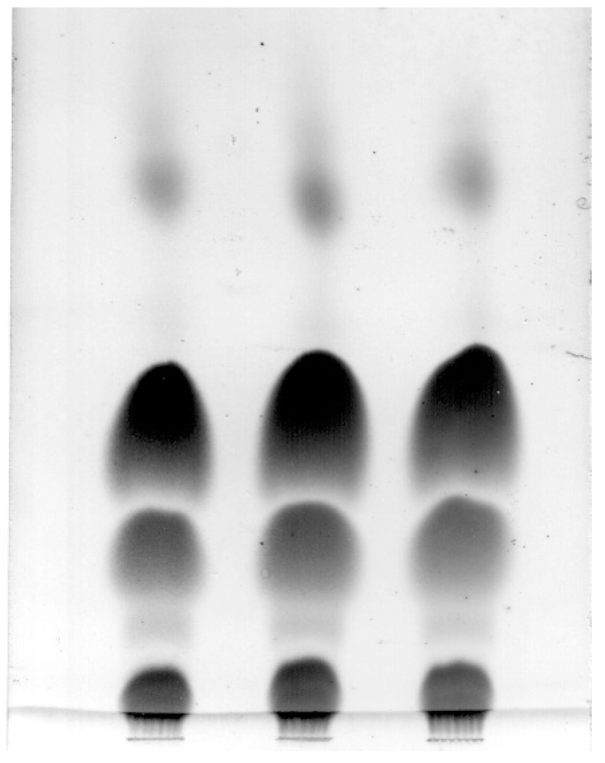
2.4. Analysis of Lipid Class Compositions

One-dimensional thin layer chromatography (TLC) using precoated silica gel plates (6 cm × 6 cm) Sorbfil PTLC-AF-V (Sorbfil, Krasnodar, Russia) was employed to analyze TL composition. The development of plates commenced with n-hexan/Et2O/AcOH (70:20:1, v/v/v) to their full length, followed with CHCl3/MeOH/C6H6/NH4OH (65:30:5:10, v/v/v) to 25% length. Following that, plates were subjected to a stream of air for drying, sprayed with 10% H2SO4 in MeOH and heated at 240 °C for 10 min. An image scanner (Epson Perfection 2400 PHOTO) operating in grayscale mode was used to record the chromatograms. An image analysis software (Sorbfil TLC Videodensitometer, Krasnodar, Russia) was used to determine percentages of lipid contents based on band intensity. Peak areas and lipid class percentages were calculated according to Hamoutene (2008) [27]. In the first and second direction of separation of polar lipids using two-dimensional silica gel TLC, CHCl3/MeOH/C6H6/28% NH4OH (65:30:10:6, v/v/v) and CHCl3/MeOH/AcOH/CH3COCH3/C6H6/H2O (70:30:4:5:10:1, v/v/v) were used, respectively. Identification of phospholipids on TLC plates was made using aforementioned authentic standards and the specific spray reagents [28]. The phospholipid content was evaluated with spectrophotometry following the digestion with perchloric acid [29].

2.5. Analysis of Lipid Class Compositions

To obtain fatty acid methyl esters (FAMEs), the lipids, contained in a screw cap vial, were treated with 2% H2SO4 in MeOH for 2 h at 80 °C under Ar. The purification was performed by TLC development in benzene. The preparation of 4,4-Dimethyloxazoline (DMOX) derivatives of FAs from FAMEs follows the study of Svetashev (2011) [30].
FAMEs were analyzed by gas chromatography (GC) employed on a Shimadzu GC-2010 chromatograph (Kyoto, Japan) at 210 °C. GC was performed in tandem with a flame ionization detector on a SUPELCOWAX 10 (Supelco, Bellefonte, PA, USA) capillary column (30 m × 0.25 mm× 0.25 µm). Temperatures of both injector and detector were maintained at 240 °C. Carrier gas was He at 30 cm/s. The identification of FAMEs was made by comparison with authentic standards (Supelco 37 Component FAME Mix; Supelco, Bellefonte, PA, USA) and a table of equivalent chain-lengths [31].
To identify structural composition of FAs, corresponding methyl esters and DMOX derivatives were analyzed by gas chromatography mass spectrometry (GC–MS) on a Shimadzu GCMS-2010 instrument (Kyoto, Japan) (electron impact at 70 eV) with a MDN-5s (Supelco, Bellefonte, PA, USA) capillary column (30 m × 0.25 mm ID). Carrier gas was He at 30 cm/s. The temperature parameter for analysis of FAMEs started at 160 °C, followed by an increase of 2 °C/min to 240 °C that was held for 20 min. Temperatures for both injector and detector temperatures were maintained at 250 °C. For analysis of GC–MS of DMOX derivatives, the temperature started at 210 °C, increased by 3 °C/min to 270 °C that was held for 40 min. The injector and detector temperatures were 250 °C for this analysis. The mass spectra of FAMEs were compared with the Mass Spectral Library: WILEY275.L and NIST 98 [32].

2.6. Analysis of Molecular Species of Polar Lipids

Determination of chemical structures and molecular species of PL was performed by high performance liquid chromatography, in conjunction with high-resolution mass spectrometry (HPLC−HRMS). In the HPLC separation of the polar lipids, content of Et3N/AcOH (0.08:1, v/v) was fixed in the solvent system [33]. This permits an efficient electrospray ionization (ESI) and stabilizes ion signal by the simultaneous registration of positive and negative ions. A Shimadzu Prominence liquid chromatograph (Kyoto, Japan) was used to perform the HPLC−HRMS analysis of polar lipids. The instrument was equipped with two LC-20AD pump units, a high pressure gradient forming module, CTO-20A column oven, SIL-20A auto sampler, CBM-20A communications bus module, DGU-20A3 degasser, and a Shim-Pack diol column (50 mm × 4.6 mm ID, 5 µm particle size) (Shimadzu, Kyoto, Japan). Two solvent mixtures, A and B, formed the binary solvent gradient for HPLC separation. The mixture A consisted of n-hexane/2-propanol/AcOH/Et3N (82:17:1:0.08, v/v/v/v) and mixture B consisted of 2-propanol/H2O/AcOH Et3N (85:14:1:0.08, v/v/v/v). The acceleration of gradient started at 5% of mixture B and reached 80% throughout the course of 25 min. After 1 min, the composition changed to 5% of mixture B over 10 min and maintained at 5% for another 4 min (the total run time was 40 min). The flow rate was 0.2 mL/min. Determination of lipids was performed by a high resolution tandem ion trap time of flight mass spectrometry with a Shimadzu LCMS-IT-TOF instrument (Kyoto, Japan). The instrument operates at both positive and negative ion mode during each analysis under ESI conditions. Temperature for ion source was set to 200 °C. The range of detection was m/z 100–1200. Negative and positive modes had their potential set at-3.5 and 4.5 kV, respectively. The drying gas (N2) pressure was 200 kPa. The nebulizer gas (N2) flow was 1.5 L/min. For identification of lipids, authentic standards were compared with the samples, which was performed using Shimadzu LCMS Solution control and processing software (v.3.60.361). Molecular species of each phospholipid class were determined by HRMS fragmentation pathways in comparison with standards [30]. Individual molecular species within each polar lipid class were quantified with respect to the peak areas for the individual extracted ion chromatograms [34].

3. Results

3.1. Total Lipid Composition

Total lipids (TL) constituted 34.0 ± 0.2% of the seed. The composition of TL is presented in Table 1 and Figure 1. The level of triacylglycerols (TG) was highest (63.2% of TL), followed by waxes (WX, 9.28% of TL), free fatty acids (FFA, 6.14% of TL), and diacylglycerols (DG, 5.17% of TL). Polar lipids constituted 11.2% of TL and contained glycolipids (GL, 6.78% of TL) and phospholipids (PL, 4.45% of TL). Five major classes were found in the total PL including Phosphatidylcholine (PC), phosphatidylethanolamine (PE), phosphatidylinositol (PI), phosphatidylglycerol (PG), and phosphatidic acid (PA). These five classes amounted to 13.2, 13.1, 7.7, 4.9, and 2.9% of total PL, respectively.

3.2. Fatty Acid Composition of Total Lipids and Lipid Fractions

The FA composition of TL and their fractions, obtained from of M. elliptica seeds, is presented in Table 2. Oleic (18:1n-9), palmitic (16:0), stearic (18:0), and linoleic (18:2n-6) acids were the major FAs, collectively composing more than 98% of the total identified FAs. In general, the prevalence of unsaturated FAs is the major characteristic of the FA composition of TL. Oleic acid was the main FA (49.5%) followed by palmitic acid. The FA profiles of TL, neutral, and polar lipid fractions were similar. In addition, low levels of myristic (14:0), α-linolenic (18:3n-3), and arachidic (20:0) acids were detected.

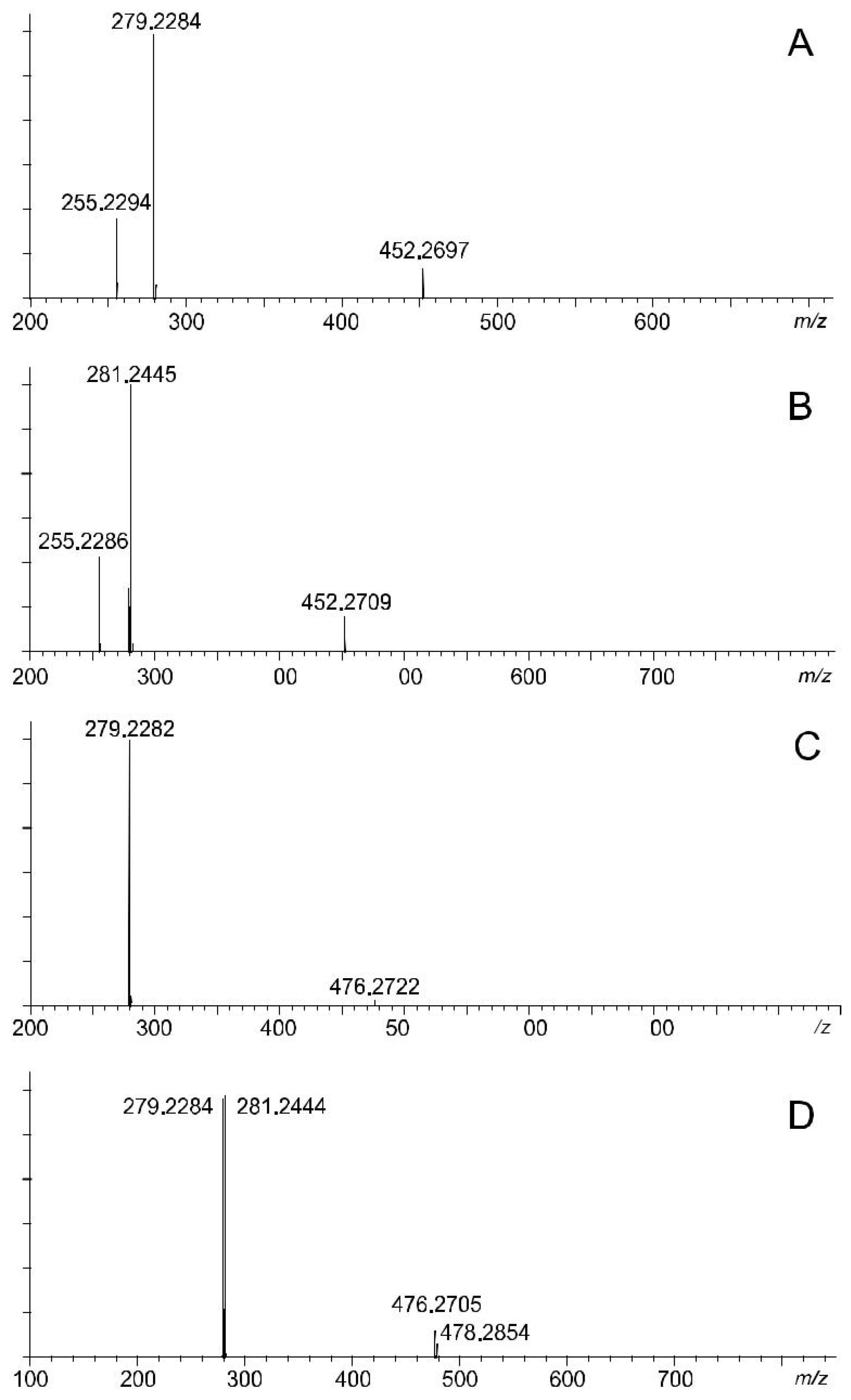
3.3. Chemical Structure of Molecular Species of Polar Lipids

The phospholipid classes of M. ellitica seeds were separated by HPLC−HRMS. The retention times of different molecular species of PE, PG, PA, PC, and PI were 5.90–6.87 min, 8.54–8.89 min, 9.18–9.37 min, 9.84–10.35 min, and 12.31–13.28 min, respectively. Among molecular species, four species including PE, PG, PA, and PI were revealed to be detectable at the MS2 stage, whereas PC species were identified at the MS3 stage [35]. HRMS spectra of PE molecular species are presented in detail as the examples of MS fragmentation (Figure 2A–D).
Phosphatidylethanolamine (PE). Six signals of negative quasi-molecular ions [M − H] at m/z 714.4988, 716.5163, 738.4975, 740.5140, 742.5296, and 744.5436, as well as six signals of positive cluster ions [M + H + (C2H5)3N]+ at m/z 817.6365, 819.6499, 841.6331, 843.6484, 845.6627, and 847.6749, were observed in the HRMS spectra of PE of M. ellitica seeds (Figure 1). Other less intensive peaks on Figure 1 corresponded to the signals of isotopic ions.
Six components were analyzed as described below. The MS2 spectrum of the ions [M − H] of component 1 (m/z 714.4988) (Table 3) contained the signal at m/z 452.2697 formed by the loss of neutral fragment of 262.2291 (C18H30O, calculated 262.2297) (Figure 2A). This neutral fragment corresponded to dehydrated 18:2 acid. The MS2 spectrum also contained the signals of carboxylate anions of 16:0 and 18:2 acids at m/z 255.2294 ([C16H31O2], calculated 255.2324) and m/z 279.2284 ([C18H31O2], calculated 279.2324), respectively. According to the elemental composition calculated and the value of monoisotopic molecular mass, component 1 was identified as palmitoyl linoleyl glycerophosphoethanolamine, 16:0/18:2 PE (Table 3). The MS2 spectrum of the ions [M − H] of component 2 (m/z 716.5163) contained the signal at m/z 452.2713 formed by the loss of neutral fragment of 264.2450 (C18H32O, calculated 264.2453) (Figure 2B). This neutral fragment corresponded to dehydrated 18:1 acid. The MS2 spectrum also contained the signals of carboxylate anions of 16:0 and 18:1 acids at m/z 255.2294 ([C16H31O2], calculated 255.2324) and m/z 281.2443 ([C18H33O2], calculated 281.2480), respectively. Component 2 was identified as palmitoyl oleoyl glycerophosphoethanolamine, 16:0/18:1 PE (Table 1).
The MS2 spectrum of the ions [M − H] of component 3 (m/z 738.4975) contained the signal at m/z 476.2778 formed by the loss of dehydrated 18:2 acid (262.2197, C18H30O, calculated 262.2297) (Figure 2C). Only one signal of carboxylate anions at m/z 279.2283 ([C18H31O2]) was found. This result showed that two acyl groups were identical. Component 3 was identified as dilinoleyl glycerophosphoethanolamine, 18:2/18:2 PE (Table 3).
The MS2 spectrum of the ions [M − H] of component 4 (m/z 740.5140) contained the signals at m/z 478.2854 and 476.2705 formed by the loss of neutral fragments of dehydrated 18:2 (262.2286, C18H30O, calculated 262.2297) and 18:1 acids (264.2435, C18H32O, calculated 264.2453) (Figure 2D). The other two signals at m/z 281.2444 and 279.2284 were attributed to the anions of 18:1 and 18:2 acids, respectively. Component 4 was identified as oleoyl linoleyl glycerophosphoethanolamine, 18:1/18:2 PE (Table 3).
The MS2 spectrum of component 5 [M − H], m/z 742.5296) contained six signals formed two compact groups (Figure 2A). Three major signals at m/z 283.2545 ([C18H35O2]), 281.2443 ([C18H33O2]), and 279.2285 ([C18H31O2]) (Figure 2B) corresponded to the anions of 18:0, 18:1, and 18:2 acids, respectively. Three weak signals at m/z 478.2866, 476.2707, and 460.2808 were formed by the loss of neutral fragments of 264.2430 (C18H32O, calculated 264.2453), 266.2589 (C18H34O, calculated 266.261), and 282.2488 (C18H34O2, calculated 282.2559), respectively.
These neutral fragments also originated from 18:1 and 18:2 acids. Component 5 was identified as a mixture of isomeric dioleoyl glycerophosphoethanolamine (18:1/18:1 PE) and stearoyl linoleyl glycerophosphoethanolamine, 18:0/18:2 PE (Table 3). The ratio between 18:1/18:1 PE and 18:0/18:2 PE was 4.4/1 according to the intensity of the corresponding ion peaks. The MS2 spectrum of the ions [M − H] of component 6 (m/z 744.5436) showed the signals at m/z 480.3046, 283.2577, and 281.2441 (Figure 2C,D). Similar to components 1 and 2, component 6 was identified as stearoyl oleoyl glycerophosphoethanolamine, 18:0/18:1 PE (Table 3).
Thus, seven PE molecular species, such as 16:0/18:2 PE, 16:0/18:1 PE, 18:2/18:2 PE, 18:1/18:2 PE, 18:1/18:1 PE, 18:0/18:2 PE, and 18:0/18:1 PE, were identified.
Phosphatidylcholine (PC). Six signals of negative acetylated molecular ions [M + CH3COO] at m/z 816.5760, 818.5889, 820.5943, 840.5712, 842.5888, and 844.6059, as well as six corresponded signals of positive quasi-molecular ions [M+ H]+, were observed in the HRMS spectra of PC of M. ellitica seeds (Table 4). The ions [M + CH3COO] of each component lost methyl acetate (CH3COOCH3) at the MS2 state. The subsequent fragmentation formed the anions characterized acyl groups of most components at the MS3 stage. In MS3 spectra of ions [M − H − CH3COOCH3], the signals at m/z 255.2320, 283.2602, 281.2474, and 279.2332 indicated the presence of acyl groups of 16:0, 18:0, 18:1, and 18:2 acids, respectively. According to the MS3 data, the elemental composition calculated, and the value of mono-isotopic molecular mass, we identified components 1, 2, and 5 as 16:0/18:2 PC, 16:0/18:1 PC, and 18:1/18:2 PC, respectively (Table 4). Component 6 were identified as a mixture of 18:1/18:1 PC and 18:0/18:2 PC with the ratio 1.58:1. We could not observe MS3 fragmentation of components 3 and 4 because of their low concentration. We suggest that these components are 16:0/18:0 PC and 18:2/18:2 PC on the base of their elemental composition and mono-isotopic molecular mass (Table 4).
Phosphatidylinositol (PI). The main molecular species of PI from M. ellitica seeds produced five negative quasi-molecular ions [M − H] at m/z 833.5188 ([C43H78O13P]), 835.5328 ([C43H80O13P]), 857.5155 ([C45H78O13P]), 861.5463 ([C45H80O13P]), 863.5635 ([C45H82O13P]) (Table 5). These five components constituted more than 96% of total PI. Positive quasi-molecular ions also formed.
MS2 fragmentation of the ions [M − H] of PI were more complex than that of PE or PC and gave more ions. Generally, the MS2 spectra the ions [M − H] of PI contained the signals of FA carboxylate anions and several fragments, which arise from the loss of each FA, each dehydrated FA, two FAs, two dehydrated FAs, and different combinations of inositol and acyl fragments.
As an example, MS2 fragmentation of the ions [M − H] of component 1 (m/z 833.5188) is explained in detail (Table 5). The ions at m/z 281.2474 [C18H33O2], 279.2307 [C18H31O2], and 255.2346 [C16H31O2] corresponded to carboxylate anions of 18:1, 18:2, and 16:0, respectively.
The loss of neutral acids 16:1, 16:0, and 18:2 gave ions at m/z 579.2901 ([M − H − C16H32O2]), 577.2784 ([[M − H − C16H30O2]), and 553.2813 ([M − H − C18H32O2], calculated 553.2783), respectively. The loss of dehydrated 18:2 and 16:0 led to the formation of ions at m/z 571.2904 ([M − H − C18H30O]) and 595.2769 ([M − H − C18H30O]), respectively. The appearance of ions at m/z 297.0388 (calculated 297.0381) was caused by the simultaneous loss of 16:0 and 18:2 (or 16:1 and 18:1). The ions at m/z 315.0464 (calculated 315.0487) were formed by the simultaneous loss of dehydrated 16:0 and 18:2 (or 16:1 and 18:1). The loss of inositol and acyl fragments, namely ([C6H10O5 + C16H30O2], ([C6H10O5 + C16H32O2], and ([C6H10O5 + C18H32O2], gave the ions at m/z 417.2418, 415.2268, and 391.2253, respectively, characteristic for PI. Thus, compound 1 was identified as a mixture of 16:1/18:1 PI and 16:0/18:2 PI. Similar to our approach described above, compounds 2, 3, and 5 were identified as individual 16:0/18:1 PI, 18:2/18:2 PI, and 18:0/18:1 PI, respectively, whereas compound 4 contained a mixture of 18:0/18:2 PI and 18:1/18:1 PI (Table 5).
Phosphatidylglycerol (PG). Molecular species of PG of M. ellitica seeds were identified according to the monoisotopic molecular mass of negative quasi-molecular ions [M − H], their elemental compositions, and MS2 fragmentation indicated acyl groups of PG molecules. Six major signals of the negative quasi-molecular ions [M − H] at m/z 745.4944, 747.5092, 749.5212, 771.5092, 773.5253, and 775.5380 were observed in the HRMS spectra (Table 6). MS2 spectra of the ions [M − H] contained the signals of carboxylate anions of 16:0 ([C16H31O2]), 18:0 ([C18H35O2]), 18:1([C18H33O2]), and 18:2 ([C18H31O2]) acids. The signals of the ions, which lost neutral FAs, or dehydrated FAs, or glycerol fragment (C3H6O2) were also observed in some MS2 spectra of the ions [M − H] of PI molecular species. According to MS/MS data, the molecular species 16:0/18:0 PG, 16:0/18:1 PG, 16:0/18:2 PG, 18:0/18:1 PG, 18:0/18:2 PG, 18:1/18:1 PG, and 18:1/18:2 PG were identified (Table 6). These seven molecular species constituted about 99% of total PG.
Phosphatidic acid (PA). Six signals of the negative quasi-molecular ions [M − H] at m/z 671.4643, 673.4798, 695.4660, 697.4803, 699.4949, and 701.5098 were detected in the HRMS spectra of molecular species of PA from M. ellitica seeds (Table 7). Similar to other phospholipid classes, MS2 spectra of the ions [M − H] of PA contained the signals of carboxylate anions of 16:0 ([C16H31O2]), 18:0 ([C18H35O2]), 18:1([C18H33O2]), and 18:2 ([C18H31O2]) acids. The signals of the ions [M − H], which lost two neutral FAs or two dehydrated FAs, were also observed in these MS2 spectra. The components, which produced ions related to three FAs, were considered as a mixture of two isotopes. For example, compound 5 (m/z 699.4949, [M − H] ) was identified as a mixture of 18:1/18:1 PA and 18:0/18:2 PA. On the whole, the molecular species 16:0/18:1 PA, 16:0/18:2 PA, 18:0/18:1 PA, 18:0/18:2 PA, 18:1/18:1 PA, 18:1/18:2 PA, and 18:2/18:2 PA were identified (Table 7).

3.4. Composition of the Molecular Species of the Phospholipids

The percentages of the individual molecular species described above within each phospholipid class (PE, PC, PI, PG, and PA) are combined in Table 8. The composition of total phospholipids (Table 1) was used to determine the percentages of the individual molecular species within total phospholipids of M. ellitica seeds (Table 8). The weight content of each molecular species was calculated with regard to the content of phospholipids in total seed lipids (44.5 g/kg). Five molecular species (16:0/18:1, 16:0/18:2, 18:1/18:1, 18:1/18:2, and 18:2/18:2) amounted about 91% of total PE. The major molecular species of PC were 16:0/18:1, 18:0/18:2, and 18:1/18:2. It was found that 16:0/18:1 was the main molecular species of both PI and PG.
About 82% of PA were comprised of four molecular species (16:0/18:1, 16:0/18:2, 18:1/18:1, and 18:1/18:2). Overall, three molecular species, namely 16:0/18:1 PC (9.05%), 18:1/18:2 PE (8.16%), and 16:0/18:1 PI (7.7%), were mostly abundant in total phospholipids. Total phospholipids contained 32.05, 38.68, 15.97, and 5.65% of the unsaturated molecular species with one, two, three, and four double bonds in their acyl groups, respectively. The content of saturated molecular species was low (2.25% of total phospholipids). Molecular species with 18:3 were not identified, probably, because of their low concentrations.

3.5. Sulfoquinovosyldiacylglycerol (SQDG)

A pure SQDG fraction was isolated from polar lipids by preparative TLC and analyzed by HPLC–HRMS. Ten major signals of the negative quasi-molecular ions [M − H].at m/z 765.4858, 793.5157, 815.4939, 817.5106, 819.5253, 839.4893, 841.5061, 843.5215, 845.5368, and 847.5527 were detected in the HRMS spectra of the SQDG fraction (Table 9). The elemental composition and the value of monoisotopic molecular mass confirmed the presence of SQDG in total lipids of M. ellitica seeds. The signals of carboxylate anions in MS2 spectra of the ions [M − H] of SQDG allowed us to identify fourteen molecular species which constituted about 99% of total SQDG. There were 14:0/16:0, 16:0/16:0, 16:0/18:1, 16:0/18:2, 16:0/18:3, 16:1/18:1, 18:0/18:1, 18:0/18:2, 18:0/18:3, 18:1/18:1, 18:1/18:2, 18:1/18:3, 18:2/18:2, and 18:2/18:3 SQDG (Table 9).
The main molecular species was 16:0/18:1 SQDG (28.09%), followed by 18:0/18:1 SQDG (17.97%), and 16:0/18:2 SQDG (12.65%). Unsaturated components predominated in the SQDG fraction, whereas the content of saturated molecular species was low (7.51% of total SQDG). Several molecular species of SQDG contained one 18:3 acyl group, but a component with two 18:3 acyl groups was not found.

4. Discussion

The seeds of Madhuca are the base of numerous food products including a variety of lipid substances. Some of which are commonly referred to as “fat”, “oil” or “butter” [13]. Technologies of their manufacturing are adopted to obtain the substances with target properties. Different methods are used for lipid analysis of these seed products and fresh seeds. Therefore, the comparable information on the lipid composition of M. ellitica seeds is limited.
The seeds of M. ellitica were found to be rich in lipids. The seed contains a considerable amount of crude non-polar compounds (up to 61% of dw), which can be extracted by organic solvents. However, the lipid percentage does not seem to exceed 50% and depends on the extraction method [10,11,13].
Classic Folch’s method yielded 34% of total lipids from fresh seeds and indicated that the lipid contents in M. ellitica seeds and other oilseed crops are similar [23]. Quality and utility of seed oils are mainly determined by their FA composition. The previous studies of the FA composition of total lipids have shown that 16:0 (11.7–25.9%), 18:0 (19.1–32.2%), 18:1n-9 (32.9–48.6%), and 18:2n -6 (9.4–15.4%) were the major FAs found in Indian M. longifolia seeds [8,13,21].
The FAs of total lipids of our samples (Table 2) contained 18:0 at low content (11.5%) and 18:1n-9 at high content (49.5%). The oils with the high content of 18:1n-9 and the very low level of 18:3n-3 (0.1%) are suitable for some cosmetic and pharmaceutical preparations. Some polyunsaturated FAs (PUFAs), such as 20:5n-3 and 22:6n-3, were earlier detected in mahua butter from the Indian buttercup [10] but were not found in the present study (Table 2). To our knowledge, these n-3 PUFAs are common for marine plants and animals. The enrichment of the polar lipid fraction with PUFAs was reported for animal lipids, but not for plant lipids (Table 2). The FA profiles of total, neutral, and polar lipids from M. ellitica seeds were quite similar (Table 2). Unusual FAs were not found in neutral lipids from M. ellitica seeds, in spite of the fact that seed TG often contain rare FAs [32]. The elevated content of 16:0 in the polar lipids (Table 2) has been previously observed in M. ellitica seeds [10]. In total lipids (TL) of oil seeds, the level of neutral lipids is highest, followed by glycolipids (GL) and phospholipids (PL). Neutral lipid classes, first of all, triacylglycerols (TG), prevailed in TL from M. ellitica seeds (Table 1). Extraction with hexane produced TL containing 91.2 and 0.2% of TG and PL, respectively [10]. The use of Folch’s method [23] showed that M. ellitica seed TL contained 4.5% of PL (Table 1). According to Ramadan and others (2006), the predominant PL subclasses were PE (57.7%) followed by PC (30.6%), while PI and PS were isolated in smaller quantities [10]. Our results confirmed that PE, PC and PI were the major PL subclasses, while PG was detected instead of PS. Additionally, PA was observed in the lipids of M. ellitica seeds. It is undoubted that the lipids of M. elliptica contained PG because its’ chemical structures were confirmed by mass-spectrometry (Table 6). Generally, both PG and PS are known to be in seed lipids [19]. The discrepancy in PL class composition of M. ellitica may be explained by different extraction methods applied for TL preparation.
Phospholipids (PL) are recognized to play multiple roles in cell processes. PL form a bilayer of cell membranes and, therefore, are involved in important functions of the cell, such as energy transduction, signal transduction, trans-membrane transport and cell-cell recognition. The wide range of these biochemical processes explains the need for high diversity in phospholipid structure [36]. At the same time, only a few molecular species within each PL class demonstrate high biological activities. A potential pharmaceutical importance of an individual molecular species mainly depends on the chemical structure of its acyl groups [37,38,39]. Thus, common chemical characteristics, such as lipid class composition and total FA composition of each lipid class, are not enough for the detailed description of polar components of seed lipids. A lipidomic study of lipid molecular species is necessary for the use of these seeds effectively [40].
Fast determination of the profile of lipid molecular species became available as a result of the development of chromatography–mass-spectrometry [41,42,43]. The amounts of neutral lipids in seed oils are known to be the highest, followed by glycolipids and phospholipids [44]. Correspondingly, analysis of molecular species of neutral lipids and glycolipids are performed on a regular basis [19,45], but data on PL molecular species are scanty [46]. For M. ellitica seeds, molecular species profiles of TG of different fat products were earlier described [21], but data on PL molecular species were absent. In the present study, the chemical structure and the content of PL molecular species of M. ellitica seeds were determined for the first time by a high-resolution tandem mass-spectrometry. This MS method allows both the detection of lipid molecular species in a presence of other low-molecular weight compounds and the determination of acyl group chemical structures of each lipid molecular species [22]. Thirty-four molecular species belonging to five PL classes were identified (Table 3). It is likely that using the molecular species profile as a description of the PL class may be preferable to using the FA profile. Indeed, total FAs of PL contained 58.3% of unsaturated FAs (Table 2), while unsaturated PL molecular species amounted to 97.7% of total PL (Table 3). In animal PL, polyunsaturated components are concentrated in position sn-2 with saturated FAs most abundant in position sn-1. In plants, the differences between the two positions are relatively minor. We did not determine the positional distributions of FAs in PL from the seeds of M. longifolia, but we showed that half of the molecular species contain two unsaturated acyl groups (Table 8). This distribution is explained by some differences between lipid metabolism in plants and other organisms [43]. The results will be important to determine nutraceutical and economical utility of M. ellitica seeds. Similar to PL, a charged group presents in molecules of sulfoquinovosyldiacylglycerol (SQDG), which is one of the important glycolipid (GL) classes in plants [47]. Membranes of chloroplasts and other plastids are enriched in GL. Thylakoid membranes of the chloroplasts are the site of the light reactions of photosynthesis. According to the important role of GL, these classes have been found in all seed oils. However, Kadri recently observed a lack of SQDG in polar lipids of Pinus halepensis seeds [19]. SQDG has been previously detected in the seeds of M. longifolia by Ramadan and others [10]. To confirm the presence and the structure of SQDG, this lipid class was obtained during the isolation of the polar lipid fraction from the seeds of M. ellitica. A profile of molecular species of SQDG was investigated similarly to that of PL. Among fourteen molecular species, three major species (16:0/18:1, 18:0/18:1, and 16:0/18:2) amounted to 58.7% of total SQDG (Table 9). The molecular species of SQDG containing two unsaturated acyl groups (for example, 18:1/18:1, 18:1/18:2, 18:2/18:2, 18:2/18:3) were also found. The compositions of the SQDG molecular species have been described in different phyla, for example, a soil bacterium [48], cyanobacteria [49,50], microalgae, seaweeds [51], Arabidopsis leaves [45], and sea urchin [52]. These molecular species were characterized by the high degree of saturation of FAs and contained one or two saturated acyl groups. Antibacterial, antitumor, and antiviral activities were reported for SQDG. Since the biological activities depend on a FA saturation degree, we suppose that unsaturated molecular species of SQDG from the seeds of M. ellitica enhance the pharmacological potential of these compounds.

5. Conclusions

The seed oil from the buttercup tree M. ellitica is widely used in India and Indochina. However, a detailed analysis of its polar lipid fractions has not been performed. A lipidomic approach showed that the seeds of M. ellitica contain a variety of polar lipid compounds with both biotechnological potential and pharmaceutical interest. Our research enhances the industrial potential of M. ellitica and shows that their seeds may be a good source of polar lipids, which contain about 95% of unsaturated components. Further studies are needed to extend knowledge concerning the distribution of SQDG molecular species between oil seeds.

Author Contributions

Investigation, D.L.P., T.Q.T., L.P.T.D., A.B.I., T.D.T., B.M. and L.G.B.; Supervision, P.Q.L. and L.M.B.; Writing—original draft, D.L.P.

Funding

This research received no external funding

Conflicts of Interest

The authors declare no conflict of interest

References

  1. Kundu, M. Madhuca longifolia (Koenig) J. F. Macb. Seed Leaflets 2012, 156. [Google Scholar]
  2. Chopra, R.N.; Nayar, S.L.; Chopra, I.C. Glossary of Indian Medicinal Plant; National Institute of Science communication and Information resource (CSIR): New Delhi, India, 1985. [Google Scholar]
  3. Benerji, D.S.N.; Rajini, K.; Rao, B.S. Studies on physico-chemical and nutritional parameter for the production of ethanol from Mahua flower (madhuca indica) using Saccharomyces cerevisiae-3090 through submerged fermentation (smf). J. Microb. Biochem. Technol. 2010, 2, 46–50. [Google Scholar] [CrossRef]
  4. Siddiqui, S.B.; Khan, S.; Kardar, M.N. A New Isoflavone from the Madhuca latifolia. Nat. Prod. Res. 2010, 24, 76–80. [Google Scholar] [CrossRef]
  5. Yoshikawa, K.; Tanaka, M.; Arihara, S.; Pal, B.C.; Roy, S.K.; Matsumura, E.; Katayama, S. New oleanene triterpenoid saponins from Madhuca longifolia. J. Nat. Prod. 2000, 63, 1679–1681. [Google Scholar] [CrossRef]
  6. Yosioka, I.; Inada, A.; Kitagawa, I. Soil bacterial hydrolysis leading to genuine aglycon. VIII. Structures of a genuine sapogenol protobassic acid and a prosapogenol of seed kernels of Madhuca longifoli. Tetrahedron 1974, 30, 707–714. [Google Scholar] [CrossRef]
  7. Siddiqui, S.B.; Khan, S.; Kardar, M.N.; Aslam, H. Chemical constituents from the fruits of Madhuca latifolia. Helventica Chim. Acta. 2004, 87, 1194–1201. [Google Scholar] [CrossRef]
  8. Ruckmini, C. Reproductive toxicology and nutritional studies on mahua oil (Madhuca latifolia). Food Chem. Toxicol. 1990, 28, 601–605. [Google Scholar] [CrossRef]
  9. Manorama, R.; Chinnasamy, N.; Rukmini, C. Multigeneration studies on red palm oil, and on hydrogenated vegetable oil containing mahua oil. Food Chem. Toxicol. 1993, 31, 369–375. [Google Scholar] [CrossRef]
  10. Ramadan, M.F.; Sharanabasappa, G.; Parmjyothi, S.; Seshagiri, M.; Moersel, J.T. Profile and levels of fatty acids and bioactive constituents in mahua butter from fruit-seeds of buttercup tree [Madhuca longifolia (Koenig)]. Eur. Food Res. Technol. 2006, 222, 710–718. [Google Scholar] [CrossRef]
  11. Singh, A.; Singh, I.S. Chemical evaluation of Mahua (Madhuca indica) seed. Food Chem. 1991, 40, 221–228. [Google Scholar] [CrossRef]
  12. Ayyanar, M.; Ignacimuthu, S. Traditional knowledge of kani tribals in Kouthalai of Tirunelveli hills, Tamil Nadu. India. J. Ethnopharmacol. 2005, 102, 246–255. [Google Scholar] [CrossRef]
  13. Yadav, S.; Suneja, P.; Hussain, Z.; Abraham, Z.; Mishra, S.K. Prospects and potential of Madhuca longifolia (Koenig) J.F. Macbride for nutritional and industrial purpose. Biomass Bioenergy 2011, 35, 1539–1544. [Google Scholar] [CrossRef]
  14. Parrota, J.A. Healing Plants of Peninsular India; CABI Publishing: London, UK, 2001. [Google Scholar]
  15. Gunstone, F.D. Phospholipid Technology and Applications; The Oily Press: Bridgwater, UK, 2008. [Google Scholar]
  16. Schwudke, D.; Schuhmann, K.; Herzog, R.; Bornstein, S.R.; Shevchenko, A. Shotgun lipidomics on high resolution mass spectrometers. Cold Spring Harb. Perspect. Biol. 2001, 3, a004614. [Google Scholar] [CrossRef]
  17. Frega, N.G.; Pacetti, D.; Boselli, E. Characterization of phospholipid molecular species by means of HPLC-tandem mass spectrometry. In Tandem Mass Spectrometry—Applications and Principles; Prasain, J., Ed.; InTech: London, UK, 2012; pp. 637–672. [Google Scholar]
  18. Gray, C.G.; Lasiter, A.D.; Li, C.; Leblond, J.D. Mono- and digalactosyldiacylglycerol composition of dinoflagellates. I. Peridinin-containing taxa. Eur. J. Phycol. 2009, 44, 191–197. [Google Scholar] [CrossRef]
  19. Kadri, N.; Khettal, B.; Yahiaoui-Zaidi, R.; Barragan-Montero, V.; Montero, J.L. Analysis of polar lipid fraction of Pinus halepensis Mill. seeds from North Algeria. Ind. Crops Prod. 2013, 51, 116–122. [Google Scholar] [CrossRef]
  20. Dodson, V.J.; Mouget, J.L.; Dahmen, J.L.; Leblond, J.D. The long and short of it: Temperature dependent modifications of fatty acid chain length and unsaturation in the galactolipid profiles of the diatoms Haslea ostrearia and Phaeodactylum tricornutum. Hydrobiologia 2014, 727, 95–107. [Google Scholar] [CrossRef]
  21. Marikkar, J.M.N.; Ghazali, H.M.; Long, K. Composition and thermal characteristics of Madhuca longifolia seed fat and its solid and liquid fractions. J. Oleo Sci. 2010, 59, 7–14. [Google Scholar] [CrossRef]
  22. Peterson, B.L.; Cummings, B.S. A review of chromatographic methods for the assessment of phospholipids in biological samples. Biomed. Chromatogr. 2006, 20, 227–243. [Google Scholar] [CrossRef]
  23. Imbs, A.B.; Dang, L.P.; Rybin, V.G.; Nguyen, N.T.; Pham, L.Q. Distribution of very-long-chain fatty acids between molecular species of different phospholipid classes of two soft corals. Biochem. Anal. Biochem. 2015, 4, 1–3. [Google Scholar]
  24. Carter, J.D.; Leblond, J.D. Red (hot) algae: Modulation of mono-and digalactosyldiacylglycerol-associated fatty acids of Polysiphonia sp. and Porphyridium sp. in response to growth temperature. Eur. J. Phycol. 2018, 53, 460–470. [Google Scholar] [CrossRef]
  25. Honda, M.; Ishimaru, T.; Itabashi, Y.; Vyssotski, M. Glycerolipid composition of the red macroalga Agarophyton Chilensis and comparison to the closely related Agarophyton vermiculophyllum producing different types of eicosanoids. Mar. Drugs 2019, 17, 96. [Google Scholar] [CrossRef] [PubMed]
  26. Folch, J.F.; Lees, M.; Sloane Stanley, G.H. A simple method for the isolation and purification of total lipids from animal tissue. J. Biol. Chem. 1957, 226, 497–509. [Google Scholar] [PubMed]
  27. Hamoutene, D.; Puestow, T.; Miller-Banoub, J.; Wareham, V. Main lipid classes in some species of deep-sea corals in the Newfoundland and Labrador region (Northwest Atlantic Ocean). Coral Reefs 2008, 27, 237–246. [Google Scholar] [CrossRef]
  28. Khotimchenko, S.V.; Kulikova, I.V. Lipids of different parts of the lamina of Laminaria japonica Aresch. Bot. Mar. 2000, 43, 87–91. [Google Scholar] [CrossRef]
  29. Vaskovsky, V.E.; Kostetsky, E.Y.; Vasendin, I.M. A universal reagent for phospholipids analysis. J. Chromatogr. 1974, 114, 129–141. [Google Scholar] [CrossRef]
  30. Svetashev, V.I. Mild method for preparation of 4,4-dimethyloxazoline derivatives of polyunsaturated fatty acids for GC–MS. Lipids. 2011, 46, 463–467. [Google Scholar] [CrossRef] [PubMed]
  31. Stransky, K.; Jursik, T.; Vitek, A. Standard equivalent chain length values of monoenic and polyenic (methylene interrupted) fatty acids. J. High Resolut. Chromatogr. 1997, 20, 143–158. [Google Scholar] [CrossRef]
  32. The AOCS Lipid Library—Mass Spectrometry of Fatty Acid Derivatives. Available online: http://lipidlibrary.aocs.org/ms/masspec.html (accessed on 16 August 2014).
  33. Postle, A.D.; Wilton, D.C.; Hunt, A.N.; Attard, G.S. Probing phospholipid dynamics by electrospray ionisation mass spectrometry. Prog. Lipid Res. 2007, 46, 200–224. [Google Scholar] [CrossRef]
  34. Imbs, A.B.; Dang, L.P.T.; Rybin, V.G.; Svetashev, V.I. Fatty acid, lipid class, and phospholipid molecular species composition of the soft coral Xenia sp. (Nha Trang Bay, the South China Sea, Vietnam). Lipids 2015, 60, 575–589. [Google Scholar] [CrossRef]
  35. Mohamed, A.; Bhardwaj, H.; Hamama, A.; Webber, C.L., III. Chemical composition of kenaf (Hibiscus cannabinus L.) seed oil. Ind. Corp. Prod. 1995, 4, 157–165. [Google Scholar] [CrossRef]
  36. Dowhan, W. Molecular basis for membrane phospholipid diversity: Why are there so many lipids? Annu. Rev. Biochem. 1997, 66, 199–232. [Google Scholar] [CrossRef] [PubMed]
  37. Postle, A.D. Phospholipid lipidomics in health and disease. Eur. J. Lipid Sci. Tech. 2009, 111, 2–13. [Google Scholar] [CrossRef]
  38. Hermansson, M.; Hokynar, K.; Somerharju, P. Mechanisms of glycerophospholipid homeostasis in mammalian cells. Prog. Lipid Res. 2011, 50, 240–257. [Google Scholar] [CrossRef]
  39. Astudillo, A.M.; Balgoma, D.; Balboa, M.A.; Balsinde, J. Dynamics of arachidonic acid mobilization by inflammatory cells. Biochim. Biophys. Acta 2012, 1821, 249–256. [Google Scholar] [CrossRef] [PubMed] [Green Version]
  40. Pacetti, D.; Boselli, E.; Lucci, P.; Frega, N.G. Simultaneous analysis of glycolipids and phospholids molecular species in avocado (Persea americana Mill.) fruit. J. Chromatogr. A 2007, 1150, 241–251. [Google Scholar] [CrossRef] [PubMed]
  41. Postle, A.D. Lipidomics. Curr. Opin. Clin. Nut. Metab. Care. 2012, 15, 127–133. [Google Scholar] [CrossRef]
  42. Brouwers, J.F. Liquid chromatographic-mass spectrometric analysis of phospholipids. Chromatography, ionization and quantification. Biochim. Biophys. Acta 2011, 1811, 763–775. [Google Scholar] [CrossRef] [PubMed]
  43. Vu, H.S.; Shiva, S.; Roth, M.R.; Tamura, P.; Zheng, L.Q.; Sarowar, S.; Li, M.; Honey, S.; McEllhiney, D.; Hinkes, P.; et al. Lipid changes after leaf wounding in Arabidopsis thaliana: Expanded lipidomic data form the basis for lipid co-occurrence analysis. Plant J. 2014, 80, 728–743. [Google Scholar] [CrossRef] [PubMed]
  44. Schmid, K.M.; Ohlrogge, J.B. Lipid metabolism in plants. In New Comprehensive Biochemistry, Biochemistry of Lipids, Lipoproteins and Membranes, 4th ed.; Vance, D.E., Vance, J.E., Eds.; Elsevier: New York, NY, USA, 2002; Volume 36, pp. 93–126. [Google Scholar]
  45. Frentzen, M. Phosphatidylglycerol and sulfoquinovosyldiacylglycerol: Anionic membrane lipids and phosphate regulation. Curr. Opin. Plant. Biol. 2004, 7, 270–276. [Google Scholar] [CrossRef]
  46. Basconcillo, L.S.; Zaheer, R.; Finan, T.M.; McCarry, B.E. A shotgun lipidomics approach in Sinorhizobium meliloti as a tool in functional genomics. J. Lipid. Res. 2009, 50, 1120–1132. [Google Scholar] [CrossRef]
  47. Kim, Y.H.; Yoo, J.S.; Kim, M.S. Structural characterization of sulfoquinovosyl, monogalactosyl and digalactosyl diacylglycerols by FAB-CID-MS/MS. J. Mass Spect. 1997, 32, 968–977. [Google Scholar] [CrossRef]
  48. Xue, C.H.; Hu, Y.Q.; Saito, H.; Zhang, Z.H.; Li, Z.J.; Cai, Y.P.; Ou, C.R.; Lin, H.; Imbs, A.B. Molecular species composition of glycolipids from Sprirulina platensis. Food Chem. 2002, 77, 9–13. [Google Scholar] [CrossRef]
  49. Li, H.Y.; Yan, X.J.; Xu, J.L.; Zhou, C.X. Precise identification of photosynthetic glycerolipids in microalga Tetraselmis chuii by UPLC-ESI-Q-TOF-MS. Sci. China. 2008, 51, 1101–1107. [Google Scholar] [CrossRef]
  50. Plouguerné, E.; da Gama, B.A.P.; Pereira, R.C.; Barreto-Bergter, E. Glycolipids from seaweeds and their potential biotechnological applications. Front. Cell Infect. Microbiol. 2014, 4, 174. [Google Scholar] [CrossRef] [PubMed]
  51. Melo, T.; Alves, E.; Azevedo, V.; Martins, A.S.; Neves, B.; Domingues, P.; Calado, R.; Abreu, M.H.; Domingues, M.R. Lipidomics as a new approach for the bioprospecting of marine macroalgae—Unraveling the polar lipid and fatty acid composition of Chondrus crispus. Algal Res. 2015, 8, 181–191. [Google Scholar] [CrossRef]
  52. Logvinov, S.V.; Denisenko, V.A.; Dmitrenok, P.S.; Moiseenko, O.P. Sulfoquinovosyldiacylglycerines from Scaphechinus mirabilis. Chem. Nat. Comp. 2012, 48, 175–179. [Google Scholar] [CrossRef]
Figure 1. Lipid classes of the M. ellitica on Sorbfil thin layer chromatography (TLC).
Processes 07 00375 g001
Figure 2. The high-resolution mass spectrometry (HRMS) spectra of (A) negative quasi-molecular ions [M − H] and (B) positive cluster ions [M + H + (C2H5)3N]+ of phosphatidylethanolamine (PE) molecular species, MS2 spectra of negative quasi-molecular ions [M − H] at m/z 714.4988 (A), 716.5163 (B), 738.4975 (C), and 740.5140 (D) corresponded to 16:0/18:2 PE, 16:0/18:1 PE, 18:2/18:2 PE, and 18:1/18:2 PE molecular species of phosphatidylethanolamine MS2 spectra of negative quasi-molecular ions [M − H] at m/z 742.5296 (A) and 744.5436 (C) corresponded to a mixture of 18:1/18:1 PE + 18:0/18:2 PE and 18:0/18:1 PE molecular species of phosphatidylethanolamine from Madhuca longifolia seeds. The sub-pictures (B) and (D) show the spectra in a large scale.
Processes 07 00375 g002
Table 1. Lipid class composition (% of total lipid) and Phospholipid (PL) class (% of total Phospholipids) obtained from Madhuca ellitica seeds.
Lipid ClassContent (%)Phospholipid (PL) ClassContent (%)
Waxes5.28 ± 0.1PC30.7 ± 0.4
Triacylglycerols63.2 ± 1.5PE29.6 ± 0.3
Diacylglycerols5.17 ± 0.9PI17.2 ± 0.1
Free fatty acids6.14 ± 0.6PG10.9 ± 0.2
Glycolipids5.78 ± 0.2PA6.8 ± 0.1
Phospholipids14.43 ± 1.3LPE4.8± 0.1
Results are given as the average of triplicate determinations ± standard deviation. PC: phosphatidylcholine; PE: phosphatidylethanolamine; PI: phosphatidylinositol; PG: phosphatidylglycerol; PA: phosphatidic acid; LPE: Lyso-PE.
Table 2. Fatty acid composition (% of total fatty acids) of total, neutral, and polar lipids obtained from Madhuca ellitica seeds.
Fatty AcidsTotal LipidsNeutral LipidsPolar Lipids
14:00.1 ± 0.10.2 ± 0.10.1 ± 0.1
16:024.4 ± 1.225.7 ± 1.428.5 ± 1.7
16:1n-70.1 ± 0.10.1 ± 0.1nd
18:011.5 ± 0.513.5 ± 2.312.5 ± 1.8
18:1n-949.5 ± 3.546.1 ± 2.844.8 ± 2.9
18:2n-613.6 ± 1.113.6 ± 0.913.2 ± 1.3
18:3n-30.1 ± 0.10.2 ± 0.10.3 ± 0.1
20:00.4 ± 0.20.4 ± 0.20.3 ± 0.1
Σ SFA36.4 ± 0.439.8 ± 0.641.4 ± 0.5
Σ UFA63.3 ± 1.760.0 ± 1.158.3 ± 1.3
SFA/UFA0.580.660.71
nd: not detected or under 0.1% of fatty acid composition in each type phospholipid; SFA: saturated fatty acids. UFA: unsaturated fatty acids.
Table 3. Molecular species of phosphatidylethanolamine (PE) from Madhuca ellitica seeds.
No.Molecular SpeciesESI-MSMonoisotopic Molecular MassMolecular FormulaMS2
[M + H + Et3N]+ m/z[M − H] m/zMeasuredCalculatedFragment Ion [M − H − X] *X
m/zComposition
116:0/18:2 PE817.6365714.4988715.5061715.5152C39H74NO8P452.2697C21H44NO7PC18H30O
279.2284C18H31O2C21H43NO6P
255.2294C16H31O2C23H43NO6P
216:0/18:1 PE819.6499716.5163717.5236717.5309C39H76NO8P452.2713C21H44NO7PC18H32O
281.2443C18H33O2C21H43NO6P
255.2294C16H31O2C23H45NO6P
318:2/18:2 PE841.6331738.4975739.5048739.5152C41H74NO8P476.2778C23H44NO7PC18H30O
279.2283C18H31O2C23H43NO6P
418:1/18:2 PE843.6484740.5140741.5213741.5309C41H76NO8P478.2854C23H46NO7PC18H30O
476.2705C23H44NO7PC18H32O
281.2444C18H33O2C23H43NO6P
279.2284C18H31O2C23H45NO6P
518:1/18:1 PE (4.4)
18:0/18:2 PE (1)
845.6627742.5296743.5369743.5465C41H78NO8P478.2866C23H46NO7PC18H32O
476.2707C23H44NO7PC18H34O
460.2808C23H44NO6PC18H34O2
283.2545C18H35O2C23H43NO6P
281.2443C18H33O2C23H45NO6P
279.2285C18H31O2C23H47NO6P
618:0/18:1 PE847.6749744.5436745.5509745.5622C41H80NO8P480.3046C23H48NO7PC18H32O
283.2577C18H35O2C23H45NO6P
281.2441C18H33O2C23H47NO6P
ESI-MS: electrospray ionization-mass spectrometry. * Precursor ion [M − H].
Table 4. Molecular species of phosphatidylcholine (PC) from Madhuca ellitica seeds.
No.Molecular SpeciesESI-MSMonoisotopic Molecular MassMolecular FormulaMS2MS3
[M + CH3COO] m/zMeasuredCalculatedFragment Ion * [M + CH3COO − C3H6O2] m/zFragmentation **
m/zComposition
116:0/18:2 PC816.5760757.5621757.5622C42H80NO8P742.5389279.2274C18H31O2
216:0/18:1 PC818.5889759.5750759.5778C42H82NO8P744.5532281.2462C18H33O2
255.2320C16H31O2
316:0/18:0 PC820.5943761.5804761.5935C42H84NO8P746.5599ND-
418:2/18:2 PC840.5712781.5573781.5622C44H80NO8P766.5309ND-
518:1/18:2 PC842.5888783.5749783.5778C44H82NO8P768.5511281.2432C18H33O2
279.2352C18H31O2
618:1/18:1 PC (1.58) 18:0/18:2 PC (1)844.6059785.5920785.5935C44H84NO8P770.5674283.2602C18H35O2
281.2474C18H33O2
279.2332C18H31O2
ND: not detected. * Precursor ion [M+CH3COO]. ** Precursor ion [M+CH3COO–C3H6O2].
Table 5. Molecular species of phosphatidylinositol (PI) from Madhuca ellitica seeds.
No.Molecular SpeciesESI-MSMonoisotopic Molecular MassMolecular FormulaMS2
[M − H] m/zMeasuredCalculatedFragment Ion * [M − H − X] m/zX
116:1/18:1 PI
16:0/18:2 PI
833.5188834.5256834.5258C43H79O13P595.2769C16H30O
579.2901C16H30O2
577.2784C16H32O2
571.2904C18H30O
553.2813C18H30O2
417.2418C22H40O7 (C6H10O5 + C16H30O2)
415.2268C22H42O7 (C6H10O5 + C16H32O2)
409.2352C24H40O6 (C6H10O5 + C18H30O)
391.2253C24H42O7 (C6H10O5 + C18H32O2)
297.0388C34H64O4 (C16H32O2 + C18H32O2)
281.2474C25H45O11P
279.2307C25H47O11P
255.2346C27H47O11P
216:0/18:1 PI835.5328836.5401836.5415C43H81O13P597.3036C16H30O
579.2939C16H32O2
571.2876C18H32O
553.2805C18H34O2
435.2438C22H40O6 (C6H10O5 + C16H30O)
417.2389C22H42O7 (C6H10O5 + C16H32O2)
409.2351C24H42O6 (C6H10O5 + C18H32O)
391.2250C24H44O7 (C6H10O5 + C18H34O2)
297.0373C34H64O4 (C16H32O2 + C18H34O2)
281.2484C25H47O11P
255.2337C27H49O11P
318:2/18:2 PI857.5155858.5228858.5258C45H79O13P577.2805C18H32O2
415.2264C24H42O7 (C6H10O5 + C18H32O2)
279.2337C27H47O11P
418:0/18:2 PI
18:1/18:1 PI
861.5463862.5536862.5571C45H81O13P599.3202C18H30O
597.3037C18H32O
595.2894C18H34O
581.3095C18H32O2
579.2945C18H34O2
577.2775C18H36O2
437.2665C24H40O6 (C6H10O5 + C18H30O)
435.2511C24H42O6 (C6H10O5 + C18H32O)
433.2306C24H44O6 (C6H10O5 + C18H34O)
419.2571C24H42O7 (C6H10O5 + C18H32O2)
417.2394C24H44O7 (C6H10O5 + C18H34O2)
415.2241C24H46O7 (C6H10O5 + C18H36O2)
297.0370C36H68O4
283.2622C27H45O11P
281.2484C27H47O11P
279.2328C27H49O11P
518:0/18:1 PI863.5635864.5708864.5728C45H83O13P701.5027C6H10O5
599.3203C18H32O
597.3032C18H34O
581.3097C18H34O2
579.2943C18H36O2
419.2573C24H44O7 (C6H10O5 + C18H34O2)
417.2393C24H46O7 (C6H10O5 + C18H36O2)
297.0376C36H70O4
283.2627C27H47O11P
281.2485C27H49O11P
* Precursor ion [M − H].
Table 6. Molecular species of phosphatidylglycerol (PG) from Madhuca ellitica seeds.
No.Molecular SpeciesESI-MSMonoisotopic Molecular MassMolecular FormulaMS2
[M − H] m/zMeasuredCalculatedFragment Ion * [M − H − X] m/zX
116:0/18:2 PG745.4944746.5017746.5098C40H75O10P391.2224C21H38O4 (C3H6O2 + C18H32O2)
279.2303C22H43O8P
255.2336C24H43O8P
216:0/18:1 PG747.5092748.5164748.5254C40H77O10P281.2482C22H43O8P
255.2323C24H45O8P
316:0/18:0 PG749.5212750.5285750.5411C40H79O10P283.2598C22H43O8P
255.2321C24H47O8P
418:1/18:2 PG771.5092772.5164772.5254C42H77O10P491.2788C18H32O2
281.2477C24H43O8P
279.2325C24H45O8P
518:1/18:1 PG
18:0/18:2 PG
773.5253774.5326774.5411C42H79O10P283.2598C24H43O8P
281.2478C24H45O8P
279.2328C24H47O8P
618:0/18:1 PG775.5380776.5524776.5567C42H81O10P493.2940C18H34O2
491.2773C18H36O2
419.2554C21H40O4 (C3H6O2 + C18H34O2)
417.2406C21H42O4 (C3H6O2 + C18H36O2)
283.2608C24H45O8P
281.2478C24H47O8P
* Precursor ion [M − H].
Table 7. Molecular species of phosphatidic acid (PA) from Madhuca ellitica seeds.
No.Molecular SpeciesESI-MSMonoisotopic Molecular MassMolecular FormulaMS2
[M − H] m/zMeasuredCalculatedFragment ion * [M − H − X] m/zX
116:0/18:2 PA671.4643672.4716672.4730C37H69O8P409.2375C18H30O
391.2271C18H32O2
279.2274C19H37O6P
255.2313C21H37O6P
216:0/18:1 PA673.4798674.4871674.4887C37H71O8P417.2394C16H32O2
409.2332C18H32O
391.2242C18H34O2
281.2469C19H37O6P
255.2318C21H39O6P
318:2/18:2 PA695.4660696.4727696.4730C39H69O8P433.2324C18H30O
415.2228C18H32O2
279.2319C21H37O6P
418:1/18:2 PA697.4803698.4876698.4887C39H71O8P435.2511C18H30O
433.2351C18H32O
417.2405C18H32O2
415.2241C18H34O2
281.2458C21H37O6P
279.2316C21H39O6P
518:0/18:2 PA 18:1/18:1 PA699.4949700.5022700.5043C39H73O8P437.2641C18H30O
435.2511C18H32O
419.256C18H32O2
417.2382C18H34O2
415.2223C18H36O2
283.2608C21H37O6P
281.2479C21H39O6P
279.2322C21H41O6P
618:0/18:1 PA701.5098702.5171702.5200C39H75O8P437.2633C18H32O
435.2503C18H34O
419.2546C18H34O2
417.2385C18H36O2
283.2621C21H39O6P
281.247C21H41O6P
* Precursor ion [M − H].
Table 8. Content of molecular species of phospholipids obtained from Madhuca ellitica seeds.
Phospholipid ClassMolecular SpeciesConcentration
% of Each Phospholipid Class% of Total Phospholipidsmg/kg of Total Lipids
PE16:0/18:1 PE19.215.772567
16:0/18:2 PE16.134.852158
18:0/18:1 PE4.641.39620
18:0/18:2 PE3.961.19529
18:1/18:1 PE17.435.232329
18:1/18:2 PE27.188.163632
18:2/18:2 PE11.443.441529
PC16:0/18:0 PC3.961.19530
16:0/18:1 PC30.119.054026
16:0/18:2 PC13.273.991774
18:0/18:2 PC17.105.142287
18:1/18:1 PC10.793.241443
18:1/18:2 PC19.785.942645
18:2/18:2 PC4.981.50666
PI16:0/18:1 PI44.187.703427
16:0/18:2 PI1.010.1878
16:1/18:1 PI20.923.651623
18:0/18:2 PI23.084.021790
18:1/18:1 PI5.250.92407
18:2/18:2 PI2.020.35157
Other PI3.540.62275
PG16:0/18:0 PG9.421.06469
16:0/18:1 PG43.474.872167
16:0/18:2 PG22.052.471099
18:0/18:1 PG7.930.89395
18:0/18:2 PG2.770.31138
18:1/18:1 PG8.320.93415
18:1/18:2 PG5.140.58256
Other PG0.900.1045
PA16:0/18:1 PA29.011.91852
16:0/18:2 PA15.981.05469
18:0/18:1 PA7.050.47207
18:0/18:2 PA4.740.31139
18:1/18:1 PA18.191.20534
18:1/18:2 PA19.621.29576
18:2/18:2 PA5.410.36159
Results are given as the average of triplicate determinations. PE: phosphatidylethanolamine; PC: phosphatidylcholine; PI: phosphatidylinositol; PG: phosphatidylglycerol; PA: phosphatidic acid.
Table 9. Molecular species of sulfoquinovosyldiacylglycerol (SQDG) from Madhuca ellitica seeds.
No.Molecular SpeciesContent, mol. %ESI–MSMonoisotopic Molecular MassMolecular FormulaMS2
MeasuredCalculatedFragment Ion a [M − H − X] m/zX
114:0/16:00.36765.4858766.4901672.4730C39H74O12S537.2731C14H28O2
509.2461C16H32O2
216:0/16:07.15793.5157794.5198794.5214C41H78O12S537.2723C16H32O2
316:0/18:32.04815.4939816.5011816.5057C43H76O12S559.2593C16H32O2
416:0/18:2 +12.65817.5106818.5179818.5214C43H78O12S563.2875C16H30O2
16:1/18:14.72 561.2734C16H32O2
537.2730C18H32O2
516:0/18:128.09819.5253820.5325820.537C43H80O12S563.2892C16H32O2
537.2747C18H34O2
618:2/18:30.77839.4893840.4965840.5057C45H76O12S561.2694C18H30O2
559.2581C18H32O2
718:2/18:2 +2.44841.5061842.5134842.5214C45H78O12S563.2841C18H30O2
18:1/18:30.75 561.2733C18H32O2
559.2638C18H34O2
818:0/18:3 +0.99843.5215844.5287844.537C45H80O12S565.3002C18H30O2
18:1/18:25.76 563.2883C18H32O2
561.2728C18H34O2
559.2573C18H36O2
918:0/18:2 +7.12845.5368846.5441846.5527C45H82O12S565.3005C18H32O2
18:1/18:18.75 563.2894C18H34O2
561.2738C18H36O2
1018:0/18:117.97847.5527848.5599848.5683C45H84O12S565.3009C18H34O2
563.2903C18H36O2
a Precursor ion [M − H].

