Review

Alternative Electron Sources for Cytochrome P450s Catalytic Cycle: Biosensing and Biosynthetic Application

by
Victoria V. Shumyantseva
1,2,*,
Polina I. Koroleva
1,
Tatiana V. Bulko
1 and
Lyubov E. Agafonova
1
1
Institute of Biomedical Chemistry, Pogodinskaya Street, 10, Build 8, 119121 Moscow, Russia
2
Department of Biochemistry, Pirogov Russian National Research Medical University, Ostrovitianov Street, 1, 117997 Moscow, Russia
*
Author to whom correspondence should be addressed.
Processes 2023, 11(6), 1801; https://doi.org/10.3390/pr11061801
Submission received: 16 May 2023 / Revised: 5 June 2023 / Accepted: 12 June 2023 / Published: 13 June 2023

Abstract

:
The functional significance of cytochrome P450s (CYP) enzymes is their ability to catalyze the biotransformation of xenobiotics and endogenous compounds. P450 enzymes catalyze regio- and stereoselective oxidations of C-C and C-H bonds in the presence of oxygen as a cosubstrate. Initiation of cytochrome P450 catalytic cycle needs an electron donor (NADPH, NADH cofactor) in nature or alternative artificial electron donors such as electrodes, peroxides, photo reduction, and construction of enzymatic “galvanic couple”. In our review paper, we described alternative “handmade” electron sources to support cytochrome P450 catalysis. Physical-chemical methods in relation to biomolecules are possible to convert from laboratory to industry and construct P450-bioreactors for practical application. We analyzed electrochemical reactions using modified electrodes as electron donors. Electrode/P450 systems are the most analyzed in terms of the mechanisms underlying P450-catalyzed reactions. Comparative analysis of flat 2D and nanopore 3D electrode modifiers is discussed. Solar-powered photobiocatalysis for CYP systems with photocurrents providing electrons to heme iron of CYP and photoelectrochemical biosensors are also promising alternative light-driven systems. Several examples of artificial “galvanic element” construction using Zn as an electron source for the reduction of Fe3+ ion of heme demonstrated potential application. The characteristics, performance, and potential applications of P450 electrochemical systems are also discussed.

1. Introduction

Cytochrome P450s (CYPs) are unique enzymes with great transforming and synthetic activities. CYPs possess unique catalytic activities such as monooxygenase, oxygen reductase or substrate reductase. CYPs participate in chemical reactions of heteroatom oxygenation and dealkylation, aromatic and aliphatic hydroxylation, and cleavage of esters and oxidation of double bonds [1,2,3]. The P450 monooxygenase system is capable of metabolizing medicinal drugs, pollutants, carcinogens, and steroid hormones. Cytochrome P450 monooxygenases exhibit great potential for application in the role of bioreactors for the decomposition of a variety of hydrophobic chemicals, including pollutants. CYPs also possess stereo specificity for the biosynthetic application and synthesis of new drugs or metabolites with a new spectrum of pharmacological activities [4,5]. The development of new innovative approaches based on the functional role of CYPs is a prospective and promising method for the practical use of these enzymes in toxicology, pharmacology, new drug metabolism, and biomedical application. However, CYPs as hemoproteins cannot implement such a great variety of biotransformation reactions alone and need protein redox partners. Type I P450 catalytic system is a three-component one, in which the redox partners represent a FAD-containing ferredoxin reductase and iron-sulfur ([Fe-S]) protein ferredoxin. The mitochondrial and bacterial CYP systems belong to a Type I system. Type II P450 catalytic system is a two-component metabolic machine that consists of FAD/FMN-containing flavoprotein, NADPH-dependent cytochrome P450-reductase (CPR), and transfers the reducing equivalents to hemoprotein. Microsomal CYP systems are Type II (class II). Type III is a self-sufficient one-component system with a reductase and heme domain on one polypeptide chain [6,7,8,9,10].
The mitochondrial (Type I) or microsomal (Type II) monooxygenase systems operate owing to the timing interaction of cytochrome P450 and redox partners protein and demonstrate nine steps of substrate metabolic biotransformation in accordance with general canonical schematic reaction [5,6,7,8,9,10,11]. Activation of oxygen for subsequent substrate hydroxylation (or other types of chemical reactions) proceeds with the participation of reduced heme iron in accordance with reaction (Fe3+) +1 electron → (Fe2+).
The electron donor in monooxygenase CYP systems is NADPH or NADH. It is well known that these cofactors are expensive and difficult to obtain [7,12]. NADPH-regenerating enzyme-based systems are also expensive, and additional enzymes for cofactor regeneration in the monooxygenase CYP systems complicate the design of the experiments and construction of potential bioreactors or biosensors.
The catalytic pathway of CYPs is complicated and consists of nine steps with substrate binding, intermediates formation, and electron donation [1,2,3,5,6,9,10]. The electron donor NADPH or NADH can be replaced by means of alternative non-enzymatic electron sources for CYPs.
From these viewpoints, alternative non-enzymatic electron sources for CYPs are in great demand for the construction of effective metabolizing or synthesizing machines. Another requirement is the ability of artificial donors not only to supply heme iron with electrons but also to serve as surrogate redox partners.
In our review paper, we concentrated on the repertoire of alternative electron sources for the supporting of sophisticated and multistep cytochrome P450 catalysis suitable for all types of systems with cytochrome P450 as an active participant in the biotransformation of exogenic and endogenic compounds. Physical-chemical methods in relation to biomolecules are able to be converted from laboratory to industry and construct P450-bioreactors for practical application.
We analyzed electrochemical reactions using modified electrodes as electron donors. Comparative analysis of surface-confined 2D and volume-confined 3D electrodes is discussed [13,14]. Solar-powered photobiocatalysis for CYP systems with photocurrents providing electrons to heme iron of CYP and photoelectrochemical biosensors are also promising alternative light-driven systems [15,16]. Several examples of artificial “galvanic element” construction using Zn as the electron source of the reduction Fe3+ ion of heme demonstrated potential application [17,18].

2. Active Metals as Electron Donor for the Reduction in Heme Iron of CYPs

The application of active metals as alternative electron sources is a prospective approach in the construction of effective biocatalytic systems. Additional equipment is not necessary for active metal application. The system only needs the appropriate protein and active metal to supply the system with electrons. The starting step in the CYP electron transfer chain is the reaction of ferric (Fe3+) with one electron leading to the formation of ferrous (Fe2+) [4,9,10]. The operating principle of a galvanic element is based on the difference in the electrode potentials of the oxidant and the reducing agent. For (Fe3+) +1 electron → (Fe2+) reaction reduction potential is +0.77 V (vs. SHE, standard hydrogen electrode) [13]. Active metals (e.g., Zn, Mg, Ti) are the most commonly used as reducing agents, while hemoproteins are used as oxidants. Hemoproteins have a more positive standard electrode potential than zinc, such as E0 (Zn0/Zn2+) = −0.763 V; therefore, zinc serves as a reducing agent in a zinc-hemoprotein “biogalvanic element” (the NADPH/NADP+ cofactor redox potential is −0.32 V (vs. SHE) in aqueous media). Scheme 1 represents a relative range of metal activity based on redox potentials vs. SHE.
For modelling CYP-catalyzed reactions, an artificial hemoprotein was designed. Upon the complex formation of human serum albumin with iron protoporphyrine IX, there occurred the incorporation of heme into the protein and the formation of a specific complex with the albumin to heme molar ratio 2:1. We used metallic zinc as an electron donor and a hemin/human serum albumin complex as an artificial hemoprotein and succeeded in effecting N-demethylation of aromatic amines [16,17]. The rates of these reactions were comparable with those of NADH-dependent CYP2B4 reactions carried out in the presence of flavin nucleotides or riboflavin as electron transfer mediators. In this case, Zn powder was used as an electron source without a mediator for the direct reduction in heme iron.
Cytochrome P450BM3 (CYP102A1) is Type III self-sufficient flavohemoprotein with heme and flavin domain on one polypeptide chain [2,3,18]. Cytochrome P450BM3 (CYP102) is a water-soluble, NADPH-dependent fatty acid hydroxylase from Bacillus megaterium that catalyzes the sub-terminal oxidation of saturated and unsaturated fatty acids. Several mutants of cytochrome P450BM3, such as P450BM3 M7 mutant (F87A V281G M354S R471C A1011T S1016G Q1022R) and P450BM3 M9 (R47F F87A M238K V281G M354S D363H W575C A595T) with improved electron transfer rate have been obtained [18]. P450BM3 M7 and P450BM3 M9 mutants were immobilized on DEAE-650S, further entrapped with k-carrageenan together with zinc dust, which functions as an electron source. For the effective reduction in CYP102 mutants, Zn/cobalt sepulchrate3+ complex (Zn/Co(III)sep) was used. Zn served as electron donor, Co(III)sep (S)-[1,3,6,8,10,13,16,19-octaazabicyclo-[6,6,6,]eicosane)cobalt(III)3+) served as electron transfer mediator. To explore the synthetic potential, continuous conversion of 3-phenoxytoluene was investigated as a model reaction. Authors optimized the experimental conditions of bioreactors, such as a concentration of 3-phenoxytoluene as a substrate, CYP102 amount, and a cobalt sepulchrate3+ concentration. Cell-free conversion CYP102-system represents a positive and efficient proof of concept that monooxygenase variants, which were regenerated for mediated electron transfer, can be applied for preparative conversions of organic compounds [18].
Despite only limited examples of “biogalvanic pair” utilization, such an approach may be prospective of the industrial and economic viewpoint component in comparison with expensive natural electron sources, such as NADPH.

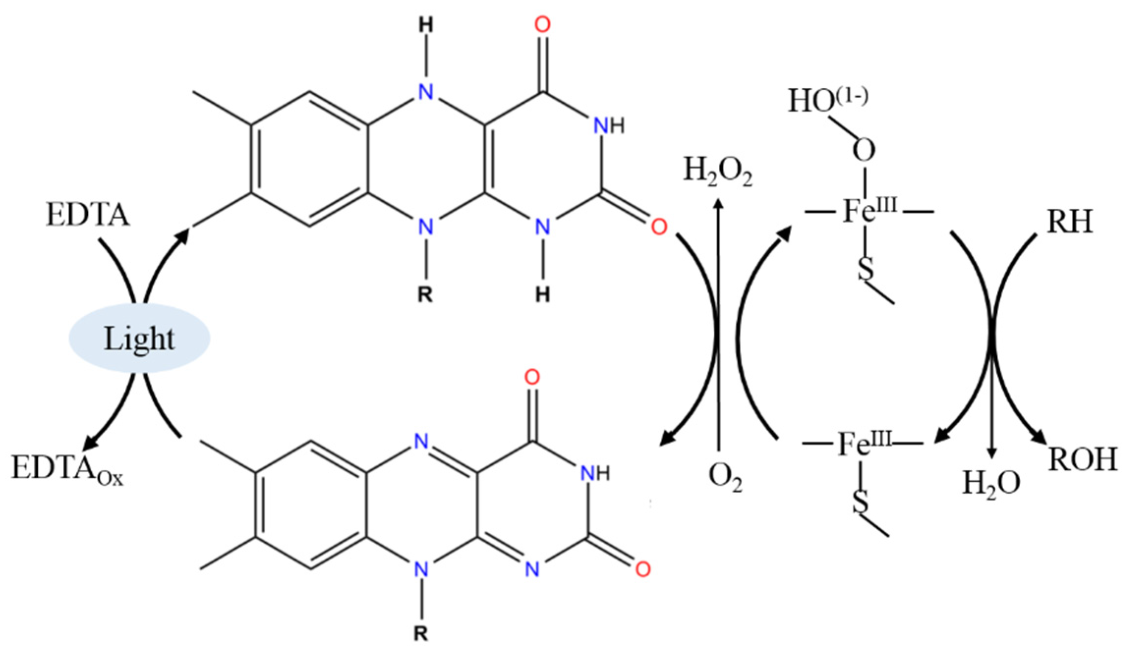
3. Light-Driven CYP Catalysis

The aim of the elaboration of CYP-based systems with alternative electro sources is to avoid expensive NADPH cofactor and protein redox partners [2,3,7]. In this part of our paper, we describe systems with CYP and photosensitive molecules (instead of the nicotinamide cofactor) able to transfer the reducing equivalents to heme iron. For the effective photochemical electron transfer, three main compounds should be present for the realization of the light-dependent P450 catalysis (Figure 1). There are photosensitizers, sacrificial electron donors, and light sources [19,20,21]. The choice of photosensitizer molecules for the effective supplying of heme iron with photo-induced electrons is the main strategy in the construction of effective photocatalytic systems [22,23,24].
Light sources → Sacrificial electron donors → Photosensitizer → Heme iron
Different types of photosensitizers have been used for the light-driven reduction of CYP enzymes, such as biological Photosystem I, nanomaterials, quantum dots (CdS, TiO2, and Ag nanoparticles), prosthetic group analogs (deazaflavins, isoalloxazine-type flavins), organic dyes (eosin Y), inorganic metal complexes, such as ruthenium polypyridine ([Ru(bpy)3]2+), zinc porphyrin, and their derivatives [19,20]. To be effective in the role of photosensitizer, these compounds must possess a visible spectrum from 400 to 710 nm. The reductive potentials of commonly used photosensitizers corresponded to −0.22 ÷ −1.28 V (vs. NHE). Photosensitizers can be divided into two groups, the first group reduces dioxygen leading to the generation of reactive oxygen species (hydrogen peroxide, mainly). The second group reduces cytochrome P450 heme iron in a manner mediated by redox partners or in a direct manner [21,22,23,24,25,26,27].
The electron transfer cascade in the photosensitizers/protein complex may be realized as a covalent or non-covalent binding, permitting the variation of the mode of interaction [1,24,27].
Sacrificial electron donors (sacrificial reductants) include ascorbic acid, triethanolamine (TEOA), diethyldithiocarbamate (DTC), 2-(N-morpholino), ethanesulfonic acid (MES), and ethylenediaminetetraacetic acid (EDTA) [22].
For effective irradiation, light sources with different power and light emitting spectrum in the visible region are used as a source of energy, and varied distances from the reaction cell must also be the subject of discussion [18,19,20,21,22,23,24,25].
Reductase cofactors, such as flavin mononucleotide (FMN), flavin adenine dinucleotide (FAD), or riboflavin, can be reduced photochemically using ethylenediaminetetraacetic acid (EDTA) as an electron donor [18]. During photo activation, reduced flavins can generate hydrogen peroxide, so peroxide shunt pathway is realized [21]. Photosensitive prosthetic group analogs such as deazariboflavin were also studied as photosensitizers [20]. Deazariboflavin undergoes a slow reaction with oxygen. Systems with deazariboflavin, bacterial holoenzyme P450BM3 (CYP102A1), demonstrated catalytic activity for the selective hydroxylation of lauric acid [20].
Bacterial cytochromes P450BM3 (CYP CYP102), CYP199A4, from Rhodopseudomonas palustris HaA2 (as T252E mutant), algal P450s, artificial hemoprotein as a complex of human serum albumin and heme, reductase-free human CYPs expressed in Escherichia coli were used in photobiocatalytic systems [16,24,25,26,27,28,29].
Whole cells expressing in Escherichia coli human CYPs 1A1, 1A2, 1B1, 2E1, and 3A4 for the bioconversion of marketed drugs and steroids were conducted to demonstrate the general applicability of the photobiocatalytic system [17,28]. Substrate conversion was registered for 4-nitrophenol as model substrate and drugs chlorzoxazone, lovastatin, simvastatin, and 17β-estradiol. For whole-cell photocatalysis, the authors observed the transport of flavins into the Escherichia coli cells producing CYP2E1 by cytometric analysis based on fluorescent intensity assay. Human CYP enzymes, in comparison with bacterial enzymes, are more sensitive to hydrogen peroxide produced by FMN/EDTA under exposure to light. Catalase is an important participant in this photocatalytic system for the prevention of enzyme inactivation by reactive oxygen species generated during photocatalysis.
Thus, photo-induced electron transport, as root of reducing equivalents, can serve as an alternative source of electrons in redox reactions provided the native or artificial hemeproteins complexed with photosensitizer molecules. This direction may be attributed to green chemistry as a cost-effective and eco-friendly way of constructing CYP-bioreactors.

4. Electrochemical Technology for Effective CYP Catalysis

Despite the great potential of cytochrome P450s, the dependence on expensive nicotinamide cofactor (NADPH) and protein redox partners, such as NADPH-P450 reductase (CPR), limits their employment in synthetic chemistry, pharmacology, nanobiotechnology and industry [1,2,3,7,8,11]. Since the catalytic cycle of cytochromes P450 is associated with the transfer of electrons [1,2,3], the use of electrochemical systems has found its practical application for modeling catalytic reactions of this class of hemoproteins. In electrochemical systems, electrodes can supply enzymes with electrons instead of NADPHs. For efficient electron transfer, the modification of electrodes and immobilization of enzymes on electrodes are necessary [30,31,32,33]. Electroanalytical methods demonstrate privileges such as high analytical sensitivity, the application of disposable or reusable electrodes that can be modified with a broad spectrum of nanocomposite materials to obtain smart electrodes, the development of both analytical and compact equipment with friendly software for the registration and analysis of the data obtained. Bioelectronics initiation of cytochrome P450s catalysis needs an appropriate type of electrode with a rational design of sensor modifiers [30]. Electrochemical methods are a modern, highly sensitive analytical tool for studying various functional aspects of cytochromes P450: the search for substrates, inhibitors, effectors, and activators as new potential drug candidates [31,32,33,34,35,36,37,38]. The analysis of the catalytic activity of cytochromes P450 can be carried out by measuring the electrochemical parameters of the enzymes themselves in the presence of substrates/inhibitors and by registering changes in the concentration of metabolites/products or substrates of electro enzymatic reactions using mass-spectrometric, spectral, fluorescent, electrochemical, and chromatographic methods [31,32,39,40,41,42]. Cytochrome P450 electrochemical setups are actively used both in the biosensor regimen for recording the substrate-inhibitory multiplicity of this class of hemoproteins and for studying the nontrivial mechanism of cytochrome P450 [33,36,37].
Recent review papers described in great detail the specificity and advances of CYP-bioelectrodes, direct catalytic and noncatalytic behavior of CYPs, and electrocatalytically driven metabolic transformation of chemicals, analysis of metabolites of CYP-electrocatalysis [31,32,33,38,39,40,41,42,43,44,45]. The functioning of cytochrome P450-electrochemical systems in the bioreactor mode is still a poorly explored area (Figure 2).
This aspect of the operation of cytochromes P450 is important for the synthesis of optically active compounds, synthetic drugs’ precursors and isomers for the production of prodrugs with greater pharmacological activity. CYPs are very important for the modification and biodegradation of toxic compounds, leading to the diversification of environmental substances. The aim of this part of the review paper is to analyze the approaches for the improvement of the efficiency of cytochrome P450 electrochemical activity.

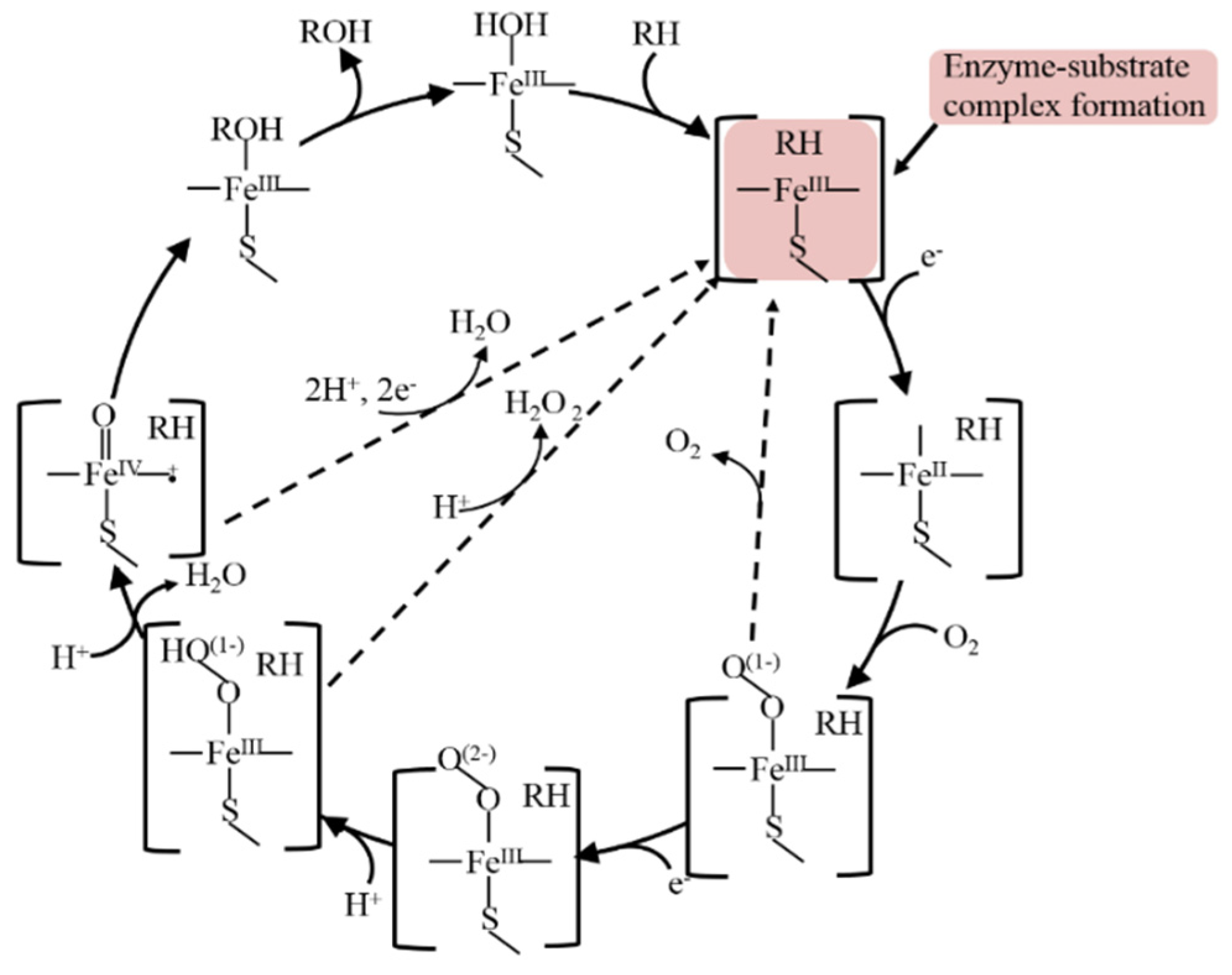
4.1. Modelling on Electrode the Catalytic Cycle of CYP3A4

The biocatalytic mechanism of cytochrome P450s is very intricate and consists of several steps with intermediates formation [1,2,7]. The classical and well-known mechanism of CYP is represented in (Figure 3).
Substrate (RH) binds to enzyme (E) with productive enzyme-substrate complex formation at the first step of CYP catalysis, with productive complex formation inducing a spin shift, which allows an Fe(III)-to-Fe(II) reduction [1,2,7,36,37,38]. The confirmation of the formation of an enzyme/substrate complex was first reaffirmed spectroscopically for the interaction of substrate 17-α-progesterone with an adrenal cortex microsomal enzyme [38].
In our experimental studies, we incubated CYP3A4 with erythromycin (ER) as a substrate for 30 min for the diffusion of the substrate into the active site of immobilized enzymes on the surface of electrodes for the productive complex formation under saturation of enzyme with substrate. We based the first step of the catalytic cycle on the enzyme-substrate complex ERH formation and on the stabilizing effect of substrate on proteins [46,47,48]. The maximum amplitude of catalytic current with the preliminary formation of the enzyme-substrate complex of ERH before electrochemical reduction increased by 17 nA (in comparison to results without (immediately after substrate addition) and with a preliminary complex formation during 30 min of erythromycin as a substrate for saturation of enzyme with substrate) [49]. In electrochemistry, the catalytic current reflects the catalytic cycle of cytochrome P450 as a flux of electrons to substrate RH + O2 + 2ē → ROH + H2O (where RH is the substrate; ROH is the product) [13,33]. When the enzyme-substrate complex formation happens exactly before receiving electrons from electrodes, it enables an enhanced yield of metabolites (in the reaction of erythromycin N-demethylation formaldehyde, it is measured by means of a Hantzsch reaction [31]). Enzyme-substrate complex formation increases the Vmax of CYP-dependent bioelectrocatalysis from 9.21 ± 1.7 × 10⁻¹¹ M/min to 1.40 ± 0.17 × 10⁻¹⁰ M/min (from 100 ± 18% to 146 ± 16%, respectively) [49]. Such an approach is the first example towards a bioreactor construction based on the employment of cytochrome P450 catalytic mechanism.

4.2. Electron Transfer Chain Optimization on CYP-Electrode

The electrochemical cytochrome P450 reactions have great potential for specific drug sensing, for searching for new drugs, and as bioreactors with broad synthetic applications. For the construction of a CYP-based electrochemical reactor, genetically engineered microsomes, human liver microsomes (HLM), and rat liver microsomes were used as the main participants of the electrochemical cell [50,51]. Microsomes consist of all proteins, which provide effective electron transfer to the heme iron ion for initiating the catalysis, such as cytochrome P450, and their redox partner protein CYP-NADPH reductase (CPR). Different types of electrodes were used for the microsomes’ utilization as bioreactors, such as a gold electrode, carbon electrode (polished basal plane pyrolytic graphite (BPG), edge plane pyrolytic graphite (EPG), glassy carbon (GC), or high-purity graphite (HPG) electrodes and modified with multiwalled carbon nanotubes EPG [50,51]. Electrolysis for the electrocatalytic conversion of testosterone to 6β-hydroxytestosterone by the designed microsomal films was carried out at an applied potential of −0.6 V vs. Ag/AgCl for 1 h under a constant supply of oxygen. Electrocatalytic testosterone hydroxylation was analyzed by means of a liquid chromatography detection of 6β-hydroxytestosterone as the metabolite of the enzymatic reaction. The CYP 2C9, 2C19, and 3A4 present in HLM have been shown to play roles in catalyzing the testosterone hydroxylation to 6β-hydroxytestosterone. Ketoconazole (100 μM), a well-known inhibitor of CYP, inhibits this catalytic reaction in the presence of 250 μM of testosterone in electrochemical cells.
Rat liver microsomes (RLMs) were used for the detection of aflatoxin B1 (AFB1) metabolites, participating in carcinogenesis [52]. Electrochemical rat liver microsome-based biosensor using a composite of gold nanoparticles adsorbed on MXene (Au@MXene) for the rapid screening of AFB1. MXene is a new two-dimensional layered material-MXene which consists of transition metal carbides, nitrides, and carbonitrides. Rat liver microsomes (RLMs) were adsorbed on the Au@MXene nanocomposite and used for the detection of aflatoxin M1 in biosensor mode with a limit of detection of 2.8 nM.
In spite of the effective substrate conversion, liver microsomes as bioreactors or biosensors possess ethical problems dealing with the liver as microsomes’ source. Fro this reason, artificial systems modeling the electron transfer chains of CYP-dependent microsomes were proposed [53]. In 1995, Estabrook et al. used the fusion protein (rFP450[mRat4Al/mRatOR]L1) or a system reconstituted with purified P4504A1 plus purified NADPH-P450 reductase in a solution with cobalt(III)sepulchrate trichloride [Co(sep)3+] as the mediator of electrons and Au electrodes as a source of electrons [53,54]. However, the authors did not describe the electroanalytical parameters of electrocatalytically driven hydroxylation of fatty acid in the solution, so such an approach was not scaled.
Efficient work of mitochondrial and microsomal cytochrome P450 systems requires additional redox proteins (diflavin reductase and cytochrome b5). NADPH-dependent cytochrome P450 reductase contains both FAD and FMN as prosthetic molecules and belongs to the flavoproteins group [1,7,8,9]. The roles of flavin nucleotides are the coupling of the reaction of hydroxylation of substrates, an increase in the efficiency of enzyme catalysis, regulation of the flow of electrons, as well as stimulation of positive conformational changes in the structure of the protein [8,49]. The main construction approach for the optimization of the electron transfer chain on electrodes as an alternative electron source involves the reductase domain or flavin cofactors into CYP and immobilization of fused protein onto the electrode surface [1,7,8,9,49]. Different approaches for the optimization of the electron transfer chain in cytochrome P450 electrochemical systems were proposed earlier [55,56,57,58,59,60,61,62,63,64,65,66].
The G. Gilardi group proposed the construction of effective electron transfer chains using the “Lego” approach, combining the heme domain of bacterial CYP102 A1 (BM3), CYP116B5 or CYP3A4 and the reductase domain of BM3 [58,59,60,61,62,63,64,65,66]. These constructs demonstrated enhanced efficiency in electrochemical systems. It was shown that interprotein electron transfer occurred from reduced flavin(s) to heme iron in flavohemeproteins [67].
Riboflavin was used as the model of reductase for the optimization and simplification of the electron transfer chain. In the presence of riboflavin as a mediator of electron flow and NADPH as an electron donor, bacterial types of cytochromes P450 CYP106A2, CYP107DY1, CYP107DY1, HmtS, HmtT, HmtN efficiently catalyzed the reaction of N-dealkylation of substrate diphenhydramine [68].
The efficiency of catalysis of covalent and non-covalent complexes of riboflavin as a simulator of flavoprotein and cytochrome P450 2B4 from rabbit liver in the presence of NADH was investigated earlier [69,70]. Based on these experiments, we used riboflavin, FMN and FAD as substitutes for reductase flavoprotein for the enhancement/improvement of electron transfer in electrochemical cytochrome P450 systems (Scheme 2). Riboflavin transfers two electrons and two protons in the oxidation process [68]. The general scheme of electrons’ transfer in the hemeprotein + flavin complex occurs as shown in the scheme: NADPH or electrons from electrode → Flavin(s) → Heme (Fe3+) [67,68].
Hepatic enzyme cytochrome P450 3A4 (CYP3A4) is involved in the metabolism of about 50% of medicinal preparations and commercial drugs such as exogenous compounds. CYP3A4 catalyzes the metabolism of macrolide antibiotics (erythromycin, clarithromycin, azithromycin), calcium channel blockers (amlodipine, etc.), HIV protease inhibitors (indinavir, etc.), statins (such as simvastatin, atorvastatin), 5α-reductase inhibitors (finasteride), immunosuppressants (cyclosporine, etc.), antihistamines (astemizole), and prokinetics (cisapride) [5,6]. CYP3A4 also catalyzes the oxidation reactions of endogenous compounds, including estradiol (2- or 4-hydroxylation), testosterone (6β-hydroxylation), cortisol (6β-hydroxylation), cholesterol (4β-hydroxylation), progesterone (21-hydroxylation), cholic acid and chenodeoxycholic (formation of 3-dehydrocholic acid), and chenodeoxycholic (6α-hydroxylation) [5,6]. The efficiency of CYP3A4 electrocatalysis was significantly enhanced using macrolide antibiotic erythromycin as a substrate when flavin cofactors were included in the system as mediators of electrons or low molecular models of reductase [50]. Screen-printed electrodes modified with membranous didodecyldimethylammonium bromide (DDAB) were used for the immobilization of CYP3A4/Flavin complexes. The efficiencies of electrocatalysis of erythromycin N-demethylation as well-known cytochrome P4503A4 substrate in the case of riboflavin, FAD and FMN (Scheme 2) as electron transfer mediators were 135 ± 6, 171 ± 15 and 203 ± 10 %, respectively (in comparison with 100 ± 18% erythromycin N-demethylation in the case of cytochrome P4503A4-electrode as the catalyst). The analysis of the reaction rate of erythromycin N-demethylation was carried out by registering formaldehyde formation as a colored product at 412 nm [31,49].
Catalytic current responses of SPE/DDAB/CYP3A4 and SPE/DDAB/CYP3A4 + FMN in the presence of 100 µM of erythromycin confirmed the enhanced catalytic activity of the optimized electron transfer method with the inclusion of FMN (Figure 4).
FMN binds to proteins by electrostatic interactions [71,72]. Riboflavin, FMN, and FAD form complexes with cytochrome P450 3A4 when the isoalloxazine ring of the riboflavin binds to the protein on the proximal side of hemeprotein, which is the place of the redox partners of hemeprotein, as was confirmed through molecular modeling [49].

4.3. Modification of Electrode Surface for the 2D → 3D Transition

The efficient electron transfer from electrodes to biomolecules requires the immobilization of the protein on the working surface of the electrode. It is well-known that there is a problem with the interaction of proteins or enzymes with flat and “hard” 2D surfaces [47,48], which can lead to deformation, distortion of the protein’s spatial structure, or denaturation of the enzyme, which does not always have a positive effect on enzymatic/catalytic activity of immobilized proteins. When working with solid electrodes, modification of the working surface (for example, by polyion films, membrane-like compounds, self-assembled monolayers, and nanomaterials) not only promotes more efficient electron transportation but can lead to the stabilization of the tertiary structure of the protein [73]. Interaction with substrates also stabilizes the enzyme and can lead to changes in both electrochemical thermodynamics and parameters [47,73]. Enzymatic reactions in confined environments mimic the membranous surroundings and partially crowded cell media [42,74,75,76,77,78,79]. The 2D → 3D transition by means of the incorporation of enzymes into 3D nanopores on a plane electrode makes it possible to study the electrochemical and catalytic activity of enzymes [76,77,78,79]. The enzyme immobilization in a volume-confined environment in a proper pore-forming nanomaterial could stabilize its conformation and reduce denaturation [76]. It was shown that mesoporous silica nanomaterial could be used for the incorporation of enzymes due to its large pore volumes, appropriate biocompatibility, adjustable surface chemistry, and comparable pore size in comparison with the diameter of enzymes [74,75]. CYP102A1 (P450BM3) is a self-sufficient single polypeptide composed of a heme monooxygenase domain and NADPH-dependent Flavin mononucleotide (FMN)/flavin adenine dinucleotide (FAD) reductase domain connected via a linker sequence [76]. P450 BM3 size is ca. 5.9 × 9.5 × 20.9 nm [75]. CYP102A1 was immobilized within confined spaces of dendritic mesoporous silica nanoparticles with a similar pore size as 28 or 31 nm but with opposite surface charge (with HO- or NH2-groups, respectively). Authors have shown that the catalytic current towards testosterone on NH2-surface was 1.81 times larger than on HO- surface using a glassy carbon electrode (GCE) as a working electrode.
In our experiments, we used porous membranes based on anodic aluminum oxide (Anodisc, Whatman, pore size 0.1 μm and 0.2 μm) attached to the surface of SPE/DDAB to form 3D pores [14,77]. These studies expand the existing ideas about methods for increasing the efficiency of electrocatalysis through directed modification of the electrode surface by creating a three-dimensional electrode structure with a nanometer pore diameter. The main advantage of the anodic aluminum oxide-based membrane is its chemical stability and its highly organized and regular nanopore structure. The cytochrome P4503A4 (CYP3A4) enzyme was trapped in the ordered nanopores of Anodisc on SPE/DDAB (Figure 5).
Nanoconfined CYP3A4 in the frame of ordered nanopores demonstrated an increase in the efficiency of electrocatalysis of CYP3A4 by a factor of 1.32–2.32, depending on the pore size of the chosen membrane in the reaction of N-demethylation of erythromycin as marker substrate of this isoform of CYP [14,77]. Therefore, the entrapment of enzymes into nanopores on electrodes has obvious advantages in comparison with enzymes immobilized on planar electrode surfaces. Nanoconfinement and the transition from 2D to 3D surface for the immobilization of cytochromes P450 enhance the efficiency of electrocatalytic events.
The pore-forming protein streptolysin O (SLO) was proposed to mimic the cellular environment for creating a more developed surface with protein cavities for effective immobilization of CYP3A4 enzyme based on hybrid biomembranes in the lipid-like bilayer of the electrode modifier didodecyldimethylammonium bromide (DDAB) [78]. The novelty of the approach proposed in this work is the enzyme incorporation in the three-dimensional composite DDAB/SLO, leading both to an improvement in the electron transfer properties and to the efficiency of CYP3A4 electrocatalysis. The confining effect when CYP3A4 is assembled inside cavities was investigated with direct non-catalytic voltammetry and electroanalysis of the enzymatic reaction, such as N-demethylation of erythromycin, occurring in the DDAB/SLO composite. We have shown that SLO on the surface of lipid-like DDAB forms a highly developed surface with cavities, which permit the confinement of the CYP3A4 enzyme for direct non-catalytic and catalytic electrochemistry. The immobilized CYP3A4 demonstrated a pair of redox peaks with a formal potential of −0.325 ± 0.024 V. Potential of E = −0.5 V was applied for substrate conversion of erythromycin registered as N-demethylation reaction. The efficiency of erythromycin electrochemical N-demethylation in SPE/DDAB/CYP3A4 and SPE/DDAB/SLO/CYP3A4 were equal to 100 ± 22% and 297 ± 7%, respectively. AFM analysis of the SPE/DDAB/SLO revealed a more developed surface with protein cavities for the effective immobilization and confinement of the CYP3A4 enzyme (Figure 5). The functioning of enzymes in cells occurs in a confined environment inside a small volume or in a molecularly crowded environment [76]. The modelling of such confined environments on electrodes by means of electrode modification with three-dimensional nanometer pore diameter is an effective method for CYP-based bioreactor construction.

5. Conclusions

Our review paper highlights the prospectives of alternative electron sources for supplying cytochromes P450 with power for the development of effective and prospective constructs or synzymes for the application of these enzymes as bioreactors and biosensors (Table 1). We analyzed active metal (Zn, for example) for the biogalvanic constructs and reduction in heme iron of CYPs. Photoreduction of CYPs with photocurrent providing electrons to hemeproteins also demonstrated potential application as an alternative for expensive NADPH cofactor towards employment of synthetic scope of these enzymes. Electrodes as direct electron sources expanded the biosynthetic prospectives of CYPs.
Most in vivo enzyme-catalyzed reactions occur in a molecularly crowded environment and/or in a confined environment. Insertion of enzymes into a small volume of nanopores or nanochannels is a suitable approach for the improvement of CYPs’ catalytic characteristics. Electrochemical reactions using modified 3D electrodes as electron donors for CYP-electrocatalysis demonstrated opportunities for the development of bioreactors. The above-mentioned approaches can be transformed into biotechnological applications for the development of effective cytochrome P450-based bioreactors for future commercialization and practical use in the industry.

Author Contributions

V.V.S. developed the concept and methodology; P.I.K. performed electrochemical experiments and carried out the reviewing and editing; T.V.B. performed electrochemical experiments and carried out the reviewing and editing; L.E.A. prepared an original draft, carried out the reviewing and editing. All authors have read and agreed to the published version of the manuscript.

Funding

This research was funded by RUSSIAN SCIENCE FOUNDATION, RSF, grant number 23-25-00064. (https://rscf.ru/en/project/23-25-00064/), accessed on 13 June 2023.

Data Availability Statement

Not applicable.

Conflicts of Interest

The authors declare no conflict of interest.

References

  1. Bernhardt, R. Cytochromes P450 as Versatile Biocatalysts. J. Biotechnol. 2006, 124, 128–145. [Google Scholar] [CrossRef] [PubMed]
  2. Bernhardt, R.; Urlacher, V.B. Cytochromes P450 as Promising Catalysts for Biotechnological Application: Chances and Limitations. Appl. Microbiol. Biotechnol. 2014, 98, 6185–6203. [Google Scholar] [CrossRef] [PubMed]
  3. Urlacher, V.B.; Girhard, M. Cytochrome P450 Monooxygenases in Biotechnology and Synthetic Biology. Trends Biotechnol. 2019, 37, 882–897. [Google Scholar] [CrossRef] [PubMed]
  4. Paine, M.J.; Scrutton, N.S.; Munro, A.W.; Gutierrez, A.; Roberts, G.C.; Wolf, C.R. Electron transfer partners of cytochrome P450. In Cytochrome P450-Structure, Mechanism, and Biochemistry; Springer: Boston, MA, USA, 2005; pp. 115–148. [Google Scholar] [CrossRef]
  5. de Montellano, P.R.O. Cytochrome P450: Structure, Mechanism, and Biochemistry; Springer: New York, NY, USA, 2015. [Google Scholar] [CrossRef]
  6. Rendic, S.; Guengerich, F.P. Survey of human oxidoreductases and cytochrome P450 enzymes involved in the metabolism of xenobiotic and natural chemicals. Chem. Res. Toxicol. 2015, 28, 38–42. [Google Scholar] [CrossRef] [Green Version]
  7. Mi, L.; Wang, Z.; Yang, W.; Huang, C.; Zhou, B.; Hu, Y.; Liu, S. Cytochromes P450 in biosensing and biosynthesis applications: Recent progress and future perspectives. Trends Anal. Chem. 2023, 158, 116791. [Google Scholar] [CrossRef]
  8. van Liempda, S.M.; Kool, J.; Reinen, J.; Schenk, T.; Meermana, J.H.N.; Irth, H.; Vermeulen, N.P.E. Development and validation of a microsomal online cytochrome P450 bioreactor coupled to solid-phase extraction and reversed-phase liquid chromatography. J. Chromatogr. A 2005, 1075, 205–212. [Google Scholar] [CrossRef]
  9. Guengerich, F.P. Intersection of the Roles of Cytochrome P450 Enzymes with Xenobiotic and Endogenous Substrates: Relevance to Toxicity and Drug Interactions. Chem. Res. Toxicol. 2017, 30, 2–12. [Google Scholar] [CrossRef] [Green Version]
  10. Guengerich, F.P. 1.19—Drug Metabolism: Cytochrome P450. In Comprehensive Pharmacology; Elsevier: Amsterdam, The Netherlands, 2022; pp. 470–508. [Google Scholar] [CrossRef]
  11. Winkler, M.; Geier, M.; Hanlon, S.P.; Nidetzky, B.; Glieder, A. Human Enzymes for Organic Synthesis. Angew. Chem. Int. Ed. 2018, 57, 13406–13423. [Google Scholar] [CrossRef] [Green Version]
  12. Lamb, D.C.; Waterman, M.R.; Kelly, S.L.; Guengerich, F.P. Cytochromes P450 and drug discovery. Curr. Opin. Biotechnol. 2007, 18, 504–512. [Google Scholar] [CrossRef]
  13. Brett, C.M.A.; Oliveira Brett, A.M. Electrochemistry: Principles, Methods, and Applications, 1st ed.; Oxford Science University Publications: Oxford, UK, 1993. [Google Scholar] [CrossRef]
  14. Shumyantseva, V.V.; Kuzikov, A.V.; Masamrekh, R.A.; Filippova, T.A.; Koroleva, P.I.; Agafonova, L.E.; Bulko, T.V.; Archakov, A.I. Enzymology on an electrode and in a nanopore: Analysis algorithms, enzyme kinetics, and perspectives. BioNanoScience 2022, 12, 1341–1355. [Google Scholar] [CrossRef]
  15. Shumyantseva, V.V.; Bulko, T.V.; Alexandrova, S.A.; Sokolov, N.N.; Schmid, R.D.; Bachmann, T.; Archakov, A.I. N-Terminal truncated cytochrome P450 2B4: Catalytic activities and reduction with alternative electron sources. Biochem. Biophys. Res. Commun. 1999, 263, 678–680. [Google Scholar] [CrossRef] [PubMed]
  16. Shumyantseva, V.V.; Bulko, T.V.; Archakov, A.I. Regulation of cytochrome P450 activity by physicochemical methods. Uspekhi Khimii 1999, 68, 967–975. [Google Scholar] [CrossRef]
  17. Shumyantseva, V.V.; Bulko, T.V.; Zimin, A.G.; Uvarov, V.Y.; Archakov, A.I. Design of an artificial hemoprotein based on human serum albumin. Biochem. Mol. Biol. Int. 1996, 39, 503. [Google Scholar] [CrossRef] [PubMed]
  18. Zhao, L.; Güven, G.; Li, Y.; Schwaneberg, U. First steps towards a Zn/Co(III)sep-driven P450 BM3 reactor. Appl. Microbiol. Biotechnol. 2011, 91, 989–999. [Google Scholar] [CrossRef]
  19. Zheng, D.; Zhang, Y.; Liu, X.; Wang, J. Coupling natural systems with synthetic chemistry for light driven enzymatic biocatalysis. Photosynth. Res. 2020, 143, 221–231. [Google Scholar] [CrossRef]
  20. Zilly, F.E.; Taglieber, A.; Schulz, F.; Hollmann, F.; Reetz, M.T. Deazaflavins as mediators in light-driven cytochrome P450 catalyzed hydroxylations. Chem. Commun. 2009, 46, 7152–7154. [Google Scholar] [CrossRef]
  21. Jensen, K.; Jensen, P.E.; Møller, B.L. Light-driven chemical synthesis. Trends Plant Sci. 2012, 17, 60–63. [Google Scholar] [CrossRef]
  22. Nielsen, A.Z. Redirecting Photosynthetic Reducing Power toward Bioactive Natural Product Synthesis. ACS Synth. Biol. 2013, 2, 308–315. [Google Scholar] [CrossRef]
  23. Le, T.-K.; Park, J.H.; Choi, D.S.; Lee, G.-Y.; Choi, W.S.; Jeong, K.J.; Park, C.B.; Yun, C.-H. Solar-driven biocatalytic C-hydroxylation through direct transfer of photoinduced electrons. Green Chem. 2019, 21, 515. [Google Scholar] [CrossRef]
  24. Lassen, L.M.; Nielsen, A.Z.; Olsen, C.E.; Bialek, W.; Jensen, K.; Møller, B.L.; Jensen, P.E. Anchoring a plant cytochrome P450 via PsaM to the thylakoids in Synechococcus sp. PCC 7002: Evidence for light-driven biosynthesis. PLoS ONE 2014, 9, 102184. [Google Scholar] [CrossRef] [Green Version]
  25. Shalan, H.; Kato, M.; Cheruzel, L. Keeping the spotlight on cytochrome P450. BBA-Proteins Proteom. 2018, 1866, 80–87. [Google Scholar] [CrossRef] [PubMed]
  26. Zheng, S.; Guo, J.; Cheng, F.; Gao, Z.; Du, L.; Meng, C.; Li, S.; Zhang, X. Cytochrome P450s in algae: Bioactive natural product biosynthesis and light-driven bioproduction. Acta Pharm. Sin. B 2022, 12, 2832–2844. [Google Scholar] [CrossRef] [PubMed]
  27. Eidenschenk, C.; Cheruzel, L. Ru(II)-diimine complexes and cytochrome P450 working hand-in-hand. J. Inorg. Biochem. 2020, 213, 111254. [Google Scholar] [CrossRef] [PubMed]
  28. Le, T.-K.; Kim, J.; Nguyen, N.A.; Nguyen, T.H.H.; Sun, E.-G.; Yee, S.-M.; Kang, H.-S.; Yeom, S.-J.; Park, C.B.; Yun, C.-H. Solar-Powered Whole-Cell P450 Catalytic Platform for C-Hydroxylation Reactions. ChemSusChem 2021, 14, 3054–3058. [Google Scholar] [CrossRef]
  29. Lee, J.H.Z.; Podgorski, M.N.; Moir, M.; Gee, A.R.; Bell, S.G. Selective Oxidations Using a Cytochrome P450 Enzyme Variant Driven with Surrogate Oxygen Donors and Light. Chem. Eur. J. 2022, 28, 202201366. [Google Scholar] [CrossRef] [PubMed]
  30. Shumyantseva, V.V.; Agafonova, L.E.; Bulko, T.V.; Kuzikov, A.V.; Masamrekh, R.A.; Yuan, J.; Pergushov, D.V.; Sigolaeva, L.V. Electroanalysis of Biomolecules: Rational Selection of Sensor Construction. Biochem. (Mosc.) 2021, 86, 140–151. [Google Scholar] [CrossRef]
  31. Masamrekh, R.A.; Kuzikov, A.V.; Haurychenka, Y.I.; Shcherbakov, K.A.; Veselovsky, A.V.; Filimonov, D.A.; Dmitriev, A.V.; Zavialova, M.G.; Gilep, A.A.; Shkel, T.V.; et al. In vitro interactions of abiraterone, erythromycin, and CYP3A4: Implications for drug-drug interactions. Fundam. Clinic. Pharmacol. 2020, 34, 120–130. [Google Scholar] [CrossRef]
  32. Shumyantseva, V.V.; Bulko, T.V.; Kuzikov, A.V.; Masamrekh, R.A.; Konyakhina, A.Y.; Romanenko, I.; Max, J.B.; Köhler, M.; Gilep, A.A.; Usanov, S.A.; et al. All-electrochemical nanocomposite two-electrode setup for quantification of drugs and study of their electrocatalytical conversion by cytochromes P450. Electrochim. Acta 2020, 336, 135579. [Google Scholar] [CrossRef]
  33. Shumyantseva, V.V.; Kuzikov, A.V.; Masamrekh, R.A.; Bulko, T.V.; Archakov, A.I. From electrochemistry to enzyme kinetics of cytochrome P450. Biosens. Bioelectron. 2018, 121, 192–204. [Google Scholar] [CrossRef]
  34. Masamrekh, R.; Filippova, T.; Haurychenka, Y.; Shumyantseva, V.; Kuzikov, A.; Shcherbakov, K.; Veselovsky, A.; Strushkevich, N.; Shkel, T.; Gilep, A.; et al. Estimation of the inhibiting impact of abiraterone D4A metabolite on human steroid 21-monooxygenase (CYP21A2). Steroids 2020, 154, 108528. [Google Scholar] [CrossRef]
  35. Kostin, V.A.; Zolottsev, V.A.; Kuzikov, A.V.; Masamrekh, R.A.; Shumyantseva, V.V.; Veselovsky, A.V.; Stulov, S.V.; Novikov, R.A.; Timofeev, V.P.; Misharin, A.Y. Oxazolinyl derivatives of [17(20)E]-21-norpregnene differing in the structure of A and B rings. Facile synthesis and inhibition of CYP17A1 catalytic activity. Steroids 2016, 115, 114–122. [Google Scholar] [CrossRef] [PubMed]
  36. Krishnan, S. Bioelectrodes for evaluating molecular therapeutic and toxicity properties. Curr. Opin. Electrochem. 2019, 19, 20–26. [Google Scholar] [CrossRef]
  37. Di Nardo, G.; Gilardi, G. Natural Compounds as Pharmaceuticals: The Key Role of Cytochromes P450 Reactivity. Trends Biochem. Sci. 2020, 45, 511–525. [Google Scholar] [CrossRef] [PubMed]
  38. Sakaki, T. Practical application of cytochrome P450. Biol. Pharm. Bull. 2012, 35, 844–849. [Google Scholar] [CrossRef] [Green Version]
  39. Kuzikov, A.; Masamrekh, R.; Shkel, T.; Strushkevich, N.; Gilep, A.; Usanov, S.; Archakov, A.; Shumyantseva, V. Assessment of electrocatalytic hydroxylase activity of cytochrome P450 3A4 (CYP3A4) by means of derivatization of 6β-hydroxycortisol by sulfuric acid for fluorimetric assay. Talanta 2019, 196, 231–236. [Google Scholar] [CrossRef]
  40. Kuzikov, A.; Filippova, T.; Masamrekh, R.; Shumyantseva, V. Biotransformation of phenytoin in electrochemically-driven CYP2C19 system. Biophys. Chem. 2022, 291, 106894. [Google Scholar] [CrossRef]
  41. Kuzikov, A.; Masamrekh, R.; Filippova, T.; Shumyantseva, V. Electrochemical Analysis of Metabolites as a Method for Cytochromes P450 Activity Determination. Biomed. Chem. Res. Methods 2022, 5, e00176. [Google Scholar] [CrossRef]
  42. Kuzikov, A.; Filippova, T.; Masamrekh, R.; Shumyantseva, V. Electroanalysis of 4′-Hydroxydiclofenac for CYP2C9 Enzymatic Assay. Electrocatalysis 2022, 13, 630–640. [Google Scholar] [CrossRef]
  43. Schneider, E.; Clark, D.S. Cytochrome P450 (CYP) enzymes and the development of CYP biosensors. Biosens. Bioelectron. 2013, 39, 1–13. [Google Scholar] [CrossRef]
  44. Panicco, P.; Castrignanò, S.; Sadeghi, S.J.; Di Nardo, G.; Gilardi, G. Engineered human CYP2C9 and its main polymorphic variants for bioelectrochemical measurements of catalytic response. Bioelectrochemistry 2021, 138, 107729. [Google Scholar] [CrossRef]
  45. Mi, L.; He, F.; Jiang, L.; Shangguan, L.; Zhang, X.; Ding, T.; Liu, A.; Zhang, Y.; Liu, S. Electrochemically-driven benzo [a] pyrene metabolism via human cytochrome P450 1A1 with reductase coated nitrogen-doped graphene nano-composites. J. Electroanal. Chem. 2017, 804, 23–28. [Google Scholar] [CrossRef]
  46. Avila-Sierra, A.; Moreno, J.A.; Goode, K.; Zhu, T.; Fryer, P.J.; Taylor, A.; Zhang, Z.J. Effects of structural and chemical properties of surface coatings on the adsorption characteristics of proteins. Surf. Coat. Technol. 2023, 452, 129054. [Google Scholar] [CrossRef]
  47. Martínez-Negro, M.; Oberländer, J.; Simon, J.; Mailänder, V.; Morsbach, S.; Landfester, K. A new methodology combining QCM-D and proteomic profiling enables characterization of protein adsorption on 2D surfaces. J. Colloid Interface Sci. 2023, 630, 965–972. [Google Scholar] [CrossRef] [PubMed]
  48. Guengerich, F.P. Drug Metabolism: Cytochrome P450. Reference Module in Biomedical Sciences; Elsevier: Amsterdam, The Netherlands, 2021. [Google Scholar] [CrossRef]
  49. Shumyantseva, V.V.; Koroleva, P.I.; Bulko, T.V.; Shkel, T.V.; Gilep, A.A.; Veselovsky, A.V. Approaches for increasing the electrocatalitic efficiency of cytochrome P450 3A4. Bioelectrochemistry 2023, 149, 108277. [Google Scholar] [CrossRef]
  50. Nerimetla, R.; Krishnan, S. Electrocatalysis by subcellular liver fractions bound to carbon nanostructures for stereoselective green drug metabolite synthesis. Chem. Commun. 2015, 51, 11681–11684. [Google Scholar] [CrossRef] [Green Version]
  51. Walgama, C.; Nerimetla, R.; Materer, N.F.; Schildkraut, D.; Elman, J.F.; Krishnan, S. A Simple Construction of Electrochemical Liver Microsomal Bioreactor for Rapid Drug Metabolism and Inhibition Assays. Anal. Chem. 2015, 87, 4712–4718. [Google Scholar] [CrossRef]
  52. Sun, X.; Sun, J.; Ye, Y.; Ji, J.; Sheng, L.; Yang, D.; Sun, X. Metabolic pathway-based self-assembled Au@MXene liver microsome electrochemical biosensor for rapid screening of aflatoxin B1. Bioelectrochemistry 2023, 151, 108378. [Google Scholar] [CrossRef]
  53. Faulkner, K.M.; Shet, M.S.; Fisher, C.W.; Estabrook, R.W. Electrocatalytically driven w-hydroxylation of fatty acids using cytochrome P450 4A1. Proc. Natl. Acad. Sci. USA 1995, 92, 7705–7709. [Google Scholar] [CrossRef] [Green Version]
  54. Estabrook, R.W.; Faulkner, K.M.; Seth, M.S.; Fisher, C.W. Application of electrochemistry for P450-catalyzed reactions. Methods Enzymol. 1996, 272, 44–51. [Google Scholar] [CrossRef]
  55. Glass, S.M.; Guengerich, F.P. Cellular retinoid-binding proteins transfer retinoids to human cytochrome P450 27C1 for desaturation. J. Biol. Chem. 2021, 297, 101142. [Google Scholar] [CrossRef]
  56. Ducharme, J.; Auclair, K. Use of bioconjugation with cytochrome P450 enzymes. BBA-Proteins Proteom. 2018, 1866, 32–51. [Google Scholar] [CrossRef] [PubMed] [Green Version]
  57. Giuriato, D.; Correddu, D.; Catucci, G.; Di Nardo, G.; Bolchi, C.; Pallavicini, M.; Gilardi, G. Design of a H2O2-generating P450SPα fusion protein for high yield fatty acid conversion. Protein Sci. 2022, 31, e4501. [Google Scholar] [CrossRef] [PubMed]
  58. Gilardi, G.; Meharenna, Y.T.; Tsotsou, G.E.; Sadeghi, S.J.; Fairhead, M.; Giannini, S. Molecular Lego: Design of molecular assemblies of P450 enzymes for nanobiotechnology. Biosens. Bioelectron. 2002, 17, 133–145. [Google Scholar] [CrossRef] [PubMed]
  59. Rua, F.; Sadeghi, S.J.; Castrignanò, S.; Di Nardo, G.; Gilardi, G. Engineering Macaca fascicularis cytochrome P450 2C20 to reduce animal testing for new drugs. J. Inorg. Biochem. 2012, 117, 277–284. [Google Scholar] [CrossRef] [PubMed]
  60. Dodhia, V.; Sassone, C.; Fantuzzi, A.; Di Nardo, G.; Sadeghi, S.J.; Gilardi, G. Modulating the coupling efficiency of human cytochrome P450 CYP3A4 at electrode surfaces through protein engineering. Electrochem. Commun. 2008, 10, 1744–1747. [Google Scholar] [CrossRef]
  61. Degregorio, D.; Sadeghi, S.J.; Di Nardo, G.; Gilardi, G.; Solinas, S.P. Understanding uncoupling in the multiredox centre P450 3A4-BMR model system. J. Biol. Inorg. Chem. 2011, 16, 109–116. [Google Scholar] [CrossRef]
  62. Castrignanò, S.; Di Nardo, G.; Sadeghi, S.J.; Gilardi, G. Influence of inter-domain dynamics and surrounding environment flexibility on the direct electrochemistry and electrocatalysis of selfsufficient cytochrome P450 3A4-BMR chimeras. J. Inorg. Biochem. 2018, 188, 9–17. [Google Scholar] [CrossRef]
  63. Castrignanò, S.; D’Avino, S.; Di Nardo, G.; Catucci, G.; Sadeghi, S.J.; Gilardi, G. Modulation of the interaction between human P450 3A4 and B. megaterium reductase via engineered loops. Biochim. Biophys. Acta Proteins Proteom. 2018, 1866, 116–125. [Google Scholar] [CrossRef]
  64. Degregorio, D.; D’Avino, S.; Castrignanò, S.; Di Nardo, G.; Catucci, G.; Sadeghi, S.J.; Gilardi, G. Human Cytochrome P450 3A4 as a Biocatalyst: Effects of the Engineered Linker in Modulation of Coupling Efficiency in 3A4-BMR Chimeras. Front. Pharmacol. 2017, 8, 121. [Google Scholar] [CrossRef] [Green Version]
  65. Fantuzzi, A.; Meharenna, Y.T.; Briscoe, P.B.; Sassone, C.; Borgia, B.; Gilardi, G. Improving catalytic properties of P450 BM3 haem domain electrodes by molecular Lego. Chem. Commun. 2006, 12, 1289–1291. [Google Scholar] [CrossRef]
  66. Correddu, D.; Catucci, G.; Giuriato, D.; Di Nardo, G.; Ciaramella, A.; Gilardi, G. Catalytically self-sufficient CYP116B5: Domain switch for improved peroxygenase activity. Biotechnol. J. 2023, 18, 2200622. [Google Scholar] [CrossRef] [PubMed]
  67. Shumyantseva, V.V.; Bulko, T.V.; Lisitsyna, V.B.; Urlacher, V.B.; Kuzikov, A.V.; Suprun, E.V.; Archakov, A.I. Electrochemical Measurement of Intraprotein and Interprotein Electron Transfer. Biophysics 2013, 58, 349–354. [Google Scholar] [CrossRef]
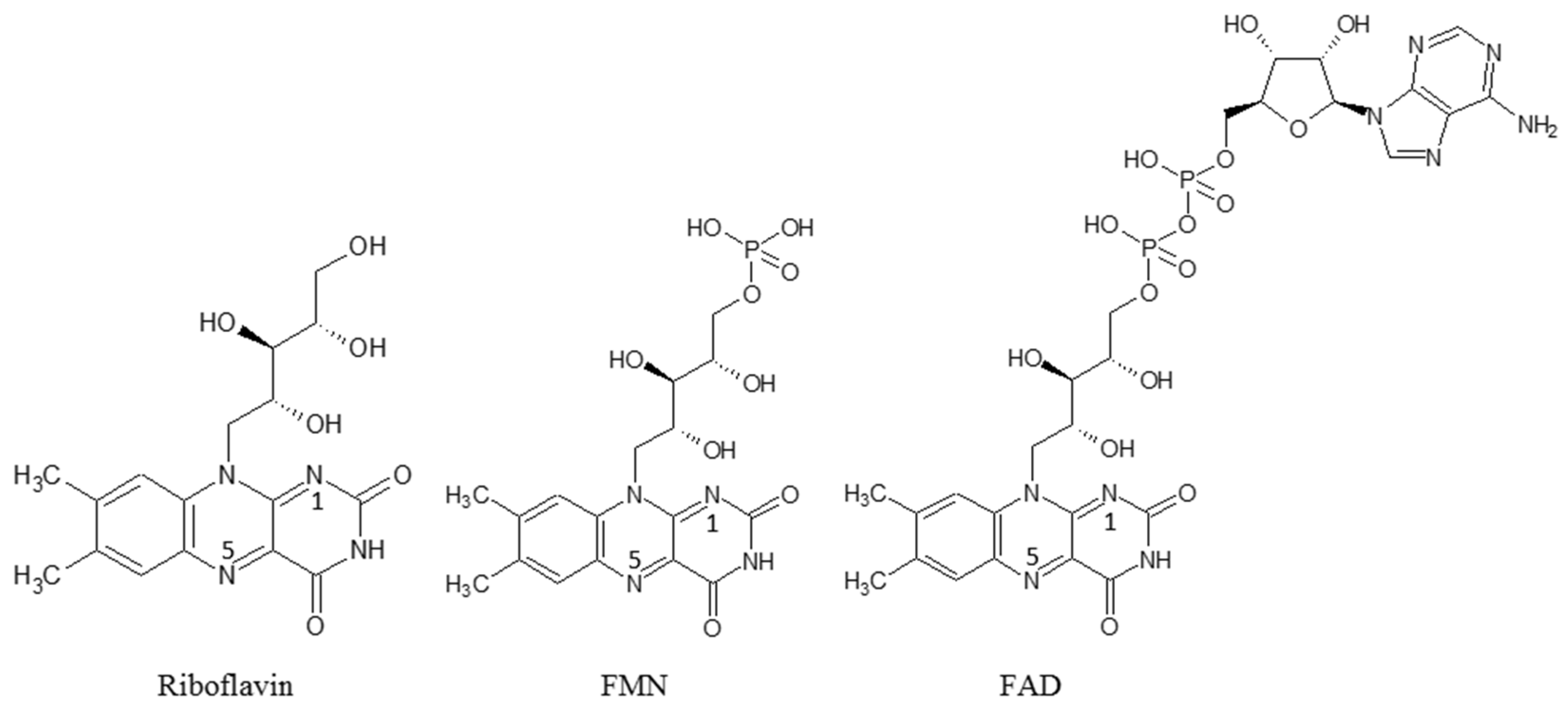
  68. Zhang, C.; Lu, M.; Lin, L.; Huang, Z.; Zhang, R.; Wu, X.; Chen, Y. Riboflavin Is Directly Involved in N-Dealkylation Catalyzed by Bacterial Cytochrome P450 Monooxygenases. Chembiochem 2020, 21, 2297–2305. [Google Scholar] [CrossRef] [PubMed]
  69. Shumyantseva, V.; Uvarov, V.; Byakova, O.; Archakov, A. Semisynthetic Flavocytochromes Based on Cytochrome P450 2B4: Reductase and Oxygenase Activities. Arch. Biochem. Biophys. 1998, 354, 133–138. [Google Scholar] [CrossRef]
  70. Shumyantseva, V.V.; Bulko, T.V.; Bachmann, T.T.; Bilitewski, U.; Schmid, R.D.; Archakov, A.I. Electrochemical Reduction of Flavocytochromes 2B4 and 1A2 and Their Catalytic Activity. Arch Biochem. Biophys. 2000, 377, 43–48. [Google Scholar] [CrossRef]
  71. Roux, Y.; Ricoux, R.; Avenier, F.; Mahy, J.-P. Bio-inspired electron-delivering system for reductive activation of dioxygen at metal centres towards artificial flavoenzymes. Nat. Commun. 2015, 6, 8509. [Google Scholar] [CrossRef] [Green Version]
  72. Sevrioukova, I.F.; Li, H.; Zhang, H.; Peterson, J.A.; Poulos, T.L. Structure of a cytochrome P450–redox partner electron-transfer complex. Proc. Natl. Acad. Sci. USA 1999, 96, 1863–1868. [Google Scholar] [CrossRef] [Green Version]
  73. Lu, J.; Shen, Y.; Liu, S. Enhanced light-driven catalytic performance of cytochrome P450 confined in macroporous silica. Chem. Commun. 2016, 52, 7703–7706. [Google Scholar] [CrossRef]
  74. Fang, X.; Qiao, L.; Yan, G.; Yang, P.; Liu, B. Multifunctional nanoreactor for comprehensive characterization of membrane proteins based on surface functionalized mesoporous foams. Anal. Chem. 2015, 87, 9360–9367. [Google Scholar] [CrossRef]
  75. Dai, Q.; Yang, L.; Wang, Y.; Cao, X.; Yao, C.; Xu, X. Surface charge-controlled electron transfer and catalytic behavior of immobilized cytochrome P450 BM3 inside dendritic mesoporous silica nanoparticles. Anal. Bioanal. Chem. 2020, 412, 4703–4712. [Google Scholar] [CrossRef]
  76. Küchler, A.; Yoshimoto, M.; Luginbühl, S.; Mavelli, F.; Walde, P. Enzymatic reactions in confined environments. Nat. Nanotechnol. 2016, 11, 409–419. [Google Scholar] [CrossRef] [PubMed]
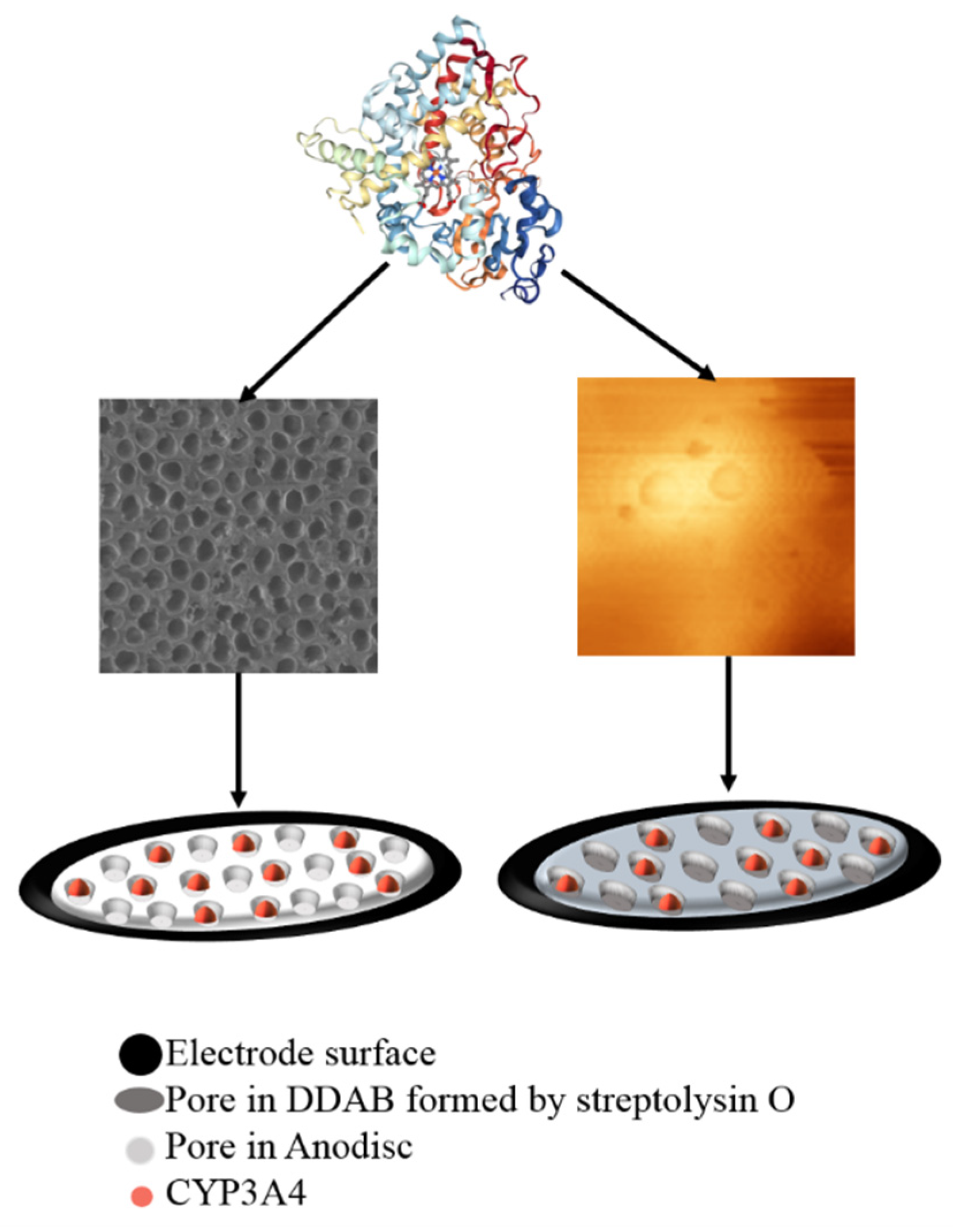
  77. Shumyantseva, V.V.; Koroleva, P.I.; Gilep, A.A.; Napolskii, K.S.; Ivanov, Y.D.; Kanashenko, S.L.; Archakov, A.I. Increasing the Efficiency of Cytochrome P450 3A4 Electrocatalysis Using Electrode Modification with Spatially Ordered Anodic Aluminum Oxide-Based Nanostructures for Investigation of Metabolic Transformations of Drugs. Dokl. Biochem. Biophys. 2022, 506, 215–219. [Google Scholar] [CrossRef] [PubMed]
  78. Koroleva, P.I.; Gilep, A.A.; Kraevskiy, S.V.; Tsybruk, T.V.; Shumyantseva, V.V. Improving the Efficiency of Electrocatalysis of Cytochrome P450 3A4 by Modifying the Electrode with Membrane Protein Streptolysin O for Studying the Metabolic Transformations of Drugs. Biosensors 2023, 13, 457. [Google Scholar] [CrossRef] [PubMed]
  79. Lin, S.; Chen, Y.; Wang, Y.; Cai, Z.; Xiao, J.; Muhmood, T.; Hu, X. Three-Dimensional Ordered Porous Nanostructures for Lithium−Selenium Battery Cathodes That Confer Superior Energy-Storage Performance. ACS Appl. Mater. Interfaces 2021, 13, 9955–9964. [Google Scholar] [CrossRef]
Scheme 1. A relative range of metal activity vs. standard hydrogen electrode.
Processes 11 01801 sch001
Figure 1. Schematic representation of the light-driven heme iron reduction. R-substrate, ROH-metabolite of CYP-dependent reaction.
Processes 11 01801 g001
Figure 2. Electrochemical systems in the CYP enzymology.
Processes 11 01801 g002
Figure 3. A common catalytic cycle of CYPs enzymes.
Processes 11 01801 g003
Scheme 2. Structures of flavin cofactors.
Processes 11 01801 sch002
Figure 4. Comparative catalytic current responses of SPE/DDAB/CYP3A4 and SPE/DDAB/CYP3A4 + FMN in the presence of 100 µM erythromycin.
Processes 11 01801 g004
Figure 5. SEM images of porous membranes based on anodic aluminum oxide (Anodisc, Whatman, pore size 0.1 μm) and scheme of immobilization of CYP3A4 on the SPE/DDAB/Anodisc (left). AFM topographic images of pore-forming protein streptolysin O (SLO) on the DDAB film and scheme of immobilization of CYP3A4 on the SPE/DDAB/SLO (right).
Processes 11 01801 g005
Table 1. Comparative characteristics of alternative electron sources for cytochrome P450s.
Electron SourceAdvantagesDisadvantages
Active metals as electron donorLabel-free approachInactivation of enzymes during electro catalysis may occur
The lack of commercial standardized sensor devices
Light-driven catalysisDoes not need additional electron sources
Can be used for broad line of enzymes
Needs additional modification of enzymes with photosensitizers and sacrificial electron donors [25]
The lack of commercial devices
Electrochemical technologyAdditional proteins or redox-active molecules are not necessary
Broad spectrum of measurement parameters (catalytic current, Icat; potential start of catalysis, Eonset; potential of catalysis, Ecat; potential of reduction, Ered; potential of oxidation, Eox) [14]
Commercial availability of different types of electrodes as 2D sensor
Inactivation of enzymes during electro catalysis may occur
Disclaimer/Publisher’s Note: The statements, opinions and data contained in all publications are solely those of the individual author(s) and contributor(s) and not of MDPI and/or the editor(s). MDPI and/or the editor(s) disclaim responsibility for any injury to people or property resulting from any ideas, methods, instructions or products referred to in the content.

