
Coarse X-ray Lumbar Vertebrae Pose Localization and Registration Using Triangulation Correspondence

 , , and
Abstract
:1. Introduction
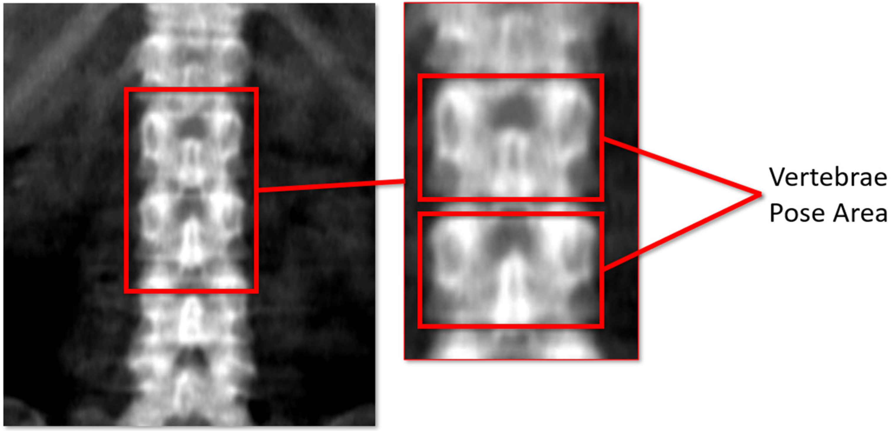
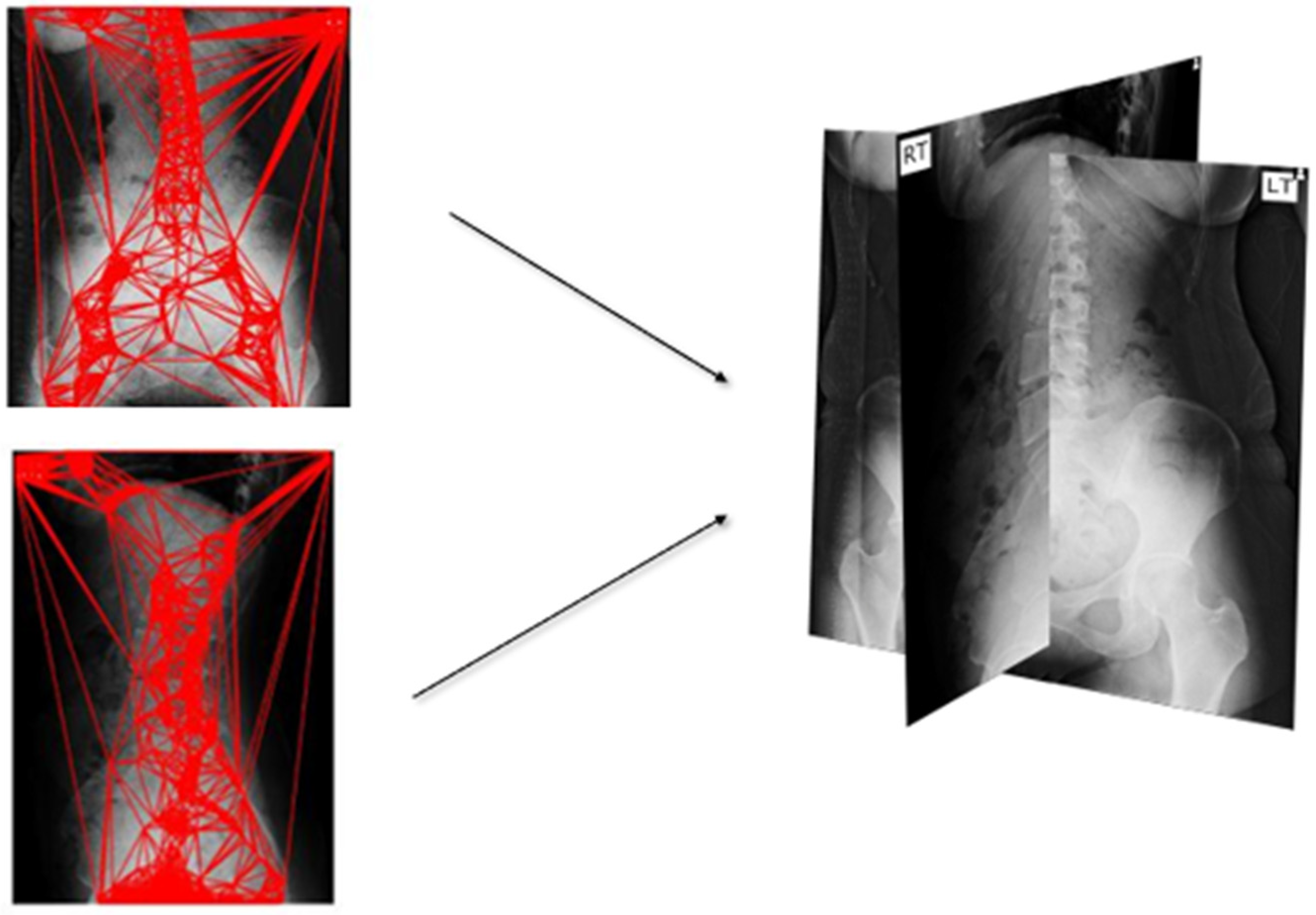
- To pinpoint the posture area of the lumbar vertebrae in environments with a low contrast. Figure 1 depicts the position of the vertebrae that must be located. Thus, each stance is observed to be difficult to execute.
- To register two sides of view for reconstructing a three-dimensional model.
2. Materials and Methods
2.1. Materials
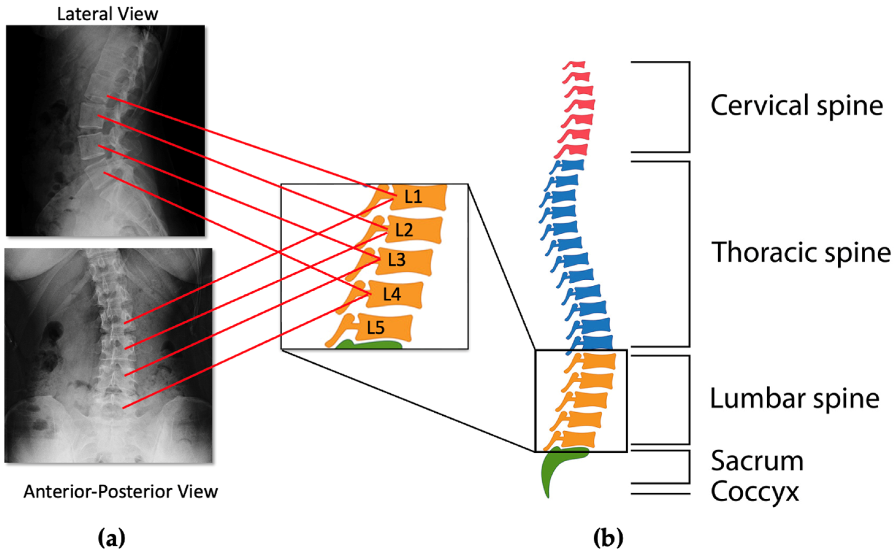
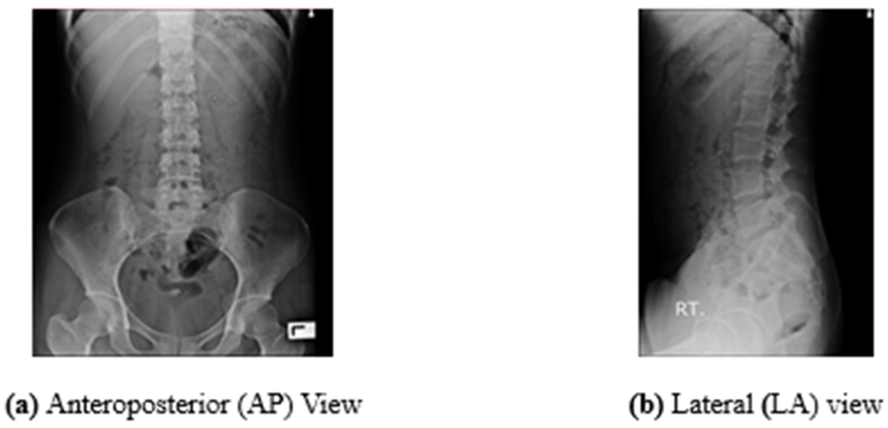
2.1.1. X-ray Image of Human Lumbar Spine
2.1.2. Speeded up Robust Features (SURF)
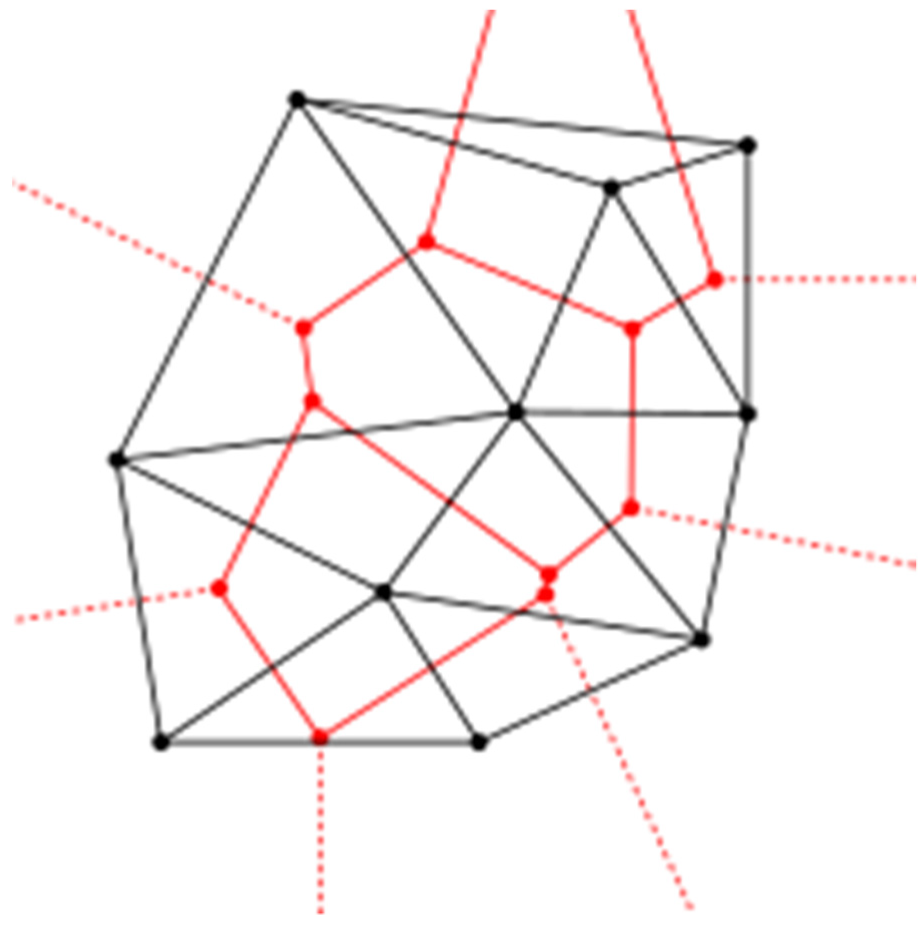
2.1.3. Delaunay Triangulation
2.1.4. Dataset
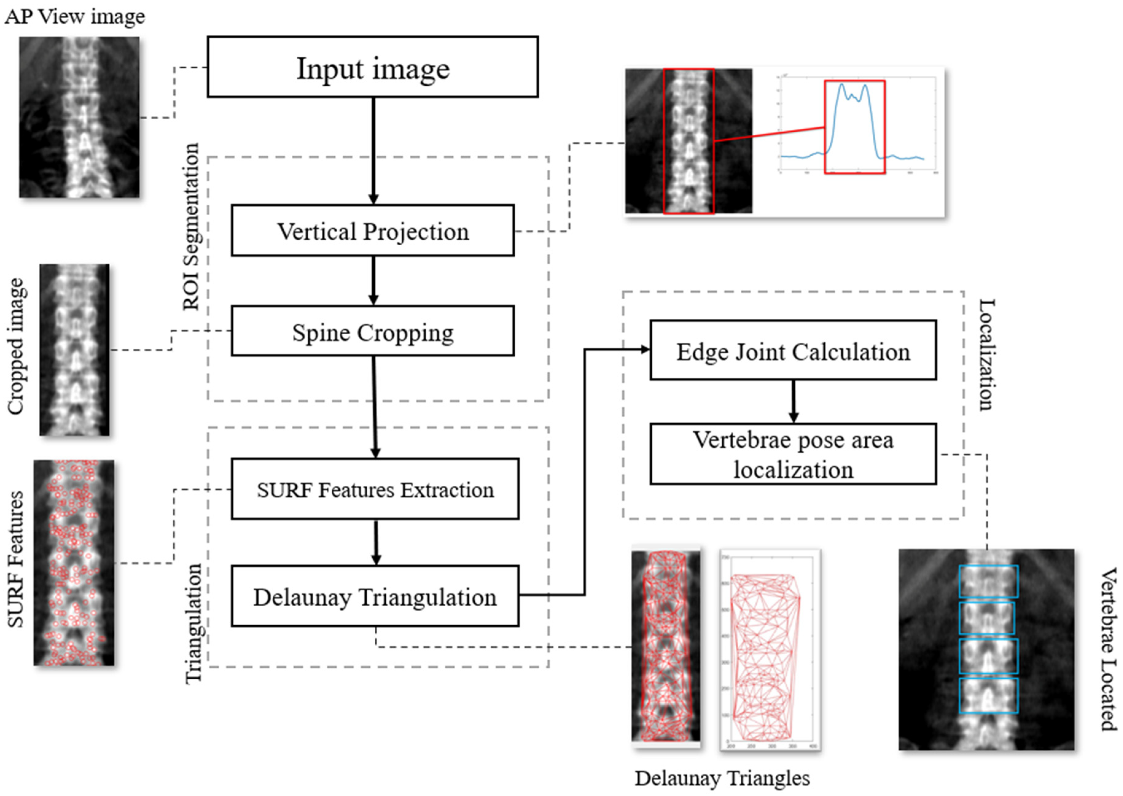
2.2. Methodology
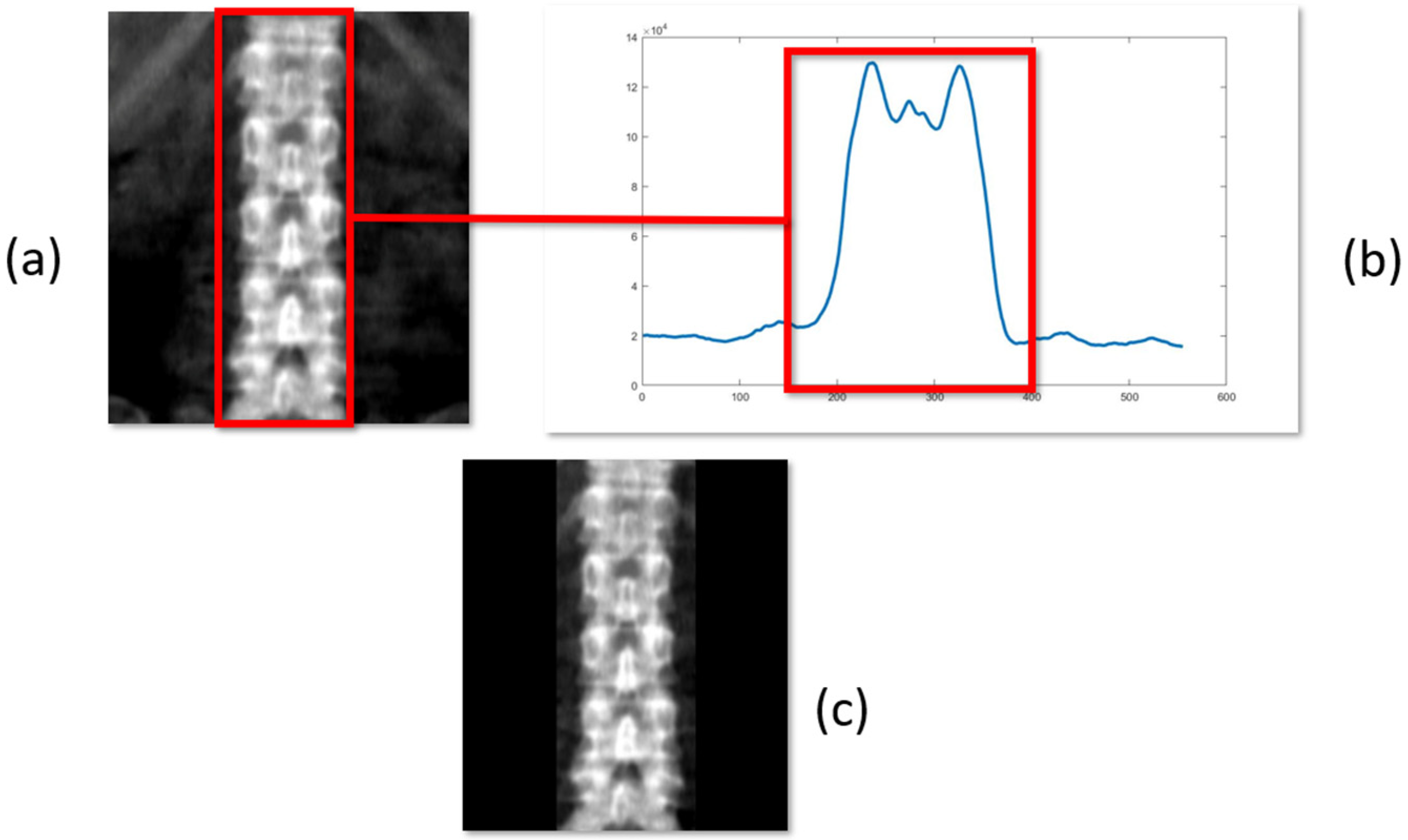
2.2.1. Region of Interest Segmentation (ROI)
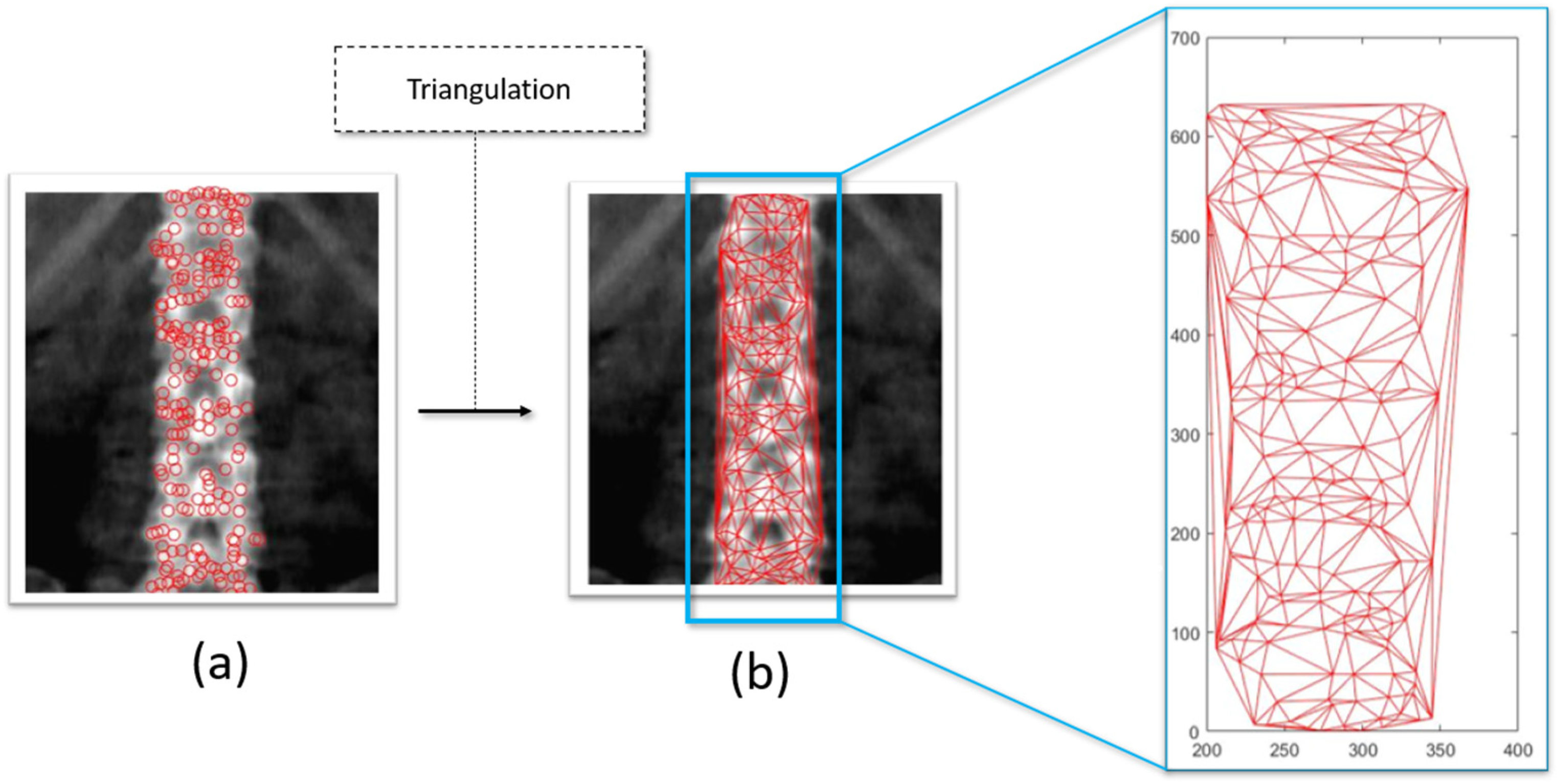
2.2.2. Triangulation Using Delaunay’s Approach
| Algorithm 1 Delaunay triangulation | |
| Algorithm | Delaunay() |
| Input | a set of point in |
| Output | |
| 1. | compute a triangulation of |
| 2. | Initialize a stack containing all the edges of |
| 3. | While stack is non-empty |
| 4. | do pop from stack and unmark it |
| 5. | if is illegal then |
| 6. | do flip to |
| 7. | for |
| 8. | do if is not marked |
| 9. | then mark and push it on stack |
| 10. | return |
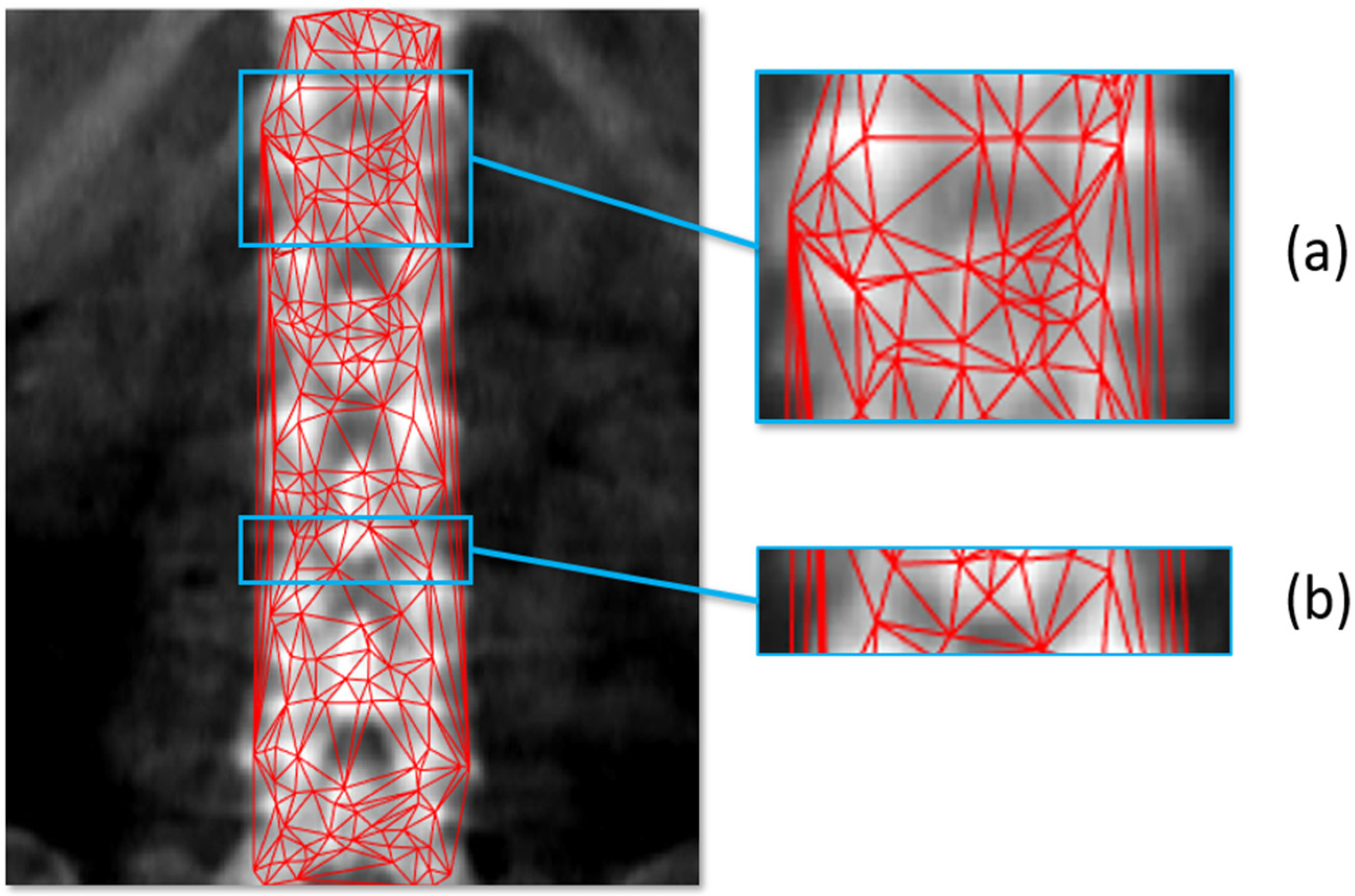
2.2.3. Vertebrae Pose Localization
| Algorithm 2 Delaunay’s edge counting algorithm | |
| Algorithm | Delaunay Edge Counting |
| input | directed graph (DT) with edge lengths |
| data | priority queue with keys , number of edge |
| 1. | initialization |
| 2. | while do |
| 3. | with minimum ; push |
| 4. | foreach vertex such that do |
| 5. | path discovery //-shorter path to ? |
| 6. | if then |
| 7. | |
| 8. | Insert/update with new key; ; |
| 9. | |
| 10. | path counting |
| 11. | if then |
| 12. | |
| 13. | append |
3. Results and Discussion
4. Conclusions
Author Contributions
Funding
Data Availability Statement
Acknowledgments
Conflicts of Interest
References
- Beutel, J.; Kundel, H.L.; Kim, Y.; Van Metter, R.L.; Horii, S.C. Handbook of medical imaging (volume 3). In A Treatise on Electricity and Magnetism, 3rd ed.; Clarendon: Oxford, UK, 1892; Volume 2, pp. 68–73. [Google Scholar]
- Horng, M.-H.; Kuok, C.-P.; Fu, M.-J.; Lin, C.-J.; Sun, Y.-N. Cobb Angle Measurement of Spine from X-ray Images Using Convolutional Neural Network. Comput. Math. Methods Med. 2019, 2019, 6357171. [Google Scholar] [CrossRef] [PubMed] [Green Version]
- Gaál, G.; Maga, B.; Lukács, A. Attention u-net based adversarial architectures for chest X-ray lung segmentation. arXiv 2020, arXiv:2003.10304. [Google Scholar]
- Vidal, P.L.; de Moura, J.; Novo, J.; Ortega, M. Multi-stage transfer learning for lung segmentation using portable X-ray devices for patients with COVID-19. Expert Syst. Appl. 2021, 173, 114677. [Google Scholar] [CrossRef] [PubMed]
- Zhuang, B.; Rohling, R.; Abolmaesumi, P. Region-of-Interest-Based Closed-Loop Beamforming for Spinal Ultrasound Imaging. IEEE Trans. Ultrason. Ferroelectr. Freq. Control. 2019, 66, 1266–1280. [Google Scholar] [CrossRef] [PubMed]
- Buerger, C.; von Berg, J.; Franz, A.; Klinder, T.; Lorenz, C.; Lenga, M. Combining deep learning and model-based segmentation for labeled spine CT segmentation. Med. Imaging 2020, 11313, 307–314. [Google Scholar] [CrossRef]
- Rak, M.; Steffen, J.; Meyer, A.; Hansen, C.; Tönnies, K. Combining convolutional neural networks and star convex cuts for fast whole spine vertebra segmentation in MRI. Comput. Methods Programs Biomed. 2019, 177, 47–56. [Google Scholar] [CrossRef]
- Shirly, S.; Ramesh, K. Review on 2D and 3D MRI Image Segmentation Techniques. Curr. Med Imaging 2019, 15, 150–160. [Google Scholar] [CrossRef]
- Natalia, F.; Meidia, H.; Afriliana, N.; Al-Kafri, A.S.; Sudirman, S.; Simpson, A.; Bashtawi, M. Development of ground truth data for automatic lumbar spine MRI image segmentation. In Proceedings of the 2018 IEEE 20th International Conference on High Performance Computing and Communications; IEEE 16th International Conference on Smart City; IEEE 4th International Conference on Data Science and Systems (HPCC/SmartCity/DSS), Exeter, UK, 28–30 June 2018; pp. 1449–1454. [Google Scholar] [CrossRef]
- Ebrahimzadeh, E.; Fayaz, F.; Ahmadi, F.; Nikravan, M. A machine learning-based method in order to diagnose lumbar disc herniation disease by MR image processing. MedLife Open Access 2018, 1, 1. [Google Scholar]
- Vania, M.; Mureja, D.; Lee, D. Automatic spine segmentation from CT images using Convolutional Neural Network via redundant generation of class labels. J. Comput. Des. Eng. 2019, 6, 224–232. [Google Scholar] [CrossRef]
- Li, Y.; Liang, W.; Zhang, Y.; Tan, J. Automatic Global Level Set Approach for Lumbar Vertebrae CT Image Segmentation. BioMed Res. Int. 2018, 6319879. [Google Scholar] [CrossRef]
- Zhou, W.; Lin, L.; Ge, G. N-Net: 3D Fully Convolution Network-Based Vertebrae Segmentation from CT Spinal Images. Int. J. Pattern Recognit. Artif. Intell. 2019, 33, 1957003. [Google Scholar] [CrossRef]
- Yang, X.; Lei, Y.; Liu, Y.; Tian, S.; Higgins, K.; Beitler, J.J.; Yu, D.S.; Jiang, X.; Liu, T.; Curran, W.J.; et al. Automatic multi-organ segmentation in thorax CT images using U-Net-GAN. Med. Phys. 2019, 10950, 1095010. [Google Scholar] [CrossRef]
- Gabriel, A.T.; Quaresma, C.; Secca, M.F.; Vieira, P. Development and clinical application of Vertebral Metrics: Using a stereo vision system to assess the spine. Med Biol. Eng. Comput. 2018, 56, 1435–1446. [Google Scholar] [CrossRef] [PubMed]
- Gabriel, A.; Quaresma, C.; Secca, M.; Vieira, P. Vertebral Metrics. In Proceedings of the International Joint Conference on Biomedical Engineering Systems and Technologies, Setubal, Portugal, 12–15 January 2016; pp. 235–240. [Google Scholar] [CrossRef] [Green Version]
- Lingayat, N.S.; Tarambale, M.R. A Computer Based Feature Extraction of Lung Nodule in Chest X-Ray Image. Int. J. Biosci. Biochem. Bioinform. 2013, 3, 624–629. [Google Scholar] [CrossRef] [Green Version]
- Nash, C.L.; Moe, J.H. A Study of Vertebral Rotation. J. Bone Jt. Surg. 1969, 51, 223–229. [Google Scholar] [CrossRef] [Green Version]
- Hemalatha, R.; Thamizhvani, T.; Dhivya, A.J.A.; Joseph, J.E.; Babu, B.; Chandrasekaran, R. Active Contour Based Segmentation Techniques for Medical Image Analysis. Med. Biol. Image Anal. 2018, 4, 2. [Google Scholar] [CrossRef] [Green Version]
- Löffler, M.T.; Sekuboyina, A.; Jacob, A.; Grau, A.-L.; Scharr, A.; El Husseini, M.; Kallweit, M.; Zimmer, C.; Baum, T.; Kirschke, J.S. A Vertebral Segmentation Dataset with Fracture Grading. Radiol. Artif. Intell. 2020, 2, e190138. [Google Scholar] [CrossRef]
- Shen, H.; Huang, J.; Zheng, Q.; Zhu, Z.; Lv, X.; Liu, Y.; Wang, Y. A Deep-Learning–Based, Fully Automated Program to Segment and Quantify Major Spinal Components on Axial Lumbar Spine Magnetic Resonance Images. Phys. Ther. 2021, 101, pzab041. [Google Scholar] [CrossRef]
- Kijowski, R.; Liu, F.; Caliva, F.; Pedoia, V. Deep Learning for Lesion Detection, Progression, and Prediction of Musculoskeletal Disease. J. Magn. Reson. Imaging 2019, 52, 1607–1619. [Google Scholar] [CrossRef]
- Grob, A.; Loibl, M.; Jamaludin, A.; Winklhofer, S.; Fairbank, J.C.T.; Fekete, T.; Porchet, F.; Mannion, A.F. External validation of the deep learning system “SpineNet” for grading radiological features of degeneration on MRIs of the lumbar spine. Eur. Spine J. 2020, 31, 2137–2148. [Google Scholar] [CrossRef]
- Goedmakers, C.M.W.; Lak, A.M.; Duey, A.H.; Senko, A.W.; Arnaout, O.; Groff, M.W.; Smith, T.R.; Vleggeert-Lankamp, C.L.A.; Zaidi, H.A.; Rana, A.; et al. Deep Learning for Adjacent Segment Disease at Preoperative MRI for Cervical Radiculopathy. Radiology 2021, 301, 664–671. [Google Scholar] [CrossRef] [PubMed]
- Mbarki, W.; Bouchouicha, M.; Frizzi, S.; Tshibasu, F.; Ben Farhat, L.; Sayadi, M. Lumbar spine discs classification based on deep convolutional neural networks using axial view MRI. Interdiscip. Neurosurg. 2020, 22, 100837. [Google Scholar] [CrossRef]
- Ghosh, S.; Malgireddy, M.R.; Chaudhary, V.; Dhillon, G. A new approach to automatic disc localization in clinical lumbar MRI: Combining machine learning with heuristics. In Proceedings of the 2012 9th IEEE International Symposium on Biomedical Imaging (ISBI), Barcelona, Spain, 2–5 May 2012; pp. 114–117. [Google Scholar] [CrossRef]
- Azevedo, T.C.S.; Tavares, J.M.R.S.; Vaz, M.A.P. 3D Object Reconstruction from Uncalibrated Images Using an off-the-Shelf Camera; Springer: Berlin/Heidelberg, Germany, 2009; pp. 117–136. [Google Scholar] [CrossRef] [Green Version]
- Azevedo, T.C.; Tavares, J.M.R.; Vaz, M.A. 3D reconstruction and characterization of human external shapes from 2D images using volumetric methods 3D reconstruction and characterization of human external shapes from 2D images using volumetric methods. Comput. Methods Biomech. Biomed. Eng. 2010, 13, 359–369. [Google Scholar] [CrossRef] [PubMed]
- Zhao, Y.J.; Shi, L.; Li, J.; Griffith, J.F.; Ahuja, A.T.; Heng, P. Vertebra segmentation of spine MRI with improved GVF snake based on shape knowledge. In Proceedings of the 2011 International Conference on Machine Learning and Cybernetics, Guangxi, China, 10–13 July 2011; Volume 4, pp. 1867–1871. [Google Scholar]
- Pintar, F.A.; Yoganandan, N.; Myers, T.; Elhagediab, A.; Sances, A., Jr. Biomechanical properties of human lumbar spine ligaments. J. Biomech. 1992, 25, 1351–1356. [Google Scholar] [CrossRef] [PubMed]
- Rahman, M.; Cao, Y.; Sun, X.; Li, B.; Hao, Y. Deep pre-trained networks as a feature extractor with XGBoost to detect tuberculosis from chest X-ray. Comput. Electr. Eng. 2021, 93, 107252. [Google Scholar] [CrossRef]


| List | Description | Unit | |
|---|---|---|---|
| 1. | Image type | Normal X-ray (Plain film) | - |
| 2. | Body part | Lumbar spine (LSPINE) | - |
| 3. | View | AP view and LA view | - |
| 4. | Numbers of patients | 3600 | records |
| 5. | Numbers of images | 7200 | images |
| 6. | Numbers of disorder patient | 621 | records |
| 7. | Numbers of spinal disorders | 788 | cases |
| 8. | Dataset size | 18.5 | GB |
| 9. | Ground truth |
| - |
| 10. | Ground truth type | Four corner coordinates points. | - |
| 11. | File types | JPG (image) and CSV (ground truth) | - |
| 12. | Locations | Thailand, Chonburi, Burapha University Hospital (BUH) | - |
| 13. | Years of records | 2000–2021 | - |
| 14. | Age range | (6–97) | years old |
| 15. | Image dimension | Original | - |
| 16. | Motivation | Delivering gold standard lumbar spine dataset of Thais for researchers around the world to develop and improve performance of the segmentation algorithms on the lumbar spine. | - |
| Dataset | Confusion Matrix | |||
|---|---|---|---|---|
| Accuracy | Recall | Precision | FNR | |
| Good | 87.60 | 88.32 | 84.24 | 11.56 |
| Medium | 81.38 | 86.55 | 83.11 | 16.20 |
| Low | 71.97 | 81.23 | 79.73 | 18.51 |
| Average | 80.32 | 85.37 | 82.36 | 15.42 |
| Method | Evaluation | ||
|---|---|---|---|
| JM | HD | PAD | |
| Proposed approach | 0.82 | 10.87 | 2.33 |
| Watershed | 0.54 | 46.28 | 5.19 |
| DRLSE | 0.77 | 27.98 | 4.63 |
| Region growing | 0.81 | 32.89 | 4.48 |
| Method | Average Time Usages (s) | |||
|---|---|---|---|---|
| Good | Medium | Lowth | Average time | |
| Proposed approach | 0.92 | 1.04 | 1.36 | 1.11 |
| Watershed | 1.33 | 1.89 | 1.92 | 1.71 |
| DRLSE | 1.74 | 1.79 | 1.81 | 1.78 |
| Region growing | 1.56 | 1.88 | 2.21 | 1.88 |
Disclaimer/Publisher’s Note: The statements, opinions and data contained in all publications are solely those of the individual author(s) and contributor(s) and not of MDPI and/or the editor(s). MDPI and/or the editor(s) disclaim responsibility for any injury to people or property resulting from any ideas, methods, instructions or products referred to in the content. |
© 2022 by the authors. Licensee MDPI, Basel, Switzerland. This article is an open access article distributed under the terms and conditions of the Creative Commons Attribution (CC BY) license (https://creativecommons.org/licenses/by/4.0/).
Share and Cite
Yookwan, W.; Limchareon, S.; Lee, S.-H.; Jang, J.-S.; Lee, D.; Chinnasarn, K. Coarse X-ray Lumbar Vertebrae Pose Localization and Registration Using Triangulation Correspondence. Processes 2023, 11, 61. https://doi.org/10.3390/pr11010061
Yookwan W, Limchareon S, Lee S-H, Jang J-S, Lee D, Chinnasarn K. Coarse X-ray Lumbar Vertebrae Pose Localization and Registration Using Triangulation Correspondence. Processes. 2023; 11(1):61. https://doi.org/10.3390/pr11010061
Chicago/Turabian StyleYookwan, Watcharaphong, Sornsupha Limchareon, Sang-Hun Lee, Jun-Su Jang, Daesung Lee, and Krisana Chinnasarn. 2023. "Coarse X-ray Lumbar Vertebrae Pose Localization and Registration Using Triangulation Correspondence" Processes 11, no. 1: 61. https://doi.org/10.3390/pr11010061