Article

Isolation, Characterization, and Breast Cancer Cytotoxic Activity of Gyrophoric Acid from the Lichen Umbilicaria muhlenbergii

by
Mahshid Mohammadi
1,
Vasudeo Zambare
2,3,
Zacharias Suntres
4 and
Lew Christopher
5,6,*
1
Biorefining Research Institute, Lakehead University, Thunder Bay, ON P7B 5Z5, Canada
2
Om Biotechnologies, Nashik 422011, India
3
Centre for Environmental Sustainability and Water Security (IPASA), Department of Water and Environmental Engineering, School of Civil Engineering, Universiti Teknologi Malaysia, Bahru 81310, Malaysia
4
Northern Ontario School of Medicine, Thunder Bay, ON P7B 5Z5, Canada
5
Biorefinery World, LLC, Rapid City, SD 57702, USA
6
Lee Enterprises Consulting, Sherwood, AR 72120, USA
*
Author to whom correspondence should be addressed.
Processes 2022, 10(7), 1361; https://doi.org/10.3390/pr10071361
Submission received: 12 June 2022 / Revised: 8 July 2022 / Accepted: 9 July 2022 / Published: 13 July 2022

Abstract

:
Lichens produce a large variety of secondary metabolites with diverse bioactivities, chemical structures, and physicochemical properties. For this reason, there is a growing interest in the use of lichen-derived bioactive molecules for drug discovery and development. Here, we report on the isolation, identification, and cytotoxic evaluation of gyrophoric acid (GA) from the lichen Umbilicaria muhlenbergii, a largely unexplored and scantly described lichen species. A simple purification protocol was developed for the fractionation of lichen crude extracts with silica gel column chromatography using solvents with changing polarity. GA was identified in one of the fractions with Fourier transform infrared spectroscopy (FTIR), ion trap mass spectrometry (MS), and nuclear magnetic resonance spectroscopy (1H-NMR and 13C-NMR). The FTIR spectra demonstrated the presence of aromatic and ester functional groups C=C, C-H, and C=O bonds, with the most remarkable signals recorded at 1400 cm−1 for the aromatic region, at 1400 cm−1 for the CH3 groups, and at 1650 cm−1 for the carbonyl groups in GA. The MS spectra showed a molecular ion [M-1] at (m/z) 467 with a molecular weight of 468.4 and the molecular formula C24H20O10. that correspond to GA. The 1H-NMR and 13C-NMR spectra verified the chemical shifts that are typical for GA. GA reduced the cell viability of breast cancer cells from the MCF-7 cell line by 98%, which is indicative of the strong cytotoxic properties of GA and its significant potential to serve as a potent anticancer drug.

1. Introduction

Cancer is a complex disease with a strong relationship between genetic and environmental factors [1]. One of the hallmarks of cancer is that cancer cells can penetrate adjacent parts of the body and lead to invasion and metastasis [2]. Although cancer research has advanced considerably over the years, cancer mortality remains high [3]. A report showed that the cancer rate has been escalating since 1990, with the most common types being lung, colorectal, and stomach cancers [4]. Between 2012 and 2020, new cancer cases increased by 37% to reach 19.3 million [5,6]. In the U.S. alone, 1.8 million new cancer cases occurred, representing 9% of the global cancer occurrence in 2020 [7]. Among cancer types, breast cancer remains one of the most common lethal types of cancer for women [8], resulting in the deaths of 1 out of 10 women diagnosed with breast cancer [9]. In 2020, there were 2.3 million women diagnosed with breast cancer and 685,000 deaths globally [10]. The conventional methods of cancer treatment include surgery, radiotherapy, and chemotherapy [11]. In this regard, investigations to identify new chemotherapeutic agents, such as natural compounds with minimum side effects, are gaining importance [12,13].
Natural bioactive molecules are becoming widely recognized for their antifungal, antibiotic, antiviral, anti-inflammatory, and antitumor activities [14]. About 60% of all natural compounds are known as pharmaceutical agents [15], with more than half of all approved drugs for cancer treatment originating from natural sources [16]. Among these natural sources, lichens are symbiotic organisms with diverse secondary metabolites [3,17,18,19]. These organisms, in addition to their well-known traditional medicinal uses for folk therapeutic and phytochemical purposes [20,21,22,23], have been also utilized in other practical areas, including dyes, perfumes, and food [24,25].
More than 1000 secondary metabolites have been identified in different lichens with various biological and pharmacological activities due to the presence of numerous aliphatic, cycloaliphatic, aromatic, and terpenoid compounds [13,26,27]. Lichen crude extracts and lichen secondary metabolites have produced apoptotic and cytotoxic effects against various cancer cell types. For example, lichen extracts and secondary metabolite parietin from Parmelia sulcate and Xanthoria parietina showed antiproliferative activity against breast cancer cells [28,29]. Similarly, the lichen Cladonia foliacea and its secondary metabolites using acid, atranorin, and fumarprotocetraric acid, as well as Lobaria pulmonaria and its metabolite static acid, were active against colon cancer cell lines [30,31]. Evidence exists for the promising anti-tumor effects of other lichen-derived secondary metabolites, such as protolichesterinic acid and physodic acid on cervical cancer [32,33], olivetoric acid on liver cancer [34], and atranorin and usnic acid on melanoma [35,36]. Overall, research on lichen-derived secondary metabolites to date has demonstrated their great potential for use as therapeutic agents. Strong evidence has been obtained for their mechanism of action on cancer cells that includes apoptosis, necrosis, or autophagy, with the cell cycle arrest at G0/G1 phases; cell cycle regulation associated with cyclin-dependent kinases (CDK4, CDK6) or cyclin D1; oxidative stress due to superoxide dismutase (SOD) or malondialdehyde (MDA); modulation of inflammatory responses via TNF-α, IL-1β, IL-6, and TGF-β1; and targeting of microRNA molecules, modulation of anti-proliferative effects by regulating signaling pathways (ERK1/2 and AKT), or proliferating protein marker Ki-67 [37,38].
The biological activities and therapeutic potential of secondary metabolites from Umbilicaria species have also been investigated. These bioactive molecules had different aromatic, aliphatic, and cyclic structures and were isolated from Umbilicaria species, such as U. crustulosa, U. cylindrical [39], U. crustulosa, U. cylindrical, U. polyphylla [40], and U. hirsuta [19]. In the present study, we report on the extraction, fractionation, purification, and cytotoxic evaluation of bioactive fr3actions from the lichen U. muhlenbergii against the breast cancer cell line MCF-7. By applying in vitro cell viability MTT assays coupled with an array of spectroscopic, physicochemical isolation, and characterization techniques, we were able to isolate and identify the secondary lichen metabolite GA as a potent and promising cytotoxic molecule. The current work is a continuation of our previous studies on U. muhlenbergii [38,41] and provides new evidence for the effects of purified GA on cancer cells. Although GA is known for its anti-tumor activity, reports on its isolation methods are scant. Our objective was to present a systematic study that describes an efficient extraction and purification protocol for the isolation of GA from U. muhlenbergii. The protocol that we have developed may be used as an alternative route for the production of GA acid as well as the isolation and purification of other polyphenolic depside molecules.

2. Materials and Methods

2.1. Collection of Lichen Specimens

Lichen samples were collected around the Tamblyn Lake of Northwest Ontario, Canada, and identified as U. muhlenbergii based on an organoleptic macroscopic examination. A voucher specimen was deposited in the Claude Garton Herbarium at Lakehead University, Thunder Bay, Ontario, Canada.

2.2. Chemicals

Dulbecco’s modified Eagle’s medium (DMEM), dimethylsulfoxide (DMSO), fetal bovine serum (FBS), 3-(4,5-dimethylthiazol-2-yl)-2,5-diphenyltetrazolium bromide (MTT), phosphate buffered saline (PBS) containing calcium, magnesium, penicillin/streptomycin, and trypsin-EDTA solution were purchased from Sigma-Aldrich (St. Louis, MO, USA). All other chemicals and regents were bought from ThermoFisher Scientific (Waltham, MA, USA).

2.3. Cell Culture and Maintenance

The human breast cancer cell line MCF-7 (purchased from American Type Culture Collection, ATCC, Manassas, VA, USA) was grown in DMEM supplemented with 10% FBS and 1% penicillin/streptomycin. The cell line was maintained in a humidified atmosphere containing 5% CO2 at 37 °C.

2.4. Cell Viability Assay and IC50

The MTT method [41] was used to measure cell viability. The MCF-7 cells were seeded in a 96-well plate (Corning@Costar, 96-well plate) at a concentration of 1 × 10⁴ cells/well and incubated in an atmosphere of 5% CO2 and 37 °C for 24 h to obtain good adherence. The initial medium was then replaced with fresh medium supplemented with 300–500 µg/mL of lichen extract. The MTT substrate, prepared in a physiologically balanced solution, was added to the cell in culture at a final concentration of 5 mg/mL and incubated at 37 °C for 4 h. Thereafter, 50 µL of DMSO was added to each well to dissolve the formazan crystals [13]. The absorbance of each well was measured with a BioTek Microplate Reader Spectrophotometer (San Francisco, CA, USA) at 490 nm (using a reference wavelength of 690 nm). The cells in the medium alone and those in hydrogen peroxide (H2O2) served as a negative and positive control, respectively [42]. The percentage cell viability was calculated as per Equation (1):
C e l l v i a b i l i t y % = A t A c / A D M S O A c × 100
where At is the absorbance of treated cells, Ac is the absorbance of background controls, and ADMSO is the absorbance of matched DMSO concentration controls. The half-maximal inhibitory concentration (IC50) was determined from the cell viability assay using 300–500 µg/mL of lichen extract [43].

2.5. Extraction and Lichenochemical Analysis

The lichen samples were air-dried and ground using a mortar and pestle. A 50 g dried lichen powder sample was soaked in 500 mL acetone (HPLC grade) and placed in an airtight screw cap bottle on an Innova 44 orbital shaker (New Brunswick Scientific, Edison, NJ, USA) at 150 rpm and at room temperature for 24 h. The crude acetone extract was then dried by vacuum evaporation using a rotary evaporator (Buchi, New Castle, DE, USA). Thereafter, the extract powder was subjected to preliminary lichenochemical screening and analysis using different reagents as described in [35] to identify the presence of phytochemicals (alkaloids, hydrocarbon, terpenoids, proteins, tannins, flavonoids, saponins, phenols, and glycosides).

2.6. Purification and Characterization of GA

As shown in Figure 1, following the initial solvent extraction of the lichen U. muhlenbergii, solvents (acetone) were removed by air-drying evaporation in a biosafety cabinet at room temperature, and the dried extracts were redissolved in fresh acetone and subjected to successive extraction on a 60 G silica gel column using solvents with increasing polarity (hexane, ethyl acetate, ethyl acetate: methanol (9:1), ethyl acetate: methanol (8:2), ethyl acetate: methanol (5:5), and pure methanol). Following solvent evaporation, the dried extracts were analyzed spectroscopically to identify the bioactive component (GA) using UV-Vis spectroscopy; Tensor37 Fourier transformer Infrared (Bruker, Billerica, MA, USA); amazonX ion trap mass spectrometry (Bruker, Billerica, MA, USA) in negative mode; and 1H and 13C NMR spectra at 500 and 125 MHz, with 16 scans using a Bruker Avance III 500 spectrometer (Bruker, Billerica, MA, USA). The 2D NOESY gHSQC and gHMBC methods were employed [19]. The purity validation of fraction F1 was carried out with high-performance liquid chromatography (HPLC, Agilent Technologies 1260) with DAD detection, Kromasil SGX C18 column (7 μm), mobile phase A (5% acetonitrile +1% (v/v) trifluoracetic acid), and mobile phase B (80% acetonitrile), employing the isocratic program at a flow rate of 0.7 mL/min: 0 min (50% A and 50% B); 25 min (0% A and 100% B); 30 min (50% A and 50% B).

2.7. Statistical Analysis

All experiments were carried out in triplicate, and each result was the average of three independent experiments. Statistical analysis was performed with Microsoft Excel 2010, and the data are presented as the mean standard deviation (±SD) of triplicates. One-way Analysis of Variance (ANOVA) with p-values were calculated from https://goodcalculators.com/one-way-anova-calculator/ (accessed on 6 June 2022).

3. Results and Discussion

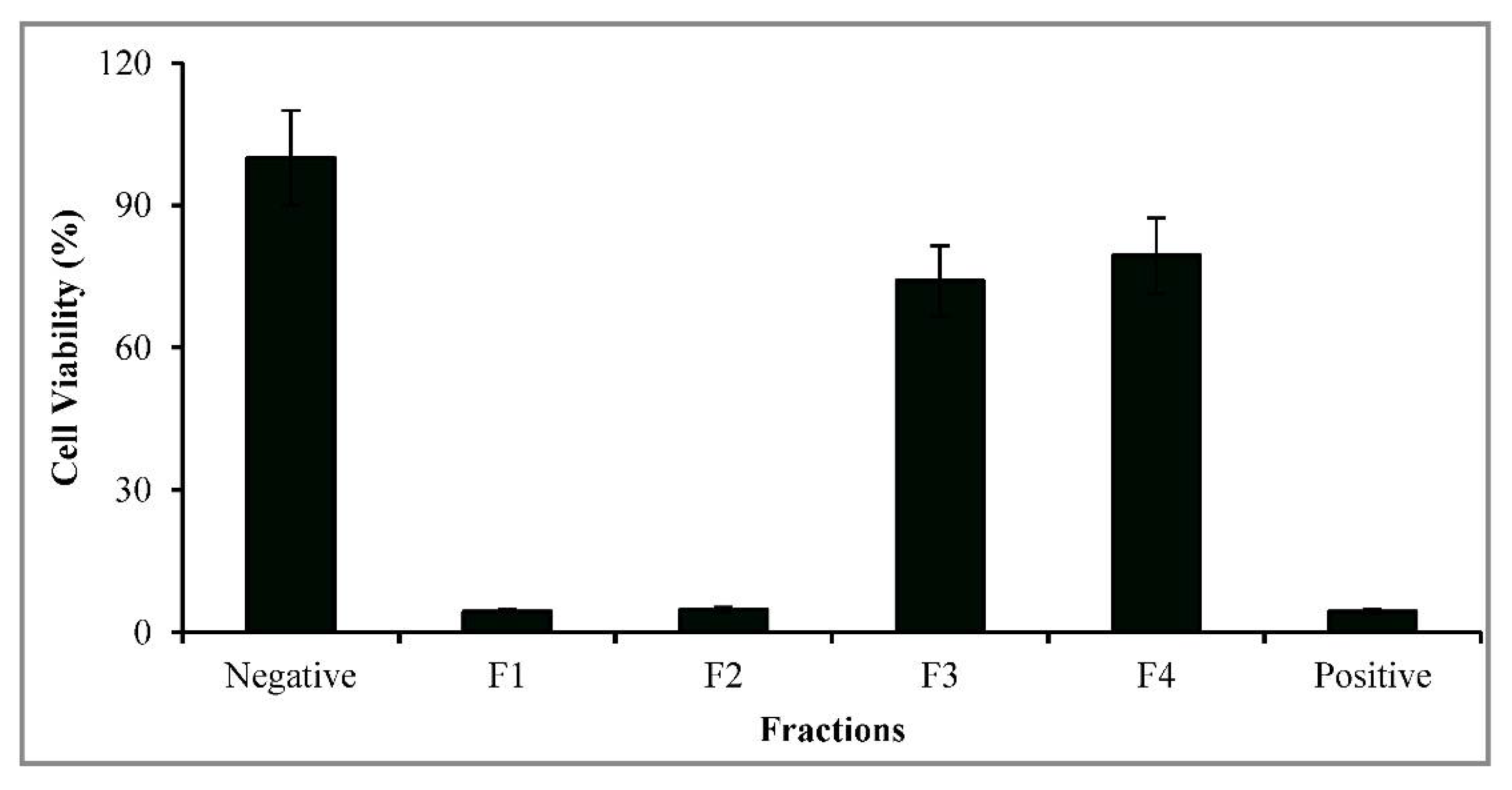
3.1. Lichen Identification and Anti-Proliferation Screening

A lichen specimen was collected from the area of Tamblyn Lake in Northwestern Ontario, and following organoleptic macroscopic examination, it was identified as Umbilicaria muhlenbergii. Acetone extraction of the lichen yielded 14.3% (w/w) dried crude acetone extract. The cytotoxic activity of acetone extracts from U. muhlenbergii against MCF-7 breast cancer cell line has been described in our previous study [41].

3.2. Lichenochemical Analysis

Lichenochemical screening studies on the crude extract of U. muhlenbergii revealed the presence of alkaloids and phenols as major components, with tannins, saponins, and hydrocarbons present in smaller quantities, whereas flavonoids, terpenoids, proteins, and glycosides were not detected (data not shown). A qualitative analysis of two Sri Lankan extracts from the lichens Parmotrema tinctorum and Parmotrema rampoddense revealed the presence of saponins, flavonoids, and polyphenols [44]. Anthracene glycosides were present in P. tinctorum but not in P. rampoddense, whereas proteins, alkaloids, tannins, reducing sugars, cyanogenic glycosides, and cardenolide glycosides were absent from both lichen extracts [44]. The presence of phenols and alkaloids with strong bioactivities is also in agreement with the published literature [17,45,46]. It has been reported that most of the lichen phenolics isolated from Parmelia caperata, Cladonia convoluta, C. rangiformis, Platisma glauca, and Ramalina cuspidata exhibited strong antioxidant activities and anticancer activities [47]. Lichen-derived compounds have been extensively discussed in previous reviews [48,49], and they provide in-depth information on their bio-therapeutic potential.

3.3. Purification and Characterization of GA

A detailed schematic diagram of purification of GA is shown in Figure 1. The bioactive compound(s) responsible for the anticancer activity of U. muhlenbergii was purified with successive solvent elution using nonpolar (hexane and ethyl ether) and polar (methanol, ethyl acetate) solvents as well as their combinations. It appeared that hexane (nonpolar solvent) and methanol (polar solvent) did not produce any bioactive fractions; however, the solvent elution with ethyl acetate alone, and in combination with methanol, yielded chemical containing fractions (F1–F4), as shown in Table 1. In many cases, the purification of secondary metabolites from lichens is tedious and complex. The product yield is affected by the type of solvent used; the presence of other compounds with similar structures and colored substances (pigments); possible interactions; and/or the formation of hydrogen bonding between the targeted molecule, the solvent, and other molecules. All this may create a masking effect due to the overlapping of compounds of different solubility and polarity with the molecule of interest, gyrophoric acid in our case, thereby altering the molecule’s polarity and reducing its purification yield [50]. A maximum eluent yield of 0.14% (w/w of lichen biomass) was obtained in fraction F1 using ethyl acetate as the solvent. Fractions F2–F4 contained extraction yields that decreased as the polarity of the mixed solvent increased.
Fractions F1-F4 were examined for their cytotoxic effect on MCF-7 breast cancer cells. Fractions F1 and F2 showed a maximum of 98% and 96% cytotoxicity, respectively (Figure 2). These results were comparable with the positive control of hydrogen peroxide (H2O2). In comparison, fractions F3 and F4 had a much lower antiproliferative activity, thereby reducing the MCF-7 cell viability by only 26% and 21%, respectively. Thus, fraction F1 was selected for further purification and identification studies as it contained the highest eluent yield (Table 1) and highest anti-proliferative activity (Figure 2). These studies included spectroscopic MS, UV, FTIR, and NMR data for F1 [19,51,52,53,54,55,56,57].
The UV absorbance of F1 (Figure S1) was measured in the wavelength range of 240–340 nm. The UV absorbance peaks for GA were detected at 210, 240, 310, and 340 nm, with high similarity for the depside groups [48] and in good agreement with previous findings [58,59,60].
The IR spectrum revealed functional groups and strength binding [13,61]. From the IR spectra at 600–4000 cm−1 (Figure S2), it is evident that the peaks at approximately 3300, 3200, and 3665 cm−1 reflect the stretching vibration of hydroxyl (OH) groups. The bands at 670–900 cm−1 result from the presence of aromatic groups and C-H bending in the compound.
The electron ionization mass spectroscopy (Figure 3) was recorded in the negative ionization mode [M-1] (m/z) at 467.0, with an error of 1.4 ppm, corresponding to GA with an exact molecular weight of 468.4 and molecular formula of C24H20O10 (Table 2). Its fragmentation produced an ion at (m/z) 316.9 due to ester cleavage, which may indicate lecanoric acid in trace amounts with an error of 1.1 ppm, as presented in Table 2 [55,62,63]. Similar fragmentation patterns for GA were also observed during the rapid identification of lichen compounds based on the structure–fragmentation relationship using ESI-MS/MS analysis [52]. Other studies also demonstrated that GA is present in high concentrations and is the prevalent depside in the Umbilicaria genus [19,53,64,65,66,67]. The chemical formula C24H20O10 for GA was verified from MS data in several reports [52,68,69].
The 1H-NMR and 13C-NMR spectra verified the chemical shifts that are typical for GA [52,61,69,70] 1H-NMR (Figure S3): (DMSO-d6, 500 MHz) δ 10.52 (hydroxyl protons), 6.66 (1H, d, J5, 3= 2.6 Hz) (aromatic protons), 6.69 (1H, d, J3, 5= 1.3 Hz), 6.49 (1H, d J3″, 5= 2.6 Hz), 2.36 (3H, s), 6.44 (1H, d, J5′, 3′ = 1.3 Hz), 6.25 (1H, d, J3, 5 = 2.6 Hz), 6.23 (1H, d, J5, 3 = 2.6 Hz), 2.35 (3H, s), 2.49 (3H,s).
13C-NMR (Figure S4): (DMSO-d6, 125 MHz) δ 108.49 (C-1), 160.0 (C-2), 100.5 (C-3), 161.12 (C-4), 109.84 (C-5), 140.20 (C-6), 167.18 (C-7), 21.28 (C-8), 118.28 (C-1′), 156.30 (C-2′), 107.22 (C-3′), 152.07 (C-4′), 114.21 (C-5′), 137.92 (C-6′), 165.78 (C-7′), 19.35 (C-8′), 116.70 (C-1″), 162.44 (C-2″), 107.17 (C-3″), 151.83 (C-4″), 113.45 (C-5″), 141.17 (C-6″), 171.47 (C-7″), 22.12 (C-8″).
Evidence for the localization of the methyl and hydroxyl groups on the aromatic rings of GA was obtained from heteronuclear multiple bond correlation (HMBC) experiments (Figure S5). The HMBC spectra showed HMBC correlations between the protons of the methyl groups/hydroxyl groups and the corresponding carbon atoms. In addition, the heteronuclear single quantum coherence (HSQC) spectrum provided data of direct determination of carbon and attached protons (hydrogen), which belong to the same spin system (Figure S6). Based on these observations, the molecular structure of GA can be presented as shown in Figure 4. The GA purity was further confirmed by HPLC (data not shown). GA, isolated from U. hirsuta, was likewise identified by 1H-NMR and 13C-NMR, and validated with HPLC [19].

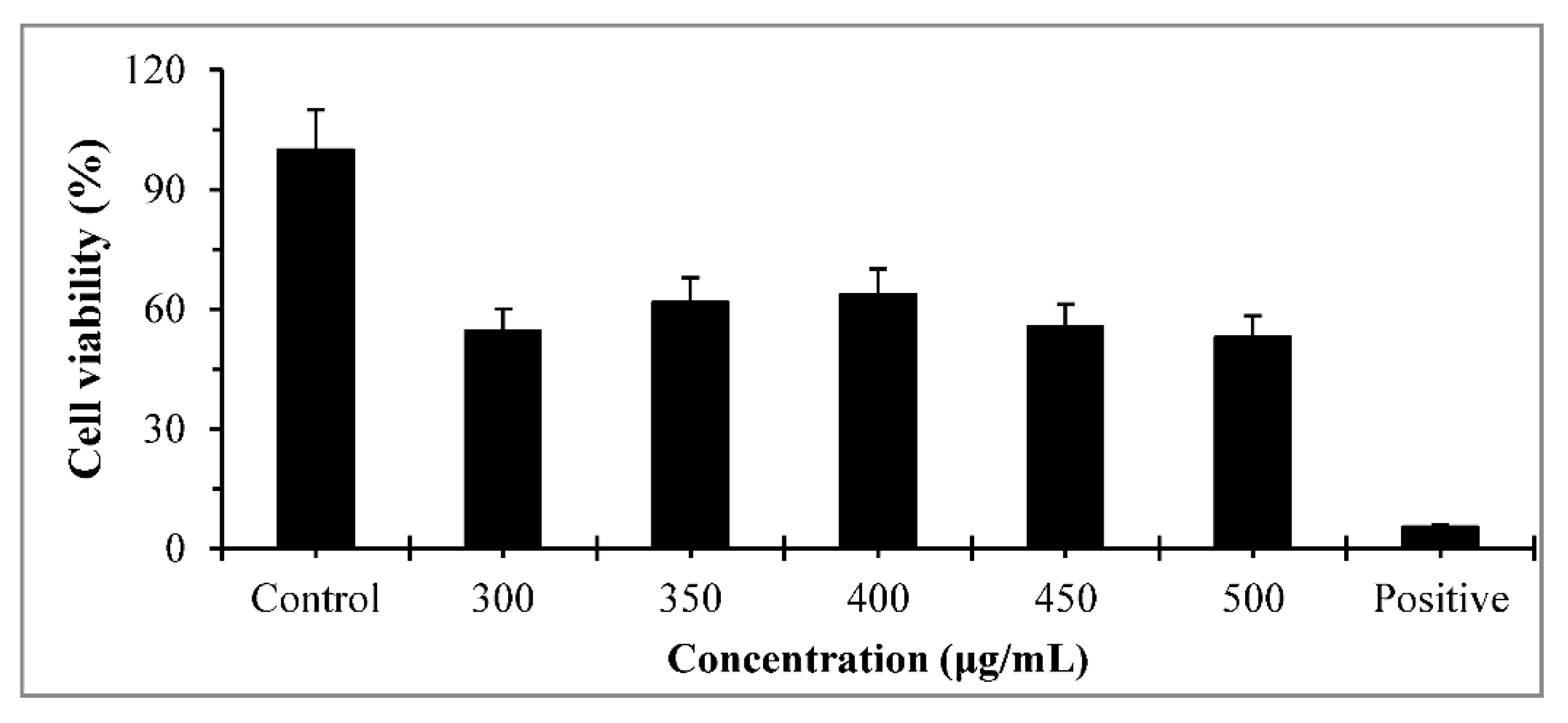
3.4. Cytotoxicity of GA

Lichen secondary metabolites are known to have diverse biological activities, including antibacterial, antitumor, antiproliferative, and cytotoxic effects [18,48]. Anticancer activities have been reported for several lichen-derived compounds, such as stictic acid [31], physodic acid [13], and parietin, to name a few. As evident from Figure 5 of this study, GA isolated from U. muhlenbergii crude extracts decreased the cell viability of MCF-7 cells. At 300 µg/mL GA, the cell proliferation was inhibited by approximately 45%, and thereafter, with a further increase in the GA dose up to 500 µg/mL, it reached a plateau. Keeping in mind the SD of the data presented in Figure 5, no significant difference in the cell viability was observed in the 300–500 µg/mL GA concentration range. This might be due to the fact that the MCF-7 cancer cells were exposed to GA for only 4 h. The low exposure time did not allow for the development of a clear dose-dependent correlation between cell viability and GA concentration using the MTT assay. Similar observations were reported for the impact of the GA dose on the inhibition of cell proliferation for HDF cells after their incubation with GA for 24 h [19]. Increasing the incubation time to 72 h produced a well-pronounced dose-dependent effect of GA on cell proliferation. The IC50 value of 478 µM of GA from U. muhlenbergii, calculated from the cell viability data on MCF-7 cells, was comparatively close to the 384 µM reported for GA from U. hirsuta on the same cell line [19]. In comparison, a standard GA showed >200 µM on the HT-29 human colon adenocarcinoma cell line [70]. Previous work has identified GA as a major bioactive compound present in 31 of the 33 studied Umbilicaria species, including U. muhlenbergii [66,71]. The present data are consistent with our previous findings that suggested the inhibition of cellular proliferation and induction of apoptosis of U. muhlenbergii crude extracts as possible causes for MCF-7 cell death [41], and they provide further evidence that GA can act as a potent anticancer agent.

4. Conclusions

This study demonstrates that the lichen U. muhlenbergii is capable of producing GA as a predominant secondary metabolite with strong anticancer activity against MCF-7 breast cancer cells. A simplified extraction and purification protocol to obtain pure GA from U. muhlenbergii crude extract was developed. The molecular structure of GA was validated through spectroscopic analyses that included UV, FTIR, MS, NMR, HSQC, and HMBC. GA, 4-({4-[(2,4-dihydroxy-6-methylphenoxy)carbonyl]-2-hydroxy-6-methylbenzoyl}oxy)-2-hydroxy-6-methylbenzoic acid, is a depside molecule that, in addition to the three aromatic rings, contains functional hydroxyl and carboxyl groups. While the polyaromatic structure of GA facilitates free radical scavenging activity, the functional side-groups impart selective reactivity of GA toward biochemical molecules, such as enzymes and cell-bound proteins, thereby promoting a dynamic interaction with different cell types. Due to the unique properties and functionalities that the molecular structure of GA presents, its potential therapeutic applications are expanding beyond the promise it holds as a cytotoxic and antitumor agent to encompass new medicinal uses that take advantage of the antioxidant, antimicrobial, and anti-inflammatory utilities of GA. Further assessment of the cytotoxic effects of GA in clinical studies is needed to establish the potential for anticancer drug development and application.

Supplementary Materials

The following are available online at https://www.mdpi.com/article/10.3390/pr10071361/s1, Figure S1: UV spectrum of fraction F1 (gyrophoric acid), Figure S2: IR spectrum of fraction F1 (gyrophoric acid); Figure S3: 1H-NMR spectrum of fraction F1 (gyrophoric acid); Figure S4: 13C-NMR spectrum of fraction F1 (gyrophoric acid); Figure S5: Selected HMBC correlations in fraction F1 (gyrophoric acid); Figure S6: HSQC spectrum of fraction F1 (gyrophoric acid).

Author Contributions

Methodology, M.M. and V.Z.; formal analysis, software and validation, M.M.; conceptualization, writing—original draft preparation, M.M., V.Z. and L.C.; final review, editing and final draft preparation, V.Z., Z.S. and L.C. All authors have read and agreed to the published version of the manuscript.

Funding

This research received no external funding.

Acknowledgments

The authors wish to thank Ladislav Malek from Lakehead University for the sample collection and identification of U. muhlenbergii. Funding from the Biorefining Research Institute (BRI) at Lakehead University, Ontario, Canada, is gratefully acknowledged.

Conflicts of Interest

The authors declare no conflict of interest.

References

  1. Ouyang, L.; Shi, Z.; Zhao, S.; Wang, F.-T.; Zhou, T.-T.; Liu, B.; Bao, J.-K. Programmed cell death pathways in cancer: A review of apoptosis, autophagy and programmed necrosis. Cell Prolif. 2012, 45, 487–498. [Google Scholar] [CrossRef] [PubMed]
  2. Greenwald, P. Cancer chemoprevention. BMJ 2002, 324, 714–718. [Google Scholar] [CrossRef] [PubMed]
  3. Nguyen, T.T.; Yoon, S.; Yang, Y.; Lee, H.-B.; Oh, S.; Jeong, M.-H.; Kim, J.-J.; Yee, S.-T.; Crişan, F.; Moon, C.; et al. Lichen secondary metabolites in Flavocetraria cucullata exhibit anti-cancer effects on human cancer cells through the induction of apoptosis and suppression of tumorigenic potentials. PLoS ONE 2014, 9, e111575. [Google Scholar] [CrossRef] [PubMed] [Green Version]
  4. Parkin, D.M.; Bray, F.; Ferlay, J.; Pisani, P. Estimating the world cancer burden: Globocan 2000. Int. J. Cancer 2001, 94, 153–156. [Google Scholar] [CrossRef]
  5. Ferlay, J.; Soerjomataram, I.; Dikshit, R.; Eser, S.; Mathers, C.; Rebelo, M.; Parkin, D.M.; Forman, D.; Bray, F. Cancer incidence and mortality worldwide: Sources, methods and major patterns in GLOBOCAN 2012. Int. J. Cancer 2015, 136, E359–E386. [Google Scholar] [CrossRef]
  6. Sung, H.; Ferlay, J.; Siegel, R.L.; Laversanne, M.; Soerjomataram, I.; Jemal, A.; Bray, F. Global Cancer Statistics 2020: GLOBOCAN Estimates of Incidence and Mortality Worldwide for 36 Cancers in 185 Countries. CA Cancer J. Clin. 2021, 71, 209–249. [Google Scholar] [CrossRef]
  7. Siegel, R.L.; Miller, K.D.; Jemal, A. Cancer statistics, 2020. CA Cancer J. Clin. 2020, 70, 7–30. [Google Scholar] [CrossRef]
  8. Azamjah, N.; Soltan-Zadeh, Y.; Zayeri, F. Global Trend of Breast Cancer Mortality Rate: A 25-Year Study. Asian Pac. J. Cancer Prev. 2019, 20, 2015–2020. [Google Scholar] [CrossRef]
  9. Alkabban, F.M.; Ferguson, T. Breast Cancer; NIH National Library of Medicine, StatPearls Publishing: Treasure Island, FL, USA, 2021; pp. 1–14. Available online: https://www.ncbi.nlm.nih.gov/books/NBK482286/#!po=89.2857 (accessed on 6 June 2022).
  10. WHO. Breast Cancer. Available online: https://www.who.int/news-room/fact-sheets/detail/breast-cancer (accessed on 6 June 2022).
  11. Sharifi-Rad, J.; Ozleyen, A.; Boyunegmez Tumer, T.; Oluwaseun Adetunji, C.; El Omari, N.; Balahbib, A.; Taheri, Y.; Bouyahya, A.; Martorell, M.; Martins, N.; et al. Natural products and synthetic analogs as a source of antitumor drugs. Biomolecules 2019, 9, 679. [Google Scholar] [CrossRef]
  12. Studzińska-Sroka, E.; Majchrzak-Celińska, A.; Zalewski, P.; Szwajgier, D.; Baranowska-Wójcik, E.; Kaproń, B.; Plech, T.; Żarowski, M.; Cielecka-Piontek, J. Lichen-Derived Compounds and Extracts as Biologically Active Substances with Anticancer and Neuroprotective Properties. Pharmaceuticals 2021, 14, 1293. [Google Scholar] [CrossRef]
  13. Cardile, V.; Graziano, A.C.E.; Avola, R.; Piovano, M.; Russo, A. Potential anticancer activity of lichen secondary metabolite physodic acid. Chem. Biol. Interact. 2017, 263, 36–45. [Google Scholar] [CrossRef] [PubMed]
  14. Azwanida, N. A review on the extraction methods use in medicinalplants, principle, strength and limitation. Med. Aromat. Plants 2015, 4, 16623297. [Google Scholar] [CrossRef]
  15. Newman, D.J.; Cragg, G.M. Natural products as sources of new drugs over the nearly four decades from 01/1981 to 09/2019. J. Nat. Prod. 2020, 83, 770–803. [Google Scholar] [CrossRef] [PubMed]
  16. Khalifa, S.A.M.; Elias, N.; Farag, M.A.; Chen, L.; Saeed, A.; Hegazy, M.-E.F.; Moustafa, M.S.; Abd El-Wahed, A.; Al-Mousawi, S.M.; Musharraf, S.G.; et al. Marine Natural Products: A Source of Novel Anticancer Drugs. Mar. Drugs 2019, 17, 491. [Google Scholar] [CrossRef] [Green Version]
  17. Kosanic, M.; Rankovic, B.; Stanojkovic, T.; Vasiljevic, P.; Manojlovic, N. Biological activities and chemical composition of lichens from Serbia. EXCLI J. 2014, 13, 1226–1238. [Google Scholar] [PubMed]
  18. Kumar, J.; Dhar, P.; Tayade, A.B.; Gupta, D.; Chaurasia, O.P.; Upreti, D.K.; Arora, R.; Srivastava, R.B. Antioxidant capacities, phenolic profile and cytotoxic effects of saxicolous lichens from trans-Himalayan cold desert of Ladakh. PLoS ONE 2014, 9, e98696. [Google Scholar] [CrossRef] [PubMed]
  19. Goga, M.; Kello, M.; Vilkova, M.; Petrova, K.; Backor, M.; Adlassnig, W.; Lang, I. Oxidative stress mediated by gyrophoric acid from the lichen Umbilicaria hirsuta affected apoptosis and stress/survival pathways in HeLa cells. BMC Complement. Altern. Med. 2019, 19, 221. [Google Scholar] [CrossRef] [PubMed]
  20. Zambare, V.P.; Zambare, A.V.; Christopher, L.P. Antioxidant and antibacterial activity of extracts from lichen Xanthoparmelia somloensis, native to the black hills, South Dakota, USA. Int. J. Med. Sci. Technol. 2010, 3, 46–51. [Google Scholar]
  21. Müller, K. Pharmaceutically relevant metabolites from lichens. Appl. Microbiol. Biotechnol. 2001, 56, 9–16. [Google Scholar] [CrossRef]
  22. Santiago, R.; Martins, M.C.B.; Vilaça, M.D.; de Barros, L.F.B.; Nascimento, T.; da Silva, N.H.; Falcão, E.P.S.; Legaz, M.-E.; Vicente, C.; Pereira, E.C. Phytochemical and biological evaluation of metabolites produced by alginate-immobilized Bionts isolated from the lichen Cladonia substellata vain. Fitoterapia 2018, 131, 23–34. [Google Scholar] [CrossRef]
  23. Yeash, E.A.; Letwin, L.; Malek, L.; Suntres, Z.; Knudsen, K.; Christopher, L.P. Biological activities of undescribed North American lichen species. J. Sci. Food Agric. 2017, 97, 4721–4726. [Google Scholar] [CrossRef] [PubMed]
  24. Yusuf, M. A Review on Trends and Opportunity in Edible Lichens. In Lichen-Derived Products; Wiley: New York, NY, USA, 2020; pp. 189–201. [Google Scholar]
  25. Elkhateeb, W.A.; El-Ghwas, D.E.; Daba, G.M. Lichens Uses Surprising Uses of Lichens that Improve Human Life. J. Biomed. Res. Environ. Sci. 2022, 3, 189–194. [Google Scholar] [CrossRef]
  26. Goga, M.; Elečko, J.; Marcinčinová, M.; Ručová, D.; Bačkorová, M.; Bačkor, M. Lichen Metabolites: An Overview of Some Secondary Metabolites and Their Biological Potential; Springer: Cham, Denmark, 2018; pp. 1–36. [Google Scholar] [CrossRef]
  27. Nayaka, S.; Haridas, B. Bioactive Secondary Metabolites from Lichens. In Plant Metabolites: Methods, Applications and Prospects; Springer: Singapore, 2020; pp. 255–290. [Google Scholar]
  28. Basile, A.; Rigano, D.; Loppi, S.; Di Santi, A.; Nebbioso, A.; Sorbo, S.; Conte, B.; Paoli, L.; De Ruberto, F.; Molinari, A.; et al. Antiproliferative, antibacterial and antifungal activity of the lichen Xanthoria parietina and its secondary metabolite parietin. Int. J. Mol. Sci. 2015, 16, 7861–7875. [Google Scholar] [CrossRef]
  29. Ari, F.; Ulukaya, E.; Oran, S.; Celikler, S.; Ozturk, S.; Ozel, M.Z. Promising anticancer activity of a lichen, Parmelia sulcata Taylor, against breast cancer cell lines and genotoxic effect on human lymphocytes. Cytotechnology 2015, 67, 531–543. [Google Scholar] [CrossRef] [PubMed] [Green Version]
  30. Yılmaz, M.; Türk, A.Ö.; Tay, T.; Kıvanç, M. The Antimicrobial Activity of Extracts of the Lichen Cladonia foliacea and Its (–)-Usnic Acid, Atranorin, and Fumarprotocetraric Acid Constituents. Z. Für Naturforsch. C 2004, 59, 249–254. [Google Scholar] [CrossRef]
  31. Pejin, B.; Iodice, C.; Bogdanović, G.; Kojić, V.; Tešević, V. Stictic acid inhibits cell growth of human colon adenocarcinoma HT-29 cells. Arab. J. Chem. 2017, 10, S1240–S1242. [Google Scholar] [CrossRef] [Green Version]
  32. Brisdelli, F.; Perilli, M.; Sellitri, D.; Bellio, P.; Bozzi, A.; Amicosante, G.; Nicoletti, M.; Piovano, M.; Celenza, G. Protolichesterinic acid enhances doxorubicin-induced apoptosis in HeLa cells in vitro. Life Sci. 2016, 158, 89–97. [Google Scholar] [CrossRef]
  33. Stojanović, I.Z.; Najman, S.; Jovanović, O.; Petrović, G.; Najdanović, J.; Vasiljević, P.; Smelcerović, A. Effects of depsidones from Hypogymnia physodes on HeLa cell viability and growth. Folia Biol. 2014, 60, 89–94. [Google Scholar]
  34. Emsen, B.; Sadi, G.; Bostanci, A.; Gursoy, N.; Emsen, A.; Aslan, A. Evaluation of the biological activities of olivetoric acid, a lichen-derived molecule, in human hepatocellular carcinoma cells. Rend. Lincei. Sci. Fis. Nat. 2021, 32, 135–148. [Google Scholar] [CrossRef]
  35. Galanty, A.; Koczurkiewicz, P.; Wnuk, D.; Paw, M.; Karnas, E.; Podolak, I.; Węgrzyn, M.; Borusiewicz, M.; Madeja, Z.; Czyż, J.; et al. Usnic acid and atranorin exert selective cytostatic and anti-invasive effects on human prostate and melanoma cancer cells. Toxicol. Vitr. 2017, 40, 161–169. [Google Scholar] [CrossRef]
  36. Galanty, A.; Zagrodzki, P.; Gdula-Argasińska, J.; Grabowska, K.; Koczurkiewicz-Adamczyk, P.; Wróbel-Biedrawa, D.; Podolak, I.; Pękala, E.; Paśko, P. A Comparative Survey of Anti-Melanoma and Anti-Inflammatory Potential of Usnic Acid Enantiomers—A Comprehensive In Vitro Approach. Pharmaceuticals 2021, 14, 945. [Google Scholar] [CrossRef]
  37. Solárová, Z.; Liskova, A.; Samec, M.; Kubatka, P.; Büsselberg, D.; Solár, P. Anticancer Potential of Lichens’ Secondary Metabolites. Biomolecules 2020, 10, 87. [Google Scholar] [CrossRef] [PubMed] [Green Version]
  38. Mohammadi, M.; Bagheri, L.; Badreldin, A.; Fatehi, P.; Pakzad, L.; Suntres, Z.; van Wijnen, A.J. Biological Effects of Gyrophoric Acid and Other Lichen Derived Metabolites, on Cell Proliferation, Apoptosis and Cell Signaling pathways. Chem. Biol. Interact. 2022, 351, 109768. [Google Scholar] [CrossRef] [PubMed]
  39. Ranković, B.; Mišić, M.; Sukdolak, S. Evaluation of antimicrobial activity of the lichens Lasallia pustulata, Parmelia sulcata, Umbilicaria crustulosa, and Umbilicaria cylindrica. Microbiology 2007, 76, 723–727. [Google Scholar] [CrossRef]
  40. Kosanić, M.; Ranković, B.; Stanojković, T. Antioxidant, antimicrobial, and anticancer activity of 3 Umbilicaria species. J. Food Sci. 2012, 77, T20–T25. [Google Scholar] [CrossRef]
  41. Letwin, L.; Malek, L.; Suntres, Z.; Christopher, L. Cytotoxic and antibiotic potential of secondary metabolites from the lichen Umbilicaria muhlenbergii. Curr. Pharm. Biotechnol. 2020, 21, 1516–1527. [Google Scholar] [CrossRef]
  42. de Souza, L.B.; de Aquino, S.G.; de Souza, P.P.C.; Hebling, J.; Costa, C.A. de S. Cytotoxic effects of different concentrations of chlorhexidine. Am. J. Dent. 2007, 20, 400–404. [Google Scholar]
  43. Altaf, M.; Casagrande, N.; Mariotto, E.; Baig, N.; Kawde, A.-N.; Corona, G.; Larcher, R.; Borghese, C.; Pavan, C.; Seliman, A.; et al. Potent in vitro and in vivo anticancer activity of new bipyridine and bipyrimidine gold (III) dithiocarbamate derivatives. Cancers 2019, 11, 474. [Google Scholar] [CrossRef] [Green Version]
  44. Shiromi, P.S.; Hewawasam, R.P.; Jayalal, R.G.; Rathnayake, H.; Wijayaratne, W.M.; Wanniarachchi, D. Chemical composition and antimicrobial activity of two Sri Lankan lichens, Parmotrema rampoddense, and Parmotrema tinctorum against Methicillin-sensitive and methicillin-resistant Staphylococcus aureus. Evid.-Based Complement. Altern. Med. 2021, 2021, 9985325. [Google Scholar] [CrossRef]
  45. Galindo, J.L.G.; García, B.F.; Torres, A.; Galindo, J.C.G.; Romagni, J.G.; Macías, F.A. The joint action in the bioactivity studies of Antarctic lichen Umbilicaria antarctica: Synergic-biodirected isolation in a preliminary holistic ecological study. Phytochem. Lett. 2017, 20, 433–442. [Google Scholar] [CrossRef]
  46. Yang, Y.; Bhosle, S.; Yu, Y.; Park, S.-Y.; Zhou, R.; Taş, İ.; Gamage, C.; Kim, K.; Pereira, I.; Hur, J.-S.; et al. Tumidulin, a lichen secondary metabolite, decreases the stemness potential of colorectal cancer cells. Molecules 2018, 23, 2968. [Google Scholar] [CrossRef] [PubMed] [Green Version]
  47. Bézivin, C.; Tomasi, S.; Lohézic-Le Dévéhat, F.; Boustie, J. Cytotoxic activity of some lichen extracts on murine and human cancer cell lines. Phytomedicine 2003, 10, 499–503. [Google Scholar] [CrossRef] [PubMed]
  48. Zambare, V.P.; Christopher, L.P. Biopharmaceutical potential of lichens. Pharm. Biol. 2012, 50, 778–798. [Google Scholar] [CrossRef] [PubMed]
  49. Mohammadi, M.; Zambare, V.; Malek, L.; Gottardo, C.; Suntres, Z.; Christopher, L. Lichenochemicals: Extraction, purification, characterization, and application as potential anticancer agents. Expert Opin. Drug Discov. 2020, 15, 575–601. [Google Scholar] [CrossRef]
  50. Alshammari, O.A.O.; Almulgabsagher, G.A.A.; Ryder, K.S.; Abbott, A.P. Effect of solute polarity on extraction efficiency using deep eutectic solvents. Green Chem. 2021, 23, 5097–5105. [Google Scholar] [CrossRef]
  51. Edwards, H.G.; Newton, E.M.; Wynn-Williams, D.D.; Coombes, S.R. Molecular spectroscopic studies of lichen substances 1: Parietin and emodin. J. Mol. Struct. 2003, 648, 49–59. [Google Scholar] [CrossRef]
  52. Musharraf, S.G.; Kanwal, N.; Thadhani, V.M.; Choudhary, M.I. Rapid identification of lichen compounds based on the structure–fragmentation relationship using ESI-MS/MS analysis. Anal. Methods 2015, 7, 6066–6076. [Google Scholar] [CrossRef]
  53. Narui, T.; Sawada, K.; Takatsuki, S.; Okuyama, T.; Culberson, C.F.; Culberson, W.L.; Shibata, S. NMR assignments of depsides and tridepsides of the lichen family umbilicariaceae. Phytochemistry 1998, 48, 815–822. [Google Scholar] [CrossRef]
  54. Salgado, F.; Albornoz, L.; Cortéz, C.; Stashenko, E.; Urrea-Vallejo, K.; Nagles, E.; Galicia-Virviescas, C.; Cornejo, A.; Ardiles, A.; Simirgiotis, M.; et al. Secondary metaboliteprofiling of species of the genus usnea by UHPLC-ESI-OT-MS-MS. Molecules 2017, 23, 54. [Google Scholar] [CrossRef] [Green Version]
  55. Sharma, P. Heteroatomic jet fuel components: Lichen substances as fuel component and potential additives. arXiv 2018, arXiv:1805.11675. [Google Scholar]
  56. Torres-Benítez, A.; Rivera-Montalvo, M.; Sepúlveda, B.; Castro, O.; Nagles, E.; Simirgiotis, M.; García-Beltrán, O.; Areche, C. Metabolomic analysis oftwo Parmotremalichens: P. robustum (Degel.) Hale and P. andinum (Mull. Arg.) Hale using UHPLC-ESI-OT-MS-MS. Molecules 2017, 22, 1861. [Google Scholar] [CrossRef] [PubMed] [Green Version]
  57. Yousuf, S.; Choudhary, M.I. Atta-ur-Rahman Lichens: Chemistry and biological activities. In Studies in Natural Products Chemistry; Rahman, A., Ed.; Elsevier: Amsterdam, The Netherlands, 2014; pp. 223–259. [Google Scholar]
  58. Rao, P.S.; Sarma, K.G.; Seshadri, T.R. The ultraviolet and infrared spectra of some lichen depsides and depsidones. Proc. Indian Acad. Sci.-Sect. A 1967, 66, 1–14. [Google Scholar] [CrossRef]
  59. Huneck, S.; Schmidt, J.; Tabacchi, R. Thermal decomposition of lichen depsides. Z. Für Naturforsch. B 1989, 44, 1283–1289. [Google Scholar] [CrossRef] [Green Version]
  60. Huneck, S.; Yoshimura, I. Identification of Lichen Substances; Springer: Berlin/Heidelberg, Germany, 1996; ISBN 978-3-642-85245-9. [Google Scholar]
  61. Huneck, S. The significance of lichens and their metabolites. Naturwissenschaften 1999, 86, 559–570. [Google Scholar] [CrossRef]
  62. Holzmann, G.; Leuckert, C. Applications of negative fast atom bombardment and MS/MS to screening of lichen compounds. Phytochemistry 1990, 29, 2277–2283. [Google Scholar] [CrossRef]
  63. Choudhary, M.I.; Ali, M.; Wahab, A.; Khan, A.; Rasheed, S.; Shyaula, S.L.; Rahman, A. New antiglycation and enzyme inhibitors from Parmotrema cooperi. Sci. China Chem. 2011, 54, 1926–1931. [Google Scholar] [CrossRef]
  64. Kärnefelt, I.; Mattsson, J.-E.; Thell, A.; Karnefelt, I. The lichen genera Arctocetraria, Cetraria, and Cetrariella (Parmeliaceae) and their presumed evolutionary affinities. Bryologist 1993, 96, 394–404. [Google Scholar] [CrossRef]
  65. Lunke, T.; Lumbsch, H.T.; Feige, G.B. Anatomical and ontogenetic studies on the lichen family Schaereriaceae (Agyriineae, Lecanorales). Bryologist 1996, 99, 53–63. [Google Scholar] [CrossRef]
  66. Schmull, M.; Miadlikowska, J.; Pelzer, M.; Stocker-Wörgötter, E.; Hofstetter, V.; Fraker, E.; Hodkinson, B.P.; Reeb, V.; Kukwa, M.; Lumbsch, H.T.; et al. Phylogenetic affiliations of members of the heterogeneous lichen-forming fungi of the genus Lecidea sensu Zahlbruckner (Lecanoromycetes, Ascomycota). Mycologia 2011, 103, 983–1003. [Google Scholar] [CrossRef] [Green Version]
  67. Posner, B.; Feige, G.B.; Huneck, S. Studies on the Chemistry of the Lichen Genus Umbilicaria Hoffm. Z. Für Naturforsch. C 1992, 47, 1–9. [Google Scholar] [CrossRef]
  68. Seriña, E.; Arroyo, R.; Manrique, E.; Sancho, L.G.; Serina, E. Lichen substances and their intraspecifie variability within eleven Umbilicaria species in Spain. Bryologist 1996, 99, 335–342. [Google Scholar] [CrossRef]
  69. Candan, M.; Yılmaz, M.; Tay, T.; Kıvança, M.; Türk, H. Antimicrobial activity of extracts of the lichen Xanthoparmelia pokornyi and its gyrophoric and stenosporic acid constituents. Z. Für Naturforsch. C 2006, 61, 319–323. [Google Scholar] [CrossRef] [PubMed]
  70. Bačkorová, M.; Bačkor, M.; Mikeš, J.; Jendželovský, R.; Fedoročko, P. Variable responses of different human cancer cells to the lichen compounds parietin, atranorin, usnic acid and gyrophoric acid. Toxicol. Vitr. 2011, 25, 37–44. [Google Scholar] [CrossRef] [PubMed]
  71. Zlatanovic, I.; Petrovic, G.; Jovanovic, O.; Zrnzevic, I.; Stojanovic, G. Isolation and identification of secondary metabolites of Umbilicaria crustulosa (Ach.) Frey. Facta Univ.-Ser. Phys. Chem. Technol. 2016, 14, 125–133. [Google Scholar] [CrossRef]
Figure 1. Schematic diagram of purification and MTT characterization of GA from U. muhlenbergii. EA, ethyl acetate; MeOH, methanol.
Processes 10 01361 g001
Figure 2. Cell viability assay via MTT for different solvent fractions (1000 µg/mL) on MCF-7 cells with negative (media and cells without substances) and positive (1mM H2O2). F1—ethyl acetate, F2—ethyl acetate: methanol (9:1), F3—ethyl acetate: methanol (8:2), and F4—ethyl acetate: methanol (5:5). Data represented in error bars are the mean values of three observations ± SD. Data were significant (p < 0.001) as confirmed with ANOVA.
Processes 10 01361 g002
Figure 3. Mass spectrum of fraction F1.
Processes 10 01361 g003
Figure 4. Molecular structure of gyrophoric acid (C24H20O10), 4-({4-[(2,4-dihydroxy-6-methylphenoxy)carbonyl]-2-hydroxy-6-methylbenzoyl}oxy)-2-hydroxy-6-methylbenzoic acid.
Processes 10 01361 g004
Figure 5. Cell viability assay via MTT for gyrophoric acid on MCF-7 cells at different concentrations (300 to 500 µg/mL), positive control (1mM H2O2), negative control (media and cells without substances). Data represented in error bars are the mean values of three observations ± SD. Data were significant (p < 0.001) as confirmed with ANOVA.
Processes 10 01361 g005
Table 1. Solvent fractionation and extraction yield of U. muhlenbergii.
FractionsSolvents UsedEluent Yields (%)
F1Ethyl acetate0.14
F2Ethyl acetate: methanol (9:1)0.06
F3Ethyl acetate: methanol (8:2)0.02
F4Ethyl acetate: methanol (5:5)0.02
Table 2. Mass spectral identification of fraction F1 with respect to fragment numbers, accurate masses, and formula.
Identified CompoundAccurate Mass (m/z)Exact MassError (ppm)Formula
Molecular ion [−1]467.0468.401.4C20H24O10
Fragment316.93181.1C16H14O7
Publisher’s Note: MDPI stays neutral with regard to jurisdictional claims in published maps and institutional affiliations.

