Article

Potential Use of Moringa oleifera Twigs Extracts as an Anti-Hyperuricemic and Anti-Microbial Source

1
School of Chemistry, Vietnam Academy of Sciences and Technology, Graduate University of Science and Technology (GUST), Hanoi 100000, Vietnam
2
Center for Research and Technology Transfer (CRETECH), Vietnam Academy of Science and Technology (VAST), Hanoi 100000, Vietnam
3
Faculty of Food Science and Technology, Ho Chi Minh University of Food Industry, 140 Le Trong Tan, Tan Phu District, Ho Chi Minh 70000, Vietnam
*
Author to whom correspondence should be addressed.
Processes 2022, 10(3), 563; https://doi.org/10.3390/pr10030563
Submission received: 15 February 2022 / Revised: 7 March 2022 / Accepted: 11 March 2022 / Published: 14 March 2022
(This article belongs to the Special Issue Total Antioxidant Capacity: Idea, Methods and Limitations)

Abstract

:
Moringa oleifera (MO) grows throughout most of the tropics and has several industrial and medicinal uses. Besides the various uses of the plant parts such as its leaves, seed kernels, roots, or stem barks, the twigs (MT) of this plant are usually regarded as excessive parts. Although there have been few studies conducted to determine the value of this plant part, in fact, its potential uses—especially the pharmaceutical effects—of this biomaterial remains an up-to-date topic for scientists to discover due to the lack of interest so far. This study aims to identify the optimized fractions of different solvents for the extraction of antioxidants, for xanthine oxidase inhibition agents, and for anti-microbial activities. The two most active fractions obtained by column chromatography were the Hexane-Ethyl Acetate elution at a 9:1 (E1) and 8:2 (E2) ratio, respectively. With regard to antioxidant activity, E1 and E2 displayed relatively high DPPH radical scavenging capacity (IC50 = 87.7 and 99.0 µg/mL), which was only four times weaker than the control BHT (IC50 = 21.4 µg/mL). The highest inhibition activity against xanthine oxidase was also observed clearly in E1 and E2, which showed relatively low IC50 (54.7 and 42.0 µg/mL, respectively). These levels were inconsiderably higher than that of the positive control (IC50 = 20.8 µg/mL), proving that E1 and E2 exerted relatively strong antioxidant activity in terms of XOD inhibition. Regarding the antimicrobial test, E2 showed the highest inhibition activities against E. coli, K. pneumoiae, L. monocytogenes, B. subtilis, and P. mirabilis. The result indicates that (1) E1 and E2 were the strongest fractions for constraining free radical agents and several bacteria, and thus, (2) Moringa oleifera twigs are also a potential source for the prevention of gout-related symptoms.

1. Introduction

In the recent era, processing industries of fruit and plant parts are witnessing rapid development as a result of the rising awareness of experts and communities [1]. By-products from plant parts are believed to be a beneficial resource due to the presence of numerous bioactive compounds [2,3]. Recovering bioactive compounds from such by-products via both conventional and innovative extraction techniques is a current goal for chemists. Therefore, applications of food by-product process in various sectors such as food, textiles, cosmetics, and pharmaceuticals are increasing rapidly [4].
Moringa oleifera (M. oleifera) is widely acknowleged for its medicinal qualities [5]. Various parts of M. oleifera are used as medicinal herbs in Asian countries such as Vietnam and Indonesia [6,7]. Leaf extracts of M. oleifera have been found to contain antioxidant, anti-inflammatory, antibacterial, and antidiabetic activities [8,9]. Traditionally, M. oleifera seed kernel has been considered a cure for diarrhea in the South of India [10]. Additionally, the stem bark extract of M. oleifera has been substantiated for its anti-inflammatory, analgesic, and anti-diabetic properties [11]. Moreover, significant therapeutic qualities of M. oleifera stem bark such as anti-tumor [12] and anthelmintic [13] activity have also been addressed. Recently, studies of the effects of anti-free radical agents from medicinal plants has become an interesting topic due to their beneficial effects on human health [14]. Thus, innovations of antioxidant research play a vital role in highlighting the importance of natural herbal medicines [15]. From a different view, it has been recognized that biofilm formation is the reason for many infectious diseases including endocarditis, dental caries, periodontitis, urinary tract infections, and chronic lung infections [16]. Several bacteria are considered to be the driving factors of biofilm formation in the human body [17]. Adapting various plant extracts, experimentations of inhibition against bacteria to prevent biofilm formation have been conducted by many researchers [18]. M. oleifera itself and its parts are the valuable subjects of several biological studies [19]. However, M. oleifera twigs, which are frequently used in addition to leaves as protein sources in subtropical and tropical agriculture [20], have been underdeveloped economically [21,22]. Moreover, the full potential of M. oleifera twigs has been unexplored so far, as not many studies on M. oleifera twigs have been conducted to investigate its bio-function against typical bacteria. Hence, adapting the biomaterial to create natural anti-biofilm products may be of interest.
Besides this, previous studies have shown that compounds with antioxidant properties and blocking capabilities that constrain the activity of the enzyme xanthine oxidase are effective in the treatment of gout [23]. Xanthine oxidase contributes significantly in catalyzing the oxidation process that forms uric acid, which is the cause of hyperuricemia and gout. When the level of uric acid in the blood increases, precipitation of urate crystals can occur mainly in the joints of the hands and feet, causing acute attacks of arthritis (acute gout attacks) [24]. An antioxidant is a type of chemical that can prevent or slow down the oxidation process by eliminating free radicals, preventing diseases and enhancing life expectancy [25,26,27]. Besides this, xanthine oxidase inhibitors are capable of reducing or removing enzyme activity, preventing the formation of urate salt crystals [28]. Therefore, xanthine oxidase inhibition is considered one of the main approaches to minimize the formation of uric acid in the blood [29].
To explore the further potential uses of M. oleifera as well as to take advantage of the maximum utilization of the plant after leaf collection, M. oleifera twigs were examined in this study to determine their total phenolic content (TPC), total flavonoid content (TFC), antioxidant activity (DPPH and ABTS activity), xanthine oxidase inhibition (XOD) activity, and antimicrobial activities (Escherichia coli, Klebsiella pneumoniae, Listeria monocytogenes, Bacillus subtilis, and Proteus mirabilis). Besides this, the optimization of the extraction efficacy, adapting different ratios of solvents (Methanol, Hexane and Ethyl Acetate) to maximize the value of the antioxidant, XOD, and microbial activities, was also investigated.

2. Materials and Methods

2.1. Materials

M. oleifera twigs (MT) obtained after removing all the leaves for food purposes were collected from Quang Ninh province, Vietnam (21°26′38.2″ N and 107°41′21.0″ E) in August 2020. The plant was identified by Dr. Nguyen The Cuong, Institute of Ecology and Biological Resources, and a voucher specimen (MT-M2020) was deposited at the Laboratory of Research and Applied Biochemistry Laboratory (CRETECH, Vietnam Academy of Science and Technology, Hanoi, Vietnam). After sterilizing with 1% NaOCl, the samples were rinsed thoroughly with tap water. Then, the MT were dehydrated in an oven at 50 °C for 10 days before the dried sample (1.55 kg) was ground into a fine powder, which was then soaked in MeOH (3 L) to produce 31.2 g of MeOH crude extract. The crude extract was suspended in water (250 mL), then fractionated in hexane and EtOAc to obtain 8.2, 6.1, and 12.4 g of water, hexane, and EtOAc extracts, respectively. Normal phase column chromatography (CC), adopting silica gel (200 g) of 70–230 mesh ASTM and LiChroprep RP-18 (40–63 mm), was employed to process the extraction of EtOAc. Hexane-EtOAc (H:E) was used as the solvent system, with a ratio-modification of 10% for each solvent for each extraction procedure. All final extracts were kept in MeOH for later analysis.

2.2. Reagents

Aluminum chloride, rutin, gallic acid, methanol, ethyl acetate, n-hexane, sodium hydroxide, sodium carbonate, Folin–Ciocalteu’s phenol, 1,1-dipheynyl2-picrylhydrazyl (DPPH), 2,2′-azinobis-(3-ethylbenzothiazoline-6-sulfonic acid; ABTS), dipotassium phosphate, monopotassium phosphate, sodium hydroxide, hydrochloric acid, dimethyl sulfoxide (DMSO), xanthine, microbial xanthine oxidase, and allopurinol were obtained from Sigma-Aldrich Pte Ltd., Singapore. All reagents used were analytical grade.

2.3. Total Phenolic Content

The phenolic content of all fractions was quantified based on the Folin–Ciocalteu method [30]. In brief, a mixture was prepared, including the prepared sample, in MeOH (200 µL–0.5 mg/L) added to 1.0 mL of Folin–Ciocalteu reagent (10%) and 0.8 mL of sodium carbonate (7.5%), respectively. The mixture was kept standing still in the shade for 30 min before measurements with a BioTek Synergy HTX microplate reader (BioTek Instruments, Inc., Winooski, VT, USA) at a wavelength of 760 nm.A calibration curve was constructed for the calculation of the total phenolic content and the absorbance was obtained at the same parameter as the sample measurement. The units for the analysis were presented as mg of gallic acid equivalent (GAE)/g dry weight (DW).

2.4. Evaluation of Total Flavonoid Content

Adapting an approach described by Matić et al. [31], the aluminum chloride (AlCl3) colorimetric method was employed to quantify the flavonoid content of all fractions. In detail, a mixture of 1 mL of the prepared sample in MeOH and 1 mL aluminum chloride (2%) was produced and then kept at laboratory temperature, in ambient conditions for 15 min. A BioTek Synergy HTX microplate reader (BioTek Instruments, Inc., USA), used at a wavelength of 430 nm, was employed for the measurement of total flavonoid. A calibration curve was constructed for the quantitative analysis and the units were interpreted as mg rutin equivalent/g DW.

2.5. Antioxidants Activity

Adapting methods from previous studies [32,33], a DPPH radical scavenging assay and an ABTS radical cation decolorization assay were used to determine the antioxidant activity of all fractions. The first experiment was conducted by alternately adding 0.25 mL DPPH (0.5 mM) and 0.5 mL acetate buffer (0.1 M—pH 5.5) to 0.5 mL of the prepared sample in MeOH. The mixture was subjected to measurements by the spectrometer at 517 nm after being kept in the shade at room temperature for 30 min. Besides this, for the ABTS assay, ABTS+ reagent was obtained by mixing 15 mL ABTS (7 mM) with 240 µL potassium persulfate and then keeping the mixture in the dark for 16 h. The obtained solution was subsequently diluted with MeOH before an aliquot of the solution (0.15 mL) was added to 0.018 mL of the prepared sample in MeOH. The final mixture was kept away from the light for 30 min. The measurements were then taken by the spectrophotometer at 734 nm.

2.6. Xanthine Oxidase Inhibition (XOD) Activity

Slight modifications from a previously reported spectrophotometric technique [34] were conducted for the measurement of inhibitory effects on xanthine oxidase (XO) of all fractions. In summary, 50 µL of sample was mixed with 30 µL of phosphate buffer (70 mM—pH 7.5) and 30 µL of enzyme solution (0.1 units/mL in the mentioned phosphate buffer). The solution was put in ambient conditions, at 25 °C for 15 min, and then added to 60 µL of substrate solution (150 µM xanthine in the mentioned phosphate buffer) and restored at 25 °C for another 30 min. Prior to analysis, 25 µL of HCl (1 M) was added to obtain the final mixture, which was measured at 290 nm via a microplate reader.

2.7. Antimicrobial Test

Following Fukuta et al. [35], the disk diffusion agar method, which employed the LB broth as a medium for the development of the bacteria, was utilized for the measurement of the antimicrobial activity of all fractions. The final population of E. coli, K. pneumoniae, L. monocytogenes, B. subtilis, and P. mirabilis was 1.26 × 10⁸, 5.2 × 10⁶, 1.8 × 10⁶, 6.0 × 10⁶, and 2.2 × 10⁶ CFU/mL, respectively. After filling each plate with LB agar, 0.1 mL of the aqueous bacteria was distributed evenly on each plate. Then, the incubation was implemented at 37 °C for 24 h by spreading filter paper dishes (6 mm diameter and impregnated with 20 µL of sample extract) on the surface of LB agar plates. The inhibition zone was recorded afterwards. Ampicillin and streptomycin (0.3 mg/disc) were used as standards in this assay.

2.8. Statistical Analysis

Means ± standard deviations (SD) was the form of the data presented in the results. Duncan’s test (α = 5%) was employed to present the significant differences in the results. Moreover, significant differences of the solvent system were determined by one-way analysis of variance (ANOVA) via the statistical software Minitab®19.2020.1 (Philadelphia, PA, USA)

3. Results

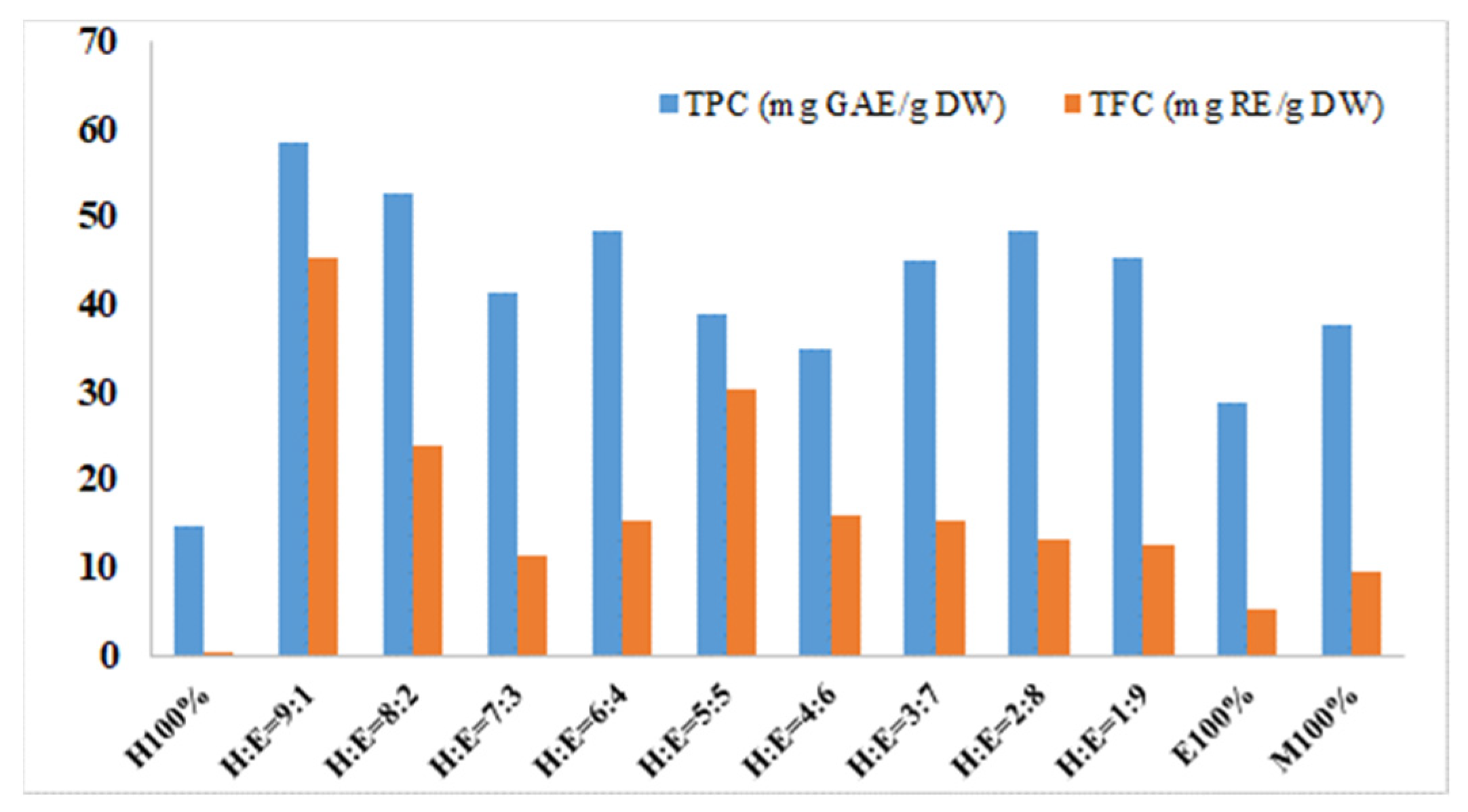
3.1. Total Phenolic Content (TPC) and Total Flavonoid Content (TFC)

The total phenolic and flavonoid contents of all the MT fractions ranged from 14.78 to 58.55 mg GAE/g DW and from 0.42 to 45.38 mg RE/g DW, respectively (Figure 1). However, the obtained value of TPC and TFC varied erratically along with the modification of the solvent ratio. The fraction of E1 yielded the highest TPC and TFC at the level of 58.55 mg GAE/g DW and 45.38 mg RE/g DW, respectively. The second highest result of TPC was 52.63 mg GAE/g DW, yielded from the fraction E2. However, that value of TFC was obtained from the fraction of E5, at a level of 30.25 mg RE/g DW. The fraction of H100 showed the lowest content of TPC and TFC, which were only 14.78 GAE/g DW and 0.42 RE/g DW, respectively. In general, the results indicated that the H:E 9:1 was the most potent ratio for the extraction of TPC and TFC.

3.2. Antioxidant Activities and In Vitro Inhibition of Xanthine Oxidase (XOD)

Antioxidant activities and inhibition of XOD are shown in Table 1.
Table 1 summarizes the antioxidant activities of the 12 fractions of MT. As can be seen in all three assays, the antioxidant activity from fractions of E1 to E100 decreased along with the reduction of Hexane and the rise of Ethyl Acetate in the solvent ratio system. However, while the highest values of antioxidant activity of DPPH scavenging and ABTS assay were obtained from fraction E1 (IC50 = 87.7 µg/mL and 219.2 µg/mL, respectively), the highest inhibitory effect on xanthine oxidase was identified in fraction E2, of which IC50 showed the lowest value of 54.7 µg/mL. Fraction H100 and M100 in the three assays displayed relatively low antioxidant capacities when the IC50 values were much higher than the control. The IC50 values of H100 and M100 were 241.7 and 203.8 µg/mL in the DPPH assay and were 604.2 and 509.6 µg/mL in the ABTS assays. In the XOD assays, the IC50 values of those two fractions resulted in levels of 1510.6 and 1274.0 µg/mL, respectively.

3.3. Antimicrobial Activity

The inhibition activities of different fractions on the development of bacteria, including E. coli, K. pneumoiae, L. monocytogenes, B. subtilis, and P. mirabilis, are illustrated in Table 2. In general, extractions from MT can produce a certain suppression on the development of the studied bacteria. Most of the examined fractions were able to generate a zone of inhibition against the bacteria, especially E. coli, and K. pneumoiae. Eleven fractions inhibited the growth of these two bacteria, but not the fraction E6. MT extracts showed the lowest inhibition activity on B. subtilis and P. mirabilis, since only six fractions (from H100 to E5) and five fractions (from E2 to E6) were efficient for these two bacteria, respectively. Compared to the controls (Ampicillin and Streptomycin), although the inhibition activity of the MT extract was relatively lower, the effect shown on the studied bacteria was considerable.
It was found that among the examined fractions, inhibition activities varied with five bacteria and were not fixed with any typical fraction. With E. coli, the largest zone of inhibition was observed with E1 (10.3 mm). Meanwhile, the largest values observed in K. pneumoiae and L. monocytogenes were detected in fraction E3 (11.3 mm) and E4 (12.5 mm), respectively. The fraction that obtained the highest inhibition activities against B. subtilis, and P. mirabilis was E2, which achieved a zone of inhibition of 10.7 mm and 9.3 mm, respectively. However, it was revealed that E2 was the fraction that showed the optimum result, as its zones of inhibition against E. coli, K. pneumoiae, L. monocytogenes, B. subtilis, and P. mirabilis were 9.1 mm, 10.2 mm, 11.2 mm, 10.7 mm, and 9.3 mm, respectively.

3.4. Correlation of TPC, TFC, Antiradical, and Antimicrobial Activity

Phenolics and flavonoids of plant extracts can be related to the antioxidant activities. There was a significant correlation between TPC and TFC, for both E1 and E2, with Pearson’s correlation coefficients of r = −0.748 and r = −0.728, respectively (Table 3).
The results in Table 3 show that there was a significant negative correlation between antioxidant activity (IC50) and TPC and TFC. This indicates the existance of large amounts of TPC and/or TFC in the samples. The TPC and TFC contents in the samples were proportional to their antioxidant activity (low IC50 value). This was positively correlated with antibacterial ability in E. coli and B. subtilis; and negatively correlated with L. monocytogenes; B. subtilis, and P. mirabilis.

4. Discussion

It is well known that plants contain effective substances and compounds for the treatments and cures of various diseases [36,37,38,39]. Additionally, investigations of antioxidants in medicinal plants are areas of interest for the development of food and medical industries [40,41,42]. Conventionally, leaves, fruits, and flowers are edible parts of plants and thus are widely consumed in many nations; these parts are a potential source of therapeutic compounds for dealing with diseases [43,44,45]. Moringa oleifera (MO) is one potential subject that contributes to the diversity of therapeutic plants [46]. MO is not only utilized as a food supplement to counteract malnourishment, particularly for infants and nursing mothers in developing countries, but its aerial parts are also considered herbal medication for combating illnesses [47,48].
However, it is a fact that MT is underestimated and usually becomes municipal waste after its leaves are separated and utilized for food and medicinal purposes. This study, however, was not conducted to discover novel constituents in MO, but to investigate its anti-oxidative activities in MT and to prove its utility for pharmaceutical purposes. The presence of phenolic substances, e.g., flavonoids, plays a vital role in exerting anti-oxidative activity [49]. The highest values of TPC and TFC found in MT were 58.55 mg GAE/g DW and 45.38 mg RE/g DW, respectively (E1; Figure 1). This finding showed higher levels of TPC and lower levels of TFC in comparison to a study conducted by Abdulkadir et al. (2015), where the level of phenolic and flavonoid compounds in MO leaves were 32.83 mg GAE/g DW and 98.67 mg QAE/g, respectively [50]. Moreover, employing an ultrasonic-assisted extraction method, Xue Lin et al. (2020) noted that the flavonoid content determined in MO leaves was 43.96 mg RE/g DW [51], which is in agreement with the TFC of this study. The comparative evaluations indicate that although MT has been used less widely than MO leaves, the former showed a certain value which can be a factor for XO inhibitory activity.
In this study, the antioxidant assay, Hexane, and EtOAc at a 9:1 (E1) in an EtOAc extract of MT had more anti-radical scavenging activity against DPPH and ABTS and β-carotene oxidation than other fractions. However, compared to the positive control BHT, E1 exhibited lower antioxidant capacities. This antioxidant property is mainly contributed by phenolic contents that can easily release hydrogen donors to naturalize free radicals. Meanwhile, in xanthine oxidase inhibition, flavonoids could be the key. Many studies have reported that compounds containing antioxidant properties and inhibition capabilities for XO activity will work in gout treatment [52,53,54,55,56]. In this study, E1 and E2 were found to inhibited XO and antioxidant activity the most. To this extent, this result also highlights the antioxidant properties of the MT extract, which has potential for further development and research in the medicinal sector. In previous studies, the antioxidant activity of MT was also noted; however, MT extracts were used as a medium for the research. Mona (2013) indicated the effects of MT extracts on increasing the levels of phenol and acid ascorbic, which investigated an important antioxidant substance in Eruca vesicaria subsp. sativa [57]. Moreover, for beneficial use in feeding dairy cows, MT was proven to have a certain impact in increasing the total antioxidant capacity (TAOC) of the plasma samples of cows, which may help in disease prevention [21].
The agar diffusion assay is one method for evaluating the inhibition capability of antibiotics against bacterial growth [58]. Specifically, the inhibition zone of antimicrobial agents is created to reduce the spread and development of a microorganism [59]. Antibacterial activity in MT has been investigated thus far using all fractions from EtOAc extracts. At the same concentration of 20 µg/disk, while inhibition zones of B. subtilis in those fractions ranged from 6.3 to 10.7 mm, the other pathogenic bacteria, E. coli, was observed to have inhibition zones of 6.7 to 10.3 mm. E4 was not sensitive at the same concentration. Serious nosocomial infections are considered to be the result of the mentioned micro-organism. Among all the fractions, E2 displayed the highest inhibition zone on the two bacteria, B. subtilis and P. mirabilis; meanwhile, K. pneumonia and L. monocytogenes were the bacteria with the lowest population constrained by E3. Both E2 and E3 are capable of blocking all the studied bacteria. The reason for this may be the possible formation of an outer membrane of gram-negative bacteria, which could be a barrier for coping with compound penetration, with the possibility of a periplasmic space containing enzymes to break down outer molecules.
A significant number of newly-invented medicines have been extracted from natural sources [60]. In this study, the method of sequential fractions with different solvent ratios (hexane and ethyl acetate) showed an abundant presence of flavonoids and phenols. Flavonoids, phenols, and other antioxidants that exist in low concentrations should be examined using analytical instruments with a higher sensitivity of detection, such as gas chromatography–mass spectrometry (GC–MS). Extracts with phenolic substance-mediated antioxidant activity were presented in conjunction with the development of lowering power. Therefore, the ethyl acetate extract may contain much higher amounts of electron donors for reducing, and they can react with free radicals to convert into more stable products, completing the radical chain reaction. The discoveries of this study suggest the potential use of MT for medicinal purposes, as it has been revealed to possess antioxidants. It is now necessary to perform the next step of searching for novel bioactive compounds in MT. However, complicated extraction processes and modern analytical instruments are required to further research and determine the chemical structures of these potential compounds, such as nuclear magnetic resonance (NMR), infrared spectroscopy (IR), liquid chromatography–mass spectrometry (LC–MS), and GC–MS. About 25,000 secondary metabolites from plants are estimated to have a chance of existing, but only about 2–3% of them have been isolated and identified [60]. Moreover, the integrated use of hexane to the extract system can further ensure the yield of various compounds with different polarities, since no single solvent can be guaranteed to extract all the antioxidant agents present in plant samples [61]. Nevertheless, the use of hexane for plant material extraction, especially in food production, should be taken into consideration due to its potential toxicity in human health. Even so, hexane is still permitted for use in food processing—especially in soybean products, which are “generally recognized as safe” by the FDA [62]. The discoveries of this study suggest the potential use of MT for medicinal purposes as it has been revealed to possess antioxidants.

5. Conclusions

This study documented the antioxidant, antimicrobial, and xanthine oxidase inhibitory activities, in vitro, of fractions from Moringa oleifera twigs. In vivo tests are recommended to affirm the bioavailability of the two fractions of Hexane and EtOAc at 9:1 (E1) and 8:2 (E2) for the development of supplements for gout prevention and/or treatment. This will also ascertain the value of this plant, leading to its sustainable cultivation. The contribution of the EtOAc extract to the biological activities of Moringa oleifera twigs should also be further investigated.

Author Contributions

T.N.M. conceived and implemented the experiments. T.N.M., P.V.T., B.Q.M. and N.Q.T. analyzed data and wrote the manuscript. T.H.M.D. and L.V.A. contributed to the antibacterial activity and xanthine oxidase inhibition activity assays. T.N.M., N.T.D., L.V.N. and N.Q.T. revised the manuscript. All authors have read and agreed to the published version of the manuscript.

Funding

This research was funded by the Graduate University of Science and Technology under grant number GUST.STS.ĐT2020-HH01.

Institutional Review Board Statement

Not applicable.

Informed Consent Statement

Not applicable.

Data Availability Statement

Not applicable.

Acknowledgments

The Center for Research and Technology Transfer, VAST is appreciated for partly supporting this research.

Conflicts of Interest

The authors declare no conflict of interest.

References

  1. Tiwari, S. Plants: A Rich Source of Herbal Medicine. J. Nat. Prod. 2008, 1, 27–35. [Google Scholar]
  2. Costa, A.S.; Alves, R.C.; Vinha, A.F.; Barreira, S.V.; Nunes, M.A.; Cunha, L.M.; Oliveira, M.B.P. Optimization of Antioxidants Extraction from Coffee Silverskin, a Roasting By-product, Having in View a Sustainable Process. Ind. Crops Prod. 2014, 53, 350–357. [Google Scholar] [CrossRef]
  3. Wong, W.H.; Lee, W.X.; Ramanan, R.N.; Tee, L.H.; Kong, K.W.; Galanakis, C.M.; Sun, J.; Prasad, K.N. Two Level Half Factorial Design for the Extraction of Phenolics, Flavonoids and Antioxidants Recovery from Palm Kernel By-product. Ind. Crops Prod. 2015, 63, 238–248. [Google Scholar] [CrossRef]
  4. Ain, H.B.U.; Saeed, F.; Barrow, C.J.; Dunshea, F.R.; Suleria, H.A.R. Food Processing Waste: A Potential Source for Bioactive Compounds. In Bioactive Compounds in Underutilized Fruits and Nuts; Springer: Cham, Switzerland, 2020; pp. 625–649. [Google Scholar]
  5. Posmontier, B. The Medicinal Qualities of Moringa oleifera. Holist. Nurs. Pract. 2011, 25, 80–87. [Google Scholar] [CrossRef] [PubMed]
  6. Hoa, N.T.; Hue, C.T. Enhanced Water Treatment by Moringa oleifera Seeds Extract as the Bio-coagulant: Role of the Extraction Method. J. Water Supply Res. Technol. 2018, 67, 634–647. [Google Scholar] [CrossRef]
  7. Sabastian, G.; Dahlia, L.; Mahrizal, M.; Mulyoutami, E.; Roshetko, J.M.; Purnomosidhi, P.; Perdana, A.; Megawati, M.; Riyandoko, R.; Maulana, H.T.; et al. Ethnobotanical use and Commercial Potential of Moringa oleifera in Indonesia: An Underused and Under-recognized Species. In Proceedings of the I International Symposium on Moringa, Manila, Philippines, 15–18 November 2015; Volume 1158, pp. 349–356. [Google Scholar]
  8. Xu, Y.-B.; Chen, G.-L.; Guo, M.-Q. Antioxidant and Anti-Inflammatory Activities of the Crude Extracts of Moringa oleifera from Kenya and Their Correlations with Flavonoids. Antioxidants 2019, 8, 296. [Google Scholar] [CrossRef] [Green Version]
  9. Tshabalala, T.; Ndhlala, A.R.; Ncube, B.; Abdelgadir, H.A.; Van Staden, J. Potential Substitution of the Root with the Leaf in the use of Moringa oleifera for Antimicrobial, Antidiabetic and Antioxidant Properties. S. Afr. J. Bot. 2020, 129, 106–112. [Google Scholar] [CrossRef]
  10. Islam, Z.; Islam, S.M.; Hossen, F.; Mahtab-ul-Islam, K.; Hasan, M.; Karim, R. Moringa oleifera is a Prominent Source of Nutrients with Potential Health Benefits. Int. J. Food Sci. 2021, 2021, 6627265. [Google Scholar] [CrossRef] [PubMed]
  11. Satish, A.; Kumar, R.P.; Rakshith, D.; Satish, S.; Ahmed, F. Antimutagenic and Antioxidant Activity of Ficus Benghalensis Stem Bark and Moringa oleifera Root Extract. Int. J. Chem. Anal. Sci. 2013, 4, 45–48. [Google Scholar] [CrossRef]
  12. Kumbhare, M.R.; Guleha, V.; Sivakumar, T. Estimation of Total Phenolic Content, Cytotoxicity and In–vitro Antioxidant Activity of Stem Bark of Moringa oleifera. Asian Pac. J. Trop. Dis. 2012, 2, 144–150. [Google Scholar] [CrossRef]
  13. Bhattacharya, A.; Tiwari, P.; Sahu, P.K.; Kumar, S. A Review of the Phytochemical and Pharmacological Characteristics of Moringa oleifera. J. Pharm. Bioallied Sci. 2018, 10, 181. [Google Scholar] [PubMed]
  14. Singh, R.; Singh, B.; Singh, S.; Kumar, N.; Kumar, S.; Arora, S. Anti-free Radical Activities of Kaempferol Isolated from Acacia Nilotica (L.) Willd. Ex. Del. Toxicol. In Vitro 2008, 22, 1965–1970. [Google Scholar] [CrossRef] [PubMed]
  15. Chang, Y.X.; Ding, X.P.; Qi, J.; Cao, J.; Kang, L.Y.; Zhu, D.N.; Zhang, B.L.; Yu, B.Y. The Antioxidant Activity Integrated Fingerprint: An Advantageous Tool for the Evaluation of Quality of Herbal Medicines. J. Chromatogr. A 2008, 1208, 76–82. [Google Scholar] [CrossRef] [PubMed]
  16. Mirzaei, R.; Mohammadzadeh, R.; Alikhani, M.Y.; Shokri Moghadam, M.; Karampoor, S.; Kazemi, S.; Barfipoursalar, A.; Yousefimashouf, R. The Biofilm-associated Bacterial Infections Unrelated to Indwelling Devices. IUBMB Life 2020, 72, 1271–1285. [Google Scholar] [CrossRef] [PubMed]
  17. Buommino, E.; Scognamiglio, M.; Donnarumma, G.; Fiorentino, A.; D’Abrosca, B. Recent Advances in Natural Product-based Anti-biofilm Approaches to Control Infections. Mini-Rev. Med. Chem. 2014, 14, 1169–1182. [Google Scholar] [CrossRef] [PubMed]
  18. Subramanian, R.; Chandra, M.; Yogapriya, S.; Aravindh, S.; Ponmurugan, K. Isolation of Methyl Gallate from Mango Twigs and its Anti-Biofilm Activity. J. Biol. Act. Prod. Nat. 2016, 6, 383–392. [Google Scholar] [CrossRef]
  19. Dhakad, A.K.; Ikram, M.; Sharma, S.; Khan, S.; Pandey, V.V.; Singh, A. Biological, nutritional, and Therapeutic Significance of Moringa oleifera Lam. Phytother. Res. 2019, 33, 2870–2903. [Google Scholar] [CrossRef] [PubMed]
  20. Mataveia, G.A. The Use of Moringa oleifera and Leucaena Leucocephala Tree Leaves to Improve Smallholder Goat Production in Mozambique. Ph.D. Thesis, University of Pretoria, Pretoria, South Africa, 2019. [Google Scholar]
  21. Zhang, T.; Si, B.; Deng, K.; Tu, Y.; Zhou, C.; Diao, Q. Effects of Feeding a Moringa oleifera Rachis and Twig Preparation to Dairy Cows on their Milk Production and Fatty Acid Composition, and Plasma Antioxidants. J. Sci. Food Agric. 2018, 98, 661–666. [Google Scholar] [CrossRef]
  22. Makkar, H.P.S.; Becker, K. Nutrients and Antiquality Factors in Different Morphological Parts of the Moringa oleifera Tree. J. Agric. Sci. 1997, 128, 311–322. [Google Scholar] [CrossRef]
  23. Kostić, D.A.; Dimitrijević, D.S.; Stojanović, G.S.; Palić, I.R.; Đorđević, A.S.; Ickovski, J.D. Xanthine Oxidase: Isolation, Assays of Activity, and Inhibition. J. Chem. 2015, 2015, 294858. [Google Scholar] [CrossRef] [Green Version]
  24. Doherty, M. New Insights into the Epidemiology of Gout. Rheumatology 2009, 48 (Suppl. S2), ii2–ii8. [Google Scholar] [CrossRef] [PubMed] [Green Version]
  25. Sohal, R.S.; Toy, P.L.; Allen, R.G. Relationship Between Life Expectancy, Endogenous Antioxidants and Products of Oxygen Free Radical Reactions in the Housefly, Musca Domestica. Mech. Ageing Dev. 1986, 36, 71–77. [Google Scholar] [CrossRef]
  26. Kitani, K.; Miyasaka, K.; Kanai, S.; Carrillo, M.C.; Ivy, G.O. Upregulation of Antioxidant Enzyme Activities by Deprenyl. Implications for Life Span Extension. Ann. N. Y. Acad. Sci. 1996, 786, 391–409. [Google Scholar] [CrossRef] [PubMed]
  27. Bize, P.; Cotting, S.; Devevey, G.; van Rooyen, J.; Lalubin, F.; Glaizot, O.; Christe, P. Senescence in Cell Oxidative Status in two Bird Species with Contrasting Life Expectancy. Oecologia 2014, 174, 1097–1105. [Google Scholar] [CrossRef] [PubMed] [Green Version]
  28. Nile, S.H.; Khobragade, C.N. Phytochemical Analysis, Antioxidant and Xanthine Oxidase Inhibitory Activity of Tephrosia purpurea Linn. Root Extract. Indian J. Nat. Prod. Resour. 2011, 2, 52–58. [Google Scholar]
  29. Bove, M.; Cicero, A.F.; Borghi, C. The Effect of Xanthine Oxidase Inhibitors on Blood Pressure and Renal Function. Curr. Hypertens. Rep. 2017, 19, 95. [Google Scholar] [CrossRef]
  30. Blainski, A.; Lopes, G.C.; De Mello, J.C.P. Application and Analysis of the Folin Ciocalteu Method for the Determination of the Total Phenolic Content from Limonium brasiliense L. Molecules 2013, 18, 6852–6865. [Google Scholar] [CrossRef] [PubMed] [Green Version]
  31. Matić, P.; Sabljić, M.; Jakobek, L. Validation of Spectrophotometric Methods for the Determination of Total Polyphenol and Total Flavonoid Content. J. AOAC Int. 2017, 100, 1795–1803. [Google Scholar] [CrossRef]
  32. Mensor, L.L.; Menezes, F.S.; Leitão, G.G.; Reis, A.S.; Santos, T.C.D.; Coube, C.S.; Leitão, S.G. Screening of Brazilian Plant Extracts for Antioxidant Activity by the use of DPPH Free Radical Method. Phytother. Res. 2001, 15, 127–130. [Google Scholar] [CrossRef]
  33. Dudonne, S.; Vitrac, X.; Coutiere, P.; Woillez, M.; Mérillon, J.M. Comparative Study of Antioxidant Properties and Total Phenolic Content of 30 Plant Extracts of Industrial Interest Using DPPH, ABTS, FRAP, SOD, and ORAC Assays. J. Agric. Food Chem. 2009, 57, 1768–1774. [Google Scholar] [CrossRef] [PubMed]
  34. Minh, T.N.; Van, T.M.; Andriana, Y.; Hau, D.V.; Duyen, D.H.; Guzman-Gelani, C.D. Antioxidant, Xanthine oxidase, α-Amylase and α-Glucosidase Inhibitory Activities of Bioactive Compounds from Rumex crispus L. Root. Molecules 2019, 24, 3899. [Google Scholar] [CrossRef] [Green Version]
  35. Fukuta, M.; Xuan, T.D.; Deba, F.; Tawata, S.; Khanh, T.D.; Chung, I.M. Comparative Efficacies In Vitro of Antibacterial, Fungicidal, Antioxidant, and Herbicidal Activities of Momilatones A and B. J. Plant Interact. 2007, 2, 245–251. [Google Scholar] [CrossRef] [Green Version]
  36. Gilani, A.H. Trends in Ethnopharmacology. J. Ethnopharmacol. 2005, 100, 43–49. [Google Scholar] [CrossRef] [PubMed]
  37. Oostendorp, M.; Kunz, W.; Dietrich, B.; Staub, T. Induced Disease Resistance in Plants by Chemicals. Eur. J. Plant Pathol. 2001, 107, 19–28. [Google Scholar] [CrossRef]
  38. Repetto, M.G.; Llesuy, S.F. Antioxidant Properties of Natural Compounds used in Popular Medicine for Gastric Ulcers. Braz. J. Med. Biol. 2002, 35, 523–534. [Google Scholar] [CrossRef] [PubMed] [Green Version]
  39. Tungmunnithum, D.; Thongboonyou, A.; Pholboon, A.; Yangsabai, A. Flavonoids and Other Phenolic Compounds from Medicinal Plants for Pharmaceutical and Medical Aspects: An Overview. Medicines 2018, 5, 93. [Google Scholar] [CrossRef] [PubMed]
  40. Djeridane, A.; Yousfi, M.; Nadjemi, B.; Vidal, N.; Lesgards, J.F.; Stocker, P. Screening of Some Algerian Medicinal Plants for the Phenolic Compounds and their Antioxidant Activity. Eur. Food Res. Technol. 2007, 224, 801–809. [Google Scholar] [CrossRef]
  41. Yeung, A.W.K.; Heinrich, M.; Atanasov, A.G. Ethnopharmacology—A Bibliometric Analysis of a Field of Research Meandering Between Medicine and Food Science? Front. Pharmacol. 2018, 9, 215. [Google Scholar] [CrossRef]
  42. Hosseinzadeh, S.; Jafarikukhdan, A.; Hosseini, A.; Armand, R. The Application of Medicinal Plants in Traditional and Modern Medicine: A Review of Thymus vulgaris. Int. J. Clin. Med. 2015, 6, 635. [Google Scholar] [CrossRef] [Green Version]
  43. Turner, N.J.; Łuczaj, Ł.J.; Migliorini, P.; Pieroni, A.; Dreon, A.L.; Sacchetti, L.E.; Paoletti, M.G. Edible and Tended Wild Plants, Traditional Ecological Knowledge and Agroecology. Crit. Rev. Plant Sci. 2011, 30, 198–225. [Google Scholar] [CrossRef]
  44. Al-Fatimi, M. Wild Edible Plants Traditionally Collected and used in Southern Yemen. J. Ethnobiol. Ethnomedicine 2021, 17, 49. [Google Scholar] [CrossRef]
  45. De Caluwé, E.; Halamouá, K.; Van Damme, P. Tamarindus Indica L.—A Review of Traditional Uses, Phytochemistry and Pharmacology. Afr. Focus 2010, 23, 53–83. [Google Scholar]
  46. Padayachee, B.; Baijnath, H. An Updated Comprehensive Review of the Medicinal, Phytochemical and Pharmacological Properties of Moringa oleifera. S. Afr. J. Bot. 2020, 129, 304–316. [Google Scholar] [CrossRef]
  47. Omodanisi, E.I.; Aboua, Y.G.; Oguntibeju, O.O. Assessment of the Anti-hyperglycaemic, Anti-inflammatory and Antioxidant Activities of the Methanol Extract of Moringa oleifera in Diabetes-Induced Nephrotoxic Male Wistar Rats. Molecules 2017, 22, 439. [Google Scholar] [CrossRef] [PubMed]
  48. Sahay, S.; Yadav, U.; Srinivasamurthy, S. Potential of Moringa oleifera as a Functional Food Ingredient: A Review. Int. J. Food Sci. Nutr. 2017, 2, 31–37. [Google Scholar]
  49. Panche, A.N.; Diwan, A.D.; Chandra, S.R. Flavonoids: An Overview. J. Nutr. Sci. 2016, 5, E47. [Google Scholar] [CrossRef] [PubMed] [Green Version]
  50. Abdulkadir, A.R.; Zawawi, D.D.; Jahan, M.S. DPPH Antioxidant Activity, Total Phenolic and Total Flavonoid Content of Different Part of Drumstic Tree (Moringa oleifera Lam.). J. Chem. Pharm. Res. 2015, 7, 1423–1428. [Google Scholar]
  51. Lin, X.; Wu, L.; Wang, X.; Yao, L.; Wang, L. Ultrasonic-assisted Extraction for Flavonoid Compounds Content and Antioxidant Activities of India Moringa oleifera L. Leaves: Simultaneous Optimization, HPLC Characterization and Comparison with Other Methods. J. Appl. Res. Med. Aromat. Plants 2021, 20, 100284. [Google Scholar]
  52. Abdulhafiz, F.; Mohammed, A.; Kayat, F.; Bhaskar, M.; Hamzah, Z.; Podapati, S.K.; Reddy, L.V. Xanthine Oxidase Inhibitory Activity, Chemical Composition, Antioxidant Properties and GC-MS Analysis of Keladi Candik (Alocasia longiloba Miq). Molecules 2020, 25, 2658. [Google Scholar] [CrossRef]
  53. Boumerfeg, S.; Baghiani, A.; Messaoudi, D.; Khennouf, S.; Arrar, L. Antioxidant Properties and Xanthine Oxidase Inhibitory Effects of Tamus communis L. Root Extracts. Phytother. Res. 2009, 23, 283–288. [Google Scholar] [CrossRef] [PubMed]
  54. Cotelle, N.; Bernier, J.L.; Catteau, J.P.; Pommery, J.; Wallet, J.C.; Gaydou, E.M. Antioxidant Properties of Hydroxy-flavones. Free Radic. Biol. Med. 1996, 20, 35–43. [Google Scholar] [CrossRef]
  55. Pacher, P.A.L.; Nivorozhkin, A.; Szabó, C. Therapeutic Effects of Xanthine Oxidase Inhibitors: Renaissance Half a Century After the Discovery of Allopurinol. Pharmacol. Rev. 2006, 58, 87–114. [Google Scholar] [CrossRef] [PubMed]
  56. Ali, F.; Ranneh, Y.; Ismail, A.; Esa, N.M. Identification of Phenolic Compounds in Polyphenols-rich Extract of Malaysian Cocoa Powder Using the HPLC-UV-ESI—MS/MS and Probing their Antioxidant Properties. J. Food Sci. Technol. 2015, 52, 2103–2111. [Google Scholar] [CrossRef] [PubMed]
  57. Mona, M.A. The Potential of Moringa oleifera Extract as a Biostimulant in Enhancing the Growth, Biochemical and Hormonal Contents in Rocket (Eruca Vesicaria Subsp. Sativa). Int. J. Plant Physiol. Biochem. 2013, 5, 42–49. [Google Scholar] [CrossRef]
  58. Abedi, D.; Feizizadeh, S.; Akbari, V.; Jafarian-Dehkordi, A. In Vitro Anti-bacterial and Anti-adherence Effects of Lactobacillus delbrueckii subsp bulgaricus on Escherichia coli. Res. Pharm. Sci. 2013, 8, 260. [Google Scholar] [PubMed]
  59. Herman, A.; Herman, A.P.; Domagalska, B.W.; Młynarczyk, A. Essential Oils and Herbal Extracts as Antimicrobial Agents in Cosmetic Emulsion. Indian J. Microbiol. 2013, 53, 232–237. [Google Scholar] [CrossRef] [PubMed] [Green Version]
  60. Rex, J.R.S.; Muthukumar, N.M.S.A.; Selvakumar, P.M. Phytochemicals as a Potential Source for Anti-microbial, Anti-oxidant and Wound Healing—A Review. MOJ Biorg. Org. Chem. 2018, 2, 61–70. [Google Scholar]
  61. Nawaz, H.; Shad, M.A.; Rehman, N.; Andaleeb, H.; Ullah, N. Effect of Solvent Polarity on Extraction Yield and Antioxidant Properties of Phytochemicals from Bean (Phaseolus Vulgaris) Seeds. Braz. J. Pharm. Sci. 2020, 56, e17129. [Google Scholar] [CrossRef] [Green Version]
  62. Markowitz, T. All About Food: The Manufacturing of Our Food Is Not Pretty. The Los Angeles Times, 27 January. 2015. Available online: https://www.latimes.com/socal/daily-pilot/entertainment/tn-hbi-et-0129-all-about-food-yucky-foods-20150127-story.html (accessed on 10 March 2022).
Figure 1. Total phenolic and flavonoids contents in extracts of M. oleifera twigs. Values are means ± SD (standard deviation; p < 0.05; n = 3).
Processes 10 00563 g001
Table 1. Antioxidant activities measured by DPPH, ABTS and XOD of EtOAc extract fractions from M. oleifera twigs in term of IC50 values.
FractionCodeIC50 (µg/mL)IC50 (µg/mL)
DPPHABTSXOD
Hexane100%H100241.7 ± 6.5 b604.2 ± 16.3 c1510.6 ± 40.7 b
H:E = 9:1E187.7 ± 2.5 i219.2 ± 6.3 g54.7 ± 4.3 h
H:E = 8:2E299.0 ± 3.2 h247.5 ± 8.1 g42.0 ± 2.8 h
H:E = 7:3E3185.2 ± 6.7 e463.0 ± 16.7 e1157.5 ± 41.6 e
H:E = 6:4E4116.8 ± 5.9 g292.0 ± 14.7 f730.0 ± 36.8 g
H:E = 5:5E5124.0 ± 5.7 f310.1 ± 14.1 f775.2 ± 35.3 f
H:E = 4:6E6205.9 ± 4.4 d514.8 ± 11.0 d1287.0 ± 27.5 d
H:E = 3:7E7235.4 ± 5.4 c588.5 ± 13.5 c1471.4 ± 33.8 c
H:E = 2:8E8237.5 ± 7.2 c593.7 ± 18.0 c1484.2 ± 45.0 c
H:E = 1:9E9321.5 ± 7.0 a803.7 ± 17.6 a2009.2 ± 44.1 a
EtOAc100%E100322.6 ± 8.0 a806.5 ± 20.1 b2016.3 ± 50.2 a
MeOH100%M100203.8 ± 0.3 d509.6 ± 0.7 d1274.0 ± 1.8 d
BHT-21.4 ± 0.3 k40.0 ± 0.6 h-
Allopurinol---20.8 ± 0.7 k
Values represent means ± SD (standard deviation). Values with similar letters in each column are not significantly different (p < 0.05; n = 3). -: measurement was not conducted; BHT: butylated hydroxytoluene.
Table 2. Antibacterial activity in term of zone of inhibition (mm) of fractions from M. oleifera twigs.
FractionZone of Inhibition (mm)
E. coliK. pneumoniaeL. monocytogenesB. subtilisP. mirabilis
Extract
Hexane100%6.7 ± 0.5 c8.5 ± 1.3 c6.6 ± 0.5 c6.3 ± 0.6 d-
H:E = 9:110.3 ± 0.6 c9.1 ± 1.7 c8.5 ± 0.5 c9.7 ± 0.6 c-
H:E = 8:29.1 ± 1.0 c10.2 ± 1.3 bc11.2 ± 1.9 bc10.7 ± 0.9 c9.3 ± 0.9 c
H:E = 7:38.3 ± 0.6 c11.3 ± 0.7 bc12.2 ± 1.1 bc8.7 ± 0.2 c6.5 ± 0.6 c
H:E = 6:48.7 ± 1.7 c6.9 ± 0.8 c12.5 ± 1.3 bc8 ± 0.7 d7.3 ± 0.3 c
H:E = 5:58.1 ± 1.7 c6.9 ± 0.3 c8.8 ± 1.0 c7 ± 0.4 d6.3 ± 0.5 c
H:E = 4:6----7.3 ± 0.6 c
H:E = 3:77.9 ± 1.5 c6.9 ± 0.2 c8.5 ± 0.5 c--
H:E = 2:87.1 ± 1.7 c-7.5 ± 0.8 c--
H:E = 1:97.4 ± 1.5 c7.4 ± 1.3 c7.9 ± 1.4 c--
EtOAc100%8.0 ± 2.1 c-6.6 ± 0.5 c--
MeOH100%7.7 ± 2.1 c8.0 ± 0.9 c9.1 ± 0.9 c--
Control
Methanol-----
Ampicillin33.9 ± 1.3 a48.7 ± 3.5 a22.5 ± 1.9 a17.3 ± 0.7 a43.3 ± 1.5 a
Streptomycin20.2 ± 1.4 b16.8 ± 0.9 b16.4 ± 1.2 b12 ± 0.3 b29.3 ± 1.2 b
Values represent means ± SD (standard deviation). Values with similar letters in each column are not significantly different (p < 0.05; n = 3).
Table 3. Correlation of TPC, TFC, antiradical, and antimicrobial activity.
TPCTFCDPPHE. cK. pL. mB. sP. m
TPC1
TFC0.57591
DPPH−0.748−0.7281
E. coli (−)0.95550.7306−0.691
K. pneumoniae (−)0.1421−0.2010.40750.16031
L. monocytogenes (+)−0.211−0.9190.5115−0.4140.27031
B. subtilis (+)0.74860.2074−0.3680.65660.67100.07431
P. mirabilis (−)−0.475−0.7610.2942−0.7010.03920.6588−0.03711
E. c: Escherichia coli; K. p: Klebsiella pneumoniae; L. m: Listeria monocytogenes; B. s: Bacillus subtilis, and P. m: Proteus mirabilis; (−): Gram-negative bacteria; (+): Gram-positive bacteria.
Publisher’s Note: MDPI stays neutral with regard to jurisdictional claims in published maps and institutional affiliations.

