Article

Bioassay-Guided Characterization, Antioxidant, Anti-Melanogenic and Anti-Photoaging Activities of Pueraria thunbergiana L. Leaf Extracts in Human Epidermal Keratinocytes (HaCaT) Cells

1
Department of Pharmaceutical & Cosmetics, Dongshin University, 185, Gunjae-ro Naju, Jeonnam 58245, Korea
2
B&Tech Co., Ltd., 584-10, Noansam-ro, Naju, Jeonnam 58250, Korea
*
Authors to whom correspondence should be addressed.
These authors contributed equally to this work.
Processes 2022, 10(10), 2156; https://doi.org/10.3390/pr10102156
Submission received: 23 September 2022 / Revised: 10 October 2022 / Accepted: 18 October 2022 / Published: 21 October 2022

Abstract

:
Although the roots and flowers of P. thunbergiana are known to have various physiologically active effects, studies on the anti-melanin production and anti-photoaging effects of its leaf extracts and cellular mechanisms are still lacking. In this study, we evaluated the possibility of using Pueraria thunbergiana leaves as a natural material for skin whitening and anti-aging-related functional cosmetics. The 30% ethyl alcohol (EtOH) extract from P. thunbergiana leaves was fractionated using n-hexane, ethyl acetate (EtOAc), butanol, and aqueous solution to measure their whitening, and anti-aging effects. The EtOAc fraction contained a high content of phenolic and flavonoids and showed higher 1,1-diphenyl-2-picryhydrazyl (DPPH) and 2,2-azino-bis (3-ethylbenzothiazoline-6-sulfonic acid) diammonium salt (ABTS) radical scavenging activities than the other fractions. It was also confirmed that the EtOAc fraction markedly inhibited α-melanocyte stimulating hormone (α-MSH)-induced melanogenesis in B16F10 melanoma cells. In addition, the EtOAc fraction showed a protective effect against ultraviolet B (UVB) in HaCaT cells and increased the collagen synthesis that was decreased due to UVB exposure. Matrix metalloproteinase-1 (MMP-1) activity and MMP-1 protein expression were reduced in human epidermal keratinocytes (HaCaT) cells. These results indicate that the EtOAc fraction has superior antioxidant activity, anti-melanogenesis, and anti-photoaging effects compared to the other fractions. Therefore, in this study, we confirmed the potential of P. thunbergiana leaf extract as a functional cosmetic ingredient, and it can be used as basic data for the physiological activity of P. thunbergiana leaf extracts.

Graphical Abstract

1. Introduction

Mammalian skin acts as a barrier to protect the body from external stimuli, such as fine dust, ultraviolet irradiation, and chemicals caused by environmental pollution [1,2,3]. Ultraviolet (UV) rays are a major environmental factor that causes skin damage, and repeated exposure to UV rays generates reactive oxygen species (ROS). Repeated exposure to UV rays induces skin aging and melanin production, which causes pigmentation [4,5].
Skin aging is divided into two types. Intrinsic aging is a phenomenon that occurs naturally as the physiological and structural functions of the skin decline, while extrinsic aging, also called photoaging, occurs due to repeated exposure to UV rays [6,7]. The main cause of skin aging is photoaging; upon exposure to UV rays, matrix proteins, such as collagen and elastin, are damaged, causing collagen deficiency and elastin denaturation [8,9]. In addition, skin aging and collagen loss are closely related. Matrix metalloproteinase-1 (MMP-1) is known as a collagen-degrading enzyme in the skin that degrades collagen in the dermal layer due to exposure to UV rays; therefore, matrix metalloproteinases (MMPs) play a very important role in exogenous aging [10,11,12]. As aging progresses due to exposure to UV rays, it induces the reduction and degradation of matrix proteins such as hyaluronic acid, elastin, and collagen, which are substances that constitute the skin, thereby reducing skin elasticity, and causing wrinkles.
Melanin is a natural pigment in animals and plants and is a main factor in deciding the color of the hair, eyes, and skin. It also plays an important role in protecting the skin because it inhibits injury to skin cells from UV rays and ROS [13,14,15,16]. However, excessive production of melanin causes various abnormal pigmentation-related conditions, such as inflammation, age spots, freckles, and melasma, and promotes skin aging by causing serious skin diseases, such as skin cancer [17,18,19].
Melanin is synthesized in melanocytes, which contain structures called melanosomes. The process of melanogenesis is regulated by several mechanisms, including transcriptional and enzyme control mechanisms [20]. In the first step of melanogenesis, the oxidative enzyme tyrosinase (TYR) catalyzes the oxidation of L-tyrosine to L-3,4-dihydroxy phenylalanine (L-DOPA) and oxidizes L-DOPA to DOPA quinone. Tyrosinase-related protein-2 (TRP-2) changes DOPA chrome to 5,6-dihydroxyindole-2-carboxylic acid (DHICA). Finally, DHICA is oxidized by tyrosinase-related protein-1 (TRP-1) to produce melanin [21] (Figure 1). In this process, tyrosinase, TRP-1, and TRP-2 are the major melanogenesis enzymes. Microphthalmia-associated transcription factor (MITF) is involved in the survival, proliferation, and differentiation of melanocytes, and it has been reported to regulate the expression of the genes involved in melanogenesis [22,23,24,25,26].
Recently, many studies have been conducted on functional substances related to skin wrinkle improvement and whitening. Currently, functional substances used to prevent or correct wrinkles include ursolic acid, epigallocatechin gallate (EGCG), retinol, retinyl palmitate, adenosine, and polyethoxylated retinamide and substances used as whitening agents include arbutin, kojic acid, and ascorbic acid, which inhibit melanogenesis by downregulating tyrosinase activity [27,28]. However, it has been reported that many of these may cause toxicity and side effects with long-term use [21,29,30]. Therefore, the need for effective functional materials using natural products and medicinal plants with high human safety and few side effects has increased, and research on the development of materials from natural substances is being actively conducted [13,22].
Kudzu (Pueraria thunbergiana) is a creeping plant that belongs to the Fabaceae family and is currently cultivated worldwide [22,31]. Its roots and flowers are used in varying ways because they have various medicinal effects. Puerarin, the main component of the root, is known to have anti-pyretic, antioxidant, anti-hypertensive, and anti-inflammatory effects [32,33,34,35,36,37]. The aerial parts of the plant contain isoflavonoids such as daidzein, daidzin, genistein, and genistin, puerarin, and they have been reported to inhibit melanogenesis and have hepatoprotective effects [38,39,40,41,42]. However, little is known about the anti- melanogenesis and anti-photoaging effects and the cellular mechanisms of the plant leaves’ extracts.
Therefore, the objective of the present study was to explore the antioxidant activity, anti-melanogenesis, and anti-photoaging effects of EtOH extracts of P. thunbergiana leaves and their fractions using B16F10 melanoma cells and HaCaT cells, and to analyze their molecular mechanisms and major components. Through these processes, we attempted to confirm whether they could be applied as physiologically active natural ingredients that exhibit skin whitening and anti-aging effects.

2. Materials and Methods

2.1. Materials

2,2-azino-bis (3-ethylbenzothiazoline-6-sulfonic acid) diammonium salt (ABTS), 1,1-diphenyl-2-picrylhydrazyl (DPPH), 3-(4,5-dimethylthiazol-2-yl)-2,5-diphenyl tetrazolium bromide (MTT), dimethyl sulfoxide (DMSO), α-melanocyte stimulating hormone (α-MSH), L-3,4-dihydroxy-phenylalanine (L-DOPA), and arbutin were purchased from Sigma-Aldrich (St. Louis, MI, USA). Daidzin and genistin were purchased from LC Laboratories (Woburn, MA, USA). Phosphate-buffered saline (PBS) and Dulbecco’s Modified Eagle Medium (DMEM) were purchased from Lonza (Walkersville, MD, USA). Penicillin/streptomycin (P/S) and fetal bovine serum (FBS) were purchased from Gibco (Thermo Fisher Scientific, Rockford, IL, USA). A Human Pro-MMP-1 Quantikine ELISA Kit (No. PDMP100) was purchased from R&D Systems (Minneapolis, MN, USA). A Procollagen Type I C-peptide (PIP) EIA Kit (No. MK101) was purchased from Takara Bio Inc. (Shiga, Japan). GAPDH was purchased from EnoGene Biotechnology (New York, NY, USA). MITF and MMP-1 antibodies were purchased from Cell Signaling Technology (Danvers, MA, USA) and Santa Cruz Biotechnology (Dallas, TX, USA). The purity of all the chemicals used in the experiment was over 97%.

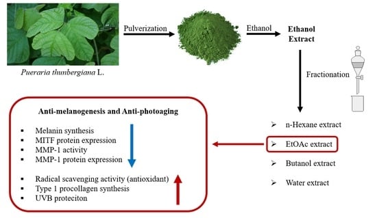
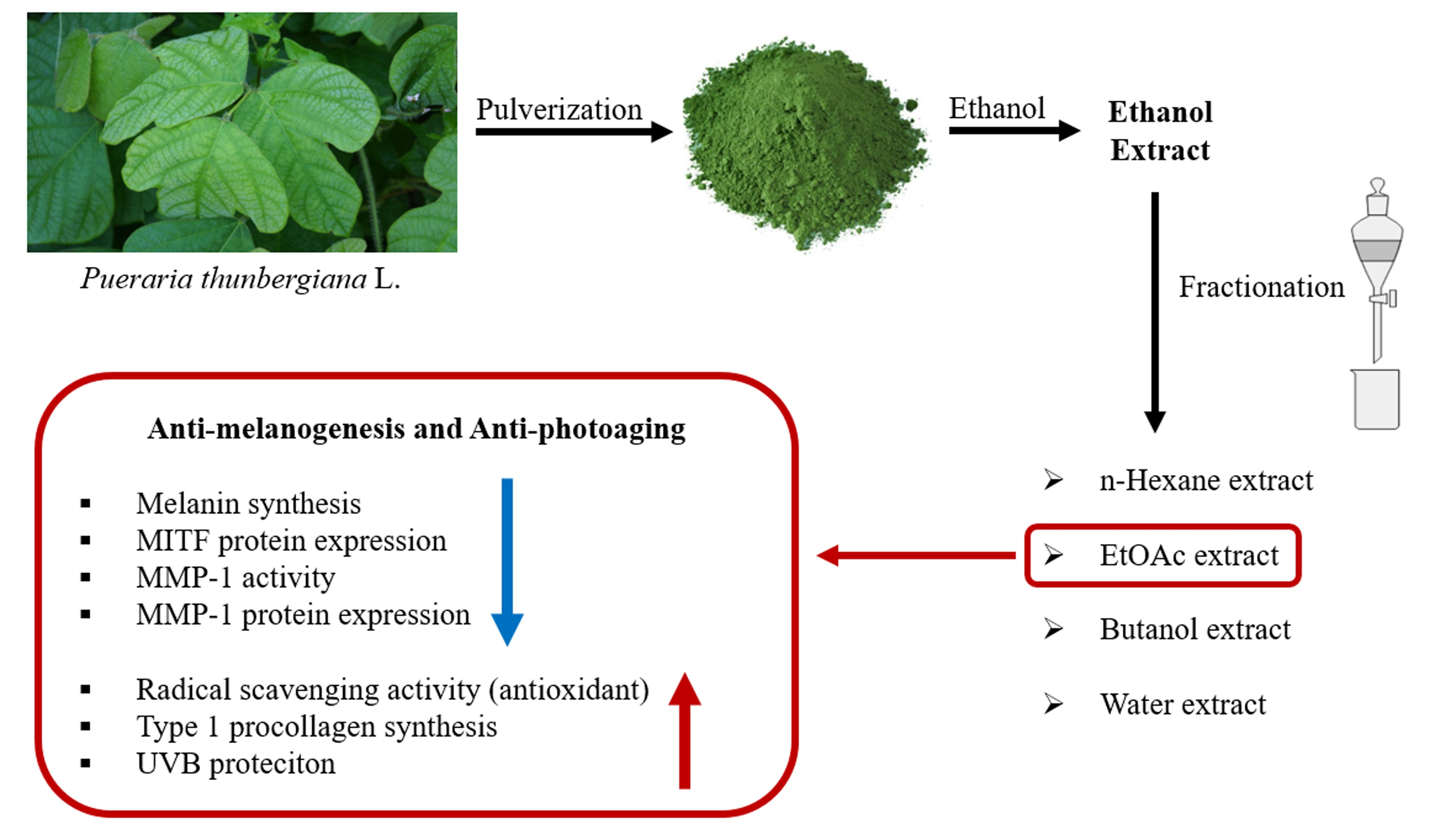
2.2. Preparation of P. thunbergiana L. Extract and Production of Different Fractionations

The P. thunbergiana leaves used in this experiment were grown in Gimhae, and purchased from Daltone (Anseong, Korea). Authentication and identification of samples were carried out for the study by Dr. Sunoh Kim at B&Tech, who has expertise in this area. The dried and pulverized sample (200 g) was placed in a round bottom flask, and 2 L of EtOH (0, 30, 50, 70, 90%) with distilled water was added. Then, the flask was fitted on a heating mantle and reflux extracted at 100 °C for 3 h. The internal temperature was maintained at 90 ± 1 °C. The extract was concentrated using a rotary evaporator (N-1000, EYELA, Tokyo, Japan). After dissolving the concentrate in distilled water, n-hexane (H), ethyl acetate (EtOAc), and butanol (B) were mixed in a 1:1 ratio, and fractionation was repeated for three times for 24 h each (Figure 1). Fractionation of the extract was performed using a separating funnel. The fractions were concentrated, lyophilized, and were stored at −20 °C until use.

2.3. Total Phenolic Content (TPC)

TPC was measured using the Folin–Ciocalteu’s reagent (FCR) method with some modifications [43]. The sample extract was mixed with 500 µL of FCR reagent, reacting for 3 min. Next, 500 µL of 10% Na2CO3 was added to the previous solution and left to react for 1 h. Gallic acid (10–60 μg/mL) was used as the standard (R2 = 0.999). The absorbance was measured at 760 nm using a UV–vis spectrophotometer (Optizen 2120 UV, Mecasys, Daejeon, Korea). A calibration curve was prepared using gallic acid as a standard. TPC was expressed as gallic acid equivalents (GAE) per mL of extract.

2.4. Total Flavonoid Content (TFC)

TFC was measured by the aluminum chloride colorimetric assay [44]. Samples (200 µL) were diluted to an appropriate concentration; 800 µL of 80% ethanol and 60 µL of 5% NaNO2 were added and left to react for 5 min. Then, 10% AlCl3 was mixed into the solution and allowed to react for another 5 min. Then, 400 µL of 1 M NaOH solution was added to the solution, and the absorbance was measured at 510 nm using a UV–vis spectrophotometer (Optizen 2120 UV, Mecasys, Daejeon, Korea). A calibration curve was prepared using catechin (50–600 μg/mL) as the standard (R2 = 0.999).

2.5. Antioxidant Activity Evaluation

DPPH radical scavenging activity was examined using the Blois method, with modifications [45]. Diverse concentrations of the extract and ascorbic acid, used as a positive control, were added to a 0.2 mM DPPH solution. The solution was mixed and incubated for 30 min. Absorbance was measured at 515 nm using a microplate spectrophotometer (Thermo Fisher Scientific, Multiskan Sky, Boseong, Korea).
ABTS radical scavenging activity was examined through a revision of the Lin Zheng method [46]. To generate ABTS radicals, 2.45 mM potassium persulfate was mixed with 7.4 mM ABTS stock solution in a 1:1 ratio. The mixture was incubated for 24 h, until ABTS radicals were generated. After this, a working solution was prepared by mixing distilled water and ABTS solution until the solution had an absorbance of approximately 0.7 (±0.2). Various concentrations of the extract and ascorbic acid, used as a positive control, were added to the ABTS working solution. The solutions were then mixed and incubated for 30 min. Absorbance was measured at 734 nm using a microplate spectrophotometer (Thermo Fisher Scientific, Multiskan Sky, Boseong, Korea).

2.6. Cell Culture

B16F10 melanoma cells and human epidermal keratinocytes (HaCaT) cells were purchased from the American Type Culture Collection (Rockville, MD, USA). B16F10 melanoma cells and HaCaT cells were cultured in DMEM containing 10% FBS and 1% penicillin and streptomycin (P/S) in a humidified atmosphere containing 5% CO2 at 37 °C.

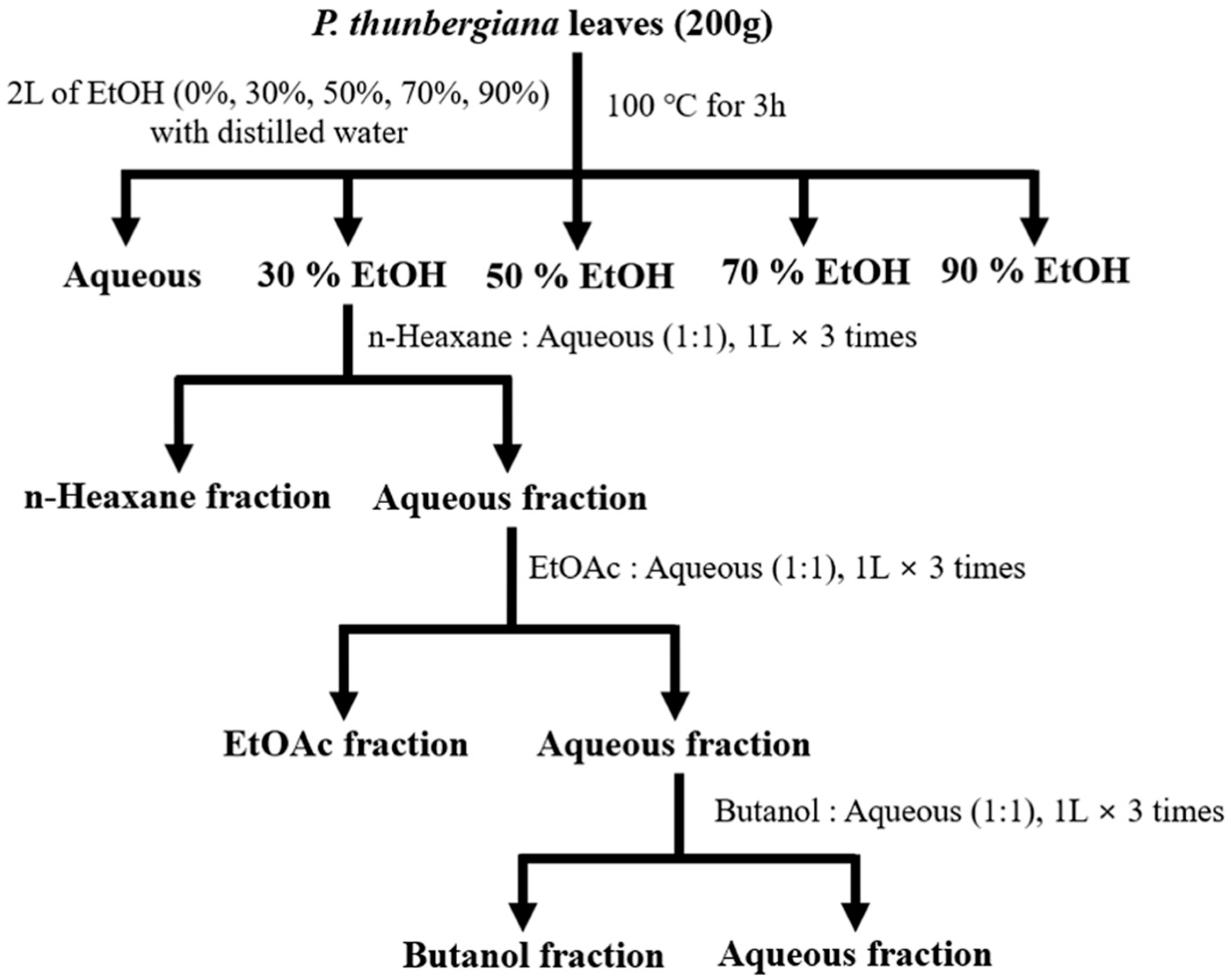
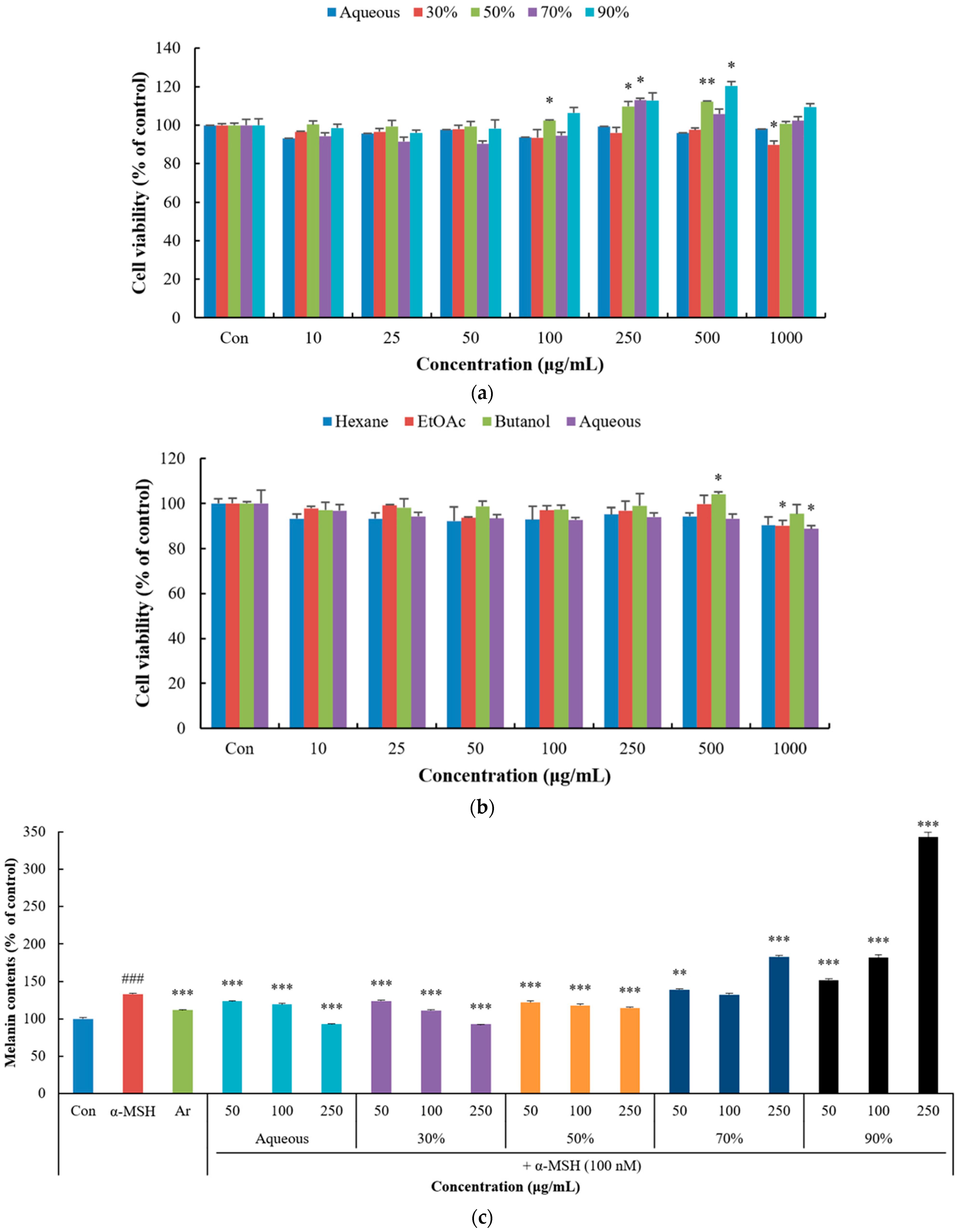
2.7. Cell Viability Assay

Cell viability was examined using the MTT method [47]. B16F10 melanoma cells and HaCaT cells were seeded at densities of 1 × 10⁴ cells/well of 96-well plates and incubated with various concentrations of extracts and fractions for 48 h, at 37 °C. After incubation, the cells were treated with 0.5 mg/mL MTT for 3 h. The media were removed and 200 µL of DMSO was added to the cells, gently shaking the plates for 15 min. Absorbance was measured at 570 nm using a microplate spectrophotometer spectrophotometer (Thermo Fisher Scientific, Multiskan Sky, Boseong, Korea).

2.8. Measurement of Melanin Content

Measurement of the inhibition of melanin biosynthesis using B16F10 cells was carried out by modifying the research method of Hosoi et al. [48]. B16F10 melanoma cells were seeded at densities of 2 × 10⁵ cells/dish in 60 mm dishes and incubated for 24 h at 37 °C. After incubation, the spent medium was replaced with fresh medium that included diverse concentrations of extracts (50–500 µg/mL), α-MSH (100 nM), and arbutin (100 µg/mL), and incubated for 48 h. The pellet obtained via centrifugation (12,000 rpm, 15 min, 23 °C) was dissolved in 1N NaOH, containing 10% DMSO, at 80 °C for 1 h. Absorbance was measured at 475 nm using a microplate spectrophotometer (Thermo Fisher Scientific, Multiskan Sky, Boseong, Korea).

2.9. UVB Protection Assay

Measurement of UVB protection using HaCaT cells was measured by modifying the research method of Wang et al. [49]. HaCaT cells were seeded at densities of 5.5 × 10⁴ cells/well of 24-well plates and incubated for 24 h at 37 °C. The extracts of various concentrations were added to the cells, irradiated with UVB (20 mJ/cm2), and incubated for 24 h. After incubation, cells were treated with 0.5 mg/mL MTT for 3 h. The media were removed and 500 µL DMSO was added, gently shaking the plate for 15 min. Absorbance was measured at 570 nm using a microplate spectrophotometer (Thermo Fisher Scientific, Multiskan Sky, Boseong, Korea).

2.10. Measurement of Type 1 Pro-Collagen Synthesis

Type 1 pro-collagen biosynthesis was examined using a Procollagen Type I C-peptide (PIP) EIA Kit. Measurements of type 1 procollagen synthesis were performed according to the kit manufacturer’s protocol. HaCaT cells were seeded at densities of 3 × 10⁵ cells/well in 6-well plates and incubated for 24 h at 37 °C. After UVB irradiation (10 mJ/cm2), the extract was incubated for 72 h. After diluting the supernatant, the antibody-POD conjugate solution and the sample were added to the wells of the plates and incubated for 3 h. The substrate solution was added, and the cells were incubated for 15 min. Finally, the reaction was stopped by adding a stop solution. Absorbance was measured at 450 nm using a microplate spectrophotometer (Thermo Fisher Scientific, Multiskan Sky, Boseong, Korea).

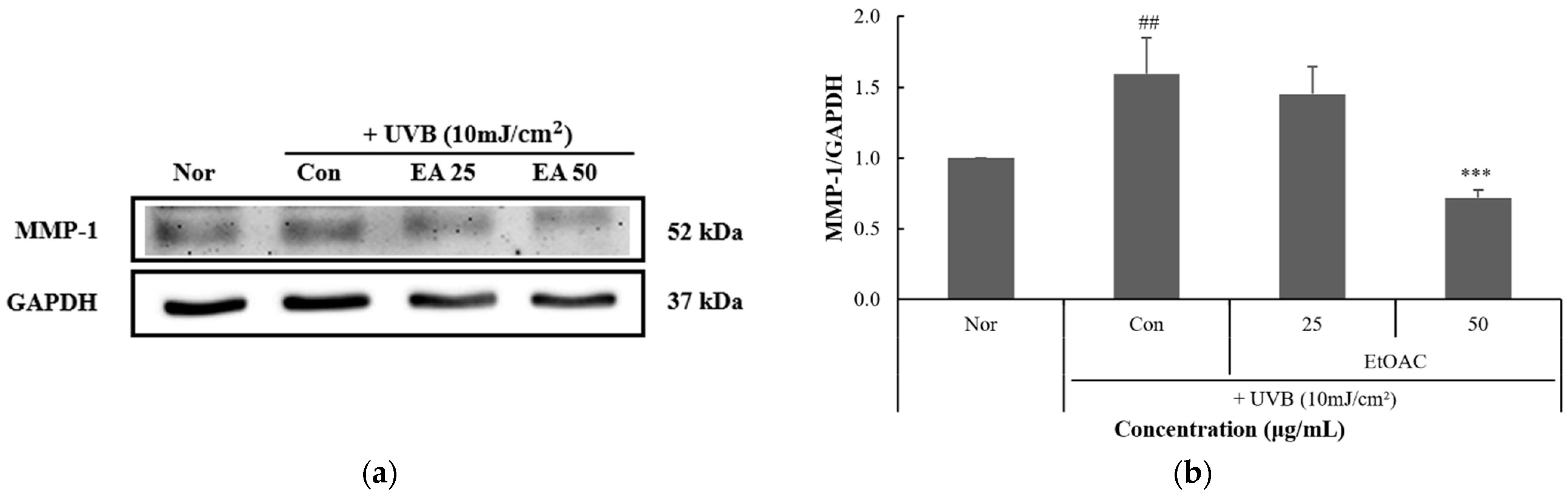
2.11. Measurement of MMP-1 Inhibitory Activity

MMP-1 inhibitory activity was examined using a Human Pro-MMP-1 Quantikine ELISA Kit. Measurements of MMP-1 inhibitory activity were performed according to the kit manufacturer’s protocol. HaCaT cells were seeded at densities of 3 × 10⁵ cells/well in 6-well plates and incubated for 24 h at 37 °C. After UVB irradiation (10 mJ/cm²), the cells were incubated with the extract for 72 h. After diluting the supernatant, diluent RD1-52 and the sample were dispensed into each cell-containing well of the plate and reacted at room temperature for 2 h. The solution contained in each well was then removed, the anti-human pro-MMP-1 conjugate was added, and the cells were allowed to react for 2 h. After removing the solution, the substrate solution was added and was reacted for 20 min. After adding the stop solution, the absorbance was measured at 450 nm and 540 nm using a microplate spectrophotometer (Thermo Fisher Scientific, Multiskan Sky, Boseong, Korea). The final absorbance was calculated by subtracting the absorbance value at 540 nm from the absorbance value at 450 nm.

2.12. Western Blot Analysis

Western blot analysis was measured by modifying the research method of Jeong et al. [50]. B16F10 melanoma cells were seeded at densities of 2 × 10⁵ cells/dish (60 mm) and incubated with the extracts, as well as α-MSH (100 nM). HaCaT cells were seeded at densities of 5.5 × 10⁵ cells/dish (60 mm), incubated with the extracts, and were subjected to UVB irradiation (10 mJ/cm2). Cells were lysed in Pro-Prep lysis solution for 15 min on ice. The cell lysates were centrifuged at 13,000 rpm, 4 °C for 5 min. Lysed supernatant protein was measured by Bradford assay. Protein (20 µg) was subjected to 10% sodium dodecyl sulfate polyacrylamide gel electrophoresis (SDS-PAGE) and transferred to a polyvinylidene difluoride (PVDF) membrane. The PVDF membranes were incubated for 1 h in blocking buffer (5% skim milk and 0.1% Tween 20 in TBS). Membranes were incubated with primary antibodies (GAPDH, MITF, MMP-1) for 1 h. GAPDH was used as an internal control. After washing four times with TBS containing 0.1% Tween 20, the membrane was incubated with the corresponding horseradish peroxidase (HRP)-conjugated anti-mouse and anti-rabbit secondary antibodies for 1 h. Protein band detection on the PVDF membranes was performed using Western BrighTM ECL reagent and the Davinch-WesternTM Imaging System (Davinch-K, Seoul, Korea).

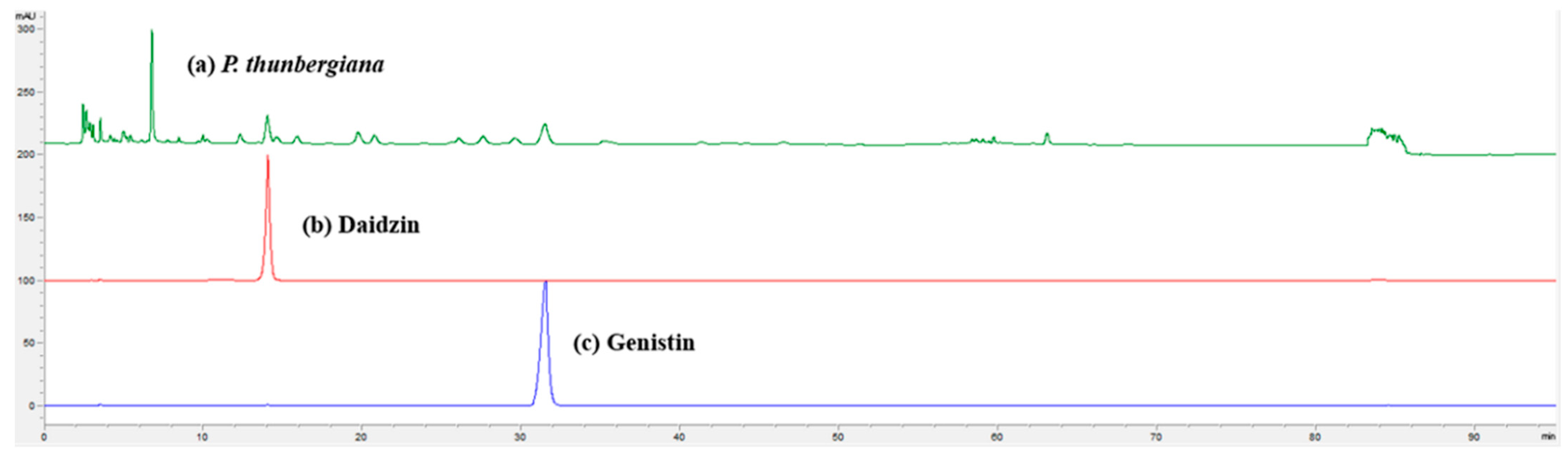
2.13. High-Performance Liquid Chromatography Analysis

The P. thunbergiana EtOAc fraction had the best antioxidant activity and inhibitory effect on melanin synthesis. We performed the components analysis of the fraction using an Agilent 1100 series HPLC (Agilent Technologies, Santa Clara, CA, USA) system and YMC ODS column (4.6 × 250 mm, Kyoto, Japan). The mobile phase consisted of water that contained 0.1% formic acid (solvent A) and acetonitrile (solvent B) with a gradient elution (a linear gradient from 15% B in 5 min, a linear gradient from 15% to 17% B in 45 min, a linear gradient from 17% to 30% B in 5 min). The injection volume of the sample was 10 μL, and the column flow rate was 0.5 mL/min. Peaks were detected by measuring absorbance at 254 nm.

2.14. Statistical Analyses

The normality test was performed using the Shapiro–Wilk test of the SPSS program. The results are presented as the mean ± standard deviation (SD) of three independent experiments, and statistical significance was determined using Student’s t-test and ANOVA. All statistical analyses were performed using SPSS statistical software 22.0. * p < 0.05, ** p < 0.01 and *** p < 0.001 were considered as significant differences.

3. Results

3.1. Total Phenolic and Flavonoid Content of P. thunbergiana L. Extracts and Fractions

First, the optimal EtOH concentration of the extract for fractionation was selected through a prior experiment. The phenol and flavonoid contents of P. thunbergiana L. EtOH extracts were measured using gallic acid and catechin as standards. Total phenol content (TPC) and total flavonoid content (TFC) were expressed as μg gallic acid equivalents (GAE) and catechin equivalents (CE) per gram of dried extract, respectively.
The 30% EtOH extract showed both the highest TPC (28 ± 0.02 μg GAE/g) and TFC (122 ± 0.80 μg CE/g), as demonstrated in Table 1. The EtOAc fraction showed the highest TPC (29 ± 0.0 μg GAE/g) and TFC (113 ± 0.00 μg CE/g).

3.2. Antioxidant Activity of P. thunbergiana L. Extracts and Fractions

DPPH radical scavenging activity was confirmed to be high in the 30% and 50% EtOH extracts (Table 2). EC50 values for DPPH radical scavenging activity of 30% and 50% EtOH extracts were 88.1 and 74.8 μg/mL, respectively. The 30% EtOH extract indicated higher ABTS radical scavenging activity than the other extracts at high concentrations and presented similar scavenging activity to ascorbic acid. EC50 values for ABTS radical scavenging activity of 30% and 50% EtOH extracts were 54.2 and 57.0 μg/mL, respectively.
The EtOAc fraction showed higher DPPH and ABTS radical scavenging activity than the other extracts. EC50 values for DPPH radical scavenging activity of EtOAc and butanol were 195.1 and 207.7 μg/mL, respectively. In addition, EC50 values for ABTS radical scavenging activity of EtOAc and butanol were 49.4 and 68 μg/mL, respectively.

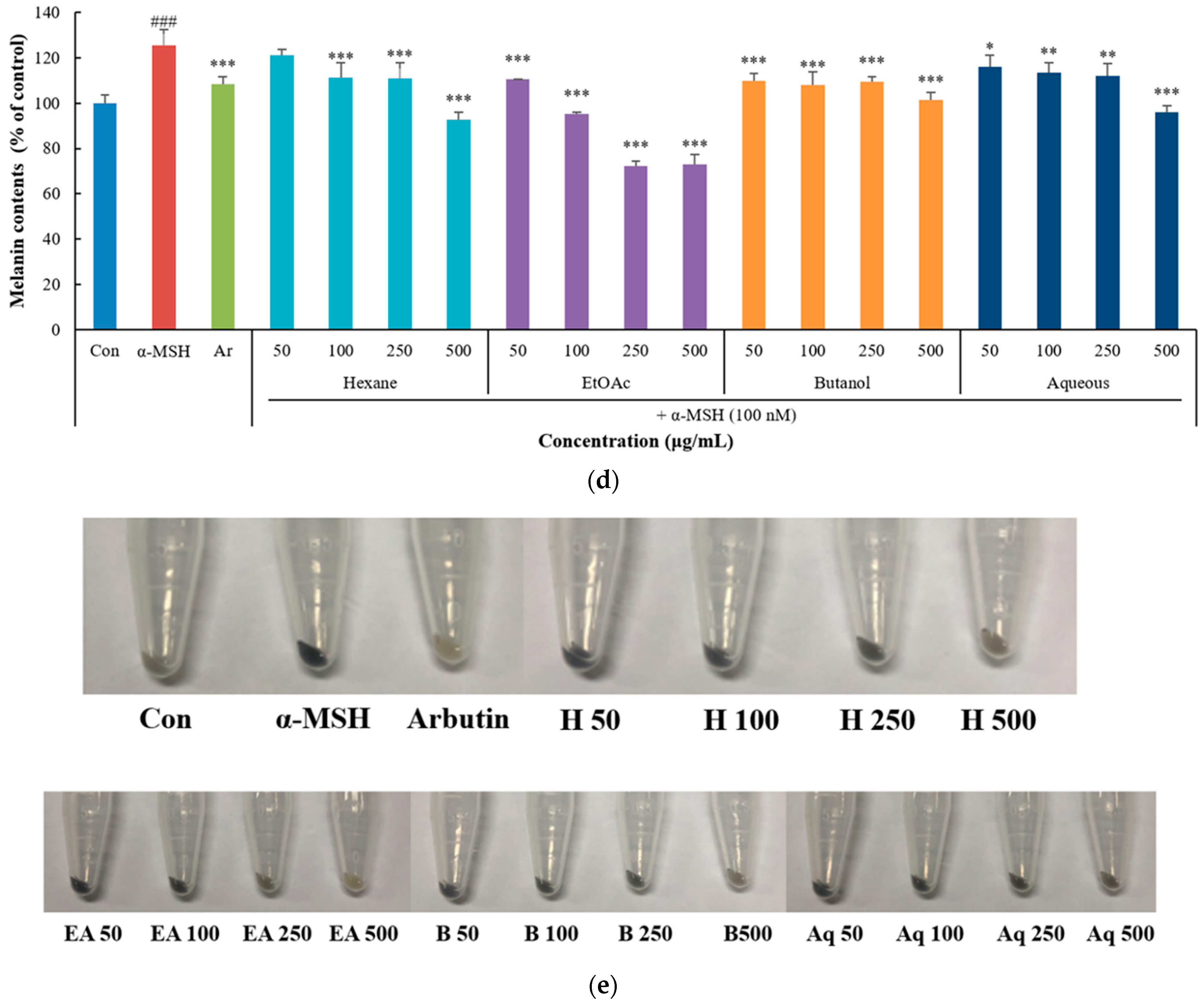
3.3. Inhibitory Effect of P. thunbergiana L. Extracts and Fractions on Melanogenesis in B16F10 Cells

Cell viability was measured in B16F10 melanoma cells using the MTT assay to evaluate the effect of treatment with the P. thunbergiana L. extracts and fractions. As a result, it did not reduce cell viability at concentrations of 10–500 µg/mL, and some extracts and fractions showed presumably the stimulation of proliferation at concentrations of 100–250 µg/mL (Figure 2a,b).
Melanin content was measured in B16F10 melanoma cells treated with α-MSH and P. thunbergiana L. extracts, using arbutin as a positive control. In particular, the 30% EtOH extracts reduced melanin content more than the other extracts and showed higher melanogenesis inhibitory activity than arbutin (Figure 2c). To confirm the effect of the P. thunbergiana L. fractions on melanogenesis, melanin content was measured in B16F10 melanoma cells treated with α-MSH and P. thunbergiana L. fractions, with arbutin as a positive control. As a result, when compared with the α-MSH alone treatment group, the groups treated with the P. thunbergiana L. fractions showed a decreased melanin content in a concentration-dependent manner. In particular, the EtOAc fraction showed higher melanogenesis inhibitory activity than the other fractions and presented superior melanogenesis inhibitory activity than arbutin at a concentration of 250 and 500 μg/mL (Figure 2d).
In addition, a color change was visually confirmed through the pellets of the treated groups. Treatment with the EtOAc fraction showed that the color intensity of the pellet decreased in a concentration-dependent manner, and it was similar to the color of the arbutin-treated pellet (Figure 2e). Taken together, the EtOAc fraction of the 30% EtOH extract showed the highest anti-melanogenesis effect.

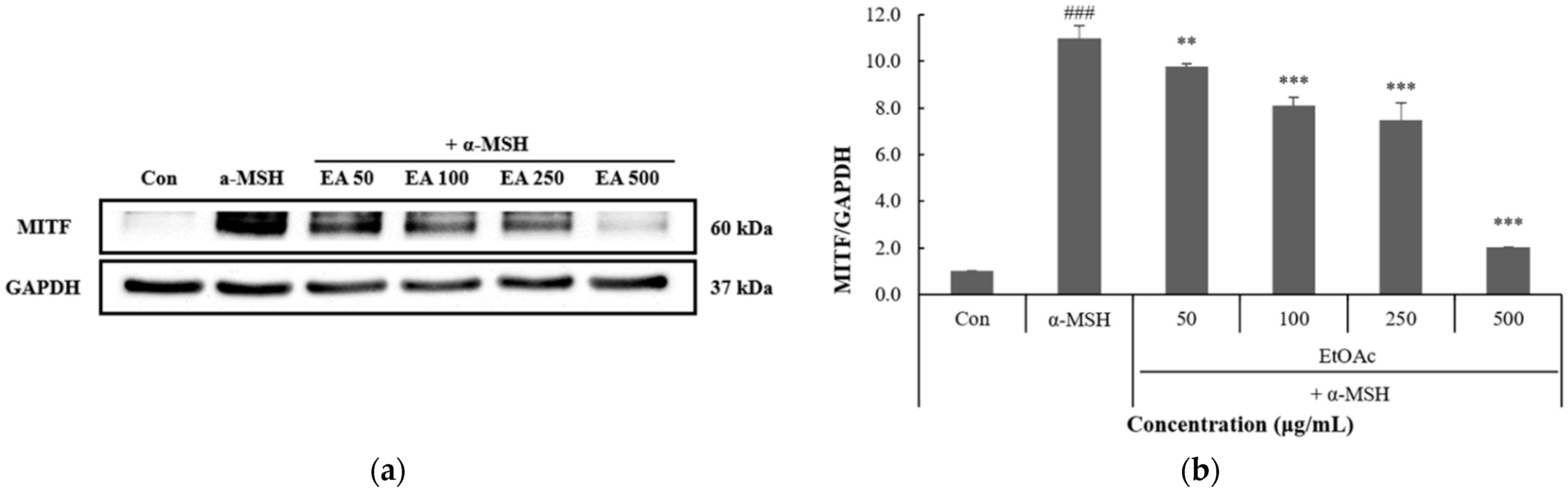
3.4. Inhibitory Effect of P. thunbergiana L. Fractions on MITF Expression in B16F10 Cells

The effect of the EtOAc fraction on the expression of transcription factors involved in melanin synthesis was determined via Western blotting. As a result, the EtOAc fraction decreased the expression of MITF induced by α-MSH in a concentration-dependent manner (Figure 3).
In addition, it was confirmed that the MITF expression was reduced by about 5 fold compared to the a-MSH treated group at a concentration of 500 ug/mL.

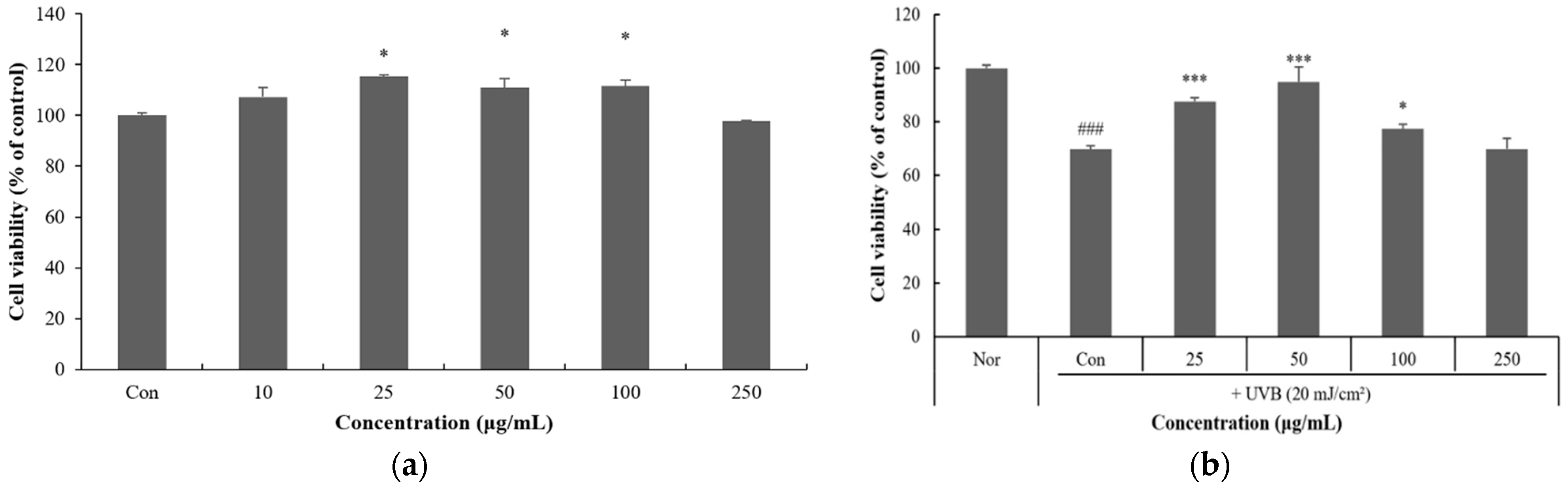
3.5. Effect of P. thunbergiana L. EtOAc Fraction on UVB-Induced Damage in HaCaT Cells

Among the fractions, only the EtOAc fraction was used in the anti-photoaging effect experiment because the EtOAc fraction showed the highest antioxidant activity and melanogenesis inhibitory effect. To assess the effect of the EtOAc fraction of P. thunbergiana L. on photoaging, cell viability of HaCaT cells was measured via the MTT assay. The EtOAc fraction did not affect cell viability at concentrations of 10–250 μg/mL (Figure 4a). Therefore, the subsequent experiments were performed using the leaf extracts at a concentration of 250 μg/mL or less.
To assess the cytoprotective effect of P. thunbergiana L. EtOAc fractions against UVB-induced damage, we confirmed the ability of HaCaT cells to proliferate even when their cell viability was reduced after UVB irradiation (20 mJ/cm2). The EtOAc fractions showed excellent cytoprotective effects against UVB at concentrations of 25 and 50 μg/mL (Figure 4b).

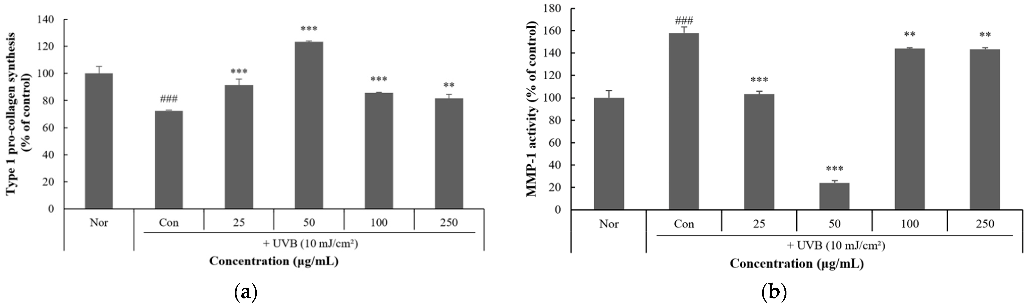
3.6. Effect of P. thunbergiana L. EtOAc Fraction on Anti-Photoaging in HaCaT Cells

To confirm the amount of pro-collagen synthesis in HaCaT cells after treatment with the P. thunbergiana L. EtOAc fraction, the cells were treated with the EtOAc fraction and were subject to UVB irradiation (10 mJ/cm2). The EtOAc fraction significantly increased pro-collagen synthesis. In particular, it was shown to increase the amount of pro-collagen by about 50% after UVB irradiation at the concentration of 50 μg/mL (Figure 5a).
To assess the inhibitory effect of the EtOAc fraction of P. thunbergiana L. on MMP-1 activity, it was measured after treatment with the EtOAc fraction and HaCaT cells were subjected to UVB irradiation (10 mJ/cm2). UVB irradiation increased MMP-1 activity, and the EtOAc fraction significantly decreased UVB-induced MMP-1 activity at concentrations of 25 and 50 μg/mL (Figure 5b).

3.7. Inhibitory Effect of P. thunbergiana L. Fractions on MMP-1 Expression in HaCaT Cells

To confirm that the EtOAc fraction of P. thunbergiana L. regulates the expression of aging-related factors, the expression of MMP-1, also known as collagenase, was confirmed via Western blotting. As a result, the EtOAc fraction decreased MMP-1 expression after UVB exposure in a concentration-dependent manner (Figure 6).

3.8. High-Performance Liquid Chromatography (HPLC) Analysis of P. thunbergiana L. EtOAc Fraction

According to the results of previous studies, it was reported that the main components of P. thunbergiana L. extract were daidzin and genistin [51]. Daidzin and genistein have excellent antioxidant activity as natural polyphenols and have been reported to exhibit tyrosinase inhibitory activity in previous reports. Therefore, we analyzed the major components of the P. thunbergiana L. EtOAc fraction by HPLC, using daidzin and genistin as standards. As a result, it was confirmed that these were two main components in the EtOAc fraction, and these results were consistent with previous studies (Figure 7).

4. Discussion

UV-induced skin pigmentation is regulated under the influence of various mechanisms, among which α-MSH binds to melanocortin 1 receptor (MC1R) and increases cytoplasmic cyclic AMP (cAMP) levels. Increased cAMP levels activate protein kinase A (PKA), which induces the expression of MITF through the phosphorylation of cAMP response element-binding protein (CREB) [52]. MITF, a main transcription factor for melanin biosynthesis, increases melanin synthesis by inducing the expression of tyrosinase-related proteins TRP-1, and TRP-2 [53]. Therefore, many studies have been conducted and showed that regulation of MITF expression may suppress melanogenesis [54].
Skin aging is closely related to the decomposition of the skin extracellular matrix. UV light activates the nuclear factor kappa-light-chain-enhancer of activated B cells (NF-κB) to decompose extracellular matrix components and increase the production of matrix metalloproteinases (MMPs), which are collagen-degrading enzymes [55]. The increase in MMP expression caused by UV rays also causes wrinkles and is used as a representative marker of skin inflammation and photoaging [56].
The currently used whitening and anti-aging functional substances cause side effects such as skin erythema and inflammation, and have low safety, which is a problem, given its potential frequent use. Therefore, research on the development of natural materials is being actively conducted to minimize their side effects and increase their safety.
P. thunbergiana is a creeper plant that belongs to the Fabaceae family, which contains isoflavonoids and diverse phenolic compounds, such as genistin, genistein, daidzin, daidzein, and puerarin [57,58]. According to modern pharmacological studies, its flowers and roots have been reported to have diverse biological activities, including antioxidant, hypoglycemic, lipid-lowering, hepatoprotective, hormonal regulation, and anti-inflammatory effects [59,60,61,62,63]. Although many studies on its flowers and roots have been reported, few studies have been conducted on the skin bioactivity of its leaf extracts.
In this study, the antioxidant activity of P. thunbergiana L. EtOAc fractions was measured, and their inhibitory activity, melanin production and MITF protein expression were confirmed in B16F10 melanoma cells. Their UVB cytoprotective effect, pro-collagen synthesis, MMP-1 inhibitory activity, and protein expression were also confirmed in HaCaT cells.
The EtOAc fraction showed a higher antioxidant effect than the other fractions. When the melanin content was measured within a concentration that did not affect cell viability in B16F10 melanoma cells, the EtOAc fraction showed a higher melanogenesis inhibitory effect than the other fractions. The EtOAc fraction reduced the protein expression of MITF, which is a well-known regulator of melanogenesis. Therefore, it was thought that the P. thunbergiana L. EtOAc fraction regulates melanogenesis by reducing the expression of MITF.
The EtOAc fraction showed a high protective effect against UVB by recovering the cell viability decreased by UVB irradiation and increased the decreased pro-collagen synthesis in HaCaT cells. In addition, the EtOAc fraction decreased the activity of collagenase MMP-1. The EtOAc fraction of P. thunbergiana L. was also thought to have an affirmative effect on skin anti-photoaging by inhibiting MMP-1 activity and protein expression.
After analyzing the components of the P. thunbergiana L. EtOAc fraction, it was found that the main components were daidzin and genistin. Daidzin and genistin are major isoflavonoids of P. thunbergiana L., which have been reported to contain radical scavengers, anti-proliferic agents against melanomas, and exhibit inhibition activity of melanin formation in vitro [64]. Their effect on tyrosinase inhibition has also been reported [65,66]. Due to these effects of daidzin and genistin, it was thought that the EtOAc fraction showed anti-melanogenesis activity.
Taken together, these results suggest that the P. thunbergiana L. EtOAC fraction inhibits melanogenesis and photoaging by down-regulating the signaling pathways MITF and MMP-1 in B16F10 mouse melanoma cells and HaCaT cells. However, additional studies are needed to fully understand the anti-melanogenesis and anti-photoaging mechanisms of the P. thunbergiana L. EtOAc fraction.

5. Conclusions

In this study, we demonstrated that the P. thunbergiana L. EtOAc fraction demonstrated melanogenesis inhibition and anti-photoaging effects in B16F10 melanoma cells and HaCaT cells. Therefore, these results show that the P. thunbergiana L. EtOAc fraction can potentially be used as an effective ingredient in whitening and anti-aging-related functional cosmetics.
In addition, further studies are needed on the purification of the compounds and their anti-melanogenic and anti-photoaging activities, as well as on their mechanisms of action.

Author Contributions

Conceptualization, methodology and writing—original draft preparation: M.J.K.; investigation and writing—review and editing: S.Y.S.; investigation and methodology: N.R.S.; methodology: S.K.; writing—review and editing: S.O.S.; project administration and writing—review and editing: K.M.P. All authors have read and agreed to the published version of the manuscript.

Funding

This research was supported by a Korea Innovation Foundation (INNIPOLIS) grant funded by the Korean government (Ministry of Science and ICT), through a science and technology project that contributes to the development of this field (grant number: 2021-DD-UP-0380).

Data Availability Statement

Not applicable.

Conflicts of Interest

The authors declare no conflict of interest.

References

  1. Wang, L.; Oh, J.Y.; Kim, Y.S.; Lee, H.G.; Lee, J.S.; Jeon, Y.J. Anti-photoaging and anti-melanogenesis effects of fucoidan isolated from Hizikia fusiforme and its underlying mechanisms. Mar. Drugs 2020, 18, 427. [Google Scholar] [CrossRef] [PubMed]
  2. Kageyama, H.; Waditee-Sirisattha, R. Antioxidative, anti-inflammatory, and anti-aging properties of mycosporine-like Amino Acids: Molecular and cellular mechanisms in the protection of skin-aging. Mar. Drugs 2019, 17, 222. [Google Scholar] [CrossRef] [Green Version]
  3. Pangestuti, R.; Siahaan, E.A.; Kim, S.K. Photoprotective substances derived from Marine Algae. Mar. Drugs 2018, 16, 399. [Google Scholar] [CrossRef] [Green Version]
  4. Perez Davo, A.; Truchuelo, M.T.; Vitale, M.; Gonzalez-Castro, J. Efficacy of an antiaging treatment against environmental factors: Deschampsia antarctica extract and high-tolerance retinoids combination. J. Clin. Aesthetic Dermatol. 2019, 12, E65–E70. [Google Scholar]
  5. Gao, W.; Lin, P.; Hwang, E.; Wang, Y.; Yan, Z.; Ngo, H.T.T.; Yi, T.H. Pterocarpus santalinus L. regulated ultraviolet B irradiation-induced procollagen reduction and matrix metalloproteinases expression through activation of TGF-β/Smad and inhibition of the MAPK/AP-1 pathway in normal human dermal fibroblasts. Photochem. Photobiol. 2018, 94, 139–149. [Google Scholar] [CrossRef] [PubMed]
  6. Shin, S.Y.; Kim, G.W.; Kang, S.W.; Cho, H.S.; Kim, E.J.; Park, K.M. Anti-aging and anti-melanogenesis effect of Cimicifuge dahurica, Coptis chinensis, Phellodendri amurense and Magnol obovata extracts mixture. J. Soc. Cosmet. Sci. Korea 2017, 43, 1–10. [Google Scholar]
  7. Papaccio, F.; D’Arino, A.; Caputo, S.; Bellei, B. Focus on the Contribution of Oxidative Stress in Skin Aging. Antioxidants 2022, 11, 1121. [Google Scholar] [CrossRef]
  8. Lee, J.H.; Park, J.H.; Shin, D.W. The Molecular Mechanism of Polyphenols with Anti-Aging Activity in Aged Human Dermal Fibroblasts. Molecules 2022, 27, 4351. [Google Scholar] [CrossRef]
  9. Zhang, Z.; Zhu, H.; Zheng, Y.; Zheng, L.; Wang, X.; Luo, Z.; Tang, J.; Lin, L.; Du, Z.; Dong, C. The effects and mechanism of collagen peptide and elastin peptide on skin aging induced by D-galactose combined with ultraviolet radiation. J. Photochem. Photobiol. B: Biol. 2020, 210, 111964. [Google Scholar] [CrossRef]
  10. Shin, J.W.; Kwon, S.H.; Choi, J.Y.; Na, J.I.; Huh, C.H.; Choi, H.R.; Park, K.C. Molecular Mechanisms of Dermal Aging and Antiaging Approaches. Int. J. Mol. Sci. 2019, 20, 2126. [Google Scholar] [CrossRef] [Green Version]
  11. Davinelli, S.; Nielsen, M.E.; Scapagnini, G. Astaxanthin in Skin Health, Repair, and Disease: A Comprehensive Review. Nutrients 2018, 10, 522. [Google Scholar] [CrossRef] [PubMed] [Green Version]
  12. Son, D.J.; Jung, J.C.; Choi, Y.M.; Ryu, H.Y.; Lee, S.; Davis, B.A. Wheat Extract Oil (WEO) Attenuates UVB-Induced Photoaging via Collagen Synthesis in Human Keratinocytes and Hairless Mice. Nutrients 2020, 12, 300. [Google Scholar] [CrossRef] [PubMed]
  13. Opperman, L.; De Kock, M.; Klaasen, J.; Rahiman, F. Tyrosinase and melanogenesis inhibition by indigenous African plants: A Review. Cosmetics 2020, 7, 60. [Google Scholar] [CrossRef]
  14. Han, H.J.; Park, S.K.; Kang, J.Y.; Kim, J.M.; Yoo, S.K.; Heo, H.J. Anti-melanogenic effect of ethanolic extract of Sorghum bicolor on IBMX–induced melanogenesis in B16/F10 melanoma cells. Nutrients 2020, 12, 832. [Google Scholar] [CrossRef] [PubMed] [Green Version]
  15. Lin, Y.S.; Wu, W.C.; Lin, S.Y.; Hou, W.C. Glycine hydroxamate inhibits tyrosinase activity and melanin contents through downregulating cAMP/PKA signaling pathways. Amino Acids 2015, 47, 617–625. [Google Scholar] [CrossRef]
  16. Boo, Y.C. p-Coumaric Acid as An Active Ingredient in Cosmetics: A Review Focusing on its Antimelanogenic Effects. Antioxidants 2019, 8, 275. [Google Scholar] [CrossRef] [PubMed] [Green Version]
  17. Del Bino, S.; Duval, C.; Bernerd, F. Clinical and biological characterization of skin pigmentation diversity and its consequences on UV impact. Int. J. Mol. Sci. 2018, 19, 2668. [Google Scholar] [CrossRef] [Green Version]
  18. Cadet, J.; Douki, T. Formation of UV-induced DNA damage contributing to skin cancer development. Photochem. Photobiol. Sci. 2018, 17, 1816–1841. [Google Scholar] [CrossRef]
  19. Passeron, T.; Krutmann, M.L.; Andersen, R.; Katta, C.C.; Zouboulis, C.C. Clinical and biological impact of the exposome on the skin. J. Eur. Acad. Dermatol. Venereol. 2020, 34, 4–25. [Google Scholar] [CrossRef]
  20. D’Mello, S.A.; Finlay, G.J.; Baguley, B.C.; Askarian-Amiri, M.E. Signaling pathways in melanogenesis. Int. J. Mol. Sci. 2016, 17, 1144. [Google Scholar] [CrossRef] [Green Version]
  21. Sim, Y.Y.; Tan, C.P.; Cheong, L.Z.; Nyam, K.L. Hibiscus cannabinus L. leaf and seed in cosmetic formulation: An integrated approach as antioxidant and melanogenesis inhibitor. Sustain. Mater. Technol. 2022, 33, e00457. [Google Scholar] [CrossRef]
  22. Kim, D.E.; Chang, B.Y.; Ham, S.O.; Kim, Y.C.; Kim, S.Y. Neobavaisoflavone inhibits melanogenesis through the regulation of Akt/GSK-3β and MEK/ERK pathways in B16F10 cells and a reconstructed human 3D skin model. Molecules 2020, 25, 2683. [Google Scholar] [CrossRef] [PubMed]
  23. Du, B.; Liu, X.; Khan, A.; Wan, S.; Guo, X.; Xue, J.; Fan, R. miRNA-183∼96∼182 regulates melanogenesis, cell proliferation and migration in B16 cells. Acta Histochem. 2020, 122, 151508. [Google Scholar] [CrossRef]
  24. Miyasaka, K.; Manse, Y.; Yoneda, A.; Takeda, S.; Shimizu, N.; Yamada, W.; Morikawa, T.; Shimoda, H. Anti-melanogenic effects of glucosylceramides and elasticamide derived from rice oil by-products in melanoma cells, melanocytes, and human skin. J. Food Biochem. 2022, 46, e14353. [Google Scholar] [CrossRef] [PubMed]
  25. Carpenter, E.L.; Becker, A.L.; Indra, A.K. NRF2 and Key Transcriptional Targets in Melanoma Redox Manipulation. Cancers 2022, 14, 1531. [Google Scholar] [CrossRef]
  26. Moon, H.R.; Jo, S.Y.; Kim, H.T.; Lee, W.J.; Won, C.H.; Lee, M.W.; Choi, J.H.; Chang, S.E. Loratadine, an H1 Antihistamine, Inhibits Melanogenesis in Human Melanocytes. BioMed Res. Int. 2019, 2019, 8. [Google Scholar] [CrossRef] [Green Version]
  27. Pillaiyar, T.; Manickam, M.; Namasivayam, V. Skin whitening agents: Medicinal chemistry perspective of tyrosinase inhibitors. J. Enzym. Inhib. Med. Chem. 2017, 32, 403–425. [Google Scholar] [CrossRef] [Green Version]
  28. Yang, X.; Niu, Z.; Wang, X.; Lu, X.; Sun, J.; Carpena, M. The Nutritional and Bioactive Components, Potential Health Function and Comprehensive Utilization of Pomegranate: A Review. Food Rev. Int. 2022, 1–27. [Google Scholar] [CrossRef]
  29. Niu, C.; Aisa, H.A. Upregulation of melanogenesis and tyrosinase activity: Potential agents for vitiligo. Molecules 2017, 22, 1303. [Google Scholar] [CrossRef] [Green Version]
  30. Draelos, Z.D. Skin lightening preparations and the hydroquinone controversy. Dermatol. Ther. 2007, 20, 308–313. [Google Scholar] [CrossRef]
  31. Kanthaliya, B.; Joshhi, A.; Meena, S.; Arora, J. Biology and Biotechnological Strategies for Conservation Management of Pueraria tuberosa, a Traditionally Established Medicinal Liana. Med. Plants 2021, 28, 693–719. [Google Scholar]
  32. Ulbricht, C.; Costa, D.; Dam, C.; D’Auria, D.; Giese, N.; Isaac, R.; Leblanc, Y.; Rusie, E.; Weissner, W.; Windsor, R.C. An evidence-based systematic review of kudzu (Pueraria lobata) by the natural standard research collaboration. J. Diet. Suppl. 2015, 12, 36–104. [Google Scholar] [CrossRef] [PubMed]
  33. Lim, D.W.; Lee, C.; Kim, I.H.; Kim, Y.T. Anti-inflammatory effects of total isoflavones from Pueraria lobata on cerebral ischemia in rats. Molecules 2013, 18, 10404–10412. [Google Scholar] [CrossRef] [PubMed]
  34. Ng, C.F.; Koon, C.M.; Cheung, D.W.S.; Lam, M.Y.; Leung, P.C.; Lau, C.B.S.; Fung, K.P. The anti-hypertensive effect of Danshen (Salvia miltiorrhiza) and Gegen (Pueraria lobata) formula in rats and its underlying mechanisms of vasorelaxation. J. Ethnopharmacol. 2011, 137, 1366–1372. [Google Scholar] [CrossRef] [PubMed]
  35. Xiong, Y.; Yang, Y.; Yang, J.; Chai, H.; Li, Y.; Jia, Z.; Wang, Z. Tectoridin, an isoflavone glycoside from the flower of Pueraria lobata, prevents acute ethanol-induced liver steatosis in mice. Toxicology 2010, 276, 64–72. [Google Scholar] [CrossRef]
  36. Yasuda, T.; Endo, M.; Kon-no, T.; Kato, T.; Mitsuzuka, M.; Ohsawa, K. Antipyretic, analgesic and muscle relaxant activities of Pueraria isoflavonoids and their metabolites from Pueraria lobata Ohwi-a traditional chinese drug. Biol. Pharm. Bull. 2005, 28, 1224–1228. [Google Scholar] [CrossRef] [Green Version]
  37. Yamazaki, T.; Hosono, T.; Matsushita, Y.; Kawashima, K.; Someya, M.; Nakajima, Y.; Narui, K.; Hibi, Y.; Ishizaki, M.; Kinjo, J.; et al. Pharmacological studies on Puerariae Flos. IV: Effects of Pueraria thomsonii dried flower extracts on blood ethanol and acetaldehyde levels in humans. Int. J. Clin. Pharmacol. Res. 2002, 22, 23–28. [Google Scholar]
  38. Park, K.H.; Gu, D.R.; Jin, S.H.; Yoon, C.S.; Ko, W.; Kim, Y.C.; Lee, S.H. Pueraria lobate Inhibits RANKL-mediated Osteoclastogenesis via downregulation of CREB/PGC1β/c–Fos/ NFATc1 signaling. Am. J. Chin. Med. 2017, 45, 1725–1744. [Google Scholar] [CrossRef]
  39. Chang, B.Y.; Lee, D.S.; Lee, J.K.; Kim, Y.C.; Cho, H.K.; Kim, S.Y. Protective activity of kudzu (Pueraria thunbergiana) vine on chemically induced hepatotoxicity: In vitro and in vivo studies. BMC Complement. Altern. Med. 2015, 16, 39. [Google Scholar] [CrossRef] [Green Version]
  40. Han, E.; Chang, B.; Kim, D.; Cho, H.; Kim, S. Melanogenesis inhibitory effect of aerial part of Pueraria thunbergiana in vitro and in vivo. Arch. Dermatol. Res. 2015, 307, 57–72. [Google Scholar] [CrossRef] [Green Version]
  41. Xu, L.; Zheng, N.; He, Q.; Li, R.; Zhang, K.; Liang, T. Puerarin, isolated from Pueraria lobata (Willd.), protects against hepatotoxicity via specific inhibition of the TGF-β1/Smad signaling pathway, thereby leading to anti-fibrotic effect. Phytomedicine 2013, 20, 1172–1179. [Google Scholar] [CrossRef] [PubMed]
  42. Tanaka, T.; Tang, H.; Yu, F.; Michihara, S.; Uzawa, Y.; Zaima, N.; Moriyama, T.; Kawamura, Y. Kudzu (Pueraria lobata) vine ethanol extracts improve ovariectomy-induced bone loss in female mice. J. Agric. Food Chem. 2011, 59, 13230–13237. [Google Scholar] [CrossRef] [PubMed]
  43. Folin, O.; Denis, W. On phosphotungstic-phosphomolybdic compounds as color reagents. J. Biol. 1912, 12, 239–243. [Google Scholar] [CrossRef]
  44. Zhishen, J.; Mengcheng, T.; Jianming, W. The determination of flavonoid contents in mulberry and their scavenging effects on superoxide radicals. Food Chem. 1999, 64, 555–559. [Google Scholar] [CrossRef]
  45. Blois, M.S. Antioxidant determinations by the use of a stable free radical. Nature 1958, 181, 1199–1200. [Google Scholar] [CrossRef]
  46. Zheng, L.; Zhao, M.; Xiao, C.; Zhao, Q.; Su, G. Practical problems when using ABTS assay to assess the radical-scavenging activity of peptides Importance of controlling reaction pH and time. Food Chem. 2016, 192, 288–294. [Google Scholar] [CrossRef] [PubMed]
  47. Mosmann, T. Rapid colorimetric assay for cellular growth and survival: Application to proliferation and cytotoxicity assays. J. Immunol. Methods 1983, 65, 55–63. [Google Scholar] [CrossRef]
  48. Hosoi, J.; Abe, E.; Suda, T.; Kuroki, T. Regulation of Melanin Synthesis of B16 Mouse Melanoma Cells by 1α, 25-Dihydroxyvitamin D3 and Retinoic Acid. Cancer Res. 1985, 45, 1474–1478. [Google Scholar]
  49. Wang, Y.S.; Cho, J.G.; Hwang, E.S.; Yang, J.E.; Gao, W.; Fang, M.Z.; Zheng, S.D.; Yi, T.H. Enhancement of Protective Effects of Radix Scutellariae on UVB-induced Photo Damage in Human HaCaT Keratinocytes. Appiled Biochem. Biotechnol. 2018, 184, 1073–1093. [Google Scholar] [CrossRef]
  50. Jeong, D.E.; Lee, Y.; Lee, S.J.V. Western Blot Analysis of C. elegans Proteins. In Methods in Molecular Biology; Huang, L., Ed.; Humana Press: New York, NY, USA, 2018; pp. 213–225. [Google Scholar]
  51. Son, E.; Yoon, J.M.; An, B.J.; Lee, Y.M.; Cha, J.; Chi, G.Y.; Kim, D.S. Comparison among activities and isoflavonoids from Pueraria thunbergiana aerial parts and root. Molecules 2019, 24, 912. [Google Scholar] [CrossRef] [Green Version]
  52. Jung, H.J.; Lee, A.K.; Park, Y.J.; Lee, S.; Kang, D.; Jung, Y.S.; Chung, H.Y.; Moon, H.R. (2E,5E)-2,5-Bis (3-hydroxy-4-methoxybenzylidene) cyclopentanone exerts anti-melanogenesis and anti-wrinkle activities in B16F10 melanoma and Hs27 fibroblast cells. Molecules 2018, 23, 1415. [Google Scholar] [CrossRef] [PubMed] [Green Version]
  53. Maddodi, N.; Jayanthy, A.; Setaluri, V. Shining light on skin pigmentation: The darker and the brighter side of effects of UV radiation. Photochem. Photobiol. 2012, 88, 1075–1082. [Google Scholar] [CrossRef] [PubMed] [Green Version]
  54. Wan, P.; Hu, Y.; He, L. Regulation of melanocyte pivotal transcription factor MITF by some other transcription factors. Mol. Cell. Biochem. 2011, 354, 241–246. [Google Scholar] [CrossRef] [PubMed]
  55. Kim, J.; Lee, C.W.; Kim, E.K.; Lee, S.J.; Park, N.H.; Kim, H.S.; Kim, H.K.; Char, K.H.; Jang, Y.P.; Kim, J.W. Inhibition effect of Gynura procumbens extract on UV-B-induced matrix-metalloproteinase expression in human dermal fibroblasts. J. Ethnopharmacol. 2011, 137, 427–433. [Google Scholar] [CrossRef]
  56. Inomata, S.; Takada, K.; Tsunenaga, M.; Fukuda, M.; Matsunaga, Y.; Amano, S.; Kobayashi, K.; Nishiyama, T.; Kohno, Y. Possible involvement of gelatinases in basement membrane damage and wrinkle formation in chronically ultraviolet B-exposed hairless mouse. J. Investig. Dermatol. 2003, 120, 128–134. [Google Scholar] [CrossRef]
  57. Kim, D.Y.; Won, K.J.; Hwang, D.I.; Yoon, S.W.; Lee, S.J.; Park, J.H.; Yoon, M.S.; Kim, B.K.; Lee, H.M. Potential skin regeneration activity and chemical composition of absolute from Pueraria thunbergiana flower. Nat. Prod. Commun. 2015, 10, 2009–2012. [Google Scholar]
  58. Park, E.K.; Shin, J.; Bae, E.A.; Lee, Y.C.; Kim, D.H. Intestinal bacteria activate estrogenic effect of main constituents puerarin and daidzin of Pueraria thunbergiana. Biol. Pharm. Bull. 2006, 29, 2432–2435. [Google Scholar] [CrossRef] [Green Version]
  59. Min, S.W.; Park, Y.J.; Kim, D.H. Kakkalide and its metabolite irisolidone ameliorate carrageenan-induced inflammation in mice by inhibiting NF-κB pathway. Inflammation 2011, 34, 344–351. [Google Scholar] [CrossRef]
  60. Cherdshewasart, W.; Sutjit, W.; Pulcharoen, K.; Chulasiri, M. The mutagenic and antimutagenic effects of the traditional phytoestrogen-rich herbs, Pueraria mirifica and Pueraria lobata. Braz. J. Med. Biol. Res. 2009, 42, 816–823. [Google Scholar] [CrossRef] [Green Version]
  61. Shin, J.E.; Bae, E.A.; Lee, Y.C.; Ma, J.Y.; Kim, D.H. Estrogenic effect of main components kakkalide and tectoridin of Puerariae flos and their metabolites. Biol. Pharm. Bull. 2006, 29, 1202–1206. [Google Scholar] [CrossRef] [Green Version]
  62. Park, K.Y.; Jung, G.O.; Choi, J.; Lee, K.T.; Park, H.J. Potent antimutagenic and their anti-lipid peroxidative effect of kaikasaponin iii and tectorigenin from the flower of Pueraria thunbergiana. Arch. Pharmacal Res. 2002, 25, 320–324. [Google Scholar] [CrossRef] [PubMed]
  63. Lee, K.T.; Sohn, I.C.; Kim, Y.K.; Choi, J.H.; Choi, J.W.; Park, H.J.; Itoh, Y.; Miyamoto, K. Tectorigenin, an isoflavone of Pueraria thunbergiana Benth., induces differentiation and apoptosis in human promyelocytic leukemia HL-60 cells. Biol. Pharmceutical Bull. 2001, 24, 1117–1121. [Google Scholar] [CrossRef] [PubMed] [Green Version]
  64. Lee, K.T.; Sohn, I.C.; Kim, D.H.; Choi, J.W.; Kwon, S.H.; Park, H.J. Hypoglycemic and hypolipidemic effects of tectorigenin and kaikasaponin III in the streptozotocin-induced diabetic rat and their antioxidant activity in vitro. Arch. Pharmacal Res. 2000, 23, 461–466. [Google Scholar] [CrossRef] [PubMed]
  65. Lewis, P.; Kaltia, S.; Wähälä, K. The phase transfer catalysed synthesis of isoflavone-O-glucosides. J. Chem. Soc. Perkin Trans. 1998, 1, 2481–2484. [Google Scholar] [CrossRef]
  66. Liu, H.; Zhu, Y.; Wang, T.; Qi, J.; Liu, X. Enzyme-site blocking combined with optimization of molecular docking for efficient discovery of potential tyrosinase specific inhibitors from Puerariae lobatae radix. Molecules 2018, 23, 2612. [Google Scholar] [CrossRef]
Figure 1. Extraction and division of fractions from P. thunbergiana L.
Processes 10 02156 g001
Figure 2. (a) Effect of P. thunbergiana L. EtOH extracts on the cell viability of B16F10 melanoma cells. Cells were treated with 10–1000 μg/mL P. thunbergiana L. EtOH extracts. (b) Effect of P. thunbergiana L. fractions on cell viability in B16F10 melanoma cells. Cells were treated with 10–1000 μg/mL P. thunbergiana L. fractions. * p < 0.05 and ** p < 0.01 compared with the control. (c) Effect of P. thunbergiana L. EtOH extracts on the melanin content in α-MSH-stimulated B16F10 melanoma cells. (d) Effect of P. thunbergiana L. fractions on melanin content in α-MSH-stimulated B16F10 melanoma cells (Ar: arbutin). Cells were exposed to α-MSH (100 nM) alone or with the different samples or arbutin (100 μg/mL) for 72 h. (e) Visual photographs of the cell pellets formed by B16F10 melanoma cells, after treatment with P. thunbergiana L. fractions (hexane: H, EtOAc: EA, butanol: B, aqueous solution: Aq). The results are expressed as the mean ± SD from three independent experiments. ### p < 0.001 compared with the control; * p < 0.05, ** p < 0.01 and *** p < 0.001 compared with α-MSH.
Processes 10 02156 g002aProcesses 10 02156 g002b
Figure 3. (a) Effect of the P. thunbergiana L. EtOAc fraction on MITF expression. (b) Band intensity compared to the control (GAPDH) was determined using TotalLab 1D software (TotalLab, Newcastle, UK). The results are expressed as the mean ± SD from three independent experiments. ### p < 0.001 compared with the control; ** p < 0.01 and *** p < 0.001 compared with α-MSH.
Processes 10 02156 g003
Figure 4. (a) Effect of P. thunbergiana L. EtOAc fraction on cell viability in HaCaT cells. Cells were treated with 10–1000 μg/mL P. thunbergiana L. EtOAc fraction. (b) Effect of P. thunbergiana L. EtOAc fraction on cell viability in UVB-irradiated (20 mJ/cm2) HaCaT cells (Nor: normal control; Con: control) The results are expressed as the mean ± SD from three independent experiments. ### p < 0.001 compared with the normal control; * p < 0.05 and *** p < 0.001 compared with the control.
Processes 10 02156 g004
Figure 5. (a) Effect of P. thunbergiana L. EtOAc fraction on type-1 procollagen synthesis in UVB-irradiated (10 mJ/cm2) HaCaT cells. (b) Effect of P. thunbergiana L. EtOAc fraction on the inhibition of MMP-1 activity in HaCaT cells. ### p < 0.001 compared with the normal control; ** p < 0.01 and *** p < 0.001 compared with the control.
Processes 10 02156 g005
Figure 6. (a) Effect of P. thunbergiana L. EtOAc fraction on expression of MMP-1 in HaCaT cells. (b) Band intensity compared to the control (GAPDH) was determined using TotalLab 1D software. The results are expressed as the mean ± SD from three independent experiments. ## p < 0.01 compared with the normal control; *** p < 0.001 compared with the control.
Processes 10 02156 g006
Figure 7. HPLC chromatogram shows peaks in extracts of P. thunbergiana EtOAc fraction, with absorbance evaluated at 254 nm. AU indicates the absorbance unit. (a) Daidzin standard, (b) genistin standard; (c) P. thunbergiana EtOAc fraction.
Processes 10 02156 g007
Table 1. Total phenolic and flavonoid content of P. thunbergiana L. extracts and fractions. Results are expressed as mean values ± standard deviation (n = 3). Means with different superscript letters (a–e) indicate statistically significant differences (p < 0.05).
SampleSolvent
Extract/Fraction
Extraction
Yield (%)
* TPC* TFC
µg GAE/gµg CE/g
ExtractsAqueous solution1823 ± 0.00 b101 ± 0.00 b
30% EtOH1628 ± 0.02 e122 ± 0.80 e
50% EtOH827 ± 0.03 d111 ± 0.22 d
70% EtOH1224 ± 0.02 c103 ± 0.67 c
90% EtOH722 ± 0.03 a77 ± 0.44 a
Fractionsn-Hexane4.516 ± 0.02 a76 ± 0.22 a
EtOAc1.429 ± 0.00 d113 ± 0.00 d
Butanol3.623 ± 0.03 c88 ± 0.22 c
Aqueous solution8.917 ± 0.07 b86 ± 0.89 b
* TPC: Total phenolic content, * TFC: total flavonoid content.
Table 2. DPPH and ABTS radical scavenging activity of P. thunbergiana L. EtOH extracts and fractions (ASA: ascorbic acid). Results are expressed as mean values ± standard deviation (n = 3). Means with different superscript letters (a–f) indicate statistically significant differences (p < 0.05).
SampleSolvent
Extract/Fraction
EC50 for DPPH (μg/mL)EC50 for ABTS (μg/mL)
ExtractsAqueous solution124 ± 0.67 e62.5 ± 0.84 b
30% EtOH88.1 ± 1.34 c54.2 ± 0.57 a
50% EtOH73.4 ± 0.91 b57.2 ± 0.8 a
70% EtOH94.3 ± 1.2 d63.5 ± 1.03 b
90% EtOH166.1 ± 5.23 f85 ± 0.24 c
ASA11.4 ± 0.77 a-
Fractionsn-Hexane290.4 ± 0.85 d79.6 ± 5.45 c
EtOAc195.4 ± 1.17 b49.4 ± 1.18 a
Butanol199.8 ± 1.13 b67.6 ± 0.43 b
Aqueous solution270.1 ± 1.2 c77.1 ± 1.16 c
ASA9.1 ± 0.9 a-
Publisher’s Note: MDPI stays neutral with regard to jurisdictional claims in published maps and institutional affiliations.

