Article

Effect of UV Light and Sodium Hypochlorite on Formation and Destruction of Pseudomonas fluorescens Biofilm In Vitro

by
Melani Sigler Zekanović
1,2,
Gabrijela Begić
2,
Silvestar Mežnarić
3,
Ivana Jelovica Badovinac
4,
Romana Krištof
5,
Dijana Tomić Linšak
3,6,† and
Ivana Gobin
2,7,*,†
1
Ilirija d.d., Tina Ujevica 7, 23210 Biograd na Moru, Croatia
2
Department of Microbiology and Parasitology, Faculty of Medicine, University of Rijeka, Brace Branchetta 10 20, 51000 Rijeka, Croatia
3
Department of Environmental Health, Teaching Institute of Public Health, Primorje-Gorski Kotar County, Krešimirova 52a, 51000 Rijeka, Croatia
4
Faculty of Physics and Centre for Micro- and Nanosciences and Technologies University of Rijeka, Brace Branchetta 10 20, 51000 Rijeka, Croatia
5
Department of Sanitary Engineering, Faculty of Health Sciences, University of Ljubljana, 1000 Ljubljana, Slovenia
6
Department for Health Ecology, Faculty of Medicine, University of Rijeka, Brace Branchetta 10 20, 51000 Rijeka, Croatia
7
Faculty of Health Studies, University of Mostar, 88000 Mostar, Bosnia and Herzegovina
*
Author to whom correspondence should be addressed.
These authors contributed equally to this work.
Processes 2022, 10(10), 1901; https://doi.org/10.3390/pr10101901
Submission received: 20 August 2022 / Revised: 12 September 2022 / Accepted: 16 September 2022 / Published: 20 September 2022
(This article belongs to the Special Issue Sanitary and Environmental Engineering: Relevance and Concerns)

Abstract

:
Pseudomonas fluorescens is one of the first colonizers of bacterial biofilm in water systems and a member of opportunistic premise plumbing pathogens (OPPPs). The aim of this study was to examine the effect of UV light and sodium hypochlorite on the formation and destruction of mature P. fluorescens biofilm on ceramic tiles. Planktonic bacteria or bacteria in mature biofilm were exposed to UV light (254 nm) for 5, 20 s. and to 0.4 mg/L sodium hypochlorite for 1 min. Mature biofilm was also exposed to increased concentration of sodium hypochlorite of 2 mg/L for 0.5, 1 and 2 h and combined with UV. Prolonged action of sodium hypochlorite and an increase in its concentration in combination with UV gave the best results in the inhibition of biofilm formation after the pre-treatment and destruction of mature biofilm. The effect of hyperchlorination in combination with UV radiation shows better results after a long exposure time, although even after 120 min there was no completely destroyed biofilm. Furthermore, the mechanism of the effect of combined methods should be explored as well as the importance of mechanical cleaning that is crucial in combating bacterial biofilm in swimming pools.

1. Introduction

Water supply systems are inhabited by the group of organisms called opportunistic premise plumbing pathogens (OPPPs) [1]. They are adapted to these systems, and can grow in changing, oligotrophic conditions [2]. OPPPs have similar characteristics, such as disinfectant resistance, biofilm formation, and amoeba digestion resistance. Model OPPPs are Legionella pneumophila, Mycobacterium avium and Pseudomonas spp., a genus for which drinking water is considered a relevant habitat. To these species, we may add Acinetobacter baumannii, Stenotrophomonas maltophila, Helicobacter pylori, Aeromonas hydrophila, and Methylobacterium spp. [3,4]. OPPPs can cause a range of transmissible and antimicrobial-resistant infections. Because of that there is a need to implement better control measurements and increase awareness [5,6,7].
Pseudomonas fluorescens is a rod-shaped aerobic, non- fermenting, gram-negative bacterium. It is widely spread in water, soil, plants, and animals [8]. Its presence in water is already well known. It can survive and replicate even in damp places, which relate to water supply sources [9,10,11,12], and it has been isolated from still, bottled water [13].
Although not considered a human pathogen, in some cases, especially in immunocompromised patients, it can cause acute diseases or outbreaks of bacteremia [8,9]. It is mostly studied as an environmental or soil bacteria, considering that Pseudomonas spp. are widely spread in the environment. But because of its ability to easily form biofilm and potentially cause infections it has been more studied as a growing concern in the food industry or in patients in medical institutions [14,15,16,17].
Biofilms are microbial communities in which cells are embedded within self-produced extracellular polymeric substances (EPS) or matrix [18]. The formation of biofilm takes place in several, usually fast phases, and P. fluorescens is frequently one of the first colonizers which adhere to the surface with the LapA protein and create microcolonies that will serve as an anchor to other microorganisms in creating a biofilm community. It can act as a “helper” as well, for other species to persist by using a P. fluorescens matrix as a shelter [18,19,20]. P. fluorescens are good biofilm producers with a strong EPS production capacity and these biofilms are characterized by an increased resistance to environmental influences and disinfectants [21,22].
Water in distribution systems or in swimming pool systems must be monitored and regulated. Disinfection is the most important process that ensures water safety. Standard doses of disinfectants, based mostly on chlorine, do not destroy OPPPs, and their number in water systems is increasing over time. Chlorine disinfection also creates a more homogeneous bacterial population, dominated by resistant Pseudomonas spp. [23,24,25]. Chemical disinfection of water contributes to the creation of harmful disinfection byproducts. Many of them have adverse health effects [26,27,28].
To avoid adaptive features of opportunistic pathogens such as resistance to disinfection or formation of biofilms, new technologies are being applied or are combining. Ultraviolet radiation (UV) is a promising technology for reducing OPPPs in water systems [29,30]. It has also been proven that combined disinfection, chlorination, and UV is an effective method to reduce concentrations of toxic byproducts [31,32,33].
The research into biofilms formed by the OPPPs group of microorganisms is scarce. Those biofilms represent potential source sites from which opportunistic pathogens are released into the aquatic environment. Therefore, in this study we isolated P. fluorescens from biofilm on the ceramic tiles of a freshwater swimming pool and examined the effect of the combined method, as well as the individual effect of UV light and sodium hypochlorite on the creation and destruction of already formed, mature P. fluorescens biofilm on ceramic tiles in vitro.

2. Materials and Methods

2.1. Bacterial Strains

P. fluorescens used in the in vitro experiment was isolated from mixed biofilm from small ceramic tiles (dimensions 2.5 cm × 2.5 cm), taken out of the freshwater swimming pool. The pool had a double disinfection method implemented, so the water was disinfected with chlorine and UV disinfection. After removal, the tile was washed and added to a tube with 10 mL of sterile water. Bacteria in biofilm were detached by treatment in an ultrasonic bath (Bactosonic, Bandelin, Germany) at 40 kHz for 1 min. Subsequently, ten-time dilutes of the sonicates were planted on a Mueller–Hinton agar (MH, Biolife, Milan, Italy) and after a 48-h incubation, suspected colonies were isolated. P. fluorescens was then identified using the API NE system (Biomerieux, Paris, France). Pure bacterial cultures were suspended in an MH broth (Biolife, Milan, Italy) of appropriate concentrations of 105 CFU/mL and used in the experiment. For this, the optical density was measured at 600 nm (OD600) (Eppendorf, Bio photometer, model #6131, Hamburg, Germany).

2.2. Mature Biofilm Formation

The method of biofilm formation was described according to the procedure developed by Ivanković et al., and modified [34]. The individual ceramic tiles were mechanically brushed, washed, and then sterilized for 1 h at 180 °C. An agar bacteriological solution (Oxoid, Basingstoke, UK) was prepared according to the manufacturer’s instructions and autoclaved at 121 °C/15 min. Three sterile tiles were placed in a Petri dish, with the ceramic surface facing up. After this, the still warm agar solution was poured, making sure that the upper ceramic area of the tiles remained uncovered. Suspensions of P. fluorescens in sterile tap water were poured onto the upper side of the tiles that were placed in agar, ensuring that they completely covered their surface, as described. Petri dishes were incubated at 35 °C for 5 days using a rotational shaker (30 rpm). In this way a mature, 5-day old biofilm was formed.

2.3. Pre-Treatments of Planktonic Bacteria with UV Light and Sodium Hypochlorite

In the pre-treatment of the planktonic bacteria, the effect of UV light, sodium hypochlorite, and their combination on the bacterial suspension was tested. The ability of bacteria to form biofilm after treatment was investigated. Bacterial suspensions of 105 CFU/mL were prepared as described, transferred to a plastic Petri dish and exposed to UV light at 254 nm (UV lamp-dual wavelength, Muttenz, Switzerland) for 5 s, 20 s, sodium hypochlorite solution 0.4 mg/L for 1 min, and a combination of UV light for 5 s, 20 s and sodium hypochlorite solution 0.4 mg/L for 1 min. A neutralizer was not used. The used UV lamp had two UV tubes for illumination (dual length), one for 254 nm and the other for 366 nm, both with 8 W of power. We only used 254 nm light for our experiment. The lamp was manually installed on two plastic stands so that the microtiter plate with suspension was one centimeter away. Treated bacterial suspension was poured over the ceramic tiles in agar, as described, followed by incubation at 35 °C for 5 days, so that mature biofilm could be formed. After washing unattached bacteria and ultrasound treatment to release bacteria in the biofilm, CFUs were determined by planting tenfold dilutions on MH agar. Treatment was performed in triplicate.
Immediately after treatment, P. fluorescens viability was tested (Live/Dead BacLight bacterial viability kit; Invitrogen, Carlsbad, CA, USA), according to the manufacturer’s protocol. Briefly, planktonic bacteria were treated as described earlier. Then two nucleic acid stains, propidium iodide (PI) and SYTO-9, were added and incubated for 15 min. The microbiological slides were prepared, and digital images were collected using a fluorescence microscope (Olympus BX51, Tokyo, Japan).

2.4. Mature Biofilm Treatments with UV Light and Sodium Hypochlorite

After incubation for 5 days, the tiles with mature biofilm were transferred to a plastic Petri dish and washed three times in sterile saline solution. After that, the mature biofilm was exposed to various treatments: UV light at 254 nm (UV lamp-dual wavelength, Muttenz, Switzerland) for 5 and 20 s, sodium hypochlorite solution (T.T.T, Sveta Nedjelja, Croatia), 0.4 mg/L for 1 min, and a combination of UV light for 5 or 20 s and sodium hypochlorite solution of 0.4 mg/L for 1 min. The lamp was manually installed on two plastic stands so that the surface of the mature biofilm was only one centimeter away. After this, sodium hypochlorite exposure, 10% sodium thiosulphate solution (Kemika, Zagreb, Croatia), was added to remove the residual sodium hypochlorite. Subsequently, tiles were washed and placed in sterile polypropylene tubes with sterile saline and treated in an ultrasonic bath (BactoSonik—Bandelin, Berlin, Germany) for 1 min/40 kHz. Tenfold serial dilutions were prepared, and samples were inoculated on MH agar. After incubation on 35 °C for 24 h, CFU/mL was determined.
Biofilm was also exposed to increased concentration of sodium hypochlorite of 2 mg/L for 1 min, 0.5, 1 and 2 h and combined with UV light for 5 and 20 s. After treatment, the tiles were processed as previously described and the number of bacteria was determined. As a control, mature biofilm was grown on the tiles under the same conditions and was not exposed to UV light and sodium hypochlorite. Each experiment was performed three times.

2.5. Scanning Electron Microscopy (SEM)

For the morphological analyses of biofilm on ceramic tiles, the scanning electron microscope Jeol JSM-7800F (JEOL Ltd., Tokyo, Japan) was used. The ability of bacteria to form biofilm after the pretreatment and destruction of mature biofilm was analyzed. Before microscopy, tiles fixation was done with 4% glutaraldehyde and 0.5% paraformaldehyde (Sigma Aldrich, Burlington, MA, USA) prepared at 4 °C in 0.1 M PBS (Sigma-Aldrich, Burlington, MA, USA). Dehydration was carried out in a series of increasing concentrations of ethanol from 50% to 100% (Sigma-Aldrich, Burlington, MA, USA) each for 20 min. Due to the increase in stability and conductivity, samples were sputtered with a gold layer.

2.6. Statistical Analyses

In order to analyze the normality of results and distribution in differently treated experimental groups Shapiro–Wilk test was used. To analyze the effect of different treatments in experimental groups, a nonparametric Mann–Whitney U-test was used for groups without normal distribution, and for groups with normal (Gaussian) distribution statistical significance was tested by a t-test. Results were expressed as means and standard deviation. Comparison with the control group was analyzed using the Wilcoxon signed-rank test. Results were considered statistically significant at p < 0.05 and are presented graphically using TIBCO Statistica 14.0, Excel office 365 and Sigmaplot 14.0.

3. Results

3.1. The Effect of UV Light and Sodium Hypochlorite on Biofilm Formation

To examine the influence of UV light (UV) and sodium hypochlorite (Cl) on the ability of P. fluorescens to form biofilms, the bacterial suspension was treated with UV for 5 and 20 s and with 0.4 mg/L Cl for 1 min. A combination of Cl and UV was also carried out, under the same described conditions. After 5 days of incubation, bacteria ability to form biofilm was determined (Figure 1).
Results showed that all treatments applied to planktonic bacteria in bacterial suspension significantly inhibited biofilm formation (5 days incubation) compared to the control (p = 0.002). The results showed that the number of bacteria in the biofilm depends on the applied treatment. Extending the exposure time of UV from 5 to 20 s significantly inhibited the number of bacteria in the biofilm (p = 0.02). Furthermore, the application of one-minute of Cl leads to a significant inhibition compared to UV radiation (p = 0.001). This was followed by a more pronounced effect of the combination of 5 s of UV and 1 min of Cl (p < 0.01). The most effective pre-treatment was the combination of 20 s of UV and 1-min Cl (p < 0.001) where 99.99% inhibition was achieved.
Dead/live staining showed that after applying individual treatments, as well as the UV 5″ + Cl 1′combined treatment, a significant number of viable cells was present and explained their ability to create biofilm after 5 days. For a UV 20″ + Cl 1′ combined treatment, there was a dominance of dead cells that were colored with red fluorescence, but there were also individual viable cells that were obviously capable of creating biofilm after 5 days of incubation (Figure 2D).

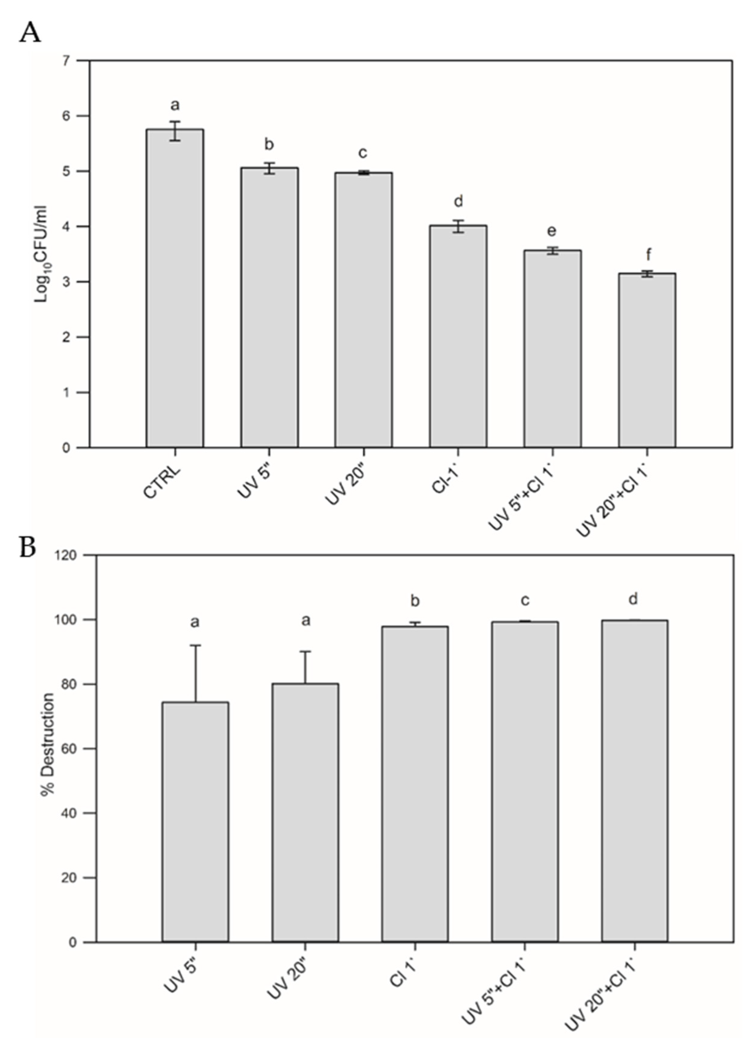
3.2. The Effect of UV Light and Sodium Hypochlorite on Mature Biofilm Destruction

Mature 5-day old biofilm was exposed to UV for 5 and 20 s, and 0.4 mg/L Cl for 1 min. A combination of UV and Cl was performed under the same described conditions. We compared the effect of individual treatments to untreated mature biofilm as a control. Treatments were also compared mutually (Figure 3.).
The results show that the applied treatments significantly reduced the number of bacteria in the biofilm compared to the untreated biofilm as follows: UV for 5 and 20 s (p = 0.0012), 1 min Cl (p = 0.0001), UV 5 s and 1 min Cl (p = 0.00018), UV for 20 s and 1 min Cl (p < 0.0001). A significant difference was also found between the effect of individual treatments, from the least effective UV to the most effective treatment, which was achieved by combining UV for 20 s with 1 min Cl. The destruction of a mature biofilm with different treatments shows results ranging from 66.67% to 99.99%. The effects of UV 5 s with reduction of 74.34% and UV 20 s with reduction of 80.19% are less successful in mature biofilm destruction then Cl alone or combined with UV. Cl 1 min treatment achieved biofilm reduction of 97.82%. With UV 5 s and UV 20 s combined with Cl for 1 min treatment, over 99% reduction was observed.

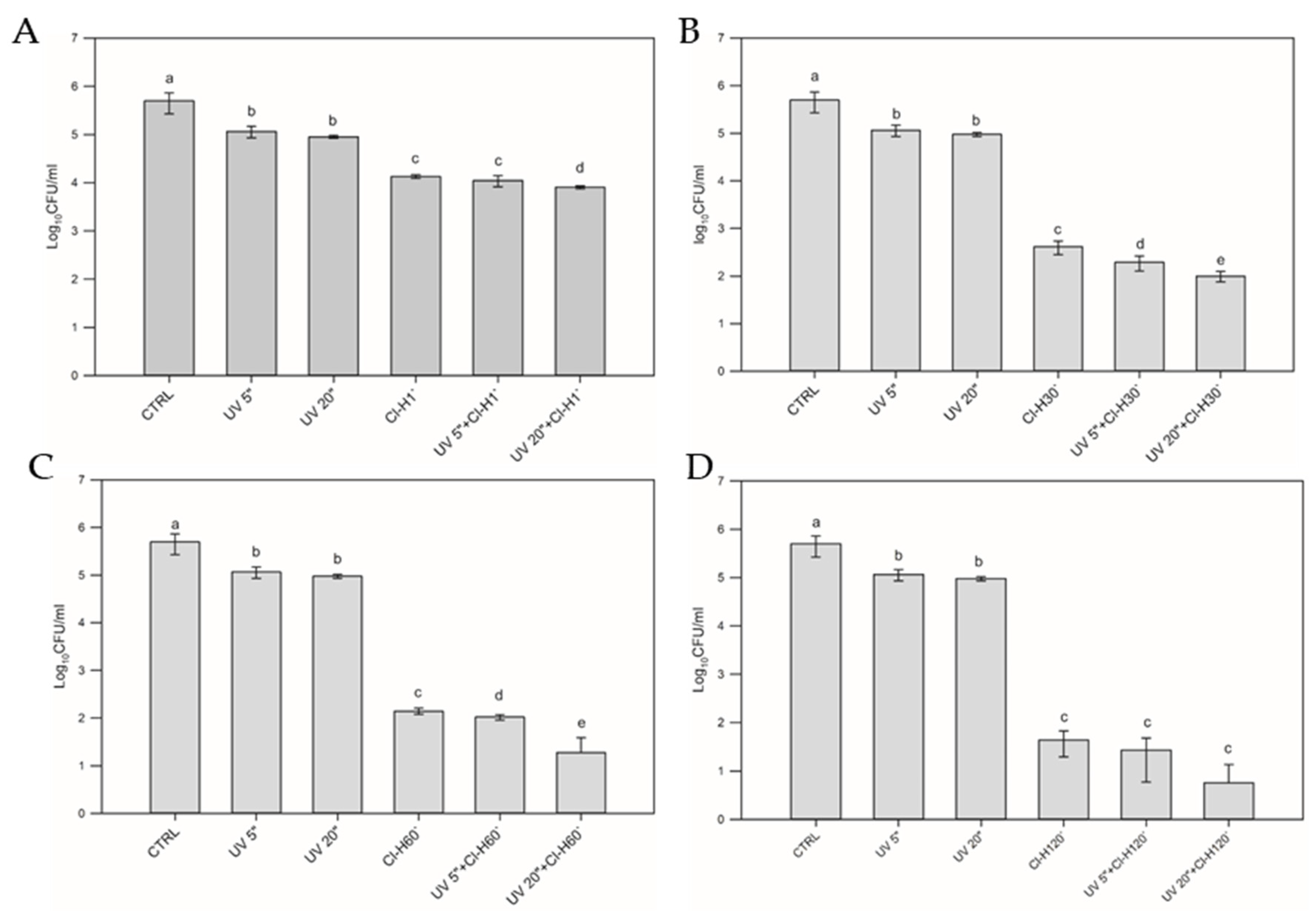
3.3. The Effect of UV Light and Hyperchlorination on Mature Biofilm Destruction

The effect of a five-time higher concentration of sodium hypochlorite of 2.0 mg/L (Cl-H) was tested on a 5-day old biofilm. Its combined effect with UV for 5 and 20 s was also examined. Afterwards, dependence of the treatment efficiency with hyperchlorination on the exposure was assayed (Figure 4).
All treatments, UV (p = 0.0012), hyperchlorination (Cl-H) lasting from 1 to 120 min (p = 0.01) and the combination of UV and Cl-H (p < 0.001) significantly reduced the number of bacteria in the biofilm compared to the untreated biofilm. As expected, hyperchlorination is significantly more effective than UV light alone. Combined exposure of the biofilm to UV for 5 s and Cl-H did not increase the effectiveness of the hyperchlorination itself, regardless of its duration. On the contrary, the duration of UV radiation had an impact on the effectiveness of the combined treatment. Exposure to UV for 20 s in combination with hyperchlorination for 1, 30, 60 or 120 min led to a significant reduction in the number of bacteria compared to almost all other treatments. Only hyperchlorination for 120 min combined with UV for 5 and 20 s did not differ significantly.
The percentage of mature biofilm destruction was also determined and results are presented in Table 1.
Hyperchlorination, Cl-H of mature biofilm for 1 min was significantly different from UV for 20 s (p = 0.034). No other statistical significance was noted in the 1-min treatment. Hyperchlorination for 30 min and UV for 20 s was different from UV for 5 s (p = 0.02). Cl-H for 30 min was significantly different from UV for 20 s (p = 0.004); a combination of UV for 20 s and Cl-H for 30 min was different from only Cl-H for 30 min (p = 0.02). Hyperchlorination for 60 min was significantly different from UV for 20 s (p < 0.001). Hyperchlorination for 120 min was significantly different from UV for 20 s (p < 0.001). No other statistical significance was noted in the 60- and 120-min treatment.

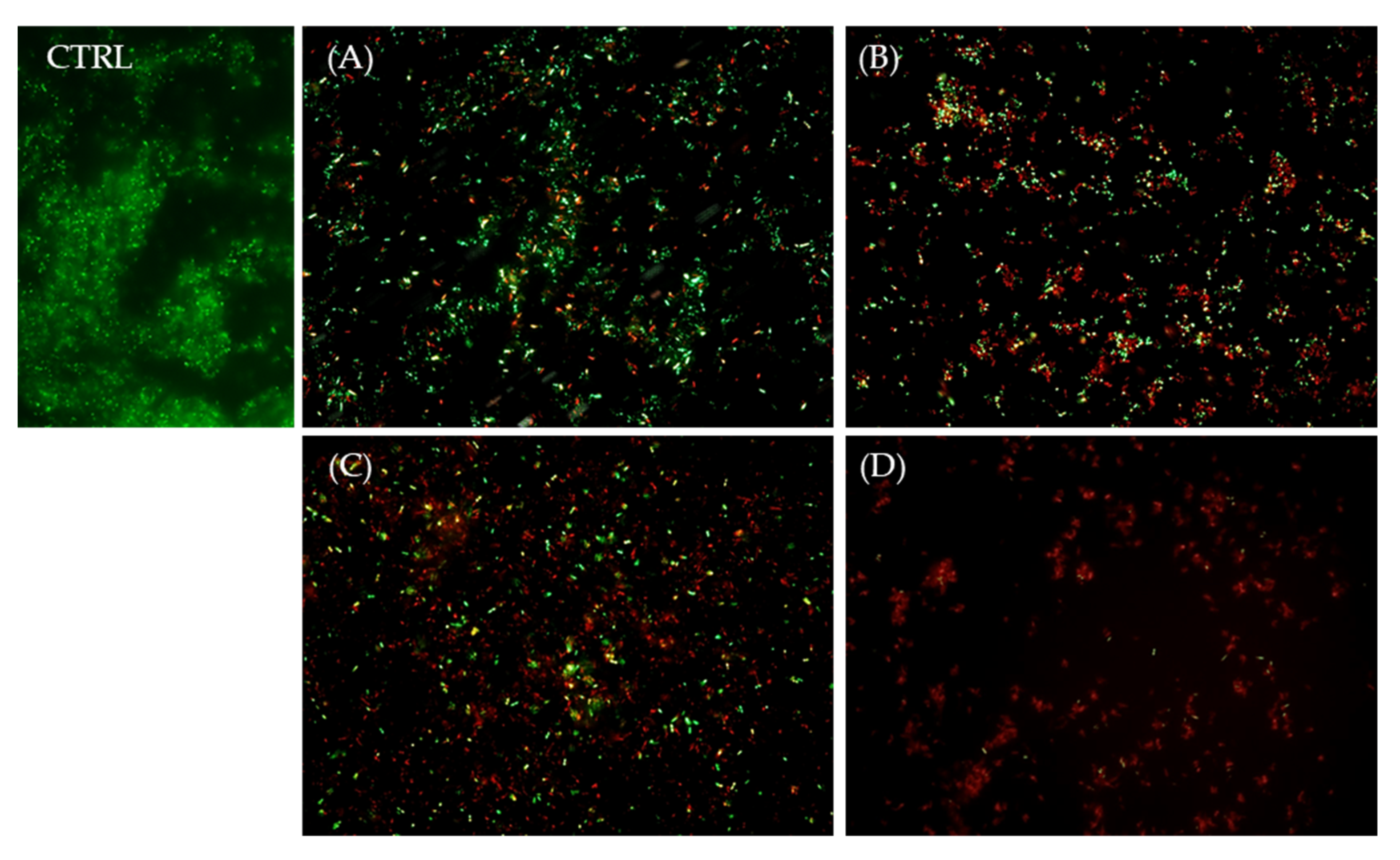
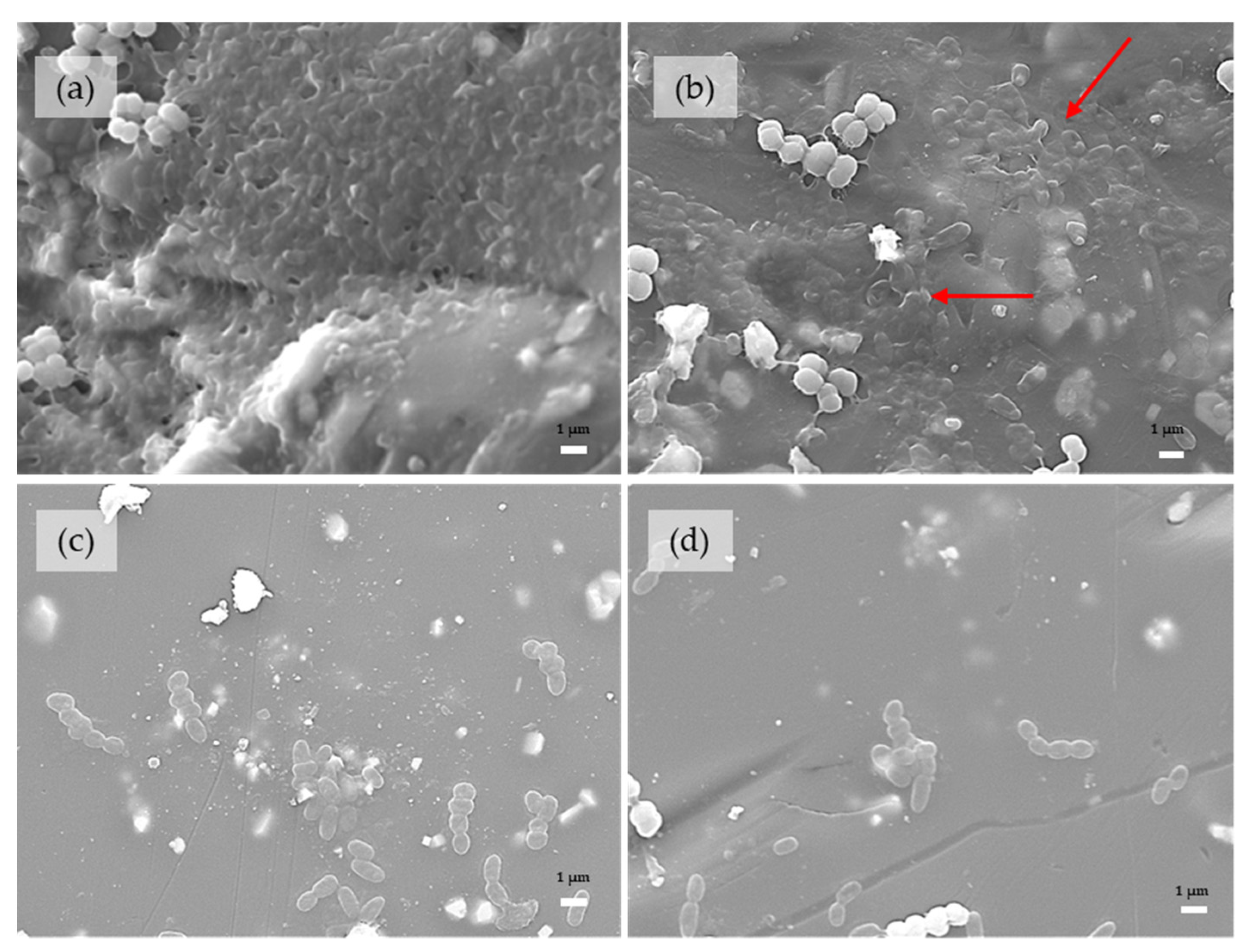
3.4. Scanning Electron Microscopy (SEM) Analysis

SEM analysis allowed a qualitative presentation of the effect of the combined treatment with UV light and chlorination with sodium hypochlorite on the mature biofilm. Representative images are shown. A dense 5-day-old biofilm with EPS is shown in pictures (Figure 5a). After UV treatments, a cluster of bacteria on the surface of the tile can be seen and they were located inside the tick EPS layer. We noticed the visible difference after Cl 1 min and fewer bacteria that are widespread on the tile surface in smaller clusters or individually can be seen without the presence of EPS. The most significant difference was seen after the combination of UV for 20 s and Cl for 1 min, where individual adhered cells or minor cell clumps can be seen on the tile surface. We did not notice the EPS layer. The figures are consistent with the result of 99% effectiveness of the study treatment described in the section (Figure 1).
Figure 6a showed the biomass with bacteria incorporated into the thick layer of EPS. After treatment of UV for 20 s, the clusters of the cells are fitted with the EPS layer and no significant changes can be seen. After 0.4 mg/L of Cl for 1-min, significant biomass destruction is visible, and fewer bacteria as well as a thinner EPS layer and EPS remains alone, were visible on the tile damage. On smooth parts of the tile, individual bacteria or cluster with fewer bacteria cells and remains of EPS can be seen. In addition to chlorination, it would be necessary to apply regular cleaning for better results. The picture (d) showed individual bacteria or small cluster of cells within a thin layer of EPS and the remains of EPS without bacteria. By combining both methods, fewer bacteria can be seen on the tiles, although EPS remains are still visible.

4. Discussion

The object of this study was to examine the effect of UV light and sodium hypochlorite on formation, as the pre-treatment and destruction of mature P. fluorescens biofilm on ceramic tiles. Our results show that different individual treatments or their combination have diverse effects on planktonic bacteria and their ability to form biofilms, as well as on the destruction of a matured, 5-day-old biofilm.
The effect of different treatments on planktonic bacterial cells has significantly affected the ability of the cells to form a biofilm.
This is consistent with studies stating that planktonic cells are considered more sensitive to external influences such as disinfectants, than those in biofilms [35,36,37]. In our study, plankton bacteria were treated with individual treatments as well as with their combinations. After the treatment, the planktonic bacteria were incubated for 5 days and their ability to create a biofilm was determined. Immediately after the treatment, we also followed the viability of the cells. Interestingly, a combined treatment with UV for 20 s and Cl for 1 min showed 99.99% inhibition of biofilm formation, but we have proven that some of the bacteria possessed an ability to form the biofilm. It seems that a small number of viable bacteria that survived the combined treatment (Figure 2D) were obviously capable of creating a biofilm, but we did not see the EPS layer present (Figure 5D). In the swimming pools, this type of disinfection of water is used and the pool water repeatedly passes through the UV lamp and is constantly exposed to chlorination. This repeated exposure would certainly be more effective than a onetime exposure. This fact should be investigated further. The mechanism of an individual effect of UV radiation and chlorination is known from earlier studies. Extended exposure to UV radiation alters the genetic material of a bacterial cell. UV affects nucleic acid molecules with wavelengths ranging from 200 to 300 nm, specifically at ~260 nm [38]. This leads to genetic mutations which cause the impossibility of DNA to replicate, and further to cell inactivation. Some bacteria can repair from UV damage and some of them enter a non-cultivable state as a response to this environmental stress [39,40,41,42]. Hijnen et al. found that UV can be effectively used for inactivation of suspended, free planktonic cells, without forming harmful disinfection by-products (DBPs) [43]. In a study conducted by Lakretz et al. [44] on Pseudomonas aeruginosa, the effectiveness of different UV wavelengths (220–280 nm) on bacterial inactivation and biofilm control was tested. The most effective ones were 254, 260 and 270 nm, because they inactivated more suspended cells, which can contribute to better biofilm control.
The effect of chlorination on the ability of planktonic bacteria to form a biofilm and results then showed that chlorination with sodium hypochlorite leads to a significant inhibition compared to UV radiation treatment or control. Chlorine, in the form of sodium hypochlorite [NaOCl], is a widely used, low-cost disinfectant with effective antimicrobial performance [45]. Chlorine is a very strong oxidant which can cause permeabilization of bacterial membranes causing leakage of protein, nucleic acid, and even lethal DNA damage [46]. Using chlorine alone cannot inactivate species such as Pseudomonas, Sphingomonas or Acinetobacter because they are known for developing resistance to chlorine-based disinfectants [47]. Studies on P. aeruginosa or Escherichia coli have shown that chlorination, as well as UV irradiation by itself, can induce cells to enter a non-cultivable state [48,49]. We accomplished damaged planktonic cells with chlorine more effectively than with UV light, but even so they can form a biofilm.
Wang et al. studied the effect of UV irradiation or chlorination alone, as well as their combined effect on the model microorganism, P. aeruginosa. They found that the number of cultivable cells was effectively reduced by using UV, chlorine, and combined UV/chlorine. This is consistent with our findings, but they also found that non-cultivable cells were present after UV and chlorination but were undetectable after UV/chlorine treatment. Bacterial reactivation was completely suppressed as bacteria were completely damaged by the combined effect of UV and chlorine. This study suggests that the UV/chlorine treatment can completely damage bacteria and is promising for opportunistic pathogen inactivation [50]. In accordance with their findings, that the combined treatment can effectively destroy bacteria or introduce them into a non-cultivable state, we can note that the combined approach to water disinfection contributes to a better reduction of chlorine-resistant opportunistic pathogens and looks promising in sustaining their presence at a barely detectable level. Joint treatments can work more effectively, and the residual effect of chlorine in their combination contributes to avoiding microbial regrowth. Biofilms differ in many ways from planktonic cells. In these complex communities, the EPS composed of extracellular DNA, proteins, and polysaccharides gives microorganisms protection from disinfectants [51,52]. Cells in biofilms may exhibit changes in their characteristics, such as reduced growth rates, because of reduced oxygen levels and lack of nutrient penetration. In addition, the frequency of physiologically resistant cells, by that chlorine resistant cells, is higher in biofilm populations [36].
Due to the above and the assumption that bacteria exhibit different characteristics in biofilms, we exposed the mature 5-day old, single species biofilm of P. fluorescens to the effect of UV light and chlorine separately and to their combination. We also examined the separate and combined effects with fivefold increased chlorine levels with prolonged exposure time in a process we labeled as hyperchlorination.
Results showed that UV did significantly affect the mature biofilm compared to control. But despite this, the impact of UV can be described as weaker compared to chlorine or the combination. Data about UV-related technologies for already formed biofilms are limited and often incoherent compared to those related to planktonic bacteria. Despite that, the effect of UV on mature biofilms is desirable and UV technologies and devices are being further developed. Sources of UV radiation that are used or examined in studies are often mercury vapor lamps emitting only 254 nm or a cluster of wavelengths. Some of the other sources that are being used, such as excimer lamps, xenon pulse lamps and light-emitting diodes (LEDs) are even showing enhanced bactericidal effects on biofilms [38,53].
So far it is known that UV minimally affects the EPS of established biofilm [54]. EPS adheres firmly to surfaces, with electrostatic forces, van der Waals forces and chemical bonding with other polymers, which are not easily interrupted with UV [55,56]. Bacteria that are good producers of EPS may have developed a protection mechanism to UV such as increasing the path length of the incident irradiation, emission of free radicals that intercept UV, usage of motility to avoid UV phototaxis, scavenging photogenic reactive oxygen species and protecting the cell structure and components from oxidative damage, light scattering caused by inorganic particles, producing UV-absorbing protective factors such as pigments [38,56,57]. Furthermore, multiple-species biofilms with different microorganisms and their protective mechanisms are less sensitive than single-species biofilms. This was demonstrated in a study conducted on natural biofilms formed on catheters and single P. aeruginosa biofilm culture which showed that multiple-species biofilms were much more tolerant of the UV effect than the single-species [58,59]. It is proven that biofilms can repair or recover themselves after irradiation has ended [56,57]. This means that the UV effect is temporary, and bacteria can regrow [44,56,57,60,61].
Formed, matured biofilm is known to be a resistant reservoir of pathogens, which can be spread in bulk water. Biofilms of water distribution systems, or generally other controlled water systems are recognized as areas of concern to be maintained safe [62]. EPS is considered as an adaptation of microorganisms to protect against disinfectants such as chlorine [61,63]. Disinfectants containing halogen species such as chlorine, with their high reactivity, are even neutralized through reaction with EPS, so have an impaired efficiency in reducing biofilm cell density [64]. The presence of EPS is not the only factor that provides biofilm resistance. Cells in biofilms can phenotypically differ from planktonic cells and develop different adaptive responses to sublethal concentrations of the disinfectant [65].
In order to effect even more efficiently on the mature biofilm, in our research we also combined the action of chlorine and prolonged hyperchlorination with UV radiation, which was proved to be the most effective method. Although we did not eradicate the biofilm completely, the synergistic action of hyperchlorination for 120 min and UV radiation for 20 s reduced the biofilm the most. Synergistic actions have been reported in various studies. A combination of UV light with chlorine dioxide was shown to be more effective in eradicating drinking water biofilms than the two treatments applied separately according to Rand et al. [66]. UV treatment systems in combination with chlorine or chlorine dioxide and monochloramine achieved greater log reductions of suspended E. coli and its biofilm, than chlorine-based disinfectants alone [67].
By using chlorine-based agents in water, such as sodium hypochlorite, relatively good disinfection results can be achieved as well as residual chlorine. Sodium hypochlorite is added to water as an oxidant that acts on organic and inorganic substances [68]. This results in creating disinfection by-products (DBPs), significantly trihalomethanes (THMs) that are becoming a serious health concern. Due to the genotoxic and carcinogenic effect of THMs, and other DBPs which have not yet been sufficiently investigated, it is preferable to use lower doses of chlorine, and one of the possible solutions is the application of combined disinfection methods, UV radiation and chlorination [68,69]. Complete eradication of biofilm was not achieved with any treatment, so it is necessary to investigate more methods, and their combination, to keep the biofilms of water systems under control. Finally, we want to emphasize that the cleaning and sanitation processes are important as well, so that disinfection methods can be more effective, and their by-products maintained at a safe level.

5. Conclusions

Our study indicates that both chlorine and UV light are effective agents in the inhibition of formation as well as in the treatment of the mature biofilm of P. fluorescens. Exposure of plankton bacteria to combined disinfection significantly affects the ability of these bacteria to create a biofilm and, by constantly repeating the procedure, with regular sanitation the formation of bacterial biofilm could be kept under control.
The application of combined methods has proven to be effective on a mature biofilm even though it has led only to its reduction. Therefore, the application of combined disinfection methods should go in the direction of treatment of planktonic bacteria with regular cleaning and sanitation. An additional application of UV radiations directly to the tiles would further facilitate maintaining the biofilm under control. Hyperchlorination has proven to be effective in destroying the biofilm, but due to the creation of by-products it should be used under defined conditions.

Author Contributions

Conceptualization, M.S.Z. and I.G.; methodology, M.S.Z., G.B., I.J.B. and R.K.; investigation, G.B. and M.S.Z.; resources, I.G.; data curation, S.M.; writing—original draft preparation, M.S.Z., G.B., D.T.L. and S.M.; writing—review and editing, I.G., R.K. and D.T.L.; visualization, I.J.B.; funding acquisition, I.G. All authors have read and agreed to the published version of the manuscript.

Funding

This research was funded by the University of Rijeka, grant number UNIRI-biomed 18-171.

Data Availability Statement

Not applicable.

Conflicts of Interest

The authors declare no conflict of interest.

References

  1. Falkinham, J.O., III. Common Features of Opportunistic Premise Plumbing Pathogens. Int. J. Environ. Res. Public Health 2015, 12, 4533–4545. [Google Scholar] [CrossRef] [PubMed]
  2. Moore, M.R.; Pryor, M.; Fields, B.; Lucas, C.; Phelan, M.; Besser, R.E. Introduction of Monochloramine into a Municipal Water System: Impact on Colonization of Buildings by Legionella spp. Appl. Environ. Microbiol. 2006, 72, 378–383. [Google Scholar] [CrossRef] [PubMed]
  3. Hayward, C.; Ross, K.E.; Brown, M.H.; Bentham, R.; Whiley, H. The Presence of Opportunistic Premise Plumbing Pathogens in Residential Buildings: A Literature Review. Water 2022, 14, 1129. [Google Scholar] [CrossRef]
  4. Vaz-Moreira, I.; Nunes, O.C.; Manaia, C.M. Diversity and antibiotic resistance in Pseudomonas spp. from drinking water. Sci. Total Environ. 2012, 426, 366–374. [Google Scholar] [CrossRef]
  5. CDC. Implications of Waterborne Disease Estimates. Available online: https://www.cdc.gov/healthywater/surveillance/burden/implications.html (accessed on 10 August 2022).
  6. Collier, S.A.; Deng, L.; Adam, E.A.; Benedict, K.M.; Beshearse, E.M.; Blackstock, A.J.; Bruce, B.B.; Derado, G.; Edens, C.; Fullerton, K.E.; et al. Estimate of Burden and Direct Healthcare Cost of Infectious Waterborne Disease in the United States. Emerg. Infect. Dis. 2021, 27, 140–149. [Google Scholar] [CrossRef]
  7. Soto-Giron, M.J.; Rodriguez-R, L.M.; Luo, C.; Elk, M.; Ryu, H.; Hoelle, J.; Santo Domingo, J.W.; Konstantinidis, K.T. Biofilms on Hospital Shower Hoses: Characterization and Implications for Nosocomial Infections. Appl. Environ. Microbiol. 2016, 82, 2872–2883. [Google Scholar] [CrossRef]
  8. Scales, B.S.; Dickson, R.P.; LiPuma, J.J.; Huffnagle, G.B. Microbiology, Genomics, and Clinical Significance of the Pseudomonas fluorescens Species Complex, an Unappreciated Colonizer of Humans. Clin. Microbiol. Rev. 2014, 27, 927–948. [Google Scholar] [CrossRef]
  9. Wong, V.; Levi, K.; Baddal, B.; Turton, J.; Boswell, T.C. Spread of Pseudomonas fluorescens Due to Contaminated Drinking Water in a Bone Marrow Transplant Unit: Table 1. J. Clin. Microbiol. 2011, 49, 2093–2096. [Google Scholar] [CrossRef]
  10. Sun, Y.-Y.; Chi, H.; Sun, L. Pseudomonas fluorescens Filamentous Hemagglutinin, an Iron-Regulated Protein, Is an Important Virulence Factor that Modulates Bacterial Pathogenicity. Front. Microbiol. 2016, 7, 1320. [Google Scholar] [CrossRef]
  11. Gershman, M.D.; Kennedy, D.J.; Noble-Wang, J.; Kim, C.; Gullion, J.; Kacica, M.; Jensen, B.; Pascoe, N.; Saiman, L.; McHale, J.; et al. Multistate Outbreak of Pseudomonas fluorescens Bloodstream Infection after Exposure to Contaminated Heparinized Saline Flush Prepared by a Compounding Pharmacy. Clin. Infect. Dis. 2008, 47, 1372–1379. [Google Scholar] [CrossRef] [Green Version]
  12. Anaissie, E.J.; Penzak, S.R.; Dignani, M.C. The hospital water supply as a source of nosocomial infections: A plea for action. Arch. Intern. Med. 2002, 162, 1483–1492. [Google Scholar] [CrossRef] [PubMed]
  13. Wilkinson, F.H.; Kerr, K.G. Bottled water as a source of multi-resistant Stenotrophomonas and Pseudomonas species for neutropenic patients. Eur. J. Cancer Care 1998, 7, 12–14. [Google Scholar] [CrossRef] [PubMed]
  14. Juyal, A.; Otten, W.; Baveye, P.C.; Eickhorst, T. Influence of soil structure on the spread of Pseudomonas fluorescens in soil at microscale. Eur. J. Soil Sci. 2021, 72, 141–153. [Google Scholar] [CrossRef]
  15. Benito, N.; Mirelis, B.; Luz Gálvez, M.; Vila, M.; López-Contreras, J.; Cotura, A.; Pomar, V.; March, F.; Navarro, F.; Coll, P.; et al. Outbreak of Pseudomonas fluorescens bloodstream infection in a coronary care unit. J. Hosp. Infect. 2012, 82, 286–289. [Google Scholar] [CrossRef]
  16. Daneshvar, A.H.E.; Truelstrup, H.L. Kinetics of biofilm formation and desiccation survival of Listeria monocytogenes in single and dual species biofilms with Pseudomonas fluorescens, Serratia proteamaculans or Shewanella baltica on food-grade stainless steel surfaces. Biofouling 2013, 29, 1253–1268. [Google Scholar] [CrossRef]
  17. Feazel, L.M.; Baumgartner, L.K.; Peterson, K.L.; Frank, D.N.; Harris, J.K.; Pace, N.R. Opportunistic pathogens enriched in showerhead biofilms. Proc. Natl. Acad. Sci. USA 2009, 106, 16393–16399. [Google Scholar] [CrossRef]
  18. Masak, J.; Cejkova, A.; Schreiberova, O.; Rezanka, T. Pseudomonas biofilms: Possibilities of their control. FEMS Microbiol. 2014, 89, 1–14. [Google Scholar] [CrossRef]
  19. Hinsa, S.M.; O’Toole, G.A. Biofilm formation by Pseudomonas fluorescens WCS365: A role for LapD. Microbiology 2006, 152, 1375–1383. [Google Scholar] [CrossRef]
  20. Puga, C.H.; Dahdouh, E.; Sanjose, C.; Orgaz, B. Listeria monocytogenes Colonizes Pseudomonas fluorescens Biofilms and Induces Matrix Over-Production. Front. Microbiol. 2018, 9, 1706. [Google Scholar] [CrossRef]
  21. Wang, H.; Cai, L.; Li, Y.; Xu, X.; Zhou, G. Biofilm formation by meat-borne Pseudomonas fluorescens on stainless steel and its resistance to disinfectants. Food Control 2018, 91, 397–403. [Google Scholar] [CrossRef]
  22. Rossi, C.; Serio, A.; Chaves-López, C.; Anniballi, F.; Auricchio, B.; Goffredo, E.; Cenci-Goga, B.T.; Lista, F.; Fillo, S.; Paparella, A. Biofilm formation, pigment production and motility in Pseudomonas spp. isolated from the dairy industry. Food Control 2018, 86, 241–248. [Google Scholar] [CrossRef]
  23. Bertelli, C.; Courtois, S.; Rosikiewicz, M.; Piriou, P.; Aeby, S.; Robert, S.; Loret, J.-F.; Greub, G. Reduced Chlorine in Drinking Water Distribution Systems Impacts Bacterial Biodiversity in Biofilms. Front. Microbiol. 2018, 9, 2520. [Google Scholar] [CrossRef] [PubMed]
  24. Rice, S.A.; Van Den Akker, B.; Pomati, F.; Roser, D. A risk assessment of Pseudomonas aeruginosa in swimming pools: A review. J. Water Health 2012, 10, 181–196. [Google Scholar] [CrossRef]
  25. Fish, K.E.; Reeves-McLaren, N.; Husband, S.; Boxall, J. Uncharted waters: The unintended impacts of residual chlorine on water quality and biofilms. NPJ Biofilms Microbiomes 2022, 12, 55. [Google Scholar] [CrossRef] [PubMed]
  26. Lee, J.; Ha, K.-T.; Zoh, K.-D. Characteristics of trihalomethane (THM) production and associated health risk assessment in swimming pool waters treated with different disinfection methods. Sci. Total Environ. 2009, 407, 1990–1997. [Google Scholar] [CrossRef] [PubMed]
  27. Villanueva, C.M.; Font-Ribera, L. Health impact of disinfection by-products in swimming pools. Annali dell’Istituto superiore di sanita 2012, 48, 387–396. [Google Scholar] [CrossRef] [PubMed]
  28. van Veldhoven, K.; Keski-Rahkonen, P.; Barupal, D.K.; Villanueva, C.M.; Font-Ribera, L.; Scalbert, A.; Bodinier, B.; Grimalt, J.O.; Zwiener, C.; Vlaanderen, J.; et al. Effects of exposure to water disinfection by-products in a swimming pool: A metabolome-wide association study. Environ. Int. 2018, 111, 60–70. [Google Scholar] [CrossRef]
  29. Haibo, W.; Chun, H.; Suona, Z.; Liu, L.; Xueci, X. Effects of O3/Cl2 disinfection on corrosion and opportunistic pathogens growth in drinking water distribution systems. J. Environ. Sci. 2018, 73, 38–46. [Google Scholar]
  30. Hongna, L.; Xiuping, Z.; Jinren, N. Comparison of electrochemical method with ozonation, chlorination and monochloramination in drinking water disinfection. Electrochim. Acta 2011, 27, 9789–9796. [Google Scholar]
  31. Karimi, B. Formation of disinfection by-products in the swimming pool water treated with different disinfection types. Desalination Water Treat. 2020, 175, 174–181. [Google Scholar] [CrossRef]
  32. Beyer, A.; Worner, H.; van Lierop, R. The Use of UV for Destruction of Combined Chlorine; Version 1.0; Wallace & Tiernan: Tonbridge, UK, 2004; Available online: https://www.pwtag.org.uk/reference/ (accessed on 15 August 2022).
  33. Cassan, D.; Mercier, B.; Castex, F.; Rambaud, A. Effects of medium-pressure UV lamps radiation on water quality in a chlorinated indoor swimming pool. Chemosphere 2006, 62, 1507–1513. [Google Scholar] [CrossRef] [PubMed]
  34. Ivanković, T.; Goić-Barišić, I.; Hrenović, J. Reduced susceptibility to disinfectants of Acinetobacter baumannii biofilms on glass and ceramic. Arch. Ind. Hyg. Toxicol. 2017, 68, 99–108. [Google Scholar] [CrossRef] [PubMed] [Green Version]
  35. Argyraki, A.; Markvart, M.; Stavnsbjerg, C.; Kragh, K.N.; Ou, Y.; Bjørndal, L.; Bjarnsholt, T.; Petersen, P.M. UV light assisted antibiotics for eradication of in vitro biofilms. Sci. Rep. 2018, 8, 16360. [Google Scholar] [CrossRef] [PubMed]
  36. Steed, K.A.; Falkinham, J.O., 3rd. Effect of Growth in Biofilms on Chlorine Susceptibility of Mycobacterium avium and Mycobacterium intracellulare. Appl. Environ. Microbiol. 2006, 72, 4007–4011. [Google Scholar] [CrossRef]
  37. Davies, D. Understanding biofilm resistance to antibacterial agents. Nat. Rev. Drug Discov. 2003, 2, 114–122. [Google Scholar] [CrossRef]
  38. Varna, K.; Jones, M. The Effects of Ultraviolet Light on Escherichia coli. J. Emerg. Investigat. 2015, 102, 23–28. [Google Scholar]
  39. Pullerits, K.; Ahlinder, J.; Holmer, L.; Salomonsson, E.; Öhrman, C.; Jacobsson, K.; Dryselius, R.; Forsman, M.; Paul, C.J.; Rådström, P. Impact of UV irradiation at full scale on bacterial communities in drinking water. NPJ Clean Water 2020, 3, 11. [Google Scholar] [CrossRef]
  40. Ben Said, M.; Masahiro, O.; Hassen, A. Detection of viable but non cultivable Escherichia coli after UV irradiation using a lytic Qβ phage. Ann. Microbiol. 2010, 60, 121–127. [Google Scholar] [CrossRef]
  41. Guo, L.; Ye, C.; Cui, L.; Wan, K.; Chen, S.; Zhang, S.; Yu, X. Population and single cell metabolic activity of UV-induced VBNC bacteria determined by CTC-FCM and D2O-labeled Raman spectroscopy. Environ. Int. 2019, 130, 104883. [Google Scholar] [CrossRef]
  42. Guo, M.; Huang, J.; Hu, H.; Liu, W.; Yang, J. UV inactivation and characteristics after photoreactivation of Escherichia coli with plasmid: Health safety concern about UV disinfection. Water Res. 2012, 46, 4031–4036. [Google Scholar] [CrossRef]
  43. Hijnen, W.A.M.; Beerendonk, E.F.; Medema, G.J. Inactivation credit of UV radiation for viruses, bacteria and protozoan (oo)cysts in water: A review. Water Res. 2006, 40, 3–22. [Google Scholar] [CrossRef] [PubMed]
  44. Lakretz, A.; Ron, E.Z.; Mamane, H. Biofouling control in water by various UVC wavelengths and doses. Biofouling 2010, 26, 257–267. [Google Scholar] [CrossRef] [PubMed]
  45. Gil, M.I.; Gómez-López, V.M.; Hung, Y.-C.; Allende, A. Potential of Electrolyzed Water as an Alternative Disinfectant Agent in the Fresh-Cut Industry. Food Bioprocess Technol. 2015, 8, 1336–1348. [Google Scholar] [CrossRef]
  46. Virto, R.; Manas, P.; Alvarez, I.; Condon, S.; Raso, J. Membrane Damage and Microbial Inactivation by Chlorine in the Absence and Presence of a Chlorine-Demanding Substrate. Appl. Environ. Microbiol. 2005, 71, 5022–5028. [Google Scholar] [CrossRef]
  47. Shrivastava, R.; Upreti, R.K.; Jain, S.R.; Prasad, K.N.; Seth, P.K.; Chaturvedi, U.C. Suboptimal chlorine treatment of drinking water leads to selection of multidrug-resistant Pseudomonas aeruginosa. Ecotoxicol. Environ. Saf. 2004, 58, 277–283. [Google Scholar] [CrossRef]
  48. Guo, M.-T.; Kong, C. Antibiotic resistant bacteria survived from UV disinfection: Safety concerns on genes dissemination. Chemosphere 2019, 224, 827–832. [Google Scholar] [CrossRef]
  49. Chen, S.; Li, X.; Wang, Y.; Zeng, J.; Ye, C.; Li, X.; Guo, L.; Zhang, S.; Yu, X. Induction of Escherichia coli into a VBNC state through chlorination/chloramination and differences in characteristics of the bacterium between states. Water Res. 2018, 142, 279–288. [Google Scholar] [CrossRef]
  50. Wang, L.; Ye, C.; Guo, L.; Chen, C.; Kong, X.; Chen, Y.; Shu, L.; Wang, P.; Yu, X.; Fang, J. Assessment of the UV/Chlorine Process in the Disinfection of Pseudomonas aeruginosa: Efficiency and Mechanism. Environ. Sci. Technol. 2021, 55, 9221–9230. [Google Scholar] [CrossRef]
  51. Lopes, F.A.; Morin, P.; Oliveira, R.; Melo, L. Impact of biofilms in simulated drinking water and urban heat supply systems. Int. J. Environ. Eng. 2009, 1, 276–294. [Google Scholar] [CrossRef]
  52. Flemming, H.-C.; Wingender, J. The biofilm matrix. Nat. Rev. Microbiol. 2010, 8, 623–633. [Google Scholar] [CrossRef]
  53. Garvey, M.; Rabbitt, D.; Stocca, A.; Rowan, N. Pulsed ultraviolet light inactivation of Pseudomonas aeruginosa and Staphylococcus aureus biofilms. Water Environ. 2015, 29, 36–42. [Google Scholar] [CrossRef]
  54. Elasri, M.O.; Miller, R.V. Study of the Response of a Biofilm Bacterial Community to UV Radiation. Appl. Environ. Microbiol. 1999, 65, 2025–2031. [Google Scholar] [CrossRef] [PubMed]
  55. Nguyen, T.; Roddick, F.A.; Fam, L. Biofouling of Water Treatment Membranes: A Review of Underlying Causes, Monitoring Techniques and Control Measures. Membranes 2012, 2, 804–840. [Google Scholar] [CrossRef] [Green Version]
  56. Song, W.; Zhao, C.; Zhang, D.; Mu, S.; Pan, X. Different Resistance to UV-B Radiation of Extracellular Polymeric Substances of Two Cyanobacteria from Contrasting Habitats. Front. Microbiol. 2016, 7, 1208. [Google Scholar] [CrossRef]
  57. Luo, X.; Zhang, B.; Lu, Y.; Mei, Y.; Shen, L. Advances in application of ultraviolet irradiation for biofilm control in water and wastewater infrastructure. J. Hazard. Mater. 2022, 421, 126682. [Google Scholar] [CrossRef] [PubMed]
  58. Yuan, L.; Sadiq, F.A.; Wang, N.; Yang, Z.; He, G. Recent advances in understanding the control of disinfectant-resistant biofilms by hurdle technology in the food industry. Crit. Rev. Food Sci. Nutr. 2021, 61, 3876–3891. [Google Scholar] [CrossRef] [PubMed]
  59. Bak, J.; Ladefoged, S.D.; Tvede, M.; Begovic, T.; Gregersen, A. Disinfection of Pseudomonas aeruginosa biofilm contaminated tube lumens with ultraviolet C light emitting diodes. Biofouling 2010, 26, 31–38. [Google Scholar] [CrossRef] [PubMed]
  60. Lakretz, A.; Ron, E.Z.; Harif, T.; Mamane, H. Biofilm control in water by advanced oxidation process (AOP) pre-treatment: Effect of natural organic matter (NOM). Water Sci. Technol. 2011, 64, 1876–1884. [Google Scholar] [CrossRef]
  61. Lakretz, A.; Ron, E.Z.; Mamane, H. Biofilm control in water by a UV-based advanced oxidation process. Biofouling 2011, 27, 295–307. [Google Scholar] [CrossRef]
  62. Clayton, G.E.; Thorn, R.M.S.; Reynolds, D.M. The efficacy of chlorine-based disinfectants against planktonic and biofilm bacteria for decentralised point-of-use drinking water. NPJ Clean Water 2021, 4, 48. [Google Scholar] [CrossRef]
  63. De Beer, D.; Srinivasan, R.; Stewart, P.S. Direct measurement of chlorine penetration into biofilms during disinfection. Appl. Environ. Microbiol. 1994, 60, 4339–4344. [Google Scholar] [CrossRef] [PubMed]
  64. Stewart, P.S.; Rayner, J.; Roe, F.; Rees, W.M. Biofilm penetration and disinfection efficacy of alkaline hypochlorite and chlorosulfamates. J. Appl. Microbiol. 2001, 91, 525–532. [Google Scholar] [CrossRef] [PubMed]
  65. Bridier, A.; Briandet, R.; Thomas, V.; Dubois-Brissonnet, F. Resistance of bacterial biofilms to disinfectants: A review. Biofouling 2011, 27, 1017–1032. [Google Scholar] [CrossRef] [PubMed]
  66. Rand, J.L.; Hofmann, R.; Alam, M.Z.B.; Chauret, C.; Cantwell, R.; Andrews, R.C.; Gagnon, G.A. A field study evaluation for mitigating biofouling with chlorine dioxide or chlorine integrated with UV disinfection. Water Res. 2007, 41, 1939–1948. [Google Scholar] [CrossRef] [PubMed]
  67. Murphy, H.M.; Payne, S.J.; Gagnon, G.A. Sequential UV- and chlorine-based disinfection to mitigate Escherichia coli in drinking water biofilms. Water Res. 2008, 42, 2083–2092. [Google Scholar] [CrossRef]
  68. Ekowati, Y.; Ferrero, G.; Farré, M.J.; Kennedy, M.D.; Buttiglieri, G. Application of UVOX Redox® for swimming pool water treatment: Microbial inactivation, disinfection byproduct formation and micropollutant removal. Chemosphere 2019, 220, 176–184. [Google Scholar] [CrossRef]
  69. Manasfi, T.; Temime-Roussel, B.; Coulomb, B.; Vassalo, L.; Boudenne, J.-L. Occurrence of brominated disinfection byproducts in the air and water of chlorinated seawater swimming pools. Int. J. Hyg. Environ. Health 2017, 220, 583–590. [Google Scholar] [CrossRef]
Figure 1. Effect of biofilm creation after pre-treatment of planktonic bacteria with UV 5 s (UV5″), UV 20 s (UV 20″), Cl for 1 min (Cl 1′), combination of UV 5 s and Cl for 1 min (UV5″ + Cl 1′), and UV for 20 s and Cl for 1 min (UV20″ + Cl 1′) and afterward incubation for 5 days (log10CFU/mL) (A). (The control is a biofilm created from untreated planktonic bacteria. The mean values are shown along with the standard deviations. (B)) % of inhibition of biofilm formation after the above treatments. Lowercase letters above the results indicate the statistical significance. Different letters indicate statistically significant differences tested by Students t-test and Mann–Whitney U test for UV 20″ + Cl 1′.
Processes 10 01901 g001
Figure 2. Representative images of dead/live staining of planktonic bacteria after treatment with: (A) UV 20 s, (B) Cl for 1 min, (C) combination of UV 5 s and Cl for 1 min, and (D) UV for 20 s and Cl for 1 min. CTRL represent control (untreated bacteria). Green fluorescence represents viable cells while red fluorescence indicates dead cells. Magnification 1000×.
Processes 10 01901 g002
Figure 3. Destruction of a 5-day-old biofilm (log10CFU/mL) (A) after treatment with UV 5 (UV 5″) and 20 s (UV 20″), 0.4 mg/L of Cl for 1 min (Cl 1′), combination of UV 5 s and Cl for 1 min (UV 5″ + Cl 1′), and UV for 20 s and Cl for 1 min. The control is represented by an untreated biofilm. The mean values are shown along with the standard deviations. (B) % of destruction of a mature biofilm after the above treatments. Lowercase letters above the results indicate the statistical significance. The same letters indicate that there is no statistically significant difference between groups while different letters indicate a statistically significant difference tested by Students t-test for UV 20″ + Cl 1′ vs. UV 5″ + Cl 1′ and UV 5″ + Cl 1′ vs. Cl 1′ and Mann–Whitney U test for other samples.
Processes 10 01901 g003
Figure 4. Destruction of a 5-day-old biofilm (log10CFU/mL) after treatment with UV 5 (UV 5″) and 20 s (UV 20″), and hyperchlorination (Cl-H) for (A) 1 min, (B) 30 min, (C) 60 min and (D) 120 min and their combinations. The control is represented by an untreated biofilm. The mean values are shown along with the standard deviations. Lowercase letters above the results indicate the statistical significance. The same letters indicate that there is no statistically significant difference between groups while different letters indicate a statistically significant difference tested by Students t-test and by Mann–Whitney U test for UV 20″ vs. UV 5″; Cl-H120′ vs. UV 20″ and UV 20″ + Cl-H120′ vs. UV 5″ + Cl-H 120′.
Processes 10 01901 g004
Figure 5. Representative scanning electron microscopy (SEM) micrographs of biofilm inhibition after treatment with UV 20 s (b), 0.4 mg/L of Cl for 1 min (c), combination of UV for 20 s and Cl for 1 min (d); (a) represented untreated control of 5-day-old biofilm. Magnifications 5000×. Red arrows point out bacteria embedded in EPS.
Processes 10 01901 g005
Figure 6. Representative scanning electron microscopy (SEM) micrographs of 5-day-old biofilm destruction after treatment with UV 20 s (b), 0.4 mg/L of Cl for 1 min (c), combination of UV for 20 s and Cl for 1 min (d); (a) represented untreated control biofilm. Magnifications (a,c) 5000×, (b) 1000× and (d) 2000×. Red arrows point out bacteria embedded in EPS.
Processes 10 01901 g006
Table 1. Destruction of mature biofilm after different hyperchlorination treatments. Results are presented as a percentage (%). Small letters above the results indicate a statistically significant difference (a according to UV 5″; b according to UV 20″; c according to Cl (p < 0.05), non-parametric Mann–Whitney U test).
Destruction of Mature Biofilm
UV 5″UV 20″Cl-H 1′UV 5″ + Cl-H 1′UV 20″ + Cl-H 1′
Hyperchlorination of mature biofilm
1 min
69.66%
(±0.20)
77.73%
(±0.12)
96.57% ab
(±0.02)
97.06% ab
(±0.02)
97.98% ab
(±0.01)
UV 5″UV 20″Cl-H 30′UV 5″ + Cl-H 30′UV 20″ + Cl-H 30′
Hyperchlorination of mature biofilm
30 min
66.67%
(±0.20)
75.74% a
(±0.11)
=99.90% ab
(±0.002)
99.94% ab
(±0.001)
99.97% abc
(±0.007)
UV 5″UV 20″Cl-H 60′UV 5″ + Cl-H 60′UV 20″ + Cl-H 60′
Hyperchlorination of mature biofilm
60 min
69.66%
(±0.10)
76.96%
(±0.12)
=99.96% ab
(±0.02)
99.97% ab
(±0.09)
99.99% ab
(±0.01)
UV 5″UV 20″Cl-H 120′UV 5″ + Cl-H 120′UV 20″ + Cl-H 120′
Hyperchlorination of mature biofilm
120 min
69.66%
(±0.10)
76.96%
(±0.12)
=99.99% ab
(±0.01)
99.99% ab
(±0.01)
99.99% ab
(±0.001)
Hyperchlorination treatments of the mature biofilm for 1, 30, 60 and 120 min have shown most efficiency combined with UV and results range from 97.06% to 99.99%.
Publisher’s Note: MDPI stays neutral with regard to jurisdictional claims in published maps and institutional affiliations.

