Review

Electrochemical Sensors for Liquid Biopsy and Their Integration into Lab-on-Chip Platforms: Revolutionizing the Approach to Diseases

1
CNR NANOTEC–Institute of Nanotechnology, Via per Monteroni, 73100 Lecce, Italy
2
Department of Mathematics and Physics “Ennio De Giorgi”, University of Salento, Via per Arnesano, 73100 Lecce, Italy
*
Authors to whom correspondence should be addressed.
Chemosensors 2023, 11(10), 517; https://doi.org/10.3390/chemosensors11100517
Submission received: 30 August 2023 / Revised: 22 September 2023 / Accepted: 26 September 2023 / Published: 1 October 2023
(This article belongs to the Special Issue Recent Developments in Electrochemical Sensing)

Abstract

:
The screening and early diagnosis of diseases are crucial for a patient’s treatment to be successful and to improve their survival rate, especially for cancer. The development of non-invasive analytical methods able to detect the biomarkers of pathologies is a critical point to define a successful treatment and a good outcome. This study extensively reviews the electrochemical methods used for the development of biosensors in a liquid biopsy, owing to their ability to provide a rapid response, precise detection, and low detection limits. We also discuss new developments in electrochemical biosensors, which can improve the specificity and sensitivity of standard analytical procedures. Electrochemical biosensors demonstrate remarkable sensitivity in detecting minute quantities of analytes, encompassing proteins, nucleic acids, and circulating tumor cells, even within challenging matrices such as urine, serum, blood, and various other body fluids. Among the various detection techniques used for the detection of cancer biomarkers, even in the picogram range, voltammetric sensors are deeply discussed in this review because of their advantages and technical characteristics. This widespread utilization stems from their ability to facilitate the quantitative detection of ions and molecules with exceptional precision. A comparison of each electrochemical technique is discussed to assist with the selection of appropriate analytical methods.

1. Introduction

A biopsy is a technique in which tissue samples are taken from the body and examined under a microscope to see if cancer (though the concept is applicable to many other diseases) or abnormal cells are present. Biopsies can be classified into the following categories based on the sample being taken (Figure 1).
In the last decades, the liquid biopsy, namely, the possibility to have a diagnosis from body fluids without resorting to a tissue biopsy, has been increasingly investigated. The possibility to detect and classify tumors or other diseases, even at a very early stage, in a minimally invasive and repeatable way could have a significant clinical impact, and significant progress has been made in the development of devices able to do this in a smarter manner compared to standard analytical methods. Despite the great advantages for patients’ compliance and the minimally invasive features, this approach has not yet attained the status of a conventional tool in the armory of clinical oncologists [1].
Biosensors are considered to be promising tools for the quantitative or semi-quantitative detection of analytes [2]. In this type of sensor, a biological molecule interacts with the analyte, previously immobilized on the biosensor, producing a physicochemical signal that is detected by the transducer. Biosensors can be divided into two categories: catalytic-based, which produce a substance starting from substrate’s compounds, and affinity-based, which directly bind the analyte. According to the type of signal being transmitted, biosensors can be classified as electrochemical, optical [3], magnetic, or piezoelectric, just to name a few [4].
Electrochemical techniques excel among these methods by offering rapid, sensitive, selective, and cost-effective detection and monitoring of a wide range of biological molecules associated with diverse diseases. Additionally, their seamless integration into portable systems enables the implementation of point-of-care diagnostic approaches [5]. An electrochemical biosensor is a compact device that utilizes both biorecognition processes and electrochemical transducers to convert biological information into electrical signals. This conversion provides either quantitative or semi-quantitative information about the analyte being detected [6].
Electrochemical biosensors were recommended by the international union of pure and applied chemistry (IUPAC). Which states that an electrochemical biosensor is a self-contained integrated device that is capable of providing specific quantitative or semi-quantitative analytical information using a biological recognition element (biochemical receptor) which is kept in direct spatial contact with an electrochemical transduction element.
Electrochemical methods, such as electrochemical impedance spectroscopy (EIS), differential pulse voltammetry (DPV), and cyclic voltammetry (CV), play a crucial role in both the development of biosensors and the evaluation of their performance [7]. These methods are highly valuable approaches in the field of liquid biopsy (Figure 2). For research purposes, the CV technique is widely used in biosensor development because it provides valuable information such as the types of redox processes present in the analysis and the reversibility of reactions. A sensing system that can identify a cell’s location within a microfluidic channel was designed by Rapier et al. The results from electrochemical impedance spectroscopy (EIS) show that cells in microfluidic channels can be positioned between different pairs of electrodes at varied locations along the device’s length. Impedance spectra distinguish among confluent, sparse, and empty microfluidic channels. A huge boost to the development of this kind of sensors as well as to their application in the liquid biopsy was given by their high suitability to miniaturization. Electrodes’ architecture, indeed, can be easily achieved through micro- and nanofabrication methods, increasing the number of sensing elements per area and allowing high-throughput performances [8].
Also, lab-on-a-chip integration is directly linked to miniaturization. The possibility to perform multiple assays by lowering the dimensions of sensing elements has led to the necessity to differently functionalize and use them for the detection of different biomarkers [9,10].
The utilization of impedance-based methods significantly facilitated the simplification of cellular assays, providing quantitative and highly sensitive results that are amenable to automation and scalability in multi-well formats. An extensive review by W. Gamal et al. shed light on their efficacy. Moreover, in the context of the real-time monitoring of a three-dimensional cell culture, dielectric spectroscopy and electrical impedance tomography emerged as promising alternatives to two-dimensional impedance sensing [11]. These impedance-based cellular assays (IBCAs) serve as label-free phenotyping assays and are gaining increasing interest in the field of regenerative medicine applications [12]. To determine the flow in a microfluidic chip, Evangelos S. et al. developed a strain-sensing module based on microfluidic and lab-on-a-chip systems that offers simple integration with most microfluidic systems. The sensor consists of interconnected platinum nanoparticles that self-assemble on flexible polyolefin substrates, which also serve as the sealing layer for the microfluidic channels. These nanoparticle networks are formed using a modified sputtering approach and are implemented on printed circuit board substrates (PCBs) through milling and computer numerical control machining. The resulting module exhibits a competitive limit of detection (LOD), cost-effectiveness, low power requirements, and seamless integration with existing microfluidic systems. It can be utilized as an independent unit or integrated into the sealing material, enabling the detection of flow rates as low as 5 μL/min (equivalent to a strain of 0.00337%). The sensor demonstrates a sensitivity of 0.021 μL per minute [13].
Lucile et al. reported another novel microfluidic method that can selectively extract, preconcentrate, and fluorescently detect IL-6 directly on the chip by the fluidization of magnetic beads. The ability to switch between packed and fluidized states allowed the authors to evaluate how the physical characteristics of the beads could be altered to increase mass transport, lessen non-specific binding, and triple the detection signal. A high dynamic range (10 pg/mL to 2 ng/mL) and a twofold reduction in LOD compared to traditional approaches were demonstrated by integrating the entire ELISA protocol into a single microfluidic chamber [14].
In this review, an overview about electrochemical methods and their applications as transduction techniques in the development of biosensors is provided. Particular attention is paid to the liquid biopsy and to the use of a miniaturized platform, allowing for the spread of point-of-care devices in this field.

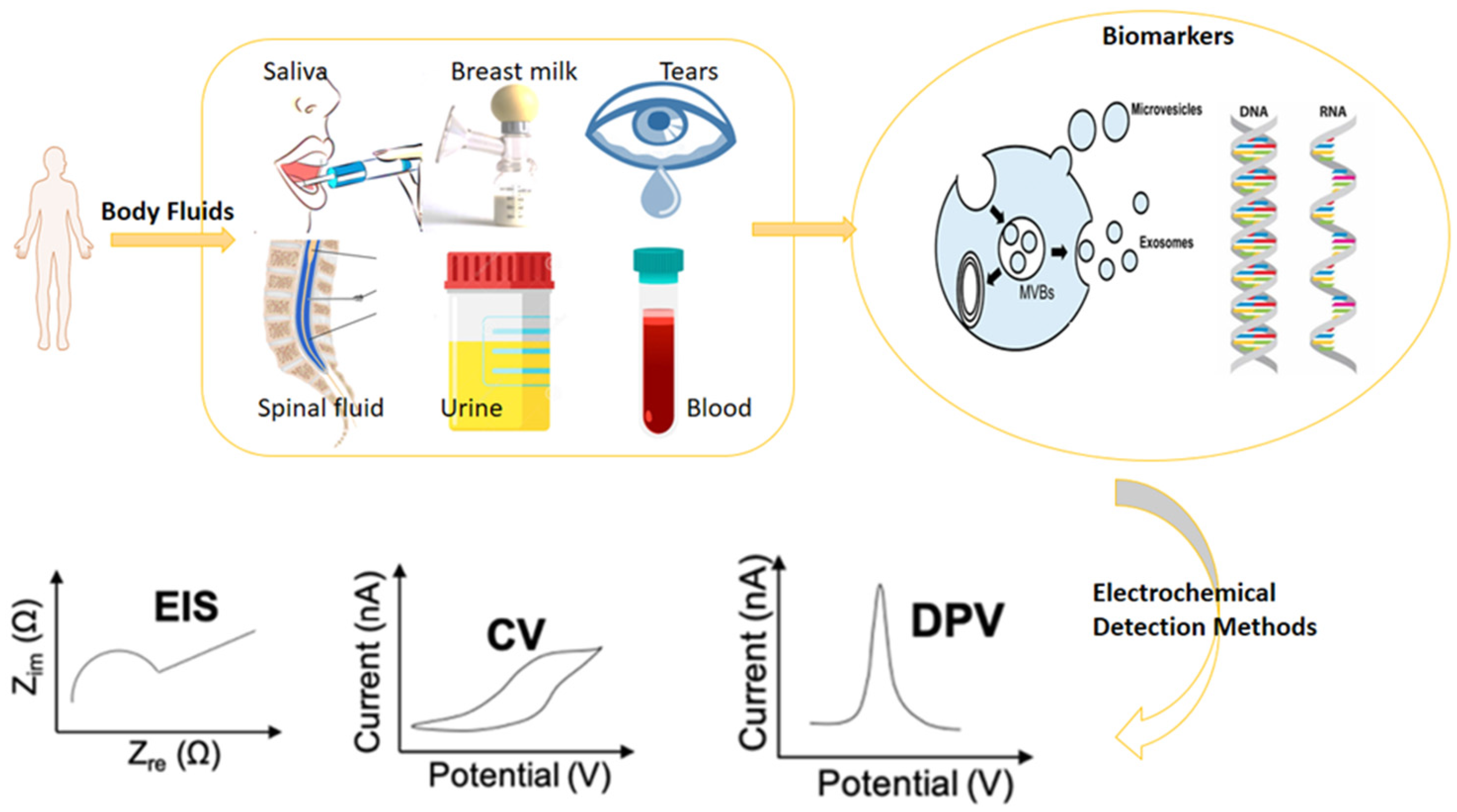
Essential Biomarkers for Liquid Biopsy Detection and Monitoring

The liquid biopsy is increasingly used for the detection, analysis, and monitoring of circulating tumor cells (CTCs), circulating tumor DNA, and circulating extracellular nucleic acids [15], in blood or other body fluids such as urine, with the main advantage of diagnosing cancer at an early stage. In contrast to a traditional invasive biopsy, which includes cells or tissues from the lesion, the liquid biopsy is a non-invasive procedure that can also be used during treatment planning as well as in the follow-up of the disease. In addition to blood, other body fluids are under consideration for the liquid biopsy: urine [16] is already used as a source of biomarkers (PCA3 in prostate and pancreatic cancer detection), and several examples of state-of-the-art detection from saliva, seminal fluid [17], or stool were described [18].
The liquid biopsy is a non-invasive method for detecting, analyzing, and monitoring cancer cells, DNA, and other nucleic acids in body fluids such as blood and urine. It offers several advantages over traditional biopsy methods, including the ability to diagnose cancer at an early stage without the need for invasive procedures. This technique can be used not only for the initial diagnosis of cancer but also during treatment planning and in follow-up procedures. Other body fluids such as urine, saliva, seminal fluid, and stool are also being studied as potential sources of biomarkers for the liquid biopsy.
Biomarkers are recognized to have a critical role in the early detection of cancer, the creation of personalized treatments, and the identification of the disease-related underlying processes. An ideal cancer biomarker would have high clinical sensitivity and specificity, rapid release in the blood for early detection, high concentration in the blood for prolonged periods of time, and the possibility to be quantified.
The detection targets of the liquid biopsy primarily encompass cells and nucleic acids found in body fluids, including circulating tumor cells (CTCs) in blood, cell-free DNA (cfDNA), circulating tumor DNA (ctDNA), exosomes, micro-RNA (miRNA), and proteins.
Circulating tumor cells are released either from primary tumors or from secondary metastatic sites. Identifying CTCs among blood cells and enumerating them has significant prognostic value [19]. Investigating their presence in blood at the early stage of tumor pathologies means that a highly sensitive method should be used, since a very low number of cells/mL should be detected. To this aim, the presence of a specific membrane biomarker can be used as the target for probes aiming to immobilize or label CTCs [20].
Protein biomarkers are widely used in the liquid biopsy, more often but not only in blood and serum. As an example, urine is already being used to detect PCA3 in prostate and pancreatic cancer, and several state-of-the-art detection methods were developed for other body fluids [21]. Typically, in the development of an electrochemical sensor, a protein biomarker can be identified using antibody/antigen probes immobilized on the surface of the electrodes, aptamers specifically designed to bind the protein, and molecularly imprinted polymers (MIP) directly synthesized on the electrodes’ surface, producing artificial antibodies [22,23,24].
Nucleic acid biomarkers include several kinds of molecules, ranging from fragments of DNA circulating in blood (i.e., cell-free DNA) to miRNA-associated (or not) to extracellular vesicles (EVs) to circulating DNA, also in this case associated with CTCs or as the cargo of EVs. The interest in these last entities is strongly increasing since EVs are identified as important messengers of information among cells and body districts. Indeed, they are physiologically produced and up taken by all types of cells and released into all biofluids. EVs are commonly divided into two classes: large EVs, including microvesicles originating from membrane budding, and small EVs, including exosomes originating from intracellular vesicular bodies. Capturing, counting, and deeply characterizing the surface and the cargo of these membrane bodies is one of the biggest challenges in molecular and cellular biology, as they could be very informative as diagnostic and prognostic biomarkers. The standard methods for the analysis of EVs suffer from strong limitations, and some biochip-based devices were developed to overcome them [25,26].
An innovative way of using the liquid biopsy, going in the direction of point-of-care medicine, is the integration of smart assays into portable devices, which need a small amount of the sample, low power to work, and minimal equipment [27]. Electrochemical methods are particularly suitable for integration into the near-the-bed platform, since most of them are label-free methods, and all the components can be easily miniaturized into lab-on-a-chip platforms. A list of examples from literature including biomarkers investigated, biofluids and electrochemical methods with related LODs is reported in Table 1.

2. Miniaturization Strategies for Biosensing

Miniaturization is the key challenge and research trend currently pursued in the field of biosensing. Moore’s law, a theory in the field of microelectronics, postulates the continuous advancement of the industry by the doubling of the number of on-chip transistors every two years. This exponential growth in the transistor count has led to a reduction in the cost per function. Miniaturizing the transducer as well as the biosensing element means a boost in the enhancing sensitivity and specificity features. This advancement will contribute to the achievement of three key performance metrics in biosensors: enhancing the limit of detection, reducing the response time, and lowering production costs.
One of the practical and technological advantages of miniaturizing the biosensing system, indeed, is the improved sensitivity of electrochemical and electronic sensors by increasing the system’s signal-to-noise ratio, reaching dimensions of the micro-structured electrodes and nanomaterials that are comparable with entities of interest (as examples: cells in the case of microstructures and protein and nucleic acids in the case of nanomaterials). Signal enhancement in this condition is achieved by the presence of nanogaps and/or nanostructured electrodes that obtain the noise reduction by making available a high surface area for biosensing interactions. This holds true for systems based on field-effect transistors as well as nanoscale electrochemical biosensors. Additionally, reducing the interelectrode spacing at the nanoscale can be also considered as a strategy to amplify the redox current and, in this way, to obtain an electrochemical single-molecule analysis by generating a significate signal-to-noise ratio [39].
The kinetics of transport reactions in biosensing are closely linked to the time needed for biorecognition events to take place. In this context, the background current associated with the charging of the double layer (capacitive current) varies in proportion to the electrode’s conductive area. In miniaturized electrochemical cells, the resistive drop is minimized by shortening the ionic current pathway. As a result, the capacitance is reduced, leading to a decreased time constant for the system. This enables faster electron-transfer kinetics compared to macroscale systems.
The possibility of reducing dimensions brings several side advantages. Among these, first of all, is adding portability and integration into a complex platform, which allows for the realization of a multiplexed analysis, including sample treatment tools and parallelized functions. This not only enables the creation of compact yet robust devices but also has the added benefit of reducing manufacturing costs by minimizing material and fabrication expenses per device. This efficiency in mass production contributes to overall cost reduction.

2.1. Micro- and Nanofabrication Methods

Several methods for the micro- and nanofabrication of electrodes are available, allowing for the production of the desired geometries of transducers and sensing elements for electrochemical biosensing. In addition to the consolidated optical and electron-beam lithography technologies, some innovative tools are gaining popularity thanks to the possibility they provide to achieve a better resolution and higher customizable processes in a relatively short time with respect to the standard techniques. These tools offer wide-range opportunities to innovate alongside Moore scaling without requiring high investment levels but offering large-feature, low-end production and high-end performances [40].
Among these technologies, maskless lithography methods based both on a direct laser writing source (for example, a femtosecond laser or UV laser) and two-photon systems are widely used for the fabrication of microelectrodes, ensuring a very high resolution of lithographic patterns even over a large surface area and in a three-dimensional operating mode [41,42]. Recently, several groups were working on these kinds of processes. Zhu and coworkers dealt with the realization of planar electrodes onto flexible substrates and realized direct laser writing on the stacked graphene multilayer of a large-area micro-supercapacitor onto a polyaniline substrate, demonstrating the possibility to fabricate pressure/gas sensors with high sensitivity for multiple applications [43]. Dotan et al. implemented a novel approach for the development of soft and flexible microelectrode structures used in electric and electrochemical sensing. Their method involved the combination of the supersonic cluster beam deposition (SCBD) of gold nanoparticles onto Polydimethylsiloxane (PDMS), followed by femtosecond (fs) laser processing. Through this technique, they successfully produced a nanocomposite film with mechanical properties comparable to those of the elastomeric substrate [44]. Two-photon lithography is a very versatile and flexible micro- and nanofabrication technology that allows for the 3D architecture of a lab-on-a-chip and an integrated platform. The possibility to combine planar and multilevel structures into the same chip thanks to two-photon lithography was explored by Luitz and coworkers, who realized complex 3D micro- and nano-objects using a platinum-containing photoresin, which can be structured via direct lithographic two-photon polymerization, paving the way for novel applications like the production of innovative metamaterials for biomedical applications, where high surface areas and the physicochemical properties of Pt are highly desirable. Moreover, with the subsequential steps of lithography, the two-photon lithography method enables the possibility of embedding sensor structures into microfluidic devices, thus obtaining a monolithic platform for on-chip sample preparation and characterization [45,46,47].
Microfluidic devices offer the ability to control the flow of fluids at the microscale, enabling the rapid and precise detection of biomolecules. When combined with nanomaterial-based sensors, the real-time monitoring of low concentrations of biomolecules in body fluids such as blood [48], urine [49], tears [50], and saliva [51] can be achieved. The available and easy methods for the fabrication of PDMS made this material emerge as the most popular polymer for the realization of microfluidic channels, which is useful for sample preparation or reaction chambers. Its property of sticking onto various substrates and the possibility to mold it in microstructures allow for its application in a wide range of research fields. Moreover, its adhesion on the surface of connection pads often does not interfere with electrical/acoustic/magnetic signals, allowing for the integration of microfluidics with sensing modules [52,53,54].
On the other hand, despite the large use of PDMS in research contexts, it has several drawbacks. First of all, its reversible hydrophilicity limits long-term experiments with biological materials and hinders the transition into industrial applications. Moreover, PDMS is not suitable for use with most commonly used solvents (ethanol, isopropanol, acetone, etc.), which could be necessary for preliminary sample treatment. The resistance of PDMS microchannels to high pressures is also limited in the case of stand-alone devices, thus forcing operators to use screws and clamps to secure and keep the watertight sealing. To overcome these aspects, some other polymers are on the rise due to their properties that are more suitable for the realization of robust monolithic devices. Among them, the list includes thermoplastic polymers (plastics) like poly(methyl methacrylate) (PMMA), polystyrene (PS), cyclic olefin copolymers (COC), and polycarbonate (PC), which allow for easy surface treatments, are biocompatible and transparent, and meet some of the industrial requirements for the LOC market [55,56].

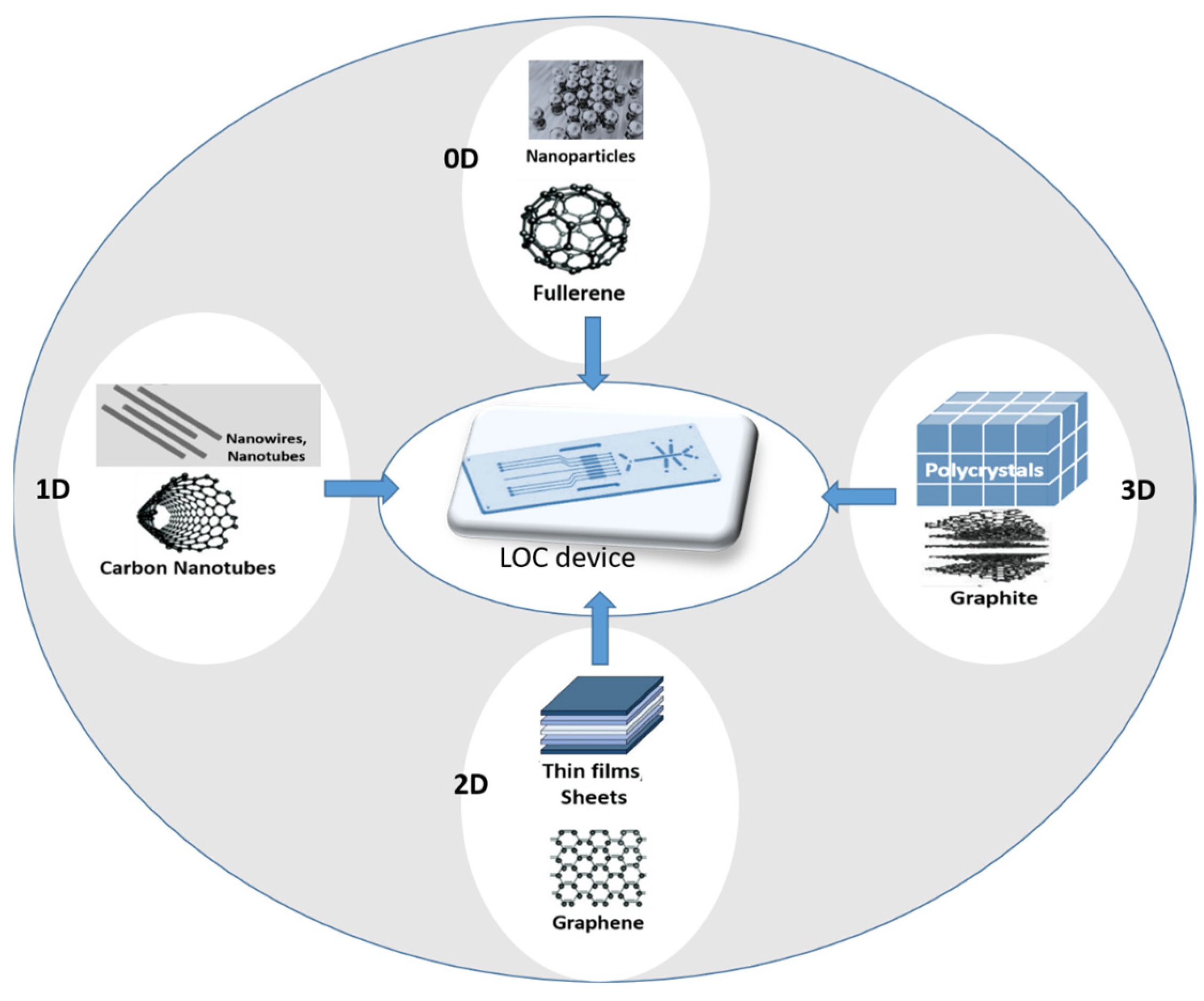
2.2. Nanomaterials in Electrochemical Sensors Integrated in LOC Device: From 2D to 3D Electrodes

Nanomaterials have emerged as a promising class of materials for sensing applications due to their unique physicochemical properties [57,58,59]. The exceptional properties exhibited by nanomaterials, including high surface area, excellent electrical and thermal conductivity, and unique optical characteristics, make them highly advantageous for seamless integration into lab-on-a-chip (LOC) devices as electrochemical sensors. This integration enables the detection of molecules in body fluids with significantly improved sensitivity and accuracy [22].
The incorporation of nanomaterials in electrochemical biosensors holds the potential to bring about a revolutionary transformation in the field of clinical diagnostics. This advancement facilitates the rapid, sensitive, and highly specific detection of biomolecules in body fluids, paving the way for significant advancements in medical diagnosis and patient care. This enhancement is achieved by either promoting electronic transfers or increasing the volume/surface area ratio [58]. This has significant implications for medical diagnosis and treatment, as it can enable the early detection and monitoring of diseases such as cancer, diabetes, and cardiovascular diseases or the timely identification of bacterial infections.
The integration of nanomaterials onto the electrode surface of microfluidic devices plays a crucial role in the advancement of high-performance electrochemical sensors. This is particularly important when electrodes are situated in less accessible locations, as often encountered in lab-on-a-chip systems. By incorporating nanomaterials, the sensitivity and overall performance of the electrochemical sensors can be significantly enhanced, enabling accurate and reliable detection in challenging sample environments. In this kind of device, nanomaterials are typically integrated onto the electrode surface using various techniques, such as in situ synthesis [60], drop casting [61], spin coating [62], electrochemical deposition [63], electrospinning [64], and inkjet printing [65]. Drop casting and spin coating are simple and cost-effective techniques that involve the deposition of nanomaterials onto the electrode surface using a dropper or a spinning device, respectively. Electrochemical deposition involves the deposition of nanomaterials onto the electrode surface by applying a voltage or a current to the electrode in the presence of the nanomaterials in solution. Inkjet printing involves the precise deposition of nanomaterials onto the electrode surface using a specialized printer. Among them, electrochemical deposition and inkjet printing are the techniques that allow for the most precise and controlled deposition of nanomaterials onto the electrode surface, which is critical for the development of high-performance electrochemical sensors.
Various types of organic and inorganic nanomaterials, including carbon nanotubes, graphene, metal and metal oxide, polymer, quantum dots, Prussian blue [66], nanorods, and tubes, are incorporated into electrochemical transducers to enhance electrochemical sensing (Figure 3).
Metallic nanostructures, such as gold [67], silver [68], and platinum [69], are widely used in electrochemical sensors and integrated into microfluidic devices for the detection of molecules in body fluids. Gold nanoparticles (AuNPs), for example, are extensively studied due to their unique electronic, optical, and surface properties, which make them ideal for use in biosensing applications [70]. AuNPs are used in a variety of electrochemical sensors for the detection of different biomolecules, such as glucose, cholesterol [71], and prostate-specific antigen (PSA) [72].
Magnetic nanoparticles (MNPs), such as iron oxide nanoparticles, are also used in electrochemical biosensors integrated into microfluidic devices for the detection of biomolecules in bodily fluids. MNPs have unique magnetic and surface properties, which make them ideal for use in biosensing applications [73]. These nanomaterials not only improve the limit of detection of the sensors but also enable the separation and transportation of bioanalytes inside the microfluidic device, thereby allowing for the miniaturization of analytical methods [74]. For example, MNPs functionalized with iridium oxide nanoparticles and tyrosinase are used for the detection and quantification of methimazole in microsystems. In the analytical measurements, a permanent magnet was used to immobilize the magnetic complex on the electrode surface. The system was highly sensitive with a low limit of detection (0.004 μM) and demonstrated effectiveness in serum samples. Interestingly, the use of a microfluidic device allows for an improved limit of detection, reusability, automation, the volume of the sample, and response time compared to batch configuration [75].
Among nonmetallic nanomaterials, carbon nanotubes (CNTs) [76], graphene [77], and quantum dots [78,79] (QDs) show high sensitivity toward various analytes, including glucose [77], cholesterol [80], and proteins, [81] with detection limits in the subnanomolar range. Moreover, carbon-based nanomaterials such as graphene and carbon nanotubes are also combined with metallic nanoparticles or polymeric layers to produce nanocomposites with improved performance [82,83,84] in terms of electronic transfer and selectivity.
One of the challenges associated with the use of nanomaterials in electrochemical sensors integrated into microfluidic devices is the reproducibility and stability of the sensors. Due to the small size of nanomaterials, synthesizing and functionalizing these materials can be challenging, leading to variations in sensor performance. Additionally, the stability of nanomaterial-based sensors can be affected by factors such as temperature, pH, and humidity, resulting in reduced sensor performance over time. Efforts are being made to address these challenges, by developing reproducible synthesis and functionalization methods and optimizing sensor design to enhance stability.
Another challenge related to the use of nanomaterials in electrochemical sensors integrated into microfluidic devices is the integration of these sensors into practical clinical applications. While many studies demonstrated the feasibility of using nanomaterial-based sensors for detecting biomolecules in body fluids, further development and optimization are required before these sensors can be widely adopted in clinical settings. This includes optimizing the sensitivity, selectivity, and stability of the sensors, as well as developing user-friendly and cost-effective instrumentation for their use. In fact, despite the enhanced sensor performance offered by nanomaterials implemented on electrode surfaces, the two-dimensional planar electrodes can still limit component and signal transmission when used in vivo, thereby affecting sensor accuracy and sensitivity [85,86]. This limitation is particularly true for complex samples such as blood or plasma used in point-of-care-devices [87,88]. Furthermore, the planar structure of two-dimensional electrodes poses challenges in achieving the adequate immobilization of active components and an efficient signal transmission, leading to potential issues in sensing accuracy. To address these limitations, the integration of macroscale three-dimensional (3D) porous materials, comprising nanomaterials combined with polymers [89,90,91,92], can be employed as electrodes. This approach facilitates expanded microfluidic transport and enables the incorporation of multianalyte detection capabilities, thereby enhancing the overall performance of the sensor system.
The incorporation of porous channels in biosensing systems offers several advantages, including increased surface area, enhanced ion/mass transport pathways, and the improved immobilization and stability of active components. In this context, graphene emerged as a promising avenue for the development of three-dimensional (3D) electrodes. Graphene can be fabricated in the form of aerogel or combined with polymers, providing an excellent opportunity to create highly efficient and versatile 3D electrode structures. Furthermore, the surface of graphene can be easily engineered with other nanomaterials and biorecognition elements. For instance, Xu et al. [93] demonstrated the use of a graphene foam (GF) modified with carbon-doped titanium dioxide nanofibers (nTiO2) as an electrochemical working electrode. The three-dimensional and porous structure of the GF facilitated the penetration and attachment of nTiO2 onto its surface, resulting in enhanced charge-transfer resistance, increased surface area, and improved access of the analyte to the sensing surface. The GF–nTiO2 composite was further functionalized with the ErbB2 antibody for the specific detection of the target ErbB2 antigen, a biomarker for breast cancer. The sensor was employed for quantification of the ErbB2 antigen using differential pulse voltammetry and electrochemical impedance spectroscopy techniques. Remarkably, both methods exhibited high sensitivity across a wide concentration range of the target antigen, demonstrating excellent specificity even in the presence of other members of the EGFR family.
In another study, Zhang et al. [94] conducted a study where they developed an enzymatic electrochemical microfluidic biosensor for glucose detection. The biosensor incorporated a three-dimensional porous graphene aerogel and glucose oxidase (GOx), taking advantage of the aerogel’s high electrical conductivity and specific surface area to enhance the immobilization of GOx. The microfluidic system implemented in the biosensor reduced sample consumption during testing. The biosensor exhibited excellent selectivity and stability and successfully monitored glucose levels in serum samples. This innovative biosensor shows promise for clinical applications in diabetes diagnosis, and the method employed for preparing the graphene-aerogel-modified electrode holds potential for broader use in diverse electrochemical sensors.
In addition to high sensitivity, nanomaterial-based sensors are highly selective, enabling the detection of specific molecules in complex biological samples. This selectivity is achieved through the functionalization and modification of nanomaterials, by attaching specific ligands (e.g., antibodies, DNA, RNA, aptamers, and enzymes) [95] through covalent or non-covalent interactions to enhance specificity and electronic transfer in sensors. For example, Fan et al. (2022) developed a smartphone-based electrochemical system composed of CNTs functionalized with gold nanoparticles, thionine, and antibodies for the detection of CA125, a biomarker for prostate cancer [96]. The biosensor exhibited high selectivity toward CA125, with no interference from the other biomolecules present in human serum.
Another approach to obtaining sensors with high specificity is the creation of molecular imprinted polymers (MIPs). MIPs are polymer-based artificial receptors with the ability to recognize different types of target molecules such as amino acids, peptides [97], pesticides [98], drugs [99], and even larger molecules such as proteins [100] and whole cells [101]. The target molecules act as a template and interact with functional monomers to form a complex during polymerization, and then the template can be removed, leaving cavities able to rebind the template molecules thanks to its geometry and chemical moieties. MIPs are largely used as recognition elements in electrochemical sensors and offer great advantages such as improved stability, cost-effectiveness, and a rapid fabrication procedure, overcoming the limitations of natural receptors (antibodies, nucleic acids, and peptides) such as sensitivity to enzymatic digestion, low preservation temperature, etc.
So, by combining the advantages of MIPs and electrochemical transducers, several sensing platforms were realized, joining the sensitivity and ease of use of electrochemical sensors with the high selectivity and stability of MIPs [102]. To realize a high-performance sensing platform based on a MIP as an artificial receptor, it is necessary to consider at least two key aspects: (i) the choice of polymer and (ii) imprinting processes. Electrochemical sensors are compatible with different imprinting approaches such as in situ bulk polymerization, surface imprinting, and electrosynthesis.
The most commonly employed approach for imprinting is bulk imprinting, wherein the transducer surface is coated with a mixture of the template and pre-polymer, which exhibit mutual interaction. Then, after polymerization, template molecules are entrapped inside the polymer matrix and can be removed by a washing step, creating cavities able to recognize the analyte in the subsequent analytic steps. To apply this technique to larger molecules is necessary to realize a very thin layer of polymer, so the imprinted binding sites are near the interface, making template removal and rebinding easier. Another possibility for the recognition of large molecules such as proteins is epitope imprinting, which consists of imprinting only a portion of the target molecules [103].
An alternative approach is based on electrosynthesis, in which polymerization is induced by applying a suitable potential range to a solution containing the monomers with the template molecules without any initiator. The characteristics of resulting films can be tuned by modulating electrochemical parameters. Conductive polymers (CP) and insulators/non-conductive polymers (NCP) can be used with different advantages and disadvantages. Non-conducting MIP films self-limit their growth, to allow a fine control on their thickness, while CPs are more flexible and offer the possibility to tune not only the thickness but also the conductive properties by changing the deposition conditions. The selection of polymers is strictly related to the detection methods: for example, capacitive [104] or impedimetric [105] sensors require nonconductive polymers, while for amperometric detection it is better to use conductive ones [106].
Surface imprinting is one of the most used techniques for the development of MIPs for large molecules, cells, and microorganisms: it consists within the template imprinting only on the MIP surface. Several techniques such as soft lithography, microcontact imprinting, and sacrificial template support methods were exploited to confine the imprinted sites on the MIP interface [105].
The analytical performances of electrochemical MIP-based sensors can be furtherly improved by combining MIP technology with nanomaterials and realizing an imprinted nanocomposite. Different nanomaterials were used for this purpose ranging from carbon-based materials (e.g., nanotubes [107,108] and graphene [109]) to metallic nanoparticles [110]: this transition toward a nanoMIP significantly improved the analytical performances of MIP-based sensors in terms of detection limits and sensitivity, and the nanostructuration of the material allowed for better diffusion of the analyte on the transducer surface, resulting in a faster response time for the sensor. Nevertheless, further efforts are still necessary to have standardized procedures for industrial applications and medical diagnostics.

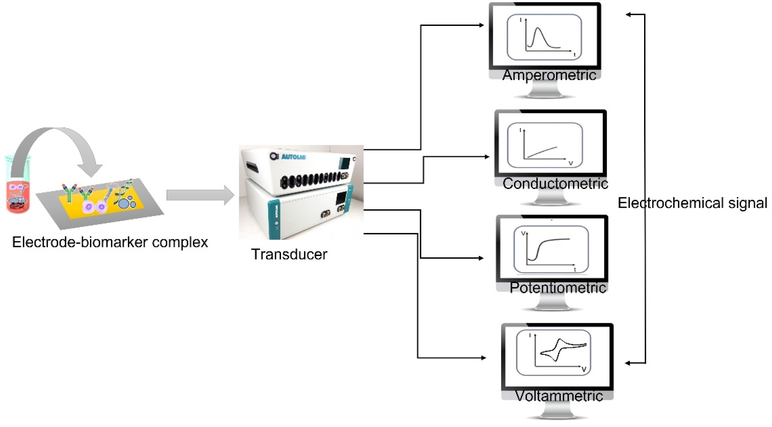
3. Electrochemical Biosensors

Electrochemical biosensors are interesting because they are simple to miniaturize and enable low-cost mass production. In the following section, the most used techniques in electrochemical biosensors and their working principles are briefly illustrated and discussed.

3.1. Amperometric Method

In amperometric biosensors, the electrode current is measured, and most amperometric electrochemical biosensors are based on an enzyme’s redox activity (most often horseradish peroxidase (HRP)). Enzymes enhance biosensing systems by catalyzing chemical reactions, thus improving sensitivity [111]. The performance of enzyme-based biosensors relies on factors such as the electrode surface, enzyme type, substrate, and mediator usage [112]. HRP is commonly employed as a secondary detection reagent, with the TMB/H2O2 substrate proving most effective. Amperometric biosensors measure current at a constant potential to detect the analyte. This method provides selectivity, as the potential used is characteristic of the analyte. The current is measured after directly setting the desired potential, enhancing the accuracy of the analysis [113].
Zhang et al. reported the development of a nonenzymatic immunosensor for the detection of SCC-Ag, utilizing rGO-TEPA and AuAg NCs [114]. In another study, a competitive RNA/RNA hybridization-assay-based biosensor was developed using Streptavidin–Horseradish Peroxidase (SA–HRP) and biotinylated capture probes. The biosensor employed H2O2 as substrate and hydroquinone (HQ) as a RedOx mediator. Two separate platforms, a screen-printed electrode (SPE) with Au NPs and a GCE with tungsten diselenide and Au NPs, were used in the biosensor [115]. Additionally, a microfluidic amperometric immunosensor was developed for the detection of the cancer biomarker CLD7 in circulating extracellular vesicles (EVs). The immunosensor was validated in colorectal cancer (CC) patients [116].
The amperometric graph in Figure 4 is used to detect the biomarker miRNA-211. The biosensor, made of a gold-nanoparticle-modified electrode with an attached RNA probe, measures the change in electric current when different concentrations of miRNA-21 are added. The current is high when no miRNA-21 is present, as all the biotinylated miRNA-21 can bind to the probe. When miRNA-21 is present, it competes with the biotinylated miRNA-21 for hybridization with the probe, reducing the current. The lower the current is, the higher the concentration of miRNA-21 is in the sample.
Chronoamperometry is a variation of an amperometric technique that involves monitoring the current generated by the faradaic process at the electrode over time. It involves applying a sufficiently large potential step to the working electrode to initiate a chemical reaction and then observing the current as a function of time.

3.2. Potentiometric Method

Potentiometric biosensors use a tiny amount of current to monitor the potential of an electrochemical cell [118]. Potentiometric sensors, employing the controlled current method, utilize an electrochemical cell containing two reference electrodes to measure the potential across an ion-selective membrane. Enzymes are commonly used to facilitate ion production, which is then detected by the supporting electrode. Controlled current methods offer the advantage of using more affordable measurement instrumentation compared to controlled potential methods. The Nernst equation relates concentration and potential in potentiometric measurements, offering a low limit of detection (LOD) for early-stage cancer diagnosis [117]. Jia et al. developed a Light Addressable Potentiometric Sensor (LAPS) for the detection of the liver cancer biomarker hPRL-3 [119]. Another study [120] utilized surface molecular imprinted self-assembled monolayers (SAM) for a potentiometric biosensor with a linear range of 2.5–250 ng/mL [121]. Label-free potentiometric detection targeted the HAPLN1 protein biomarker in MPM, achieving a pM-range LOD. Goda et al. created a hybridization-based potentiometric microarray for exosomal miRNA identification [28].
Potentiometric methods were employed to target malignant cells, investigating their electrochemistry in microenvironments affected by lactate release and pH fluctuations. Shaibani et al. achieved an LOD of 103 cells per ml, revealing pH flux alterations around cancer cells and their connection to altered cell metabolism [122]. The selective detection of circulating tumor cells (CTCs) in prostate cancer was achieved by utilizing an anti-EpCAM functionalized graphene oxide potentiometric biosensor based on the Light Addressable Potentiometric Sensor (LAPS) approach [123].
Based on pH monitoring, a sensor-integrated microfluidic technique is employed to find cancer cells. In this instance, the CTC’s metabolic alteration resulted in a decrease in the pH of the surrounding environment. Potentiometric methods with Ag/AgCl and ZnO electrodes were employed to assess pH variations and cell modifications in vitro, utilizing three cell lines (A549, A7r5, and MDCK) in a microfluidic setup (Figure 5) [124].
The potentiometric curve in Figure 5 shows the effect of the flow rate on the potential difference between the working and reference electrodes in the microfluidic device. The authors tested four different flow rates from 24 to 120 μL/min with four different pH buffer solutions from 1.68 to 9.18. The results indicated that the potential difference was not significantly affected by the flow rate, which implies that the microfluidic device has high stability and reliability over a wide range of flow speeds. The authors also observed a minimal drift of ±3 mV/h for each pH solution, which is acceptable for many applications.

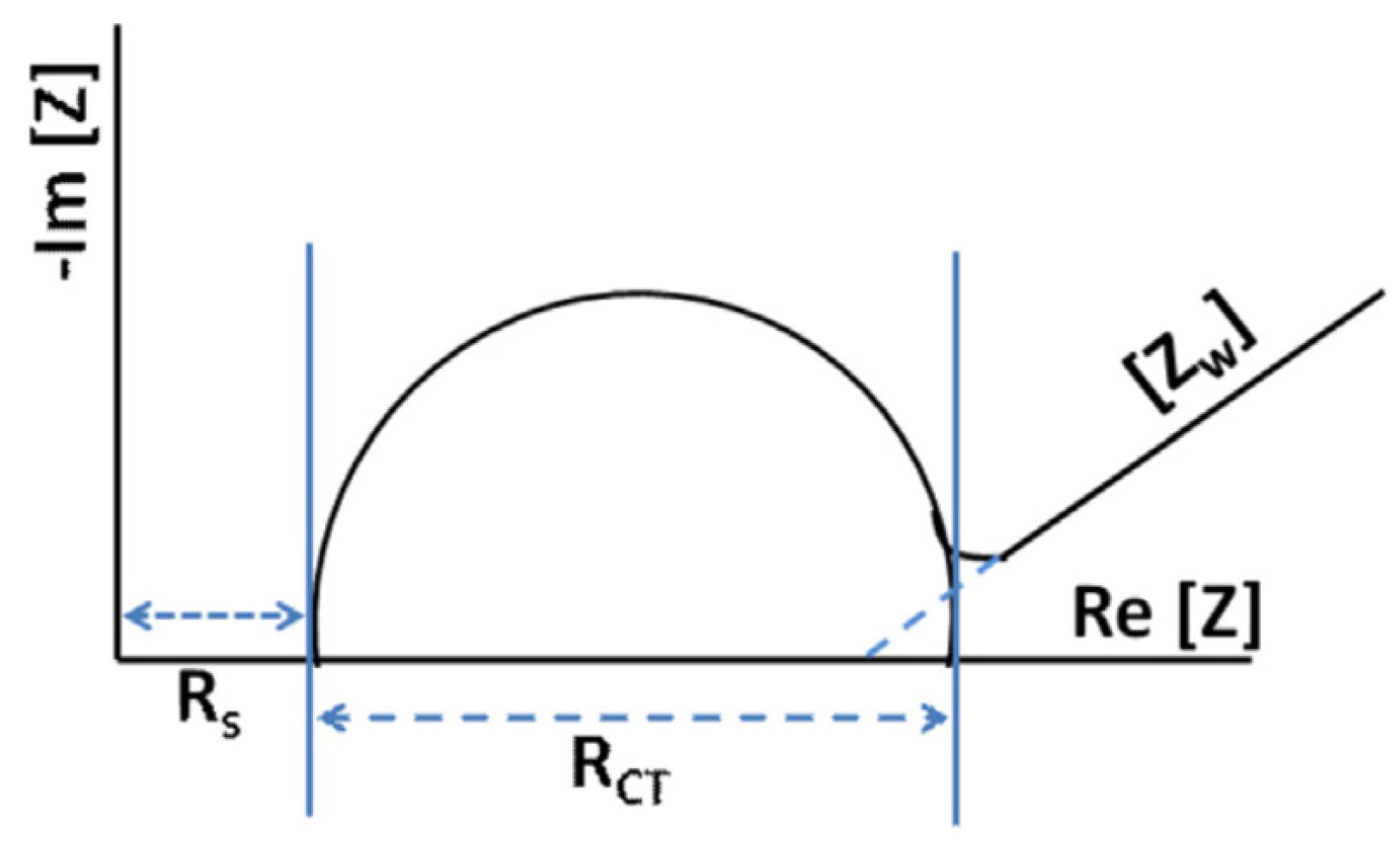
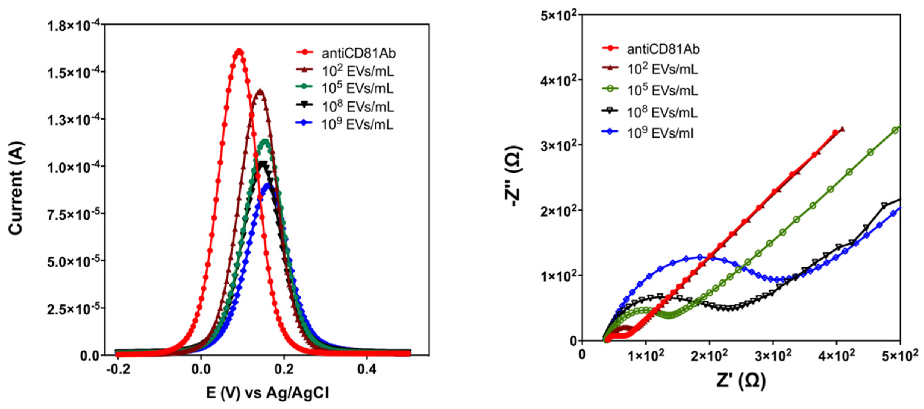
3.3. Impedimetric Method

Electrochemical impedance spectroscopy (EIS) is a technique that examines the resistive and capacitive characteristics of a system by applying an AC excitation signal with varying frequencies. By analyzing the impedance spectra, it is possible to determine the resistive and capacitive components of the system based on the in-phase and out-of-phase current responses. At higher frequencies, the migration rate of redox species can become rate limiting, resulting in a frequency-dependent phase lag when analytes impede access to the electrode surface.
Electrochemical impedance spectroscopy (EIS) evaluates the interfacial characteristics, ion passage, and biomolecule interactions with electrode surfaces. It involves applying the AC potential to an electrochemical cell and measuring the resulting current signal. The resulting frequency-dependent impedance is represented on a Nyquist plot using a Randles circuit, showing semicircles at higher frequencies for electron transfer restrictions and a linear line at lower frequencies indicating the diffusion-limited electron transfer (Figure 6).
The Rct value reflects electron-transfer kinetics, while RS represents bulk electrolyte characteristics, and ZW represents diffusion. The Nyquist plot helps calculate ZW, represented by a 45-degree sloped straight line intercept. EIS is utilized for the label-free detection of cancer cells.
Elshafey et al. developed a label-free impedimetric biosensor for detecting the cancer biomarker EGFR. The biosensor utilized protein G and gold nanoparticles on a modified gold electrode for efficient immobilization. The calibration curve exhibited a wide dynamic range, from 1 pg/mL to 1 g/mL, with a low detection limit of 0.34 pg/mL in PBS and 0.88 pg/mL in human plasma. Interference from various substances in human plasma led to slight variations in the electrochemical signal during real-world experiments [125]. Han et al. developed a label-free cytosensor for cancer cell identification using phage display technology and EIS, demonstrating rising Rct values with an increasing cell concentration, indicating reduced electron transfer efficiency. The approach employed a specific phage immobilized on a gold electrode, with [Fe(CN)6] as the redox probe indicator, offering high specificity and repeatability and eliminating the need for complex steps of purification of recognition elements [126].
Hu et al. utilized EIS for the detection of liver cancer cells. They immobilized a mannose-specific lectin (con A) on a gold electrode, leading to changes in the charge-transfer resistance that correlated with the concentration of cancer cells (Bel-7404). This label-free approach directly targeted cancer cells, providing a direct, selective, and sensitive method with a detection limit of 234 cells/mL, eliminating the requirement for probe labeling [127]. Azzouzi et al. developed an impedimetric electrochemical biosensor using a biotinylated DNA/LNA molecular beacon (MB) probe linked with gold nanoparticles (AuNPs) for miRNA-21 detection in blood samples. This biosensor demonstrated high selectivity, good repeatability, and a wide linear detection range of 1–1000 pM, with a low detection limit of 0.3 pM. The use of neutravidin as a recognition element on the electrode surface enhanced biosensing properties, including sensitivity [128]. Kilic et al. introduced a label-free electrochemical sensor to compare the secretion levels of extracellular vesicles (EVs) in hypoxia and normoxic MCF-7 cells. The sensor utilized functionalized gold electrodes and employed differential pulse voltammetry (DPV) and electrochemical impedance spectroscopy (EIS) for EV detection (Figure 7). The sensor exhibited a linear operating range of 102–109 EVs/mL, with a limit of detection (LOD) of 77 EVs/mL. Selectivity was assessed using the RhD protein, and the results were compared with ELISA and Nanoparticle Tracking Analysis (NTA) [129].

3.4. Conductometric Method

Conductometric biosensors offer exciting possibilities for advanced bioanalytical detection. They measure changes in electrical conductivity during chemical reactions, using enzymes to modify the ionic strength of the sample solution. These biosensors are advantageous due to their miniaturization potential, low voltage requirement, and lack of a reference electrode. They are promising for applications in healthcare, environmental monitoring, and food safety [130]. Capacitive biosensors have the benefit of their capacitance measurements being more informative about the biosensor’s insulating qualities [131]. Even slight sensor layer desorption generally results in a rise in the capacitance baseline. Furthermore, nonspecific binding is less likely with capacitive sensors.
Liang et al. established a conductometric immunoassay for the detection of alpha-fetoprotein (AFP) in the serum of liver cancer patients. The assay demonstrated robust conductometric responses, achieving a low detection limit of 4.8 pg/mL across a dynamic linear range of 0.01–100 ng/mL [132]. For the quantification of prostate cancer antigen, Bhardwaj et al. [133] introduced a conductometric immunosensing platform utilizing tetracyanoquinodimethane (TCNQ)-doped thin films of copper MOF, Cu3(BTC)2. The platform exhibited a dynamic linear range of 0.1–100 ng/mL for PSA detection, with a limit of detection as low as 0.06 ng/mL. Lin and coauthors devised conductometric sensors based on silicon nanowires for the detection of apolipoprotein A1, a biomarker associated with bladder cancer. The sensors demonstrated a wide dynamic range spanning from 0.2 ng/mL to 10 µg/mL, with a detection limit of approximately 1 ng/mL [134].

3.5. Voltammetric Method

Voltammetric biosensors measure current intensity by applying potential between a working and a reference electrode, detecting analyte concentration. They offer high sensitivity and multiplexed biomarker detection and serve as valuable point-of-care diagnostic devices [135].
Voltammetry is widely used for characterizing reaction kinetics and obtaining qualitative and quantitative information about analytes. It provides valuable insights into complex electrode reactions through current measurements. Voltammetry offers technical advantages and fewer limitations compared to other techniques for quantitative determination. It is commonly employed for detecting biomarkers during the liquid biopsy. The voltammetric methods broadly used for the detection of biomarkers under the liquid biopsy are described in the following sections.

3.5.1. Cyclic Voltammetry (CV)

Cyclic voltammetry (CV) is a widely used method for investigating redox processes, monitoring reaction intermediates, and assessing reaction product stability. It involves measuring the potential between the working electrode and reference electrode, while measuring the current between the working electrode and counter electrode. CV plots the electrochemical current on the y-axis and the working electrode potential on the x-axis, with the potential cycling back to its starting value. It provides information on the oxidation and reduction of redox species [136]. A partial cycle, a complete cycle, or a series of cycles might be performed depending on the results of the study. The electrons are transferred from the analyte to the WE or from the electrodes to the analyte during the redox reaction.
Kumar et al. utilized nanostructured zirconia (nZrO2) as a transducer surface in a CV-based technique for detecting the oral cancer biomarker CYFRA. The immobilized receptor antibody (anti-CYFRA) on amine-functionalized nZrO2 showed proportional electrochemical current changes, enabling detection in the range of 2–16 ng/mL with a sensitivity of 2.2 mA mL/ng [137]. Later on, Wang et al. developed an electrochemical sensor using a glassy carbon electrode modified with silver hybridized mesoporous silica nanoparticles (Ag@MSNs) to detect the prostate cancer biomarker PSA. The sensor exhibited improved bioreceptor adsorption and electron-transfer rates, utilizing hydroquinone as a redox probe. PSA detection was achieved in a wide concentration range of 0.05 to 50.0 ng/mL, with a detection limit of 15 pg/mL [138]. A study conducted by Kumar et al. was based on using two-dimensional Ti3C2-MXene nanosheets to detect the carcinoembryonic antigen (CEA) biomarker with a detection range of 0.0001–2000 ng/mL [139].
Taleat et al. employed a sandwich technique for detecting the MUC1 protein, a key contributor to tumor development in various cancers. They combined MUC1 monoclonal antibody immobilized on a poly-aminobenzoic-acid-modified graphite screen-printed electrode with a methylene-blue-modified aptamer (specific ss-DNA) [140]. Feng et al. coupled CV and electrochemiluminescence (ECL) techniques to achieve the simultaneous detection of AFP and CEA by tagging the detection antibody and using methylene blue as an electrochemical indicator that binds directly to aptamers’ G base for MUC1 protein concentration identification [141].

3.5.2. Differential Pulse Voltammetry (DPV)

Differential pulse voltammetry (DPV) involves the scanning potential with small amplitude pulses while measuring the current at two points before and after each pulse. The difference in current measurements is calculated and plotted as a function of the base potential, allowing for analysis of non-faradaic current decay.
DPV is a widely used electrochemical procedure known for its sensitivity and speed. It involves applying fixed-amplitude electrochemical pulses on a slowly increasing base potential and recording the resulting current differential. DPV is utilized to detect early cancer and study drug performance in cancer. Lin et al. developed a reusable biosensor using a magnetic graphene-oxide-modified gold electrode (MGO-Au) to detect VEGF in human plasma for cancer detection [142]. The DPV-based sensor demonstrated effective sensitivity, a rapid reaction time, and a wide linear detection range, outperforming the ELISA approach.
Amjadi et al. studied the influence of doxorubicin (DOX) and a flavonoid-modified drug (FMD) on lung cancer cells (A549) using the DPV approach, revealing that the FMD had a stronger effect on cancer cells compared to DOX, as demonstrated by the reduction in electrochemical reactivity with increasing drug concentration [143]. Additionally, Pacheco et al. and Wang et al. utilized electrochemical techniques (CV and DPV) with breast cancer cells immobilized on working electrodes to quantify cancer cells in unknown samples using a known cancer cell calibration curve [144,145].
The fabrication and alteration of electrodes are important in electrochemical measurement. The screen printing technique is widely employed in this context for the manufacturing of portable low-cost electronics, particularly disposable electrodes. Compared to conventional electrode fabrication methods, this technology presents numerous advantages, encompassing the precise manipulation of electrode dimensions, a wide range of electrode designs, compact device sizes, reduced production expenses, user-friendly operation, and the capability to create diverse arrays of electrodes [146]. Furthermore, screen-printed electrodes allow for additional customization of the electrode surface by altering it with different nanomaterials, resulting in an increased surface area, increased biomolecule immobilization efficiency, and unique electrochemical characteristics.
Using eight disposable screen-printed microelectrode arrays as the transducer surface, Zani et al. established a sensitive and easy PSA detection technique. Magnetic beads were employed to collect the main PSA antibody in this experiment. The electrochemical measurements were taken using DPV after the collected beads were washed with the antibody-labeled enzyme alkalinephosphatase (AP). The detection range of this biosensor was linear (0–20 ng/mL), with a lower detection limit of 1.4 ng/mL [147]. Erdem et al. described an electrochemical biosensor based on a multichannel screen-printed array of electrodes (MUX-SPE16) for assessing the nucleic acid hybridization of distinct miRNA sequences (miRNA-16, miRNA-15a, and miRNA-660). In this study, streptavidin-coated magnetic beads were placed on the electrode surface before a biotinylated DNA probe was immobilized. Following the hybridization procedure, the electrochemical response was measured using the DPV method on the guanine oxidation signal [148].
Furthermore, due to the limited sensitivity and specificity of a single biomarker, measuring or tracking it is insufficient for reliable cancer diagnosis. Therefore, researchers are interested in the simultaneous detection of numerous tumor markers to obtain more accurate and dependable results. Serum VEGF-C had a specificity of 68% and a sensitivity of 85%, whereas MMP-9 had a specificity of 75% and a sensitivity of 63%. Similarly, VEGF had a specificity of 59% and a sensitivity of 80%, but the combination of these three markers had a greater sensitivity and specificity (83% and 76%, respectively) than the single-biomarker strategy for lung cancer detection [149]. CEA was also demonstrated to enhance cancer prediction when combined with other biomarkers. When CEA was combined with CA 15-3, for example, its sensitivity rose from 89% to 96% [150].
Wu et al. introduced a novel approach for the concurrent detection of CA 19-9 and CA 125 cancer biomarkers, which involved the utilization of a disposable two-throughput immune-electrode array. The researchers applied a cellulose acetate membrane onto the graphite working electrodes (W1 and W2) of a screen-printed chip, followed by the co-immobilization of thionine/CA 19-9 and thionine/CA 125 on separate electrodes. Antibodies labeled with HRP were then detected on these working electrodes. By establishing an electron shuttle mechanism facilitated by the immobilized thionine, the enzymatic reduction of H2O2 by HRP resulted in the generation of electrochemical signals, enabling the simultaneous detection of both biomarkers [151,152].
Two separate cancer biomarkers (CEA and AFP) were concurrently identified utilizing DPV technology and a metal-ion-tagged immunocolloidal gold nanocomposite as a signal tag in another manner. The signal antibody was modified with two metal ions (AuNPs/anti-CEA/Cu21 and AuNPs/anti-AFP/Pb21) in this manner. The authors leveraged the intrinsic electrochemical characteristics of metal ions in this study to obtain the multiplex detection of cancer biomarkers on a single platform with high sensitivity. The findings were also confirmed using a conventional ELISA, indicating that they might be used in clinical settings [153].
In the realm of quantitative real-time evaluation and early cancer detection, circulating tumor markers (CTMs) such as extracellular vesicles (microvesicles and exosomes), circulating tumor cells (CTC), and circulating nucleic acids in the blood emerged as valuable indicators. These CTMs offer a range of advantages, including their potential as cost-effective, reproducible, dynamic, and non-invasive diagnostic tools for both cancer diagnosis and the monitoring of disease progression during the early stages. Several detection techniques were employed to identify CTMs, including quartz crystal measurement (QCM), microcantilevers, colorimetric assays, the enzyme-linked immunosorbent assay (ELISA), surface-enhanced Raman scattering (SERS), surface plasmon resonance, the polymerase chain reaction (PCR), and electrochemical methods [154].
Moscovici et al. developed a microfabricated glass chip with gold apertures for cell counting using DPV, enabling the specific detection of 125 prostate cancer cells in 15 min, even in complex cell populations [155]. Yang et al. demonstrated a microelectrode-based electrochemical biosensor for micro-RNA detection using nanostructured palladium electrodes, achieving detection as low as 10 aM of a target with enhanced signals using the DPV approach and Fe(III) regeneration of Ru(III) for amplification [156].
Zhang et al. utilized DPV to detect capecitabine in serum specimens without the need for labels, using an electrochemical biosensor based on stacked graphene nanofibers (SGNF) and gold nanoparticles (AuNPs). The biosensor showed a wide linear detection range of 0.05–80.00 M and an exceptional detection limit of 0.017 M for capecitabine electrochemical reduction [157]. Venu et al. published an electrochemical biosensor based on a ZrO2/rGO nanocomposite for the detection of an anticancer medication (regorafenib; REG). The fabricated biosensor had a wider linear detection range of 11–343 nM, with a remarkable lower detection limit of 59 and a remarkable limit of quantifications of 59 and 17 nM. For the accurate assessment of REG, the biosensor’s effectiveness was also good in both blood samples and pharmaceutical formulations. The biosensor was also useful for detecting REG, uric acid, and ascorbic acid all at the same time [158].

3.5.3. Linear Sweep Voltammetry (LSV)

Throughout the scan, the electrode potential is adjusted at a constant rate, and the resultant current is recorded in LSV. Yan et al. developed an immunosensor of carbon combining screen printing with an excellent material, vegetable parchment. The proposed immunosensor, involving linear sweep voltammetry as the electrochemical method and prostate specific antigen (PSA) as a model analyte, showed a limit of detection of 2 pg/mL [159].
In their study, Bo et al. presented an electrochemical immunosensor that employed a double signal amplification strategy utilizing enzyme-encapsulated liposomes and biocatalytic metal deposition. This innovative approach was specifically developed for the detection of human prostate-specific antigen (PSA). Linear sweep voltammetry (LSV) was employed to measure the quantity of deposited silver, which served as an indicator of the target analyte. The experimental findings demonstrated a linear relationship between the anodic stripping peak current and the concentration of PSA within the range of 0.01–100 ng/mL. Impressively, the detection limit achieved by the sensor was as low as 0.007 ng/mL, thereby illustrating its high sensitivity for PSA detection.

3.5.4. Square Wave Voltammetry (SWV)

The excitation signal in SWV consists of a symmetrical square-wave pulse with an amplitude, Esw, superimposed over a staircase waveform with step height E, where the waveform’s forward pulse corresponds to the staircase step. The difference between the forward and reverse currents is used to calculate the net current, which is centered on the redox potential. SWV provides various benefits, including high sensitivity, speed, and non-faradic current discrimination.
The combination of autocatalytic deposition and square-wave stripping voltammetry with enlarged gold nanoparticles labeled on goat anti-rabbit immunoglobulin G enabled the detection of the rabbit immunoglobulin G (RIgG) analyte with a remarkably low limit of 1.6 fM, highlighting the sensitivity enhancement of the electrochemical immunoassay (GaRIgG-Au) method [160]. In a subsequent study, Liu et al. conducted a comparative analysis between square-wave voltammetry (SWV) and electrochemical impedance spectroscopy (EIS) for the development of a label-free electrochemical immunosensor targeting the hormone estradiol. The researchers aimed to assess the performance of these two techniques in terms of sensitivity. Notably, the results revealed that SWV outperformed EIS, with a lower detection limit of 18 pg/mL for estradiol, compared to 26 pg/mL achieved by EIS. This comparative evaluation highlighted the superior sensitivity of SWV in the context of estradiol detection, suggesting its potential for enhanced analytical applications in hormone analysis [161]. Zhang et al. detected CTCs using SWV based on catalytic amplification, and the limit of detection of was 1 cell/mL.

3.5.5. Stripping Voltammetry (SV)

This technique has the lowest detection limits with respect to the commonly used electrochemical techniques. SV is also known as a pre-concentration technique, and the most commonly used stripping voltametric techniques involve anodic stripping voltammetry, cathodic stripping voltammetry, and adsorptive stripping voltammetry.
Despite the fact that each approach has its own distinct characteristics, they all follow the same two procedures. The target analyte is first concentrated onto the working electrode in the sample solution. In the second phase, the potential is used to remove the preconcentrated analyte off the electrode surface, which is then measured. In the stripping process, potential waveforms such as the linear sweep, differential pulse, and square wave can be employed. Due to their ability to distinguish against charging current, differential pulses and square waves are the most prevalent. In addition, compared to the differential pulse, the square wave offers the advantages of a faster scan rate and greater sensitivity.
The Joseph Wang group introduced a novel electrically heated carbon paste electrode specifically designed for conducting adsorptive stripping measurements of trace amounts of nucleic acids. This groundbreaking approach combines the principles of electrochemistry with electrically heated electrodes and adsorptive constant current stripping chronopotentiometry. The integration of these techniques brings forth notable advantages when it comes to the precise detection and quantification of nucleic acids at trace levels. This innovative coupling of electrochemistry with electrically heated electrodes presents a promising avenue for achieving enhanced accuracy and sensitivity in nucleic acid analysis [162].
A recapitulation of the working principle of a biosensor with electrochemical methods available for biosensors is reported in Figure 8.

4. Sensor Integration

In the advancement of point-of-care and wearable biosensors, miniaturization plays a pivotal role in achieving portability, user-friendliness, and cost-effectiveness as well as maintains high sensitivity with rapid response times. The downsizing of sensing electrodes or their constituents to the nanoscale, as seen in nanoelectrode, nanotextured, nanogap, and field-effect transistor-based biosensors, can significantly enhance the signal-to-noise ratio. It is worth noting, however, that this enhancement can potentially introduce an undesired increase in the biosensor’s response time. Therefore, while nanoscale miniaturization offers various advantages, careful consideration must be given to strike a balance between improved sensitivity and response time to ensure the optimal performance of the biosensor.
The above-mentioned electrochemical methods, discussed in the related paragraphs, are well-suited for implementation in miniaturized systems. Achieving miniaturization, along with the features discussed in Section 2, is essential for creating compact, user-friendly, and cost-effective point-of-care, portable, and wearable biosensors. These biosensors offer competitive limits of detection and rapid response times while maintaining their convenient and accessible nature.

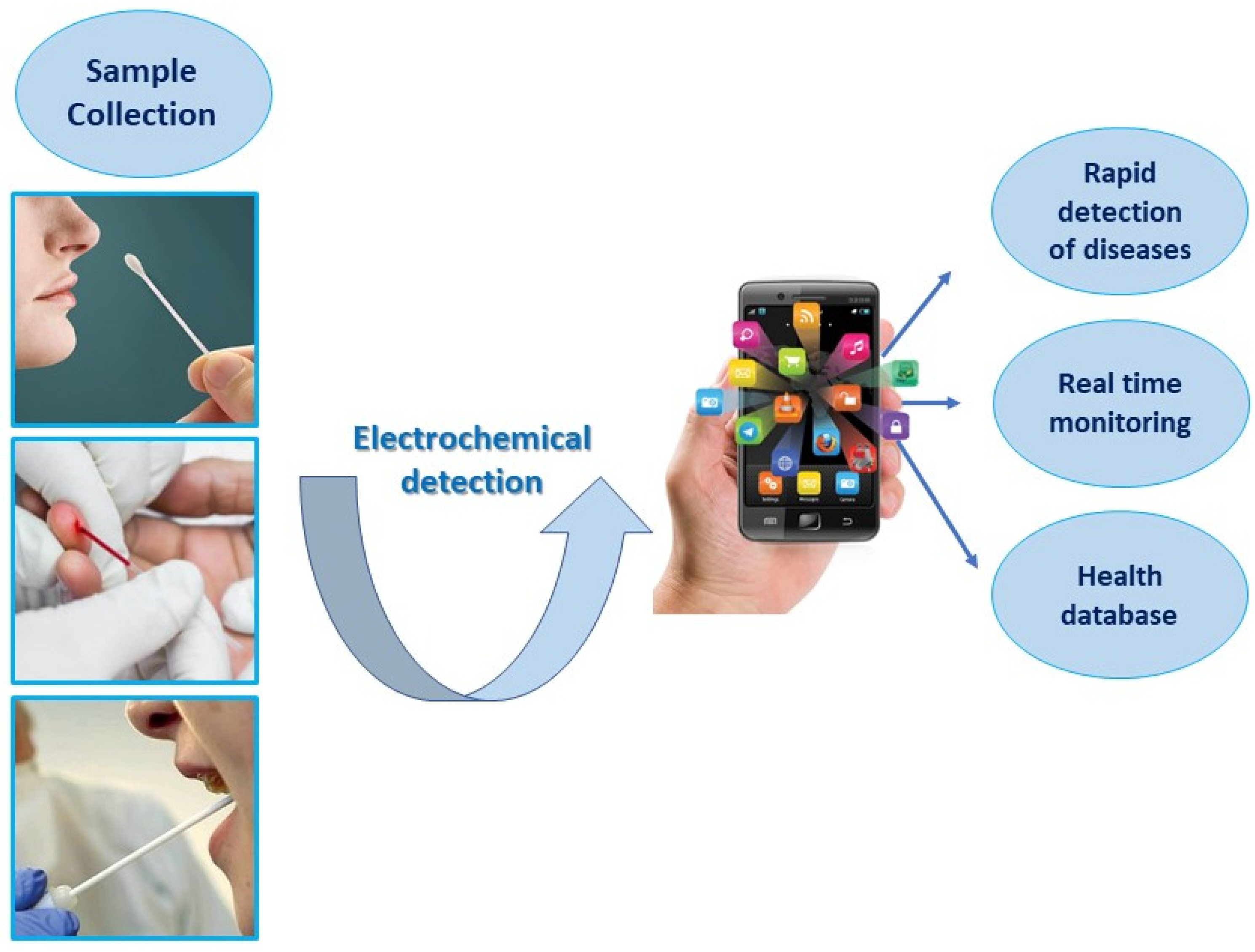
Lab-on-a-Chip Platforms: Wearable and Portable Devices for PoC

Lab-on-a-chip (LOC) devices, notably multitasking devices, show the most attractive advantages for performing several lab procedures on a single chip with small volumes of chemicals and great efficiency. One of the most challenging features of LOCs, also known as a micro total analysis system (μTAS), is to fully integrate a microelectromechanical system (MEMS) with automated microfluidic tools to allow their widespread use in medical applications (liquid biopsy, therapeutic follow-ups, and health and disease monitoring). These user-friendly, sensitive, and portable LOC sensors for real-time analysis have various benefits over traditional analytical techniques. In this review, we list several examples of sensors suitable for integration into LOC devices. Nevertheless, the step over technological gap to overcome limitations in the widespread diffusion of such devices is still linked to the scalability of systems and the poor affordability of highly integrated platforms. A few examples, indeed, are the commercially available platforms including electrochemical systems, and their use for monitoring health parameters is going in the direction of self-diagnostics and fitness applications but not toward becoming a gold standard in clinical practices.
Recently, Atkcakoca and coworkers reported on the realization of an electrochemical biosensor with an integrated microheater improving the performance of the nucleic acid hybridization assay based on electrochemical impedance spectroscopy, paving the way for the development of highly sensitive and specific integrated label-free biosensors [163]. Kasturi and collaborators developed a microfluidic channel with integrated valves and an electrochemical biosensor for the detection of beta-amyloid, a biomarker for Alzheimer’s disease. Gold microelectrodes are the transducer holding the antibody–antigen complexes inside the fluidic chambers governed by pneumatic valves. A linear response of the sensor, through DPV measurements, was reported for beta-amyloid antigen concentrations from 2.2 pM to 22 μM [164]. One of the most promising applications of an integrated LOC for diagnostic purposes is the realization of smartphone-based and wearable devices, which can really realize all the features of portability, low-cost, and the rapid response needed to fully embody the point-of-care concept.
Flexible electronic devices, making use of specific methods of fabrication and detection, have properties such as flexibility, wearability, conductivity, stretchability, mechanical resistance, and biocompatibility. In this scenario, a plethora of materials suitable as substrates for electrochemical transducers were considered [165]: cellulose-based, polyaniline (PANI), polyimide (PI), and specific fabrication methods were demonstrated. Most of them were able to electrochemically detect ion concentrations from skin and sweat as well as monitor fitness parameters like heartbeat, blood pressure, body temperature, and oxygen concentration [166,167].
Sempionatto and collaborators recently developed a non-invasive device for the simultaneous monitoring of blood pressure and heart rate via ultrasonic transducers and multiple biomarkers via electrochemical sensors. They conformally optimized the integrated device to curved skin, thus ensuring mechanical resistance and the reliable sensing of several compounds: glucose in interstitial fluid; lactate, caffeine, and alcohol in sweat [168].
The integration of small microfluidic channels into wearable devices is also a challenging topic for the development of such devices, since biological fluids, easily exploitable for the liquid biopsy (sweat and interstitial fluid), could be conveyed toward the sensing areas of the devices.
Electrochemical sensors measure the reaction caused by the interaction between the sensing surface and the analytes, and the corresponding response is then converted into electric signals that can be monitored by potentiometry, amperometry, and conductometry measurements [169]. The integration of electrochemical biosensors into point-of-care (PoC) platforms, especially when combined with smartphones, emerged as a powerful tool for personalized health monitoring, thus enhancing the practicality of diagnosis compared to the traditional laboratory-based diagnostic methods [170]. In the last years, smartphone-based biosensors have been widely employed for human health PoC testing to improve the diagnosis and treatment of several diseases, thanks to their cost-effectiveness, ease of use, and portability. Figure 9 illustrates the diagnosis steps in smartphone-based electrochemical biosensors.
One of the first applications of a smartphone-based electrochemical biosensor system was described as amperometric sensing [171]. Amperometry is a type of voltametric technique in which a constant voltage is applied to the working electrode, and the current provided by the oxidation/reduction of an electroactive analyte is then measured as a function of time. Liu et al. reported an amperometric aptasensor integrated with a smartphone-assisted portable wireless biochip for the simultaneous real-time monitoring of insulin and glucose in saliva with the lowest detectable concentration of 0.85 nM and 0.8 nM, respectively. The sensing platform combined with a Bluetooth transmission system to generate the digital diagnosis by a smartphone signal readout, providing a PoC testing tool for the diagnosis of diabetes and other insulin-resistance-associated diseases [172]. Voltammetry is another electrochemical technique that includes different type of measurements such as differential pulse voltammetry (DPV), cyclic voltammetry (CV), square wave voltammetry (SWV), and linear sweep voltammetry (LSV) and is widely used in smartphone-integrated electrochemical sensors [173]. In 2020, Low et al. developed a DPV-based electrochemical sensor combined with a smartphone for the detection of the circulating miRNA-21 biomarker in saliva. They demonstrated that the smartphone-based biosensing system, equipped with a specific Android application, displayed comparable performances with commercial workstations for the detection of miRNA-21 [174]. Combining a screen-printed immunosensor with a smartphone-based electrochemical system, Fan et al. reported the detection of cancer antigen 125 (CA125) by DPV measurements. Data were transmitted to a smartphone by Bluetooth, acting as the interface for communication with a remote medical center via the Internet. The developed system provided a sensitive detection of CA 125 with an LOD of 2 mU mL−1 [96].
Electrochemical impedance spectroscopy (EIS), in which the impedance of the system is measured as a function of frequency, is also widely used in the fabrication of EIS-based electrochemical sensors combined with smartphones. For example, Talukder et al. reported a portable system for the personalized monitoring of the blood cell count, consisting of a smartphone-based microfluidic impedance cytometer [175].
The recent advances in this field highlight the advantages of electrochemical sensing systems integrated with a smartphone, thanks to their high simplicity of fabrication [176]. Moreover, the development of multiplexing smartphone-based electrochemical systems can be used for the simultaneous detection of various biomarkers, thus enabling the remote control of diseases by doctors and accelerating the diagnosis and treatment of a pathology.

5. Comparison of Liquid Biopsy Electrochemical Methods: Advantages and Limitations

Nowadays, cancer is the major disease affecting human health and life, and its large diffusion requires the development of simple, practical, and facile diagnosis methods for simplifying its treatment and improving its cure rate. Compared with medical imaging and a pathological examination, which are the most common cancer diagnosis methods, the liquid biopsy represents a promising strategy for cancer biomarker detection, opening the way to direct and rapid diagnostic methods with high efficacy [177,178]. Among several biomarkers, circulating tumor cells (CTCs) are well-established as promising targets for the detection of tumors via the liquid biopsy. Since tumor cells can be shed into the blood before the formation of visible solid tumor lesions, detecting CTCs before the imaging findings or clinical manifestations is an efficient method for the early diagnosis and monitoring of cancer [179]. However, the content of CTCs in peripheral blood circulation is very small, and the techniques used for its detection (fluorescence imaging, magnetic resonance imaging, and cytological detection [180,181,182]) have several shortcomings, such as high cost, a large amount of time, low sensitivity, and a lack of specificity, thus limiting their use in clinical applications [183].
In the last years, electrochemical sensing technology has been widely investigated as a good alternative method for the detection of CTCs because of its advantages of high sensitivity, good selectivity, low cost, easy portability, and rapid detection. Compared with traditional detection techniques, electrochemical methods demonstrate competitive results in terms of the LOD and selectivity. CTCs can be electrochemically detected by using two common types of approaches. The first one is often related to impedimetric sensors and exploits the change in the electron transfer produced by CTCs captured on the electrode, usually conjugated to various recognitive materials including antibodies [184], aptamers [185], and receptors [186]. However, this type of sensor usually needs just one electrode to work, thus resulting in a lack of capture efficiency. To overcome this limit, modifying electrodes with nanomaterials can be a good strategy to enhance the capture efficiency for CTCs. For example, Wang et al. conjugated gold nanostars with a high surface area, with CTCs’ specific aptamer. Owing to this design, the sensor showed a sensitive detection limit of 5 cells/mL [187]. Cai et al. developed a dual-recognition electrochemical cytosensor for the detection of CTCs. The sensor, based on Cabot carbon black (BP2000)/AuNPs anchoring anti-epithelial cell adhesion molecule (anti-EpCAM) antibodies as capture probes and novel branched PtAuRh trimetallic nanospheres (b-PtAuRh TNS), linked with aptamers targeting mucin1 (MUC1) as signal probes, exhibited a wide linear range of 5–1 × 10⁶ cells per mL−1 and a low detection limit of 1 cell per mL−1 [188]. In another work, Zhang et al. exploited the reaction of LiFePO4 with sodium molybdate to generate an electrochemical signal for detecting CTCs. In particular, they captured CTCs from sample by using Fe3O4 magnetic nanospheres (MNs) modified with the EpCAM antibody, while gold-nanoparticle-modified LiFePO4 (LiFePO4/Au) was used as an electrochemical probe. The assay presented a detection range from 3 to 10,000 cells per mL−1, with a detection limit of 1 cell per mL−1 [189]. Although several studies demonstrated the advantages of electrochemical sensing technology for the detection of CTCs and the significant advances in the biosensing research area thanks to immunotechnology, microfluidics, and nanotechnology, the clinical use of such biosensors is still limited. In fact, the number of CTCs in the peripheral blood circulation is very little, and detection can be very difficult. Moreover, the existing detection techniques use nucleic acids and antibodies as target molecules, which, lacking specificity for the classification of captured CTCs, cannot be utilized to give precise information about patient-specific tumor biology. Thus, combining advanced technologies such as microfluidics and the DNA walker and exploring more cell-specific targets could be a significative strategy to improve the sensitivity and specificity of such biosensors. Recently, Ming et al. developed a new on-skin optoelectronic biosensor based on cyclic voltammetry that can measure various vital signs related to blood flow and oxygenation in a non-invasive and continuous way [190].
As voltammetry is the most commonly used electrochemical technique for liquid biopsies, this review primarily focuses on the DPV technique, a subtype of voltammetry, because DPV is considered to be an important electrochemical method for liquid biopsy applications because it offers high sensitivity, selectivity, a wide dynamic range, rapid analysis, and minimal interference from other components in body fluids.
Scientists can determine which electrochemical methods are best-suited for particular applications by comparing the various electrochemical techniques used in liquid biopsy biosensing, depending on elements like the type of biomarker being analyzed, the concentration range, and the complexity of the sample matrix.
Such a comparison can help to guide the development of new biosensors for liquid biopsy analysis as well as to optimize existing methods for improved performance and sensitivity. Additionally, understanding the advantages and limitations of different electrochemical methods can aid in the interpretation of experimental results and can inform the selection of appropriate analytical methods for a given research question. These aspects have been summarized in Table 2.
Each electrochemical technique has advantages and disadvantages of its own. The choice of method depends on the specific analyte of interest and the requirements of the analysis.
The desire to manufacture micro total analysis systems, low-cost point-of-care diagnostics, and environmental monitoring devices sparked the creation of tiny and portable biosensor devices. So, for the development of such a biosensor, it is essential to understand the electrochemical method on which this biosensor operates.
The efficient transducer surface or immobilization matrix is the most significant step in the fabrication of a miniaturized electrochemical biosensor. For optimal biosensor performance, we need to carefully select the materials, electrochemical methodology, and manufacturing process. The PoC devices used for the liquid biopsy might benefit from a wise device design and efficient detection procedures. Research needs to be conducted to create combinatorial electrochemical biosensors with a high throughput and low cost for cancer diagnosis, therapy, and monitoring, utilizing the liquid biopsy. The commercialization of biosensors will increase when an electrochemical-based biosensing platform effectively works in a real-world sample environment with excellent selectivity, sensitivity, and stability.

6. Future Perspectives and Concluding Remarks

This review paper highlights the significance of screening and early diagnosis in disease management, particularly in the context of cancer. It emphasizes the importance of non-invasive analytical methods capable of detecting biomarkers to facilitate successful treatments and improve patient survival rates. The focus of the study is on the electrochemical methods used for the development of biosensors in the liquid biopsy, owing to their ability to offer a rapid response, precise detection, and low detection limits. This review discusses the advancements in electrochemical biosensors, which hold the potential to enhance the specificity and sensitivity of conventional analytical techniques. Electrochemical biosensors demonstrate the ability to detect minute quantities of analytes, including proteins, nucleic acids, and circulating tumor cells, even in complex bodily fluids such as urine, serum, and blood. Among the various detection techniques explored for cancer biomarker detection, voltammetric sensors are extensively discussed due to their advantages and technical characteristics, which led to their widespread use in the quantitative detection of ions and molecules.
This review also provides a comprehensive comparison of different electrochemical techniques to aid in the selection of the appropriate analytical methods based on the specific requirements. This comparative analysis helps researchers and clinicians identify the most suitable approach for their intended applications. Looking toward the future, the development and refinement of electrochemical biosensors hold tremendous potential for advancing diagnostic capabilities in the field of the liquid biopsy. Further advancements in sensor design and surface modification techniques and integration with emerging technologies like nanomaterials and microfluidics are expected to enhance the performance, reliability, and multiplexing capabilities of electrochemical biosensors. These developments will likely contribute to improved disease detection and monitoring, enabling personalized and targeted treatment strategies.
However, it is important to acknowledge that while electrochemical biosensors offer great promise, there are still challenges to overcome. Some of these challenges include improving the selectivity and stability of sensors, standardizing protocols for clinical use, and addressing the complexities associated with analyzing biomarkers in diverse biological matrices. The continuous innovation and optimization of electrochemical biosensing technologies are expected to play a vital role in improving disease diagnosis, monitoring treatment efficacy, and, ultimately, enhancing patient outcomes.
The potential and ambition of the liquid biopsy pose significant challenges and opportunities for personalized medicine and point-of-care diagnostics and follow-ups. Within this context, the utilization of miniaturized and rapid detection tools, such as electrochemical sensors, holds great promise for advancing the field. These devices can be seamlessly integrated into everyday objects like smartphones, smartwatches, and more. Furthermore, as individuals become increasingly health conscious and emphasize early disease screening, the introduction and widespread use of user-friendly sensors, lab-on-a-chip devices, and similar tools for manipulating and analyzing biofluids are poised to greatly benefit self-awareness and disease detection.
Despite the significant advances in electrochemical biosensors for the liquid biopsy, there are still some challenges and limitations that need to be addressed. These include the optimization of the biosensor design, the selection of the most suitable biomarkers and detection techniques, the validation of biosensor performance in clinical samples, and the standardization of biosensor fabrication and operation. Moreover, there is a need for more interdisciplinary collaboration among researchers from different fields, such as chemistry, biology, engineering, and medicine, to develop innovative and effective solutions for the liquid biopsy. Furthermore, there is the potential for combining electrochemical biosensors with other analytical methods, such as optical or magnetic sensors, to achieve complementary and synergistic results. Electrochemical biosensors for the liquid biopsy have a bright future ahead, as they can revolutionize the approach to diseases and improve the quality of life of patients.

Author Contributions

Conceptualization: M.S.C., F.F. and S.U.; methodology: E.P., A.T., S.U. and G.S.; investigation: G.S., I.T. and S.U.; original draft preparation: S.U., G.S., A.T., E.P., M.S.C. and F.F.; supervision: M.S.C.; funding acquisition: M.S.C., F.F. and A.T. All authors have read and agreed to the published version of the manuscript.

Funding

This research was funded by Italian Ministry of University and Research (MUR), Progetto PON ARS01_00906 “TITAN: Nanotecnologie per l’ImmunoTerapia dei Tumori”, funded within FESR Programme PON “Ricerca e Innovazione” 2014–2020 Azione II-OS 1.b); PRIN 2022 Projects- “RESOLVE—innovative platfoRm based on fiEld-flow-fractionation and Sample On-chip detection to unravel extraceLlular Vesicles hEterogeneity” (grant number: 202233FTW8), “MAGELLANO—MAnufacturing systems for Graphene nanocompositEs and their appLications as embedded eLectrochemical seNsors in Lab-On-chip” (grant number: 2022NRMS4X), and Regione Puglia within “Tecnopolo per la medicina di precisione” (TecnoMed Puglia): DGR n.2117 del 21/11/2018, CUP: B84I180 0 0540 0 02 and by Italian Ministry of Enterprises and Made in Italy: “MSP4WATER” Project funded within the frame of the Programme “Fondo per la Crescita Sostenibile—Accordi per l’innovazione DM 31/12/2021” (grant number: F/310200/01-04/X56).

Institutional Review Board Statement

Not applicable.

Informed Consent Statement

Not applicable.

Data Availability Statement

Not applicable.

Acknowledgments

The authors acknowledge the Italian Ministry of Research (MUR), Italian Ministry of Enterprises and Made in Italy, and Regione Puglia for the financial support.

Conflicts of Interest

The authors declare no conflict of interest.

References

  1. Ferrara, F.; Zoupanou, S.; Primiceri, E.; Ali, Z.; Chiriaco, M.S. Beyond liquid biopsy: Toward non-invasive assays for distanced cancer diagnostics in pandemics. Biosens. Bioelectron. 2022, 196, 113698. [Google Scholar] [CrossRef] [PubMed]
  2. Nič, M.; Jirát, J.; Košata, B.; Jenkins, A.; McNaught, A. (Eds.) Compendium of Chemical Terminology; Blackwell Scientific Publications: Oxford, UK, 1997. [Google Scholar]
  3. Ragavan, K.V.; Kumar, S.; Swaraj, S.; Neethirajan, S. Advances in biosensors and optical assays for diagnosis and detection of malaria. Biosens. Bioelectron. 2018, 105, 188–210. [Google Scholar] [CrossRef] [PubMed]
  4. Sadasivuni, K.K.; Ponnamma, D.; Kim, J.; Cabibihan, J.J.; AlMaadeed, M.A. (Eds.) Biopolymer Composites in Electronics; Elsevier: Amsterdam, The Netherlands, 2017; p. 544. [Google Scholar]
  5. Kelley, S.O. Disease Detector. Sci. Am. 2015, 313, 48–51. [Google Scholar] [CrossRef]
  6. Thevenot, D.R.; Toth, K.; Durst, R.A.; Wilson, G.S. Electrochemical biosensors: Recommended definitions and classification. Biosens. Bioelectron. 2001, 16, 121–131. [Google Scholar] [CrossRef] [PubMed]
  7. Brazaca, L.; Ribovski, L.; Janegitz, B.; Zucolotto, V. Medical Biosensors for Point of Care (POC) Applications; Elsevier: Amsterdam, The Netherlands, 2017; pp. 229–254. [Google Scholar]
  8. Naresh, V.; Lee, N. A Review on Biosensors and Recent Development of Nanostructured Materials-Enabled Biosensors. Sensors 2021, 21, 1109. [Google Scholar] [CrossRef] [PubMed]
  9. Garzarelli, V.; Ferrara, F.; Primiceri, E.; Chiriacò, M.S. Biofluids manipulation methods for liquid biopsy in minimally-invasive assays. MethodsX 2022, 9, 101759. [Google Scholar] [CrossRef]
  10. de Andrade, A.C.; Resende, R.R.; Marques, A.P.A. Lab-on-chip technologies for point-of-care diagnostics: Advances and challenges for clinical applications. Biosens. Bioelectron. 2020, 1, 63–73. [Google Scholar]
  11. Primiceri, E.; Chiriacò, M.S.; Ionescu, R.E.; D’Amone, E.; Cingolani, R.; Rinaldi, R.; Maruccio, G. Development of EIS cell chips and their application for cell analysis. Microelectron. Eng. 2009, 86, 1477–1480. [Google Scholar] [CrossRef]
  12. Gamal, W.; Wu, H.; Underwood, I.; Jia, J.; Smith, S.; Bagnaninchi, P.O. Impedance-based cellular assays for regenerative medicine. Phil. Trans. R. Soc. B 2018, 373, 20170226. [Google Scholar] [CrossRef]
  13. Evangelos, S.; Evangelos, A.; George, K.; Emmanouil, A.V.K.; Angeliki, T.; Dimitris, T. Flow determination via nanoparticle strain sensors for easy Lab on Chip integration. Sens. Actuators A. Phys. 2022, 344, 113765. [Google Scholar]
  14. Lucile, A.; Amel, B.; Iago, P.; Madad, A.; Simon, D.; Laurent, M.; Thanh, D.M.; Stéphanie, D. Modular microfluidic system for on-chip extraction. preconcentration and detection of the cytokine biomarker IL-6 in biofluid. Sci. Rep. 2022, 12, 9468. [Google Scholar]
  15. Shen, C.; Liu, S.; Li, X.; Yang, M. Electrochemical Detection of Circulating Tumor Cells Based on DNA Generated Electrochemical Current and Rolling Circle Amplification. Anal. Chem. 2019, 91, 11614–11619. [Google Scholar] [CrossRef] [PubMed]
  16. Daulton, E.; Wicaksono, A.N.; Tiele, A.; Kocher, H.M.; Debernardi, S.; Crnogorac-Jurcevic, T.; Covington, J.A. Volatile organic compounds (VOCs) for the non-invasive detection of pancreatic cancer from urine. Talanta 2021, 221, 121604. [Google Scholar] [CrossRef]
  17. Gilany, K.; Minai-Tehrani, A.; Savadi-Shiraz, E.; Rezadoost, H.; Lakpour, N. Exploring the human seminal plasma proteome: An unexplored gold mine of biomarker for male Infertility and male reproduction disorder. J. Reprod. Infertil. 2015, 16, 61–71. [Google Scholar] [PubMed]
  18. Park, J.; Kim, N.E.; Yoon, H.; Shin, C.M.; Kim, N.; Lee, D.H.; Park, J.Y.; Choi, C.H.; Kim, J.G.; Kim, Y.K. Fecal Microbiota and Gut Microbe-Derived Extracellular Vesicles in Colorectal Cancer. Front. Oncol. 2021, 11, 650026. [Google Scholar] [CrossRef]
  19. Sun, Y.; Haglund, T.A.; Rogers, A.J.; Ghanim, A.; Sethu, P. Review: Microfluidics technologies for blood-based cancer liquid biopsies. Anal. Chim. Acta 2018, 1012, 10–29. [Google Scholar] [CrossRef]
  20. Liu, J.; Bao, N.; Liu, X.; Ding, S.-N. Nonenzymatic amperometric Aptamer cytosensor for ultrasensitive detection of circulating tumor cells and dynamic evaluation of cell surface N-Glycan expression. ACS Omega 2018, 3, 8595–8604. [Google Scholar] [CrossRef] [PubMed]
  21. Cimmino, I.; Bravaccini, S.; Cerchione, C. Urinary Biomarkers in Tumors: An Overview. In Springer EBooks; Springer: Berlin/Heidelberg, Germany, 2021; pp. 3–15. [Google Scholar]
  22. Paul, A.; Chiriaco, M.; Primiceri, E.; Srivastava, D.N.; Maruccio, G. Picomolar detection of retinol binding protein 4 for early management of type II diabetes. Biosens. Bioelectron. 2019, 128, 122–128. [Google Scholar] [CrossRef]
  23. Siciliano, G.; Chiriaco, M.; Ferrara, F.; Turco, A.; Velardi, L.; Signore, M.A.; Esposito, M.; Gigli, G.; Primiceri, E. Development of a MIP based electrochemical sensor for TGF-β1 detection and its application in liquid biopsy. Analyst 2023, 148, 4447–4455. [Google Scholar] [CrossRef]
  24. Radi, A.-E.; Abd-Ellatief, M.R. Electrochemical APTasensors: Current status and future Perspectives. Diagnostics 2021, 11, 104. [Google Scholar] [CrossRef]
  25. Gao, Y.; Qin, Y.; Wan, C.; Sun, Y.; Meng, J.; Huang, J.; Hu, Y.; Jin, H.; Yang, K. Small extracellular vesicles: A novel avenue for cancer management. Front. Oncol. 2021, 11, 638357. [Google Scholar] [CrossRef]
  26. Chiriaco, M.; Bianco, M.; Nigro, A.; Primiceri, E.; Ferrara, F.; Romano, A.; Quattrini, A.; Furlan, R.; Arima, V.; Maruccio, G. Lab-on-Chip for exosomes and microvesicles detection and characterization. Sensors 2018, 18, 3175. [Google Scholar] [CrossRef]
  27. Garzarelli, V.; Chiriacò, M.S.; Cereda, M.; Gigli, G.; Ferrara, F. Ultrasensitive qPCR platform for rapid detection of bacterial contamination of raw biological samples at the point of care. Heliyon 2023, 9, e16229. [Google Scholar] [CrossRef] [PubMed]
  28. Goda, T.; Masuno, K.; Nishida, J.; Kosaka, N.; Ochiya, T.; Matsumoto, A.; Miyahara, Y. A label-free electrical detection of exosomal microRNAs using microelectrode array. Chem. Commun. 2012, 48, 11942–11944. [Google Scholar] [CrossRef]
  29. Bari, S.M.I.; Hossain, F.B.; Nestorova, G.G. Advances in Biosensors Technology for Detection and Characterization of Extracellular Vesicles. Sensors 2021, 21, 7645. [Google Scholar] [CrossRef] [PubMed]
  30. Zhuang, L.; You, Q.; Su, X.; Chang, Z.; Ge, M.; Mei, Q.; Yang, L.; Dong, W.; Li, L. High-Performance Detection of Exosomes Based on Synergistic Amplification of Amino-Functionalized Fe3O4 Nanoparticles and Two-Dimensional MXene Nanosheets. Sensors 2023, 23, 3508. [Google Scholar] [CrossRef]
  31. Jeong, S.; Park, J.; Pathania, D.; Castro, C.M.; Weissleder, R.; Lee, H. Integrated Magneto−Electrochemical Sensor for Exosome Analysis. ACS Nano 2016, 10, 1802–1809. [Google Scholar] [CrossRef]
  32. Ruiyi, L.; Ling, L.; Hongxia, B.; Zaijun, L. Nitrogen-doped multiple graphene aerogel/gold nanostar as the electrochemical sensing platform for ultrasensitive detection of circulating free DNA in human serum. Biosens. Bioelectron. 2016, 79, 457. [Google Scholar] [CrossRef] [PubMed]
  33. Wang, K.; Peng, Z.; Lin, X.; Nian, W.; Zheng, X.; Wu, J. Electrochemical Biosensors for Circulating Tumor DNA Detection. Biosensors 2022, 12, 649. [Google Scholar] [CrossRef] [PubMed]
  34. Su, S.; Cao, W.; Liu, W.; Lu, Z.; Zhu, D.; Chao, J.; Weng, L.; Wang, L.; Fan, C. Dual-mode electrochemical analysis of microRNA-21 using gold nanoparticle-decorated MoS2 nanosheet. Biosens. Bioelectron. 2017, 94, 552. [Google Scholar] [CrossRef]
  35. Gurudatt, N.G.; Chung, S.; Kim, J.M.; Kim, M.H.; Jung, D.K.; Han, J.Y.; Shim, Y.B. Separation detection of different circulating tumor cells in the blood using an electrochemical microfluidic channel modified with a lipid-bonded conducting polymer. Biosens. Bioelectron. 2019, 146, 111746. [Google Scholar] [CrossRef]
  36. Tian, L.; Qi, J.; Qian, K.; Oderinde, O.; Liu, Q.; Yao, C.; Song, W.; Wang, Y. Copper (II) oxide nanozyme based electrochemical cytosensor for high sensitive detection of circulating tumor cells in breast cancer. J. Electroanal. Chem 2018, 812, 1–9. [Google Scholar] [CrossRef]
  37. Tang, S.; Shen, H.; Hao, Y.; Huang, Z.; Tao, Y.; Peng, Y.; Guo, Y.; Xie, G.; Feng, W. A Novel cytosensor based on Pt@Ag nanoflowers and AuNPs/acetylene black for ultrasensitive and highly specific detection of circulating tumor cells. Biosens. Bioelectron. 2018, 104, 72–78. [Google Scholar] [CrossRef]
  38. Aydın, M.; Aydın, E.B.; Sezgintürk, M.K. A highly selective electrochemical immunosensor based on conductive carbon black and star PGMA polymer composite material for IL-8 biomarker detection in human serum and saliva. Biosens. Bioelectron. 2018, 117, 720–728. [Google Scholar] [CrossRef]
  39. Soleymani, L.; Li, F. Mechanistic challenges and advantages of biosensor miniaturization into the nanoscale. ACS Sens. 2017, 2, 458–467. [Google Scholar] [CrossRef]
  40. Martinsson, H.; Sandstrom, T.; Bleeker, A.J.; Hintersteiner, J.D. Current status of optical maskless lithography. J. Micro-Nanolithogr. Mems Moems 2005, 4, 011003. [Google Scholar] [CrossRef]
  41. Chen, C.; Liu, Y.J.; Jiang, Z.-Y.; Shen, C.; Zhang, Y.; Zhong, F.; Chen, L.; Zhu, S.; Liu, H. Large-area long-wave infrared broadband all-dielectric metasurface absorber based on maskless laser direct writing lithography. Opt. Express 2022, 30, 13391. [Google Scholar] [CrossRef] [PubMed]
  42. Heiskanen, S.; Maasilta, I. Superconducting tunnel junction fabrication on three-dimensional topography based on direct laser writing. Appl. Phys. Lett. 2020, 117, 232601. [Google Scholar] [CrossRef]
  43. Ye, J.; Tan, H.; Wu, S.; Ni, K.; Pan, F.; Liu, J.; Tao, Z.; Qu, Y.; Ji, H.; Simon, P.; et al. Direct Laser Writing of Graphene Made from Chemical Vapor Deposition for Flexible, Integratable Micro-Supercapacitors with Ultrahigh Power Output. Adv. Mater. 2018, 30, 1801384. [Google Scholar] [CrossRef] [PubMed]
  44. Dotan, T.; Berg, Y.; Migliorini, L.; Villa, S.; Santaniello, T.; Milani, P.; Shacham-Diamand, Y. Soft and flexible gold microelectrodes by supersonic cluster beam deposition and femtosecond laser processing. Microelectron. Eng. 2021, 237, 111478. [Google Scholar] [CrossRef]
  45. Van Der Velden, G.; Fan, D.; Staufer, U. Fabrication of a microfluidic device by using two-photon lithography on a positive photoresist. Micro Nano Eng. 2020, 7, 100054. [Google Scholar] [CrossRef]
  46. Viehrig, M.; Thilsted, A.H.; Matteucci, M.; Wu, K.; Catak, D.; Schmidt, M.S.; Zór, K.; Boisen, A. Injection-Molded Microfluidic Device for SERS Sensing Using Embedded Au-Capped Polymer Nanocones. ACS Appl. Mater. Interfaces 2018, 10, 37417–37425. [Google Scholar] [CrossRef] [PubMed]
  47. Perrone, E.; Cesaria, M.; Zizzari, A.; Bianco, M.; Ferrara, F.; Raia, L.; Guarino, V.; Cuscunà, M.; Mazzeo, M.; Gigli, G.; et al. Potential of CO2-laser processing of quartz for fast prototyping of microfluidic reactors and templates for 3D cell assembly over large scale. Mater. Today Bio 2021, 12, 100163. [Google Scholar] [CrossRef] [PubMed]
  48. Timilsina, S.S.; Durr, N.; Yafia, M.; Sallum, H.M.; Jolly, P.; Ingber, D.E. Ultrarapid Method for Coating Electrochemical Sensors with Antifouling Conductive Nanomaterials Enables Highly Sensitive Multiplexed Detection in Whole Blood. Adv. Healthc. Mater. 2021, 11, 2102244. [Google Scholar] [CrossRef]
  49. Li, X.-L.; Zhan, C.; Qiqi, H.; He, M.; Yang, C.; Yang, C.; Huang, X.; Chen, M.; Xie, X.; Chen, H.-J. Smart Diaper Based on Integrated Multiplex Carbon Nanotube-Coated Electrode Array Sensors for In Situ Urine Monitoring. ACS Appl. Nano Mater. 2022, 5, 4767–4778. [Google Scholar] [CrossRef]
  50. Tseng, C.C.; Kung, C.-T.; Chen, R.; Tsai, M.-H.; Chao, H.-R.; Wang, Y.; Fu, L.-M. Recent advances in microfluidic paper-based assay devices for diagnosis of human diseases using saliva, tears and sweat samples. Sens. Actuators B-Chem. 2021, 342, 130078. [Google Scholar] [CrossRef]
  51. Dong, T.; Pires, N.; Yang, Z.; Jiang, Z. Advances in Electrochemical Biosensors Based on Nanomaterials for Protein Biomarker Detection in Saliva. Adv. Sci. 2022, 10, 2205429. [Google Scholar] [CrossRef]
  52. Destgeer, G.; Lee, K.H.; Jung, J.H.; Alazzam, A.; Sung, H.J. Continuous separation of particles in a PDMS microfluidic channel via travelling surface acoustic waves (TSAW). Lab Chip 2013, 13, 4210. [Google Scholar] [CrossRef]
  53. Go, D.B.; Atashbar, M.Z.; Ramshani, Z.; Chang, H.-C. Surface acoustic wave devices for chemical sensing and microfluidics: A review and perspective. Anal. Methods 2017, 9, 4112–4134. [Google Scholar] [CrossRef]
  54. Cardoso, S.; Leitao, D.C.; Dias, T.; Valadeiro, J.; Silva, M.; Chícharo, A.; Silverio, V.; Gaspar, J.; Freitas, P.P. Challenges and trends in magnetic sensor integration with microfluidics for biomedical applications. J. Phys. D 2017, 50, 213001. [Google Scholar] [CrossRef]
  55. Gonzalez-Gallardo, C.L.; Díaz, A.; Casanova-Moreno, J.R. Improving plasma bonding of PDMS to gold-patterned glass for electrochemical microfluidic applications. Microfluid. Nanofluidics 2021, 25, 20. [Google Scholar] [CrossRef]
  56. Zoupanou, S.; Chiriaco, M.; Tarantini, I.; Ferrara, F. Innovative 3D Microfluidic Tools for On-Chip fluids and Particles manipulation: From design to experimental validation. Micromachines 2021, 12, 104. [Google Scholar] [CrossRef]
  57. Guascito, M.R.; Filippo, E.; Mattei, G.; Manno, D.; Serra, A.; Turco, A. A new amperometric nanostructured sensor for the analytical determination of hydrogen peroxide. Biosens. Bioelectron. 2008, 24, 1057–1063. [Google Scholar] [CrossRef] [PubMed]
  58. De Benedetto, G.E.; Corvaglia, S.; Pompa, P.P.; Mattei, G. An innovative and simple all electrochemical approach to functionalize electrodes with a carbon nanotubes/polypyrrole molecularly imprinted nanocomposite and its application for sulfamethoxazole analysis. J. Colloid Interface Sci. 2021, 599, 676–685. [Google Scholar]
  59. Guascito, M.R.; Chirizzi, D.; Mattei, G.; Mazzotta, E.; Siciliano, M.; Siciliano, T.; Tepore, A.; Turco, A. Low-potential sensitive H2O2 detection based on composite micro tubular Te adsorbed on platinum electrode. Biosens. Bioelectron. 2011, 26, 3562–3569. [Google Scholar] [CrossRef]
  60. Zhang, Q.; Xu, J.-J.; Liu, Y.; Chen, H.-Y. In-situ synthesis of poly(dimethylsiloxane)–gold nanoparticles composite films and its application in microfluidic systems. Lab Chip 2008, 8, 352–357. [Google Scholar] [CrossRef]
  61. Anshori, I.; Harimurti, S.; Rizalputri, L.N.; Hartono, M.S.; Althof, R.R.; Handayani, M.; Mengko, T.L.R.; Yuliarto, B. Modified screen-printed electrode using graphene ink for electrochemical sensor application. J. Phys. Conf. Ser. 2021, 1912, 012022. [Google Scholar] [CrossRef]
  62. Pachauri, N.; Lakshmi, G.B.V.S.; Sri, S.; Gupta, P.K.; Solanki, P.R. Silver molybdate nanoparticles based immunosensor for the non-invasive detection of Interleukin-8 biomarker. Mater. Sci. Eng. C 2020, 113, 110911. [Google Scholar] [CrossRef]
  63. Ko, E.; Tran, V.-K.; Geng, Y.; Chung, W.C.; Park, C.S.; Kim, M.; Jin, G.H.; Seong, G.H. Continuous electrochemical detection of hydrogen peroxide by Au-Ag bimetallic nanoparticles in microfluidic devices. J. Electroanal. Chem. 2017, 792, 72–78. [Google Scholar] [CrossRef]
  64. Xuecui, M.; Jiao, Y.; Xinge, Y.; Zhengchun, P.; Guanghui, Z.; Yingchun, L. Wearable molecularly imprinted electrochemical sensor with integrated nanofiber-based microfluidic chip for in situ monitoring of cortisol in sweat. Sens. Actuators B Chem. 2023, 381, 133451. [Google Scholar]
  65. Zheng, F.; Pu, Z.; He, E.; Huang, J.; Yu, B.; Li, D.; Li, Z. From functional structure to packaging: Full-printing fabrication of a microfluidic chip. Lab Chip 2018, 18, 1859–1866. [Google Scholar] [CrossRef]
  66. Cinti, S.; Arduini, F.; Moscone, D.; Palleschi, G.; Gonzalez-Macia, L.; Killard, A.J. Cholesterol biosensor based on inkjet-printed Prussian blue nanoparticle-modified screen-printed electrodes. Sens. Actuators B-Chem. 2015, 221, 187–190. [Google Scholar] [CrossRef]
  67. Lee, C.-W.; Chang, H.-Y.; Wu, J.-K.; Tseng, F.-G. Ultra-sensitive electrochemical detection of bacteremia enabled by redox-active gold nanoparticles (raGNPs) in a nano-sieving microfluidic system (NS-MFS). Biosens. Bioelectron. 2019, 133, 215–222. [Google Scholar] [CrossRef] [PubMed]
  68. Beck, F.; Horn, C.; Baeumner, A.J. Dry-reagent microfluidic biosensor for simple detection of NT-proBNP via Ag nanoparticles. Anal. Chim. Acta 2021, 1191, 339375. [Google Scholar] [CrossRef] [PubMed]
  69. Gu, S.; Lu, Y.; Ding, Y.; Li, L.; Song, H.; Wang, J.; Wu, Q. A droplet-based microfluidic electrochemical sensor using platinum-black microelectrode and its application in high sensitive glucose sensing. Biosens. Bioelectron. 2014, 55, 106–112. [Google Scholar] [CrossRef]
  70. Sun, J.; Xianyua, Y.; Jiang, X. Point-of-care biochemical assays using gold nanoparticle-implemented microfluidics. Chem. Soc. Rev. 2014, 43, 6239–6253. [Google Scholar] [CrossRef] [PubMed]
  71. Aravamudhan, S.; Kumar, A.; Mohapatra, S.S.; Bhansali, S. Sensitive estimation of total cholesterol in blood using Au nanowires based micro-fluidic platform. Biosens. Bioelectron. 2007, 22, 2289–2294. [Google Scholar] [CrossRef] [PubMed]
  72. Wang, X.; He, X.; He, Z.; Liwei, H.; Ge, C.; Wang, L.; Li, S.; Xu, Y. Detection of prostate specific antigen in whole blood by microfluidic chip integrated with dielectrophoretic separation and electrochemical sensing. Biosens. Bioelectron. 2022, 204, 114057. [Google Scholar] [CrossRef]
  73. Vural, T.; Yaman, Y.T.; Ozturk, S.; Abaci, S.; Denkbaş, E.B. Electrochemical immunoassay for detection of prostate specific antigen based on peptide nanotube-gold nanoparticle-polyaniline immobilized pencil graphite electrode. J. Colloid Interface Sci. 2018, 510, 318–326. [Google Scholar] [CrossRef]
  74. Sharma, S.; Bhatia, V. Magnetic nanoparticles in microfluidics-based diagnostics: An appraisal. Nanomedicine 2021, 16, 1329–1342. [Google Scholar] [CrossRef]
  75. Kurbanoglu, S.; Mayorga-Martinez, C.C.; Medina-Sánchez, M.; Rivas, L.; Ozkan, S.A.; Merkoçi, A. Antithyroid drug detection using an enzyme cascade blocking in a nanoparticle-based lab-on-a-chip system. Biosens. Bioelectron. 2015, 67, 670–676. [Google Scholar] [CrossRef]
  76. Liu, Z.; Jin, M.; Cao, J.-P.; Ruiwen, N.; Li, P.; Zhou, G.; Yu, Y.; Van Den Berg, A.; Shui, L. Electrochemical sensor integrated microfluidic device for sensitive and simultaneous quantification of dopamine and 5-hydroxytryptamine. Sens. Actuators B-Chem. 2018, 273, 873–883. [Google Scholar] [CrossRef]
  77. Yang, J.; Yu, J.H.; Strickler, J.R.; Chang, W.-J.; Gunasekaran, S. Nickel nanoparticle–chitosan-reduced graphene oxide-modified screen-printed electrodes for enzyme-free glucose sensing in portable microfluidic devices. Biosens. Bioelectron. 2013, 47, 530–538. [Google Scholar] [CrossRef] [PubMed]
  78. Cincotto, F.H.; Fava, E.L.; Moraes, F.; Fatibello-Filho, O.; Faria, R.C. A new disposable microfluidic electrochemical paper-based device for the simultaneous determination of clinical biomarkers. Talanta 2019, 195, 62–68. [Google Scholar] [CrossRef]
  79. Chunhua, W.; Zhang, Y.; Tang, W.; Wang, C.; Han, Y.; Qiang, L.; Gao, J.; Liu, H.; Heinegård, D. Ultrasensitive, high-throughput and multiple cancer biomarkers simultaneous detection in serum based on graphene oxide quantum dots integrated microfluidic biosensing platform. Anal. Chim. Acta 2021, 1178, 338791. [Google Scholar]
  80. Wisitsoraat, A.; Sritongkham, P.; Karuwan, C.; Phokharatkul, D.; Maturos, T.; Tuantranont, A. Fast cholesterol detection using flow injection microfluidic device with functionalized carbon nanotubes based electrochemical sensor. Biosens. Bioelectron. 2010, 26, 1514–1520. [Google Scholar] [CrossRef] [PubMed]
  81. Materon, E.M.; Lima, R.A.; Joshi, N.; Shimizu, F.M.; Oliveira, O.N. Graphene-Containing Microfluidic and Chip-Based Sensor Devices for Biomolecules. In Elsevier EBooks; Elsevier: Amsterdam, The Netherlands, 2019; pp. 321–336. [Google Scholar]
  82. Narang, J.; Malhotra, N.; Singhal, C.; Mathur, A.; Chakraborty, D.; Anil, A.; Ingle, A.; Pundir, C.S. Point of care with micro fluidic paper based device integrated with nano zeolite–graphene oxide nanoflakes for electrochemical sensing of ketamine. Biosens. Bioelectron. 2017, 88, 249–257. [Google Scholar] [CrossRef]
  83. Dolati, A.; AbdelFatah, T.; Sanati, A.; Jalali, M.; Flynn, S.J.; Mahshid, S.; Mahshid, S. A Nanostructured Gold/Graphene Microfluidic Device for Direct and Plasmonic-Assisted Impedimetric Detection of Bacteria. ACS Appl. Mater. Interfaces 2020, 12, 23298–23310. [Google Scholar]
  84. Zhang, S.; Zahed, A.; Sharifuzzaman, M.; Yoon, S.; Hui, X.; Barman, S.C.; Sharma, S.; Yoon, H.J.; Park, C.; Park, J.Y. A wearable battery-free wireless and skin-interfaced microfluidics integrated electrochemical sensing patch for on-site biomarkers monitoring in human perspiration. Biosens. Bioelectron. 2021, 175, 112844. [Google Scholar] [CrossRef]
  85. Ciftci, S.; Cánovas, R.; Neumann, F.; Paulraj, T.; Nilsson, M.; Crespo, G.A.; Madaboosi, N. The sweet detection of rolling circle amplification: Glucose-based electrochemical genosensor for the detection of viral nucleic acid. Biosens. Bioelectron. 2020, 151, 112002. [Google Scholar] [CrossRef]
  86. Liu, J.; Zhang, Y.; Xie, H.; Li, H.; Zheng, L.; Ye, H. Applications of Catalytic Hairpin Assembly Reaction in Biosensing. Small 2019, 15, 1902989. [Google Scholar] [CrossRef]
  87. He, R.; Niu, Y.; Li, Z.; Li, A.; Yang, H.-Y.; Xu, F.; Li, F. A Hydrogel Microneedle Patch for Point-of-Care Testing Based on Skin Interstitial Fluid. Adv. Healthc. Mater. 2020, 9, 1901201. [Google Scholar] [CrossRef] [PubMed]
  88. Lin, Y.; Bariya, M.; Nyein, H.Y.Y.; Kivimäki, L.; Uusitalo, S.; Jansson, E.; Ji, W.; Yuan, Z.; Happonen, T.; Liedert, C.; et al. Porous Enzymatic Membrane for Nanotextured Glucose Sweat Sensors with High Stability toward Reliable Noninvasive Health Monitoring. Adv. Funct. Mater. 2019, 29, 1902521. [Google Scholar] [CrossRef]
  89. Zhong, R.; Tang, Q.; Wang, S.; Zhang, H.; Zhang, F.; Xiao, M.; Man, T.; Qu, X.; Li, L.; Zhang, W.; et al. Self-Assembly of Enzyme-Like Nanofibrous G-Molecular Hydrogel for Printed Flexible Electrochemical Sensors. Adv. Mater. 2018, 30, 1706887. [Google Scholar] [CrossRef] [PubMed]
  90. Zhao, F.; Shi, Y.; Pan, L.; Yu, G. Multifunctional Nanostructured Conductive Polymer Gels: Synthesis, Properties, and Applications. Acc. Chem. Res. 2017, 50, 1734–1743. [Google Scholar] [CrossRef] [PubMed]
  91. Li, L.; Wang, Y.; Pan, L.; Shi, Y.; Cheng, W.; Shi, Y.; Yu, G. A Nanostructured Conductive Hydrogels-Based Biosensor Platform for Human Metabolite Detection. Nano Lett. 2015, 15, 1146–1151. [Google Scholar] [CrossRef] [PubMed]
  92. Kayser, L.V.; Lipomi, D.J. Stretchable Conductive Polymers and Composites Based on PEDOT and PEDOT:PSS. Adv. Mater. 2019, 31, 1806133. [Google Scholar] [CrossRef]
  93. Xu, J.; Xu, K.; Han, Y.; Wang, D.; Li, X.; Hu, T.; Yi, H.; Ni, Z. A 3D porous graphene aerogel@GOx based microfluidic biosensor for electrochemical glucose detection. Analyst 2020, 145, 5141–5147. [Google Scholar] [CrossRef]
  94. Zhang, Q.; Ma, S.; Zhang, K.; Zhang, L.; Liu, C.; Shi, H.; Wang, C.; Wang, N.; Zhu, A. A facile integrated microfluidic chip based on Chitosan-Gold Nanoparticles-Anchored Three-Dimensional graphene fiber film for monitoring prostate specific antigen. Microchem. J. 2023, 184, 108171. [Google Scholar] [CrossRef]
  95. Chand, R.; Neethirajan, S. Microfluidic platform integrated with graphene-gold nano-composite aptasensor for one-step detection of norovirus. Biosens. Bioelectron. 2017, 98, 47–53. [Google Scholar] [CrossRef]
  96. Fan, Y.; Shi, S.; Ma, J.; Guo, Y. Smartphone-based electrochemical system with multi-walled carbon nanotubes/thionine/gold nanoparticles modified screen-printed immunosensor for cancer antigen 125 detection. Microchem. J. 2021, 174, 107044. [Google Scholar] [CrossRef]
  97. Di Giulio, T.; Barca, A.; Verri, T.; De Gennaro, M.; Giancane, G.; Mazzotta, E.; Malitesta, C. Molecular imprinting based on metal-ion mediated recognition: Electrosynthesis of artificial receptors for the selective detection of peptides. Sens. Actuators B-Chem. 2023, 383, 133589. [Google Scholar] [CrossRef]
  98. Mazouz, Z.; Rahali, S.; Fourati, N.; Zerrouki, C.; Aloui, N.; Zerrouki, C.; Kalfat, R.; Chehimi, M.M.; Othmane, A.; Kalfat, R. Highly Selective Polypyrrole MIP-Based Gravimetric and Electrochemical Sensors for Picomolar Detection of Glyphosate. Sensors 2017, 17, 2586. [Google Scholar] [CrossRef] [PubMed]
  99. Yarman, A.; Kurbanoglu, S.; Jetzschmann, K.J.; Ozkan, S.A.; Wollenberger, U.; Scheller, F.W. Electrochemical MIP-Sensors for Drugs. Curr. Med. Chem. 2017, 25, 4007–4019. [Google Scholar] [CrossRef]
  100. Mazouz, Z.; Mokni, M.; Fourati, N.; Zerrouki, C.; Barbault, F.; Zerrouki, C.; Kalfat, R.; Kalfat, R.; Omezzine, A.; Bouslema, A.; et al. Computational approach and electrochemical measurements for protein detection with MIP-based sensor. Biosens. Bioelectron. 2020, 151, 111978. [Google Scholar] [CrossRef]
  101. Liustrovaite, V.; Pogorielov, M.; Boguzaite, R.; Ratautaite, V.; Ramanavicius, A.; Pilvenyte, G.; Holubnycha, V.; Korniienko, V.; Diedkova, K.; Viter, R.; et al. Towards Electrochemical Sensor Based on Molecularly Imprinted Polypyrrole for the Detection of Bacteria—Listeria monocytogenes. Polymers 2023, 15, 1597. [Google Scholar] [CrossRef]
  102. Wang, L.; Pagett, M.; Zhang, W. Molecularly imprinted polymer (MIP) based electrochemical sensors and their recent advances in health applications. Sens. Actuators Rep. 2023, 5, 100153. [Google Scholar] [CrossRef]
  103. Li, M.; Wang, X.; Zhang, L.; Wei, X.-P. A high sensitive epitope imprinted electrochemical sensor for bovine serum albumin based on enzyme amplifying. Anal. Biochem. 2017, 530, 68–74. [Google Scholar] [CrossRef]
  104. Majd, S.M.; Mirzapour, F.; Shamsipur, M.; Manouchehri, I.; Babaee, E.; Pashabadi, A.; Moradian, R. Design of a novel aptamer/molecularly imprinted polymer hybrid modified Ag–Au@Insulin nanoclusters/Au-gate-based MoS2 nanosheet field-effect transistor for attomolar detection of BRCA1 gene. Talanta 2023, 257, 124394. [Google Scholar] [CrossRef]
  105. Choi, D.J.; Yang, J.; Hong, S.W.; Park, J.Y. Molecularly imprinted polymer-based electrochemical impedimetric sensors on screen-printed carbon electrodes for the detection of trace cytokine IL-1β. Biosens. Bioelectron. 2022, 204, 114073. [Google Scholar] [CrossRef]
  106. Ratautaite, V.; Boguzaite, R.; Brazys, E.; Plausinaitis, D.; Ramanavicius, S.; Samukaite-Bubniene, U.; Bechelany, M.; Ramanavicius, A. Evaluation of the interaction between SARS-CoV-2 spike glycoproteins and the molecularly imprinted polypyrrole. Talanta 2023, 253, 123981. [Google Scholar] [CrossRef]
  107. Shumyantseva, V.V.; Bulko, T.V.; Sigolaeva, L.V.; Kuzikov, A.V.; Pogodin, P.V.; Archakov, A.I. Molecular imprinting coupled with electrochemical analysis for plasma samples classification in acute myocardial infarction diagnostic. Biosens. Bioelectron. 2018, 99, 216–222. [Google Scholar] [CrossRef]
  108. Hussein, H.; Kandeil, A.; Gomaa, M.R.; Nashar, R.M.E.; El-Sherbiny, I.M.; Hassan, R.Y.A. SARS-CoV-2-Impedimetric Biosensor: Virus-Imprinted Chips for Early and Rapid Diagnosis. ACS Sens. 2021, 6, 4098–4107. [Google Scholar] [CrossRef] [PubMed]
  109. Liu, W.; Ma, Y.-K.; Sun, G.; Wang, S.; Deng, J.-Y.; Wei, H. Molecularly imprinted polymers on graphene oxide surface for EIS sensing of testosterone. Biosens. Bioelectron. 2017, 92, 305–312. [Google Scholar] [CrossRef] [PubMed]
  110. Motia, S.; Bouchikhi, B.; Bari, N.E. An electrochemical molecularly imprinted sensor based on chitosan capped with gold nanoparticles and its application for highly sensitive butylated hydroxyanisole analysis in foodstuff products. Talanta 2021, 223, 121689. [Google Scholar] [CrossRef] [PubMed]
  111. Shoaie, N.; Daneshpour, M.; Azimzadeh, M.; Mahshid, S.; Khoshfetrat, S.M.; Jahanpeyma, F. Electrochemical sensors and biosensors based on the use of polyaniline and its nanocomposites: A review on recent advances. Microchim. Acta 2019, 186, 465. [Google Scholar] [CrossRef]
  112. Shoaie, N.; Forouzandeh, M.; Omidfar, K. Voltammetric determination of the Escherichia coli DNA using a screen-printed carbon electrode modified with polyaniline and gold nanoparticles. Microchim. Acta 2018, 185, 217. [Google Scholar] [CrossRef]
  113. Eggins, B.R. Chemical Sensors and Biosensors; John Wiles & Sons: New York, NY, USA, 2002. [Google Scholar]
  114. Zhang, X.; Li, F.; Wei, Q.; Du, B.; Wu, D.; Li, H. Ultrasensitive nonenzymatic immunosensor based on bimetallic gold–silver nanoclusters synthesized by simple mortar grinding route. Sens. Actuator B-Chem 2014, 194, 64–70. [Google Scholar] [CrossRef]
  115. Zouari, M.; Campuzano, S.; Pingarrón, J.M.; Raouafi, N. Competitive RNA-RNA hybridization-based integrated nanostructured disposable electrode for highly sensitive determination of miRNAs in cancer cells. Biosens. Bioelectron. 2017, 91, 40–45. [Google Scholar] [CrossRef]
  116. Francisco, G.O.; German, E.G.; Chiara, B.; Ines, C.G.; Carmen, G.N.; Richard, F.D.; María, P.M.V.; Maria, J.S.; Germ, A.M.; Jose, E.H.; et al. Microfluidic amperometric immunosensor based on porous nanomaterial towards claudin7 determination for colorectal cancer diagnosis. Talanta 2022, 251, 123766. [Google Scholar]
  117. Grieshaber, D.; MacKenzie, R.; Vörös, J.; Reimhult, E. Electrochemical Biosensors—Sensor Principles and Architectures. Sensors 2008, 8, 1400–1458. [Google Scholar] [CrossRef] [PubMed]
  118. Bard, A.J.; Faulkner, L.R. Electrochemical Methods: Fundamentals and Applications, 2nd ed.; Wiley: New York: NY, USA, 2008. [Google Scholar]
  119. Jia, Y.; Qin, M.; Zhang, H.; Niu, W.; Li, X.; Wang, L.; Li, X.; Bai, Y.; Cao, Y.; Feng, X. Label-free biosensor: A novel phage modified Light Addressable Potentiometric Sensor system for cancer cell monitoring. Biosens. Bioelectron. 2007, 22, 3261. [Google Scholar] [CrossRef] [PubMed]
  120. Wang, Y.; Zhang, Z.; Jain, V.; Yi, J.; Mueller, S.; Sokolov, J.; Liu, Z.; Levon, K.; Rigas, B.; Rafailovich, M.H. Potentiometric sensors based on surface molecular imprinting: Detection of cancer biomarkers and viruses. Sens. Actuators B Chem. 2010, 146, 381. [Google Scholar] [CrossRef]
  121. Mathur, A.; Blais, S.; Goparaju, C.M.V.; Neubert, T.; Pass, H.; Levon, K. Development of a Biosensor for Detection of Pleural Mesothelioma Cancer Biomarker Using Surface Imprinting. PLoS ONE 2013, 8, 1. [Google Scholar] [CrossRef]
  122. Shaibani, P.M.; Etayash, H.; Naicker, S.; Kaur, K.; Thundat, T. Metabolic Study of Cancer Cells Using a pH Sensitive Hydrogel Nanofiber Light Addressable Potentiometric Sensor. ACS Sens. 2017, 2, 151. [Google Scholar] [CrossRef]
  123. Gu, Y.; Ju, C.; Li, Y.; Shang, Z.; Wu, Y.; Jia, Y.; Niu, Y. Detection of circulating tumor cells in prostate cancer based on carboxylated graphene oxide modified light addressable potentiometric sensor. Biosens. Bioelectron. 2015, 66, 24. [Google Scholar] [CrossRef]
  124. Mani, G.K.; Morohoshi, M.; Yasoda, Y.; Yokoyama, S.; Kimura, H.; Tsuchiya, K. ZnO-Based Microfluidic pH Sensor: A Versatile Approach for Quick Recognition of Circulating Tumor Cells in Blood. ACS Appl. Mater Interfaces 2017, 9, 5193–5203. [Google Scholar] [CrossRef]
  125. Elshafey, R.; Tavares, A.C.; Siaj, M.; Zourob, M. Electrochemical impedance immunosensor based on gold nanoparticles–protein G for the detection of cancer marker epidermal growth factor receptor in human plasma and brain tissue. Biosens. Bioelectron. 2013, 50, 143149. [Google Scholar] [CrossRef]
  126. Han, L.; Liu, P.; Petrenko, V.A.; Liu, A. A label-free electrochemical impedance cytosensor based on specific peptide fused phage selected from landscape phage library. Sci. Rep. 2016, 6, 22199. [Google Scholar] [CrossRef]
  127. Hu, Y.; Zuo, P.; Ye, B.C. Label-free electrochemical impedance spectroscopy biosensor for direct detection of cancer cells based on the interaction between carbohydrate and lectin. Biosens. Bioelectron. 2013, 43, 7983. [Google Scholar] [CrossRef]
  128. Azzouzi, S.; Mak, W.C.; Kor, K.; Turner, A.; Ali, M.; Beni, V. An integrated dual functional recognition/amplification bio-label for the one-step impedimetric detection of Micro-RNA-21. Biosens. Bioelectron. 2017, 92, 154–161. [Google Scholar] [CrossRef]
  129. Kilic, T.; De Sousa Valinhas, A.T.; Wall, I.; Renaud, P.; Carrara, S. Label-free detection of hypoxiainduced extracellular vesicle secretion from MCF-7 cells. Sci. Rep. 2018, 9, 27203. [Google Scholar]
  130. Ronkainen, N.J.; Halsall, H.B.; Heineman, W.R. Electrochemical biosensors. Chem. Soc. Rev. 2010, 39, 1747–1763. [Google Scholar] [CrossRef]
  131. Labib, M.; Hedström, M.; Amin, M.; Mattiasson, B. A capacitive immunosensor for detection of cholera toxin. Anal. Chim. Acta 2009, 634, 255–261. [Google Scholar] [CrossRef]
  132. Liang, J.; Wang, J.; Zhang, L.; Wang, S.; Yao, C.; Zhang, Z. Conductometric immunoassay of alpha-fetoprotein in sera of liver cancer patients using bienzymefunctionalized nanometer-sized silica beads. Analyst 2019, 144, 265. [Google Scholar] [CrossRef]
  133. Bhardwaj, S.K.; Sharma, A.L.; Bhardwaj, N.; Kukkar, M.; Gill, A.A.S.; Kim, K.H.; Deep, A. TCNQ-doped Cu-metal organic framework as a novel conductometric immunosensing platform for the quantification of prostate cancer antigen. Sens. Actuators B Chem. 2017, 240, 10. [Google Scholar] [CrossRef]
  134. Lin, Y.H.; Lin, W.S.; Wong, J.C.; Hsu, W.C.; Peng, Y.S.; Chen, C.L. Bottom-up assembly of silicon nanowire conductometric sensors for the detection of apolipoprotein A1, a biomarker for bladder cancer. Microchim. Acta 2017, 184, 2419. [Google Scholar] [CrossRef]
  135. Pourali, A.; Rashidi, M.R.; Barar, J.; Pavon-Djavid, G.; Omidi, Y. Voltammetric biosensors for analytical detection of cardiac troponin biomarkers in acute myocardial infarction. TrAC Trends Anal. Chem. 2021, 2134, 116123. [Google Scholar] [CrossRef]
  136. Malhotra, B.D. Biosensors: Fundamentals Applications; Smithers Rapra Technology Ltd. Shawbury: Shrewsbury, UK, 2017. [Google Scholar]
  137. Kumar, S.; Kumar, S.; Tiwari, S.; Srivastava, S.; Srivastava, M.; Yadav, B.K.; Kumar, S.; Tran, T.T.; Dewan, A.K.; Mulchandani, A.; et al. Biofunctionalized nanostructured zirconia for biomedical application: A smart approach for oral cancer detection. Adv. Sci. 2015, 2, 1500048. [Google Scholar] [CrossRef]
  138. Wang, H.; Zhang, Y.; Yu, H.; Wu, D.; Ma, H.; Li, H.; Du, B.; Wei, Q. Label-free electrochemical immunosensor for prostate-specific antigen based on silver hybridized mesoporous silica nanoparticles. Anal. Biochem. 2013, 434, 123127. [Google Scholar] [CrossRef]
  139. Kumar, S.; Lei, Y.; Alshareef, N.H.; Quevedo-Lopez, M.; Salama, K.N. Biofunctionalized two-dimensional Ti3C2 MXenes for ultrasensitive detection of cancer biomarker. Biosens. Bioelectron. 2018, 121, 243249. [Google Scholar] [CrossRef] [PubMed]
  140. Taleat, Z.; Cristea, C.; Marrazza, G.; Mazloum-Ardakani, M.; Săndulescu, R. Electrochemical immunoassay based on aptamer–protein interaction and functionalized polymer for cancer biomarker detection. J. Electroanal. Chem. 2014, 717, 119124. [Google Scholar] [CrossRef]
  141. Feng, X.; Gan, N.; Zhang, H.; Yan, Q.; Li, T.; Cao, Y.; Hu, F.; Yu, H.; Jiang, Q. A novel strategy for multiplexed immunoassay of tumor markers based on electrochemiluminescence coupled with cyclic voltammetry using graphene-polymer nanotags. Electrochim. Acta 2015, 170, 292299. [Google Scholar] [CrossRef]
  142. Lin, C.W.; Wei, K.C.; Liao, S.S.; Huang, C.Y.; Sun, C.L.; Wu, P.J.; Lu, Y.J.; Yang, H.W.; Ma, C.C. A reusable magnetic graphene oxide-modified biosensor for vascular endothelial growth factor detection in cancer diagnosis. Biosens. Bioelectron. 2015, 67, 431437. [Google Scholar] [CrossRef]
  143. Amjadi, M.; Khoshraj, J.M.; Majidi, M.R.; Baradaran, B.; de la Guardia, M. Evaluation of Flavonoid Derivative and Doxorubicin Effects in Lung Cancer Cells (A549) Using Differential Pulse Voltammetry Method. Adv. Pharm. Bull. 2018, 8, 637–642. [Google Scholar] [CrossRef]
  144. Wang, K.; He, M.Q.; Zhai, F.H.; He, R.H.; Yu, Y.L. A novel electrochemical biosensor based on polyadenine modified aptamer for label-free and ultrasensitive detection of human breast cancer cells. Talanta 2017, 166, 8792. [Google Scholar] [CrossRef]
  145. Pacheco, J.G.; Silva, M.S.; Freitas, M.; Nouws, H.P.; Delerue-Matos, C. Molecularly imprinted electrochemical sensor for the point-of-care detection of a breast cancer biomarker (CA 15-3), Sens. Actuators B Chem. 2018, 256, 905912. [Google Scholar] [CrossRef]
  146. Fakunle, E.S.; Fritsch, I. Low-temperature co-fired ceramic microchannels with individually addressable screen-printed gold electrodes on four walls for self-contained electrochemical immunoassays. Anal. Bioanal. Chem. 2010, 398, 26052615. [Google Scholar] [CrossRef] [PubMed]
  147. Zani, A.; Laschi, S.; Mascini, M.; Marrazza, G. A New Electrochemical Multiplexed Assay for PSA Cancer Marker Detection. Electroanalysis 2011, 23, 9199. [Google Scholar] [CrossRef]
  148. Erdem, A.; Congur, G. Label-free voltammetric detection of microRNAs at multi-channel screen printed array of electrodes comparison to graphite sensors. Talanta 2014, 118, 713. [Google Scholar] [CrossRef]
  149. Ahn, J.; Cho, J.Y. Current serum lung cancer biomarkers. J. Mol. Biomark. Diagn 2013, 4, 2. [Google Scholar]
  150. Alatas, F.; Alatas, Ö.; Metintas, M.; Colak, O.; Harmanci, E.; Demir, S. Diagnostic value of CEA, CA 15-3, CA 19-9, CYFRA 21-1, NSE and TSA assay in pleural effusions. Lung Cancer 2001, 31, 916. [Google Scholar] [CrossRef]
  151. Wu, J.; Zhang, Z.; Fu, Z.; Ju, H. A disposable two-throughput electrochemical immunosensor chip for simultaneous multianalyte determination of tumor markers. Biosens. Bioelectron. 2007, 23, 114120. [Google Scholar] [CrossRef] [PubMed]
  152. Wu, D.; Guo, A.; Guo, Z.; Xie, L.; Wei, Q.; Du, B. Simultaneous electrochemical detection of cervical cancer markers using reduced graphene oxide-tetraethylene pentamine as electrode materials and distinguishable redox probes as labels. Biosens. Bioelectron. 2014, 54, 634639. [Google Scholar] [CrossRef]
  153. Xu, T.; Jia, X.; Chen, X.; Ma, Z. Simultaneous electrochemical detection of multiple tumor markers using metal ions tagged immunocolloidal gold. Biosens. Bioelectron. 2014, 56, 174179. [Google Scholar] [CrossRef] [PubMed]
  154. Zhou, Y.G.; Kermansha, L.; Zhang, L.; Mohamadi, R.M. Miniaturized electrochemical sensors to facilitate liquid biopsy for detection of circulating tumor markers. In Applications of Microfluidic Systems in Biology and Medicine; Tokeshi, M., Ed.; Springer: Singapore, 2019; p. 7198. [Google Scholar]
  155. Moscovici, M.; Bhimji, A.; Kelley, S.O. Rapid and specific electrochemical detection of prostate cancer cells using an aperture sensor array. Lab Chip 2013, 13, 940946. [Google Scholar] [CrossRef]
  156. Yang, H.; Hui, A.; Pampalakis, G.; Soleymani, L.; Liu, F.F.; Sargent, E.H.; Kelley, S.O. Direct, electronic microRNA detection for the rapid determination of differential expression profiles. Angew. Chem. Int. Ed. 2009, 48, 84618464. [Google Scholar] [CrossRef]
  157. Zhang, Q.; Shan, X.; Fu, Y.; Liu, P.; Li, X.; Liu, B.; Zhang, L.; Li, D. Electrochemical determination of the anticancer drug capecitabine based on a graphene-gold nanocomposite-modified glassy carbon electrode. Int. J. Electrochem. Sci. 2017, 12, 1077310782. [Google Scholar] [CrossRef]
  158. Venu, M.; Venkateswarlu, S.; Reddy, Y.V.M.; Seshadri Reddy, A.; Gupta, V.K.; Yoon, M.; Madhavi, G. Highly sensitive electrochemical sensor for anticancer drug by a zirconia nanoparticle-decorated reduced graphene oxide nanocomposite. ACS Omega 2018, 3, 1459714605. [Google Scholar] [CrossRef] [PubMed]
  159. Yan, M.; Zang, D.; Ge, S.; Ge, L.; Yu, J. A disposable electrochemical immunosensor based on carbon screen-printed electrodes for the detection of prostate specific antigen. Biosens. Bioelectron. 2012, 38, 355–361. [Google Scholar] [CrossRef]
  160. Liao, K.; Huang, H. Femtomolar immunoassay based on coupling gold nanoparticle enlargement with square wave stripping voltammetry. Anal. Chim. Acta 2005, 538, 159–164. [Google Scholar] [CrossRef]
  161. Liu, X.; Duckworth, P.A.; Wong, D.K.Y. Square wave voltammetry versus electrochemical impedance spectroscopy as a rapid detection technique at electrochemical immunosensors. Biosens. Bioelectron. 2010, 25, 1467–1473. [Google Scholar] [CrossRef]
  162. Anderson, J.L.; Coury, L.A.; Leddy, J. Dynamic electrochemistry: Methodology and application. Anal. Chem. 1998, 70, 519–589. [Google Scholar] [CrossRef]
  163. Akcakoca, I.; Ghorbanpoor, H.; Blair, E.O.; Öztürk, Y.; Dizaji, A.N.; Kocagöz, T.; Avci, H.; Corrigan, D.K.; Guzel, F.B. An electrochemical biosensor with integrated microheater to improve the sensitivity of electrochemical nucleic acid biosensors. J. Micromech. Microeng. 2022, 32, 045008. [Google Scholar] [CrossRef]
  164. Kasturi, S.; Torati, S.R.; Eom, Y.; Kim, C. Microvalve-controlled miniaturized electrochemical lab-on-a-chip based biosensor for the detection of β-amyloid biomarker. J. Ind. Eng. Chem. 2021, 97, 349–355. [Google Scholar] [CrossRef]
  165. Liu, E.; Cai, Z.; Ye, Y.; Zhou, M.; Liao, H.; Yi, Y. An Overview of Flexible Sensors: Development, Application, and Challenges. Sensors 2023, 23, 817. [Google Scholar] [CrossRef] [PubMed]
  166. Chen, S.-W.W.; Qi, J.; Fan, S.; Qiao, Z.; Yeo, J.G.; Lim, C.T. Flexible Wearable Sensors for Cardiovascular Health Monitoring. Adv. Healthc. Mater. 2021, 10, 2100116. [Google Scholar] [CrossRef] [PubMed]
  167. Vaghasiya, J.V.; Mayorga-Martinez, C.C.; Pumera, M. Wearable sensors for telehealth based on emerging materials and nanoarchitectonics. Npj Flex. Electron. 2023, 7, 26. [Google Scholar] [CrossRef]
  168. Sempionatto, J.R.; Lin, M.; Yin, L.; De La Paz, E.; Pei, K.; Sonsa-Ard, T.; De Loyola E Silva, A.N.; Khorshed, A.A.; Zhang, F.; Tostado, N.; et al. An epidermal patch for the simultaneous monitoring of haemodynamic and metabolic biomarkers. Nat. Biomed. Eng. 2021, 5, 737–748. [Google Scholar] [CrossRef]
  169. Subramaniam, S. The smartphone biosensors for point-of-care detection of human infectious diseases: Overview and perspectives—A systematic review. Curr. Opin. Electrochem. 2021, 32, 100925. [Google Scholar]
  170. Rauf, S.; Lahcen, A.A.; Aljedaibi, A.; Beduk, T.; De Oliveira Filho, J.P.; Salama, K.N. Gold nanostructured laser-scribed graphene: A new electrochemical biosensing platform for potential point-of-care testing of disease biomarkers. Biosens. Bioelectron. 2021, 180, 113116. [Google Scholar] [CrossRef] [PubMed]
  171. Chugh, B.; Thakur, S.; Singh, A.K.; Joany, R.M.; Rajendran, S.; Nguyen, T.V. Electrochemical sensors for agricultural application. In Elsevier EBooks; Elsevier: Amsterdam, The Netherlands, 2022; pp. 147–164. [Google Scholar]
  172. Liu, S.; Shen, Z.; Deng, L.; Liu, G. Smartphone assisted portable biochip for non-invasive simultaneous monitoring of glucose and insulin towards precise diagnosis of prediabetes/diabetes. Biosens. Bioelectron. 2022, 209, 114251. [Google Scholar] [CrossRef] [PubMed]
  173. Umapathi, R.; Ghoreishian, S.M.; Sonwal, S.; Rani, G.M.; Yu, J.S. Portable electrochemical sensing methodologies for on-site detection of pesticide residues in fruits and vegetables. Coord. Chem. Rev. 2022, 453, 214305. [Google Scholar] [CrossRef]
  174. Low, S.S.; Pan, Y.; Ji, D.; Li, Y.; Lu, Y.; He, Y.; Chen, Q.; Liu, Q. Smartphone-based portable electrochemical biosensing system for detection of circulating microRNA-21 in saliva as a proof-of-concept. Sens. Actuators B-Chem. 2020, 308, 127718. [Google Scholar]
  175. Talukder, N.; Furniturewalla, A.; Le, T.A.; Chan, M.T.V.; Hirday, S.; Cao, X.; Xie, P.; Lin, Z.; Gholizadeh, A.; Orbine, S.; et al. A portable battery powered microfluidic impedance cytometer with smartphone readout: Towards personal health monitoring. Biomed. Microdevices 2017, 19, 36. [Google Scholar] [CrossRef]
  176. Rosati, G.; Urban, M.; Zhao, L.; Yang, Q.; De Carvalho Castro E Silva, C.; Bonaldo, S.; Parolo, C.; Nguyen, E.P.; Ortega, G.; Fornasiero, P.; et al. A plug, print & play inkjet printing and impedance-based biosensing technology operating through a smartphone for clinical diagnostics. Biosens. Bioelectron. 2022, 196, 113737. [Google Scholar]
  177. Tang, X.-Y.; Li, Y.; Ma, J.; Wang, X.; Zhao, W.; Hossain, A.; Yang, Y. Adenovirus-mediated specific tumor tagging facilitates CAR-T therapy against antigen-mismatched solid tumors. Cancer Lett. 2020, 487, 1–9. [Google Scholar] [CrossRef]
  178. Kumar, A.; Deep, G. Exosomes in hypoxia-induced remodeling of the tumor microenvironment. Cancer Lett. 2020, 488, 1–8. [Google Scholar] [CrossRef]
  179. Gai, P.; Ji, Y.; Wang, W.; Song, R.; Zhu, C.; Chen, Y.; Zhang, J.-R.; Zhu, J.-J. Ultrasensitive self-powered cytosensor. Nano Energy 2016, 19, 541–549. [Google Scholar] [CrossRef]
  180. Wang, M.; Liu, Y.-X.; Shao, B.; Liu, X.; Hu, Z.; Wang, C.; Li, H.; Zhu, L.; Li, P.; Yang, Y. HER2 status of CTCs by peptide-functionalized nanoparticles as the diagnostic biomarker of breast cancer and predicting the efficacy of anti-HER2 treatment. Front. Bioeng. Biotechnol. 2022, 10, 1015295. [Google Scholar] [CrossRef]
  181. Zoupanou, S.; Volpe, A.; Primiceri, E.; Gaudiuso, C.; Ancona, A.; Ferrara, F.; Chiriacò, M.S. SMILE Platform: An Innovative Microfluidic Approach for On-Chip Sample Manipulation and Analysis in Oral Cancer Diagnosis. Micromachines 2021, 12, 885. [Google Scholar] [CrossRef] [PubMed]
  182. Tran, H.V.; Ngo, N.; Medhi, R.; Srinoi, P.; Liu, T.; Rittikulsittichai, S.; Lee, T.R. Multifunctional Iron Oxide Magnetic Nanoparticles for Biomedical Applications: A Review. Materials 2022, 15, 503. [Google Scholar] [CrossRef] [PubMed]
  183. Khoshfetrat, S.M.; Mehrgardi, M.A. Amplified detection of leukemia cancer cells using an aptamer-conjugated gold-coated magnetic nanoparticles on a nitrogen-doped graphene modified electrode. Bioelectrochemistry 2017, 114, 24–32. [Google Scholar] [CrossRef]
  184. Salahandish, R.; Ghaffarinejad, A.; Naghib, S.M.; Majidzadeh-A, K.; Zargartalebi, H.; Sanati-Nezhad, A. Nano-biosensor for highly sensitive detection of HER2 positive breast cancer. Biosens. Bioelectron. 2018, 117, 104–111. [Google Scholar] [CrossRef]
  185. Shen, C.; Zhong, L.; Xiong, L.; Liu, C.; Yu, L.; Chu, X.; Luo, X.; Zhao, M.; Liu, B. A novel sandwich-like cytosensor based on aptamers-modified magnetic beads and carbon dots/cobalt oxyhydroxide nanosheets for circulating tumor cells detection. Sens. Actuators B-Chem. 2021, 331, 129399. [Google Scholar] [CrossRef]
  186. Chen, Y.; Peng, J.; Lai, Y.; Wu, B.; Sun, L.; Weng, J. Ultrasensitive label-free detection of circulating tumor cells using conductivity matching of two-dimensional semiconductor with cancer cell. Biosens. Bioelectron. 2019, 142, 111520. [Google Scholar] [CrossRef]
  187. Wang, S.; Zhao, X.; Liu, F.-F.; Younis, M.; Xia, X.-H.; Wang, C. Direct Plasmon-Enhanced Electrochemistry for Enabling Ultrasensitive and Label-Free Detection of Circulating Tumor Cells in Blood. Anal. Chem. 2019, 91, 4413–4420. [Google Scholar] [CrossRef] [PubMed]
  188. Cai, J.; Shen, H.; Wang, Y.; Peng, Y.; Tang, S.; Zhu, Y.; Liu, Q.; Li, B.; Xie, G.; Feng, W. A dual recognition strategy for accurate detection of CTCs based on novel branched PtAuRh trimetallic nanospheres. Biosens. Bioelectron. 2021, 176, 112893. [Google Scholar] [CrossRef]
  189. Zhang, W.; Chen, H.; Yang, M.; Liao, L. Electrochemical assay for detection of circulating tumor cells based on LiFePO4 as electrochemical probe. Mater. Lett. 2020, 276, 128219. [Google Scholar] [CrossRef]
  190. Ming, X.; Yu, T.; Jiang, Y.; Tao, R.; Li, J.; Ran, F.; Zhu, T.; Huang, J.; Zhang, J.; Zhang, J.-H.; et al. Multi-vital on-skin optoelectronic biosensor for assessing regional tissue hemodynamics. SmartMat 2022, 4, e1157. [Google Scholar]
Figure 1. Classification of biopsies: understanding the different categories.
Chemosensors 11 00517 g001
Figure 2. Overview of the electrochemical biosensors’ operation.
Chemosensors 11 00517 g002
Figure 3. An overview of the nanomaterials used for biosensing.
Chemosensors 11 00517 g003
Figure 4. Direct competitive hybridization assay developed for miRNA determination shown schematically (Reprinted with permission from Ref. [117]). The competitive assay is obtained incubating a thiolated RNA complementary to the target miRNA assembled onto an AuNPs-modified SPCE with a biotinylated, short-stranded RNA whose sequence is identical to that of the target miRNA (a). Conversely in (b), a mix of biotinylated and target miRNA is present. The higher the concentration of the target miRNA the lower amperometric response was measured as a consequence of the smaller number of biotin-miRNA molecules attached to the electrode.
Chemosensors 11 00517 g004
Figure 5. A microfluidics-based pH sensor (A) was successfully created with the use of rf sputtered ZnO thin films and Ag/AgCl ink. The potentiometric curve (B) obtained using this sensor. (Reprinted with permission from Ref. [124]. Copyright 2017 American Chemical Society.).
Chemosensors 11 00517 g005
Figure 6. The Nyquist plot exhibits a depressed arc, indicating polarization caused by a combination of kinetic and diffusion processes.
Chemosensors 11 00517 g006
Figure 7. Differential pulse voltammograms recorded for various EVs concentrations (10–1010 EVs/mL) (left) and EIS measurements in the concentration range of 102–5 × 10⁶ EVs/mL (right). (Reprinted from Ref. [129]).
Chemosensors 11 00517 g007
Figure 8. A general presentation illustrating diverse electrochemical techniques employed in biosensing.
Chemosensors 11 00517 g008
Figure 9. Smartphone-based electrochemical biosensors for health monitoring.
Chemosensors 11 00517 g009
Table 1. Some common biomarkers used for liquid biopsy, the biofluid in which they are detected, and electrochemical method used.
BiomarkerBiofluid/SampleElectrochemical MethodLODRef.
ExosomesPlasmaPotentiometric20 pM[28]
106 mL−1[29]
43 particles μL−1[30]
<105 vesicles/10 μL[31]
Circulating
nucleic acids
  • Circulating tumor DNA (ctDNA)
  • Circulating microRNA (miRNA)
Human serum

Serum
DPV

DPV and EIS
3.9 × 10⁻²² g/mL

0.45 fM
[32]
[33]
[15]
[34]
Circulating
tumor cells (CTCs)
BloodAmperometry5 cells/mL[35]
BloodDPV27 cells/mL[36]
Peripheral bloodDPV3 cells/ml[37]
ProteinsHuman serum and saliva CV and EIS3.3 fg m/L[38]
Table 2. A comparison of various electrochemical methods employed in biosensing applications for liquid biopsy analysis.
EC MethodAdvantagesLimitations
Potentiometric
High selectivity for specific analytes through the use of ion-selective electrodes (ISEs)
Wide range of analytes that can be detected using ISEs, including ions, gases, and molecules
Simple instrumentation, with ISEs often consisting of a single electrode and a reference electrode
Non-destructive, as the sample is not consumed during the analysis
Limited sensitivity compared to other electrochemical methods
Limited dynamic range, as ISEs typically have a limited linear response range
Interference from other ions or molecules in the sample can affect the accuracy of the analysis
Slow response time compared to other electrochemical methods
Impedimetric
High sensitivity for certain analytes
Can measure non-faradaic processes such as adsorption and desorption
Can provide information on both the electron-transfer kinetics and the charge-transfer resistance of the system
Requires complex instrumentation and data analysis
Limited dynamic range compared to other electrochemical methods
Sensitive to electrode fouling and surface defects
Conductometric
High sensitivity, as changes in conductivity can be highly sensitive to analyte concentration
Wide range of analytes that can be detected, including ions, gases, and molecules
Simple instrumentation, with conductometric biosensors often consisting of a pair of electrodes and a transducer
Non-destructive, as the sample is not consumed during the analysis
Limited selectivity compared to other electrochemical methods
Interference from other ions or molecules in the sample can affect the accuracy of the analysis
May be affected by changes in temperature, humidity, and other environmental factors
May require the optimization of electrode and transducer properties to achieve the desired sensitivity and selectivity
Cyclic Voltammetry (CV)
High sensitivity for certain analytes
Simple instrumentation and low cost
Can measure both oxidation and reduction reactions
Limited selectivity; it can be affected by interfering species
Low resolution; the current signal can be difficult to interpret
Slow scan rate that can limit the speed of analysis
Differential Pulse Voltammetry (DPV)
High sensitivity and selectivity for certain analytes
Wide dynamic range
Rapid analysis
Minimal interference from other components in the sample
Limited applicability to certain types of analytes (e.g., those with weak redox activity)
Requires the careful optimization of parameters such as pulse width and amplitude
High background noise can be a problem in complex samples
Stripping Voltammetry (SV)
High sensitivity and selectivity for analytes, like heavy metals and trace elements
Wide range of analytes that can be detected, including ions, gases, and molecules
Non-destructive, as the sample is not consumed during the analysis
Can be used for both qualitative and quantitative analysis
Requires pre-concentration of the analyte before measurement, which can be time-consuming and may limit the speed of analysis
Can be affected by interference from other species in the sample
Limited dynamic range, particularly for quantitative analysis
May require specialized instrumentation, such as a mercury electrode
Disclaimer/Publisher’s Note: The statements, opinions and data contained in all publications are solely those of the individual author(s) and contributor(s) and not of MDPI and/or the editor(s). MDPI and/or the editor(s) disclaim responsibility for any injury to people or property resulting from any ideas, methods, instructions or products referred to in the content.

