Review

Review—Recent Progress in Graphene Based Modified Electrodes for Electrochemical Detection of Dopamine

by
Zouhour Hsine
1,
Rym Mlika
1,
Nicole Jaffrezic-Renault
2 and
Hafsa Korri-Youssoufi
3,*
1
Laboratory of Interfaces and Advanced Materials, Faculty of Science of Monastir, University of Monastir, Monastir 5019, Tunisia
2
Institute of Analytical Sciences, University of Lyon, 69100 Villeurbanne, France
3
Université Paris-Saclay, CNRS, Institut de Chimie Moléculaire et des Matériaux d’Orsay (ICMMO), Equipe de Chimie Biorganique et Bioinorganique (ECBB), Bât 420, 2 Rue du Doyen Georges Poitou, 91400 Orsay, France
*
Author to whom correspondence should be addressed.
Chemosensors 2022, 10(7), 249; https://doi.org/10.3390/chemosensors10070249
Submission received: 12 May 2022 / Revised: 22 June 2022 / Accepted: 23 June 2022 / Published: 28 June 2022
(This article belongs to the Collection Women Special Issue in Chemosensors and Analytical Chemistry)

Abstract

:
Graphene and its derivatives have been widely used for the electrochemical detection of dopamine (DA) neurotransmitter, thanks to its high surface area and excellent conductivity. Modified graphene and graphene-based nanocomposites have shown improved catalytic activity towards DA detection. Various modification approaches have been taken, including heteroatom doping and association with other nanomaterials. This review summarizes and highlights the recent advances in graphene-based electrodes for the electrochemical detection of DA. It also aims to provide an overview of the advantages of using polymer as a linker platform to form graphene-based nanocomposites applied to electrochemical DA sensors.

1. Introduction

Neurotransmitters are the endogenous substance that allows the transmission of nerve impulses between neurons through synapses. Of the neurotransmitters, dopamine (DA) is the most abundant catecholamine, and, as a major neuromodulator, it affects neuronal plasticity, as well as many brain functions [1]. Its role is not limited to that: it controls stress responses, consciousness, information flow and attention span, learning, the sleep–wake cycle, motivation, motions, and memory formation [2]. For a healthy individual, the concentration of DA in the extracellular fluid is in the range of 0.01–1 μM [3]. Higher or lower concentrations of DA produced in the central nervous system is linked to neurological and physiological illnesses, such as Parkinson’s disease, schizophrenia, HIV infection, and addictive behavior [4]. Consequently, precise and periodic diagnosis of DA in physiological fluids is recognized as important for therapeutic implications. Many analytical approaches have been developed for the determination of DA in the past decades, in which the most-used techniques in clinical laboratories are high performance liquid chromatography (HPLC), fluorescence spectrometry, capillary electrophoresis, and ultraviolet visible spectrophotometry. These techniques are highly sensitive; however, they require large, expensive equipment, considerable time, and skillful operators. Compared with the techniques mentioned above, electrochemical techniques are simple to operate, produce a quick response at a low cost and with good selectivity, and do not require large equipment or highly qualified technicians [5]. In addition, electrochemical techniques are based on the electrical signals generated by the Faradaic process, which makes them the best suited for the detection of DA since it is an electroactive substance that can be easily oxidized [4]. Many researchers have developed electrochemical sensors with good affinity towards DA, but the detection range and the limit of detection (LOD) still require further improvement for a practical clinical diagnosis of patients with neurological diseases. The selectivity of the electrochemical DA sensor is another issue that needs to be resolved, because when dealing with complex biological matrices, unmodified electrodes are easily poisoned or fouled due to the accumulation of interferents on the surface. This results in a loss of reusability, selectivity, and reproducibility of these electrode surfaces. Of the various biomolecules in physiological fluids, ascorbic acid (AA) and uric acid (UA) molecules are considered to interfere the most with DA, due to the closeness of the oxidation potentials of these compounds that results in difficulties in distinguishing the individual peak potentials [6]. These limitations can be overcome by choosing an appropriate material to modify the electrode surface. Lately, carbon-based nanomaterials have often been used to modify the electrode surface of DA electrochemical sensors. These carbon nanomaterials generally have excellent conductivity, biocompatibility, and a large specific surface area [7]. Of these carbon nanomaterials, graphene and its derivatives have been widely used for the design of DA sensors thanks to their better macroscopic scale conductivity and electrocatalytic activity than other carbon-based materials [8]. Graphene has also been incorporated into nanocomposites and showed high performance in the electrochemical determination of DA [9,10]. This enhanced performance originates from the good interaction between nanocomposite matrixes. Current progress has proven that chitosan, a non-toxic and natural polymer, has great potential to link graphene-based nanocomposite materials [11], thanks to its abundance of hydroxyl and amine groups that can easily interact with negatively charged membranes via hydrogen bonding and electrostatic interactions [12]. This results in the creation of a new nanocomposite with good dispersion properties, enhanced catalytic activity, and biocompatibility for application in real media. The present review summarizes the latest studies using graphene and modified graphene electrodes that have proven to be effective for the determination of DA, in the presence of AA, and UA (Figure 1).

2. Overview of Graphene Synthesis and Electrochemical Properties

Graphene is a two-dimensional, single-layered sp2-hybrid carbon atomic sheet tightly packed in a hexagonal lattice structure. This innovative 2D nanocarbon material has become a fast-growing star in the area of the science of materials after it was discovered, thanks to its superior physical and electronic characteristics compared to various other carbon-based nanomaterials [13]. In particular, graphene’s chemical stability, high electron transfer mobility, large surface area to volume ratio, and great potential window make it an ideal candidate for constructing electrode sensing materials for hosting a large variety of analytes [14]. In addition, the low cost and non-toxicity of graphene facilitate the fabrication of an eco-friendly and inexpensive electrochemical sensor with a high analytical performance [9]. Thus, various electrochemical sensors based on graphene and modified graphene have been developed, where, the sensitivity, limit of detection, linear range, repeatability, and reproducibility have been used to evaluate and validate their analytical performance. The electroanalytical properties of 2D graphene depend on their synthesis methods and the conditions of deposition.

2.1. Synthesis Methods

Graphene production over time split into two key categories, top-down (exfoliation from graphite) and bottom-up (formation of graphene atom by atom), which offered from the simplest to state-of-the-art technologies for graphene synthesis methods. Exfoliation includes chemical, electrochemical, and mechanical methods, and the bottom up method consists of biomass pyrolysis, thermal annealing, epitaxial growth, and chemical vapor deposition [15]. Each of the methods mentioned presents several extraordinary advantages in graphene synthesis but also some substantial drawbacks.
The first experimental synthesis of 2D graphene, conducted in 2004, was via the mechanical exfoliation of graphite. Exfoliation consists in separating a few layers from the bulk material by overcoming the strong van der Waals attractions between adjacent layers. Mechanical exfoliation is a method that requires mechanical energy in order to exfoliate graphite. This method offers a high quality of graphene but only small-scale production [16]. The electrochemical exfoliation process of graphene production implies the immersion of electrodes in an electrolyte solution with the application of an electrical current between the electrodes. This method produces high-quality graphene; however, it generates hydroxyl and other oxygen radicals (defects) that can affect the electrochemical response [17]. Chemical exfoliation is considered as the best approach for graphene synthesis [15,18]. This method involves dispersing graphite in a solvent mediated by an exfoliation to reduce interlayer van der Waals forces, in order to enhance interlayer spacing. This process leads to the extraction of individual layers of graphene sheet. The main disadvantage of using this method is the toxicity and high boiling point of the solvent involved. The chemical synthesis technique is based on the reduction of graphene oxide under low temperature conditions, making it more comfortable to fabricate graphene on different substrates at room temperature. Nevertheless, graphene produced under these conditions is non–uniform [18]. This method can reduce oxygen functionalization, but also induce the creation of other functional groups arising from the solvent and lead to instability in the RGO for sensing applications [19]. Chemical vapor deposition (CVD) is a method that resolves the problem of non-uniformity: it involves depositing a solid material from a gaseous phase at transition-metal substrates, such as Ni, Pd, Ru, Ir, and Cu [20]. Graphene synthesized by CVD is formed through the decomposition of a carbon containing feedstock on a substrate at high temperature. This method produces graphene with high conductivity and few defects [21]. Unfortunately, it uses complex equipment, with a very high cost. Epitaxial graphene growth on SiC is another graphene synthesis method. In this method, single layer and/or multilayer graphene can be grown by sublimating Si atoms from SiC substrates at high temperature [22], but the main drawback of this technique is that it is performed under high temperature conditions. Rapid thermal annealing is one more method of graphene synthesis that is often used in semiconductor device fabrication. This method promotes the film growth of graphene through substrate or material heating [23]. One of the biggest disadvantages of this method is a lack of scale-up synthesis since graphene thin films were predominantly from several hundred nanometers to few micrometers in diameter, which can restrict their wider application. Laser-induced graphene is another method that allows the organic films to be converted to 3D porous graphene structures. It is obtained by pulsed laser irradiation of organic compounds in ambient conditions. This process allows the formation of a few layers of graphene with high production efficiency. Graphene can be generated directly in a surface covered with carbon sources, opening the way for their application in various electronic devices and sensors and biosensors [24].
Unlike the usual use of commercially available chemicals for graphene synthesis, graphene synthesis from the biomass involves natural materials. Since the first mention of graphene synthesis using carbonization of the biomass and bio-waste materials in 2011, there have been few studies on this method, and the quality and properties of graphene produced are still being studied and will be introduced further.

2.2. Electrochemical Properties

Better knowledge of graphene’s fundamental electrochemical characteristics is highly relevant today due to the perspectives of graphene use in electrochemical sensors and biosensor devices [25]. The heterogeneous electron transfer (ET) rate (ks) between graphene surfaces and a redox mediator is an essential parameter to evaluate the effectiveness of the electrochemical properties of graphene as an electrode material. According to the literature, the electron transfer kinetics on different forms of graphene has been calculated using principally two redox couples in aqueous solution, namely ferro/ferricyanide ([Fe(CN)6]3−/4−) and hexaammine-ruthenium ([Ru(NH3)6]2+/3+). [Fe(CN)6]3−/4− is classified among inner sphere redox mediators and it is termed surface sensitive: the electron transfer rate constant, ks, is strongly influenced by the state of the electrode surface (surface chemistry and microstructure) [26]. Conversely, [Ru(NH3)6]2+/3+, which is known as an outer-sphere electrochemical redox probe, involves electron transfer on the bulk of graphitic electrodes. Consequently, the reaction rate is unaffected by any surface modification, implying that electron transfer is sensitive to the interactions with a surface site or a functional group. The density of electronic states (DOS) in the bulk of the nanomaterial membrane is the most critical element controlling the reaction rate [27]. On the other hand, it is well established that the graphene edges are more defective than graphene basal-plane due to the existence of rich carbon dangling bonds at the edge sites [28]; thus, graphene basal and edge planes possess different surface morphologies. A host of theoretical and experimental research papers resulted in a dominant opinion that the electron transfer kinetics of the redox processes are greatly enhanced at graphene edge sites due to the high density of defects and corrugations [29,30,31]. Zhang et al. [32] have demonstrated these properties by measuring the electron transfer (ET) kinetics of inner and outer sphere redox couples in aqueous solution (IrCl6 2−/3−, Ru(NH3)6 3+/2+ and Fe(CN)6 4−/3−) on different grades of graphene layers obtained from highly oriented pyrolytic graphite (HOPG). Highly oriented pyrolytic graphite (HOPG) is a highly pure and ordered form of synthetic graphite and graphene layers are obtained by mechanical exfoliation of HOPG using the scotch tape method [33]. This method involves scotch tape that is placed to the HOPG substrates and imparts a simple stress force repeatedly many times; the graphitic layer continues to become gradually thinner until it eventually forms single-layer graphene [34]. The authors have studied the charge transfer kinetics on high and low quality HOPG samples, namely, AM HOPG, ZYB HOPG, and SPI-3 HOPG. In this study, AM HOPG is mainly covered by the basal plane surface with low step coverage (ca. 0.09%), ZYB has more step edges (0.3–0.8%), while SPI-3 HOPG is covered by the most step edges (ca. 30%). The electrochemical results have revealed higher charge transfer kinetics for SPI-3 grade HOPG than AM and ZYB grades. This result has been explained by the highest zigzag step edge sites at SPI-3 HPOG.
Ashwini et al. [35] have synthesized graphene oxide (GO) using a mechanical milling of graphite at different time intervals. Milling induces edge plane defects on graphitic sheets which influence the degree of oxidation, sp3/sp2 hybridization and functional groups constitution on the GO produced. The GO produced from milled graphite at different time intervals showed a better HET rate constant ks (ks of GO-10 h = 9.33 × 10⁻² cm s−1, ks of GO-20 h = 8.49 × 10⁻² cm s−1 and ks of GO-50 h = 12.31 × 10⁻² cm s−1), compared to the GO from un-milled graphite (ks of un-milled graphite = 4.44 × 10⁻² cm s−1). In addition, the electrochemical activity of GO produced by milled graphite has shown an enhanced sensitivity and detection limit in the detection of H2O2 as compared to the GO from un-milled graphite. This study has demonstrated that the degree of defect density in the edge of GO enhances its electron transfer rate as well as its electrocatalytic activity.
Kislenko et al. [36] have theoretically investigated the kinetics of an outer-sphere nonadiabatic electron transfer reaction on the defective graphene. This work has demonstrated that the intrinsic defect of graphene-induced mid-gap states, catalyze the electron transfer reaction, which is useful for applications in selective electrocatalysis and electrochemical sensors.
Yadav et al. [37] present the electrochemistry at a photolithographically created isolated monolayer graphene edge (GrEdge). The photolithographic method realizes a single graphene edge (GrEdge) by appropriate passivation of the basal plane. The graphene edge is usually composed of a random mixture of zigzag and armchair configurations. The authors have observed high electron transfer rates at GrEdge electrodes, at least 14 cm s−1 for the outer-sphere ferrocene-methanol probe. This result may be explained by the zigzag or armchair configuration of the graphene edge which enhances the electron transfer of the graphene edge.
In the following section, we will present the various sensors based on graphene, doped graphene and the nanocomposite based on graphene and polymers, including chitosan.

2.3. Electrochemical Techniques of Dopamine Detection

Since dopamine is easily oxidized, numerous electrochemical characterization techniques such as cyclic voltammetry, differential pulse voltammetry, square wave voltammetry and amperometry have been developed. In fact, when dopamine biomolecules are captured by a sensing material deposited on the working electrode, an electrochemical signal is generated as a function of the applied potential and informs about the oxidation/reduction reaction (Figure 2). The obtained signal reflects the quantity of the dopamine present in the sample. Of the electrochemical techniques, voltammetric techniques are the most commonly used in DA sensing. These voltammetric techniques are based on the application of a constant, scanning, or pulsing potential to a working electrode (WE), which is the sensor, versus a reference electrode, and the current is measured between the working and counter electrode. The obtained current is proportional to the concentration of dopamine molecules in the sample solution.

2.3.1. Cyclic Voltammetry (CV)

CV is the most often utilized method because it can provide quick and useful information regarding the nature of redox processes. In addition, it provides data on the kinetics of heterogeneous electron transfer reactions, coupled chemical reactions, and the adsorption processes. A characteristic CV experiment consists of scanning from an initial potential at a fixed rate to a switching potential at which the scan direction is reversed toward a final potential, and measuring the resulting current. The voltammogram is obtained by plotting current as a function of applied potential. This voltammogram has the advantage of providing both qualitative information related to the redox potential deduced from the potential value of the oxidation and reduction peaks, and quantitative information deduced from the intensity of the peak current.

2.3.2. Differential Pulse Voltammetry (DPV) and Square Wave Voltammetry (SWV)

In DPV, a base potential is selected at which no Faradaic reaction occurs, and this potential is applied to the electrode. Between pulses, the base potential is increased in equal increments. The current is measured right before each potential change, and the difference between the current and the potential is measured. The influence of the charging current is reduced when the current is sampled right before the potential is changed. In the linear sweep (LSV) approach, an oxidation process causes the voltammogram to create a wave, whereas in the DPV technique, an oxidation process causes the voltammogram to form a peak. This sharper shape facilitates the interpretation of the voltammogram and renders DPV more accurate than linear voltammetry. This is especially beneficial in the case of electrochemical biosensors.
Square wave voltammetry (SWV) is a form of linear potential sweep voltammetry that uses a combined square wave and staircase potential applied to a stationary electrode. In the process of SWV reaction, the square wave pulse voltage and the ramp-like step potential are used to modulate the potential. The waveform in SWV combines the advantages of both linear sweep voltammetry and pulse techniques.

2.3.3. Amperometric i-t Curve

An amperometric i-t curve is frequently used as a means of electrochemical detection. With the amperometric i-t curve, a constant potential is applied to the substance to be measured, which will undergo an oxidation or a reduction reaction, and then the Faraday current is measured versus the time. The intensity of the current is proportional to the concentration of the analyte. By studying the evolution of the intensity of the oxidation peak present in the amperometric i-t curve for different dopamine concentrations, it is possible to draw calibration curves and quantify this compound.

3. Application of Graphene in Electroanalytical Detection of DA

3.1. Reduced Graphene Oxide

Among the graphene family, reduced graphene oxide (rGO) is widely used as an electrode for electrochemical sensors [38]. The reduced graphene oxide is obtained after the reduction of graphene oxide (GO). GO is an oxygenated graphene sheet with high percentages of –OH, –COOH, –CO, and epoxy groups obtained by graphite exfoliation or oxidation. Despite the hydrophilic nature of GO, their low conductivity restricts their use in electrochemical sensor applications [39]. In contrast, its partial reduction to RGO leads to higher conductivity due to the restoration of conductive carbon conjugated networks [40]. After the reduction step, RGO still has low percentages of oxygenated groups, which helps its dispersion in aqueous solution, promotes the electron transfer process during electrochemical detection, and favors the formation of composites in situ [1,41,42]. In fact, RGO synthesized by the most widely used Hummer’s methods has been extensively used for the electroanalytical detection of DA [43,44,45,46]. However, other methods and forms of reduced graphene have been synthetized and used as modified electrodes for electroanalytical detection of DA. For example, Mohammadi et al. [47] describe sensors based on electrochemically-reduced graphene nanoribbons (ERGNRs) on modified screen-printed carbon electrodes (SPCE) for the individual and simultaneous electrochemical determination of DA and UA. In this research, the graphene oxide nanoribbons (GONRs) have been substantially synthesized by the oxidative longitudinal unzipping of the multi-walled carbon nanotubes (MWCNTs). Then the oxygen groups of GONR modified SPCE (SPCE/GONR) were electrochemically modified until electrochemically-reduced graphene nanoribbons (ERGNRs) were produced. The results obtained in terms of the electroanalytical detection of DA show that the peak currents of DA and UA were enhanced 2.8- and 2.2-fold, respectively, after modifying SPCE with ERGNRs in comparison with the unmodified electrode. In addition, the values of the peak potential of the two analytes have been found to be more negative at SPCE/ERGNRs compared to the peak potential attributed to the unmodified SPCE. This result demonstrates that ERGNRs act as an electrocatalyst thanks to their excellent electron transfer property. The proposed SPCE/ERGNRs sensor has resolved the problem of overlapping peak potentials of UA and DA by providing good peak to peak separations ( Δ DA-UA = 245 mV). The SPCE/ERGNRs sensor exhibited selective simultaneous determination of DA and UA with LOD of 0.15 µM (S/N = 3) and 0.3 µM (S/N = 3) for DA and UA, respectively. This new sensor has also shown satisfactory results when used to analyze DA and UA in DA injections and urine samples.
Another example of graphene called graphene nanomesh (GNM), which is known for its numerous holes, highly active edge plane and accessible vertical pathways between the layers has been used by Gong et al. and applied to the electrochemical detection of dopamine [48]. The authors developed a fast and convenient strategy to prepare three-dimensional graphene nanomesh entitled 3D-GNM, which possesses in-plane nanopores, artificial function edges, and electrocatalytic properties for the electrochemical detection of dopamine. Moreover, 3D-GNM is easily prepared by simple electrochemical polarization of 3D-G, including anodic oxidation and subsequent cathodic reduction. This method has demonstrated that 3D-GNM can not only be prepared through a simple, fast process, but also significantly improve its hydrophilicity, electroactive area, and electrocatalytical performance. In this study, 3D-GNM electrode has demonstrated well-separated oxidation peaks towards dopamine (DA), uric acid (UA), and ascorbic acid (AA). In addition, the authors have compared the analytical performance of 3D-GNM with that of three-dimensional graphene (3D-G) and they have found that the oxidation potentials of DA, UA, and AA on 3D-GNM have respective cathodic shifts of 180 mV, 130 mV, and 290 mV respectively, and the current intensities of DA and UA increase by 52 times and 12 times, compared with that of 3D-G. This result has been explained by the improved electrocatalysis by oxygenated groups, edge defects and hybridized IL present on 3D-GNM.
Baig and al [49], adapted a new reduction method of graphene oxide and applied it to the detection of DA. The graphene oxide was dispersed in an ionic medium and deposited on the surface of a GPE electrode. The reduction of graphene oxide is achieved on the electrode by direct electrochemical reduction of the ionic medium supporting the graphene oxide, offering direct reduction in a short time. The electroanalytical properties of the obtained (im-rGO) are improved compared to the usual water medium dispersed graphene-oxide (w-GO). In fact, the ionic medium reduced graphene oxide electrode (im-rGO/GPE) has been shown to provide a greater electroactive surface area compared to the water medium reduced graphene-oxide electrode (w-rGO/GPE). The im-rGO/GPE sensor has demonstrated a very high sensitivity, with a LOD of 95 nM. The effectiveness of this new platform was proven for measuring DA in a human urine sample without any significant interference. These examples highlight the effects of synthesis methods to obtain reduced graphene on its electroanalytical properties.
The chemical nature of the electrode surface can also affect the electroanalytical properties of graphene. Ahammad et al. [44] describe a modified electrode formed with reduced graphene oxide (rGO) screen-printed on F-doped tin oxide (FTO) (rGO-SP-FTO) followed by sintering at 450 °C in argon. The obtained electrodes are employed for detecting DA and UA simultaneously. In this work, RGO has been produced through a widely used chemical process (Hummer’s methods); however, the deposit method as well as the substrate chosen in this work have made the difference so that the resulting film was a ∼5 μm thick microporous layer of rGO on the FTO surface. The electrochemical phenomenon at rGO-SP-FTO was studied by EIS analysis, in which the bare FTO electrode showed Rct of 160 Ω while the rGO-SP-FTO showed only 50 Ω indicating small electron transfer resistance for K3[Fe(CN)6]3−/4−. Consequently, this result demonstrated that screen-printing of rGO onto the surface of FTO led to enhanced electron transfer kinetics toward Fe(CN)6 3−/4− system compared with unmodified bare FTO. The modified electrode was able to separate DA and UA signals completely in a solution containing DA, UA, and AA. The rGO-SP-FTO electrode has shown a good linear response in the range of 0.5–50.0 μM and 5.0–300 μM with detection limits (S/N = 3) of 0.07 μM and 0.39 μM for DA and UA, respectively. Furthermore, it has been possible to detect both DA and UA in real samples with high accuracy.
Another interesting work based on an approach using an easy and scalable post-deposition annealing process of commercial graphene ink has been used as a graphene platform for DA detection [50]. In this work, it has been demonstrated that commercially available graphene ink deposited on a silicon-on-insulator (SOI) substrate exhibited a selective and highly sensitive detection of DA by tuning the surface chemistry using a simple, one-step annealing process (Figure 3). Under optimal annealing conditions (30 min at 300 °C under 3% H2 + Ar), the graphene ink sensor has demonstrated highly electroanalytical properties. A sensitive and selective electrochemical response to DA down to 5 pM was obtained, which is the lowest reported among existing graphene-based electrodes for electrochemical DA sensors. In addition, this sensor is able to determine DA in a complex sample matrix such as serum with an analytical performance comparable to the results obtained with DA in PBS solution.

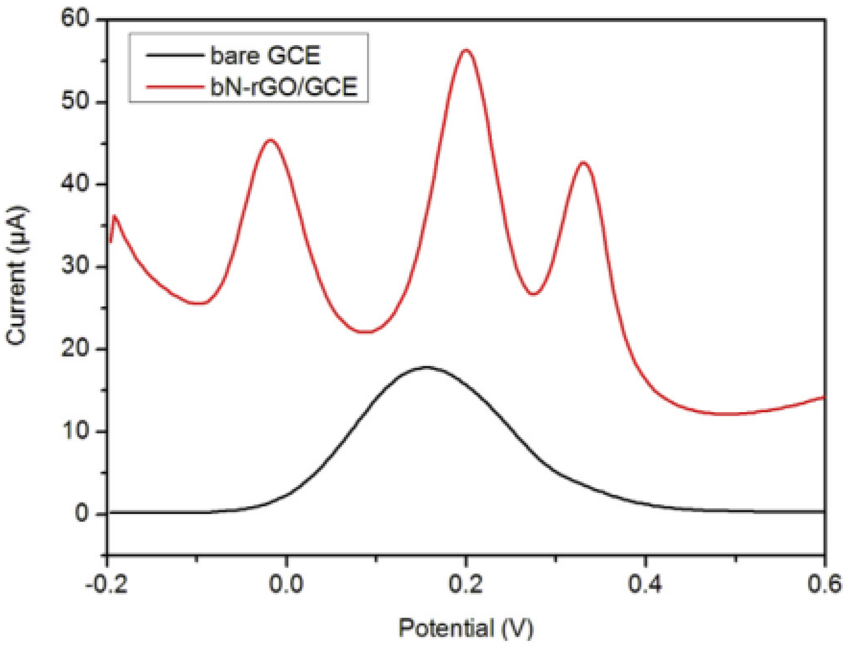
3.2. Doped Reduced Graphene Oxide

The electrochemical performance of RGO modified electrodes is usually discounted due to the graphene sheet aggregation and non-uniform dispersion issues [51]. To improve the electrochemical performance of graphene, much research effort has been devoted to the introduction of abundant defects, functional groups or doping agents [52]. Heteroatom doping of graphene has often been used as an effective strategy to create defects that not only prevent the aggregation of graphene sheet layers, but also to enhance their conductivity, resulting in an improvement in the electrochemical response [53,54]. Among the numerous potential dopants, such as boron, phosphor, sulfur, and so on, nitrogen is considered to be an excellent element for the chemical doping of carbon materials because its size is similar to a carbon atom and it can bond to carbon atoms through its five valence electrons [55]. Consequently, the N-doped reduced graphene oxide (N-rGO) is found to be the mostly used for the electrochemical detection of DA, with a significantly improved performance compared with un-doped rGOs. For this purpose, various synthesis methods of N-rGO were investigated using chemical or hydrothermal methods, and the obtained nanomaterials are applied as modified electrodes for DA sensing.
Wiench et al. [56] have investigated two different N-doping agents using urea and amitrole during hydrothermal methods to obtain various distributions of nitrogen in N-rGO. They demonstrate that urea leads to N-rGO with a pyrrolic structure where amitrole gives N-rGO with a pyridinic structure. The two nanomaterials are studied for their electrochemical sensing properties for DA detection. They show that pyrrolic N-rGO exhibits a higher electrochemical performance for DA sensing with a LOD of 335 nM and sensitivity of 3.51 μA μM−1 compared to pyridinic-N-rGO with a LOD of 500 nM and sensitivity of 2.24 μA μM−1. In addition, selectivity in the presence of AA and UA is also improved on the pyrrolic N-rGO electrode. These results show that the chemical structure of nitrogen in N-rGO has a strong influence on the electroanalytical performance, opening the possibility of investigating other types of nitrogen structure configurations in RGO such as quaternary ammonium, amine, or imidazole.
Other methods were also studied to introduce nitrogen in N-rGO. For example, Zhang et al. [57] have used a pyrolysis method of preformed nanocomposites of rGO and poly(p-phenylenediamine) by heating the nanocomposites to 800 °C in N2 atmosphere. They obtain N-rGO with a high ratio of nitrogen and show that this method has the advantage of eliminating the organic compounds by their carbonization. The proposed modified electrode GCE/N-rGO showed a sensitive and selective response for the simultaneous determination of AA, DA, and UA with three well-separated peaks ( Δ AA-DA = 220 mV, Δ DA-UA = 130 mV) (Figure 4). This was explained as the successful distribution of N-containing groups in rGO leading to enhanced electron transfer ability and increased surface area for the adsorption of DA during the electrochemical oxidation process. The GCE/N-rGO sensor designed with this method has proven superior analytical performance toward the simultaneous detection of AA, DA, and UA compared to other reported sensors based on N-rGO (Table 1).
Other N-rGO synthesis methods have also been developed and studied for DA electrochemical sensing. Soni et al. [58] have recently developed a simple, cost-effective and low-temperature synthesis route to N-rGO using a photocatalytic approach. For the photocatalytic synthesis of N-rGO, aqueous GO was mixed with ammonia solution which is used as a precursor of an N dopant, then the mixture was stirred followed by UV illumination for 60 min to achieve the photo-catalytic reduction of RGO. This new approach does not require high temperatures which are responsible for graphene sheet aggregation. The as-synthesized N-rGO coated on GCE showed a rapid electron transfer and high activity towards the oxidation of DA as compared with rGO modified GCE. The DPV measurements demonstrated a wider linear range (from 100 to 3000 μM) and a lower limit of detection of 57 nM.
The structuring of N-rGO was also demonstrated to improve electroanalytical properties. Feng et al. [59], synthesized a holey nitrogen-doped graphene aerogel (HNGA) and applied it to the electrochemical determination of ascorbic acid (AA), dopamine (DA), and uric acid (UA). The holey graphene hydrogel was synthesized by a hydrothermal reaction in the presence of H2O2, which was subsequently lyophilized and further annealed in a mixture of ammonia and argon gases to obtain HNGA. This study demonstrated that 800 °C is the optimal annealing condition and gives a high content of pyridinic N, which is known to have higher electrocatalytic activity for the detection of AA, DA, and UA. Thus, a HNGA modified electrode showed an excellent electrochemical response to the three substances detected individually, as well as in a mixture without mutual interference (Figure 5). The fast heterogeneous electron-transfer dynamics of AA, DA, and UA on HNGA/GCE has been explained by the special three-dimensional porous structure of HNGA that enables the analyte to spread to stacked graphene layers. The nitrogen doping improves the free carrier density of graphene, and then enhances its conductivity. Moreover, the proposed sensor has also been used with urine sample analysis, and satisfactory results were obtained.
Other structures of graphene have been developed for electrochemical detection of DA. Jiang et al. [60] have prepared, via a hydrothermal reaction and a freeze-drying treatment, a three-dimensional nitrogen-doped graphene (3D-NG) network. In this work, the authors have used a valid approach to assemble 2D graphene sheets into three-dimensional (3D) architectures in order to avoidthe strong π−π stacking interaction between 2D graphene sheets that cause agglomeration and hinder the utilization of the graphene’s outstanding performances. The 3D-G was then doped by nitrogen (3D-NG) and used to construct a quadruplet electrochemical sensor for the simultaneous detection of ascorbic acid (AA), dopamine (DA), uric acid (UA), and acetaminophen (AP). The authors demonstrated that synthesized 3D graphene networks create a reticular-like porous structure that produces multidimensional electron transfer pathways that provide outstanding electrical conductivity and solve the problem of agglomeration and the strong π−π stacking obtained in 2D graphene sheets. In addition, taking advantage of the synergistic effect of the porous framework and nitrogen doping, 3D-NG-based electrodes showed well-resolved oxidation peaks and enhanced response currents of AA, DA, UA, and AP. The 3D-NG-based electrodes showed enhanced electrocatalytic activities for the individual and simultaneous determination of AA, DA, UA, and AP with high sensitivity, excellent selectivity, and favorable practicality (Table 1). Moreover, this sensitive sensing platform has been successfully used to monitor AA, DA, UA, and AP in human urine samples.

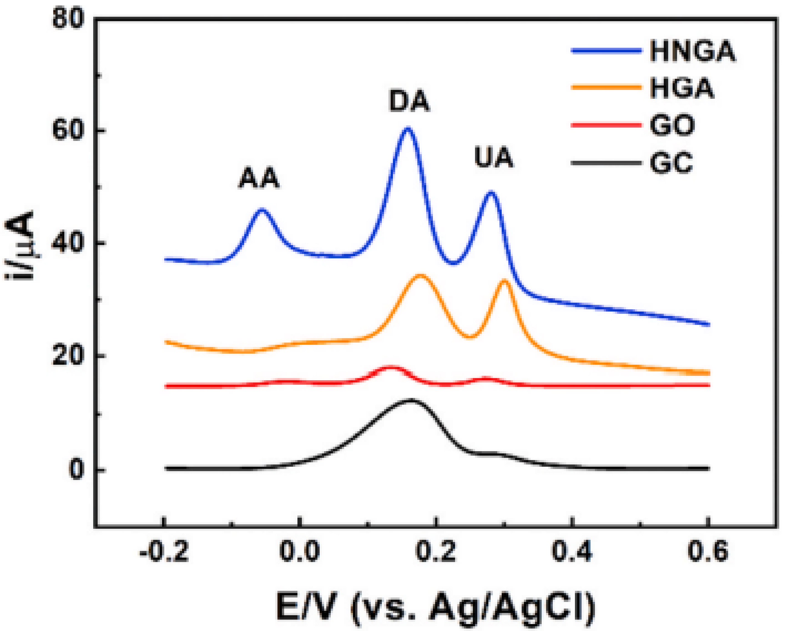
3.3. Graphene Nanocomposite

Graphene can be easily functionalized through chemical bonding or strong physical adsorption arising from the π π interaction between the functionalized molecular and graphene single layer, which makes graphene nanocomposite formation possible. In fact, a blend of graphene has demonstrated excellent physicochemical features that cannot be achieved by the respective individual constituents [10]. Lots of recent studies have demonstrated that graphene nanohybrids form stabler dispersion than graphene solution alone due to the uniform distribution and intercalation of material between graphene layers [61,62]. In addition, graphene nanocomposites not only enhance the dispersibility of graphene, but also increase its electroanalytical properties and sensing ability [63]. Among graphene nanocomposites, metallic graphene nanocomposites and polymeric graphene nanocomposites have recently gained increasing interest as they help to enhance electron transfer kinetics and electrocatalytic activity towards the oxidation of DA [1]. Below, some representative literature examples of graphene/metal nanoparticles and graphene/polymer nanocomposites for electrochemical sensing of DA are illustrated.

3.3.1. Graphene/Metal Oxide

Subramaniam and coworkers [64] have, for the first time, developed a nanocomposite with the metal oxide delafossite (CuAlO2)-encapsulated reduced graphene oxide (rGO) by a simple wet-chemical process. The as-prepared rGO/CuAlO2 nanocomposite outperformed the benchmark electrocatalyst for the detection of DA with enhanced electrochemical performance compared to pure CuAlO2 electrode. They explained this behavior by the interaction energy that occurs between graphene sheets and CuAlO2 nanostructures, that increases the conductivity and so the heterogenous electron-transfer ability. The synchronized activity of CuAlO2 and rGO is also attributed to the simple wet-chemical route increasing the porous nature of the nanocomposite. The proposed rGO/CuAlO2/GCE electrode revealed high analytical performance with a LOD in the nanomolar range and avoiding the interference of UA and AA. It also exhibited excellent linear response, good stability, reproducibility, and repeatability.
Wang et al. [65] have doped graphene oxide (GO) by Fe–Ni bimetal oxides (Fe2O3-NiO) to offer a platform called magnetic graphene nanocomposite (Fe2O3-NiO@GO) for the electrochemical detection of DA. The electrodeposition method was used for the synthesis of magnetic graphene nanocomposite based on Fe–Ni bimetal oxides (Fe2O3-NiO) and graphene oxide (GO) on a glassy carbon electrode (GCE). The authors have demonstrated the good affinity of Fe2O3-NiO@GO/GCE towards the oxidation of DA with a linear range of 10 to 1500 μM, detection limit of 0.005 μM, and sensitivity of 0.16812 μA/µΜ. They have also shown that a Fe2O3-NiO@GO/GCE platform presents comparable detection limit values and a broader linear range of DA determination compared to other reported graphene based electrochemical sensors of DA. This result has been explained by the great catalytic capability of the carbon nanostructures and magnetic nanoparticles due to the synergistic effect between GO and Fe–Ni bimetal oxides in magnetic nanocomposite. In addition, the practical capability of Fe2O3-NiO@GO/GCE for the determination of DA has been proved in prepared real samples of human blood serum.
Another example of amplified electrocatalytic activity towards the simultaneous detection of DA and UA is demonstrated by using the nanocomposite of reduced graphene oxide nano-octahedral shaped Mn3O4 (rGO/Mn3O4) [66]. Among different combinations between graphene and Mn3O4, the molar ratio of 1/10 in a nanocomposite of rGO/Mn3O4 exhibits enhanced electrocatalytic activity in terms of peak current response and shift in potential, with well separated oxidation peaks of DA and UA. Such a nanocomposite ensured excellent catalytic activity toward the simultaneous detection of DA and UA with a large linear range and the lowest LOD. This superior catalytic activity is attributed to the synergistic effect that arises from the controlled surface structure of Mn3O4 and a large specific surface area contributed by graphene sheets, which boost the interfacial charge carrier transport considerably, and enhanced electrocatalytic oxidation of DA and UA. The proposed rGO/Mn3O4/GCE electrode revealed sound stability, acceptable reproducibility, good selectivity, and detection capability for DA and UA in pharmaceutical samples and physiological residues.
Li et al. [67] have reported manganese dioxide (MnO2) combined with nitrogen-doped graphene (NG) for the simultaneous detection of trace DA and UA. In this study, various MnO2 nanoshapes, including nanowires (MnO2NWs), nanorods (MnO2NRs), nanotubes (MnO2NTs), and nanoflowers (MnO2NFs), have been synthesized and formed a composite with NG. They have shown that MnO2NFs/NG display the most sensitive response signals for DA and UA. This nanocomposite demonstrates super sensing properties with a large linear range and low LOD. The remarkable electrocatalytic activity of the nanocomposite was attributed to the synergistic effect between MnO2NFs and NG resulting in a unique porous structure, a large electroactive area, and a low charge transfer resistance (Rct), which facilitated the electron transfer between electrode and analytes and thus boosted the electrochemical responses. In addition, the proposed MnO2NFs/NG/ GCE was successfully applied for the detection of trace of DA and UA in human serum samples.

3.3.2. Graphene/Metal Nanoparticles

Metal nanoparticles are also combined with graphene to improve their catalytic properties.
Minta et al. [68], describes nanocomposites formed with N-doped reduced graphene oxide/gold nanoparticles (N-rGO/AuNPs) as the sensing platform for the simultaneous detection of DA, AA, and UA. In this study, AuNPs were electrodeposited onto the surface of GCE/N-rGO to form GCE/N-rGO-Au. The modified electrode exhibits a suitable electrocatalytic oxidation activity towards the three analytes, showing a well-resolved anodic peaks and high peak to peak separation ( Δ AA-DA = 0.34 V and Δ DA-UA = 0.20 V). These results are explained by the fact that N-doping RGO facilitates the electrodeposition of homogeneously and well distributed AuNPs due to the high affinity of the metallic particles to the nitrogenated areas of the graphene material. Consequently, the uniform distribution of AuNPs on the surface of the N-rGO improved electrical conductivity and catalytic activity, and sensing properties towards AA, DA, and UA.

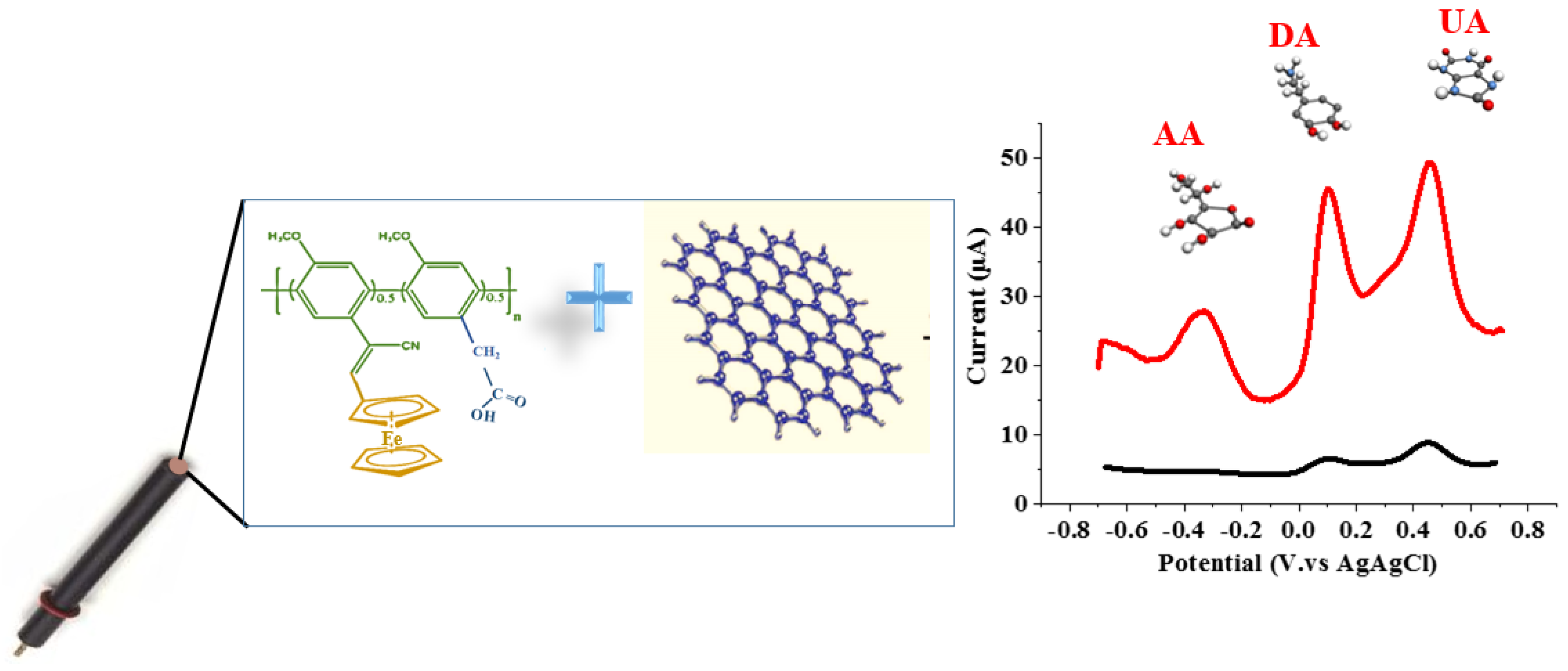
3.3.3. Graphene/Conducting Polymers

Composites formed with graphene and conducting polymers have been also developed by taking advantage of some functionality of the conducting polymers. Many studies demonstrate that the association with conducting polymers brings new properties that improve electroanalytical activity towards DA. Hsine et al. [27] explored nanohybrids based on modified poly(para-phenylene) and chemical reduced graphene oxide (Fc-ac-PPP/CRGO) as a platform for the individual and simultaneous detection of AA, DA, and UA (Figure 6). The poly(para-phenylene) carries ferrocene and carboxylic groups that bring new properties to the nanocomposite, such as redox conduction and anionic form, due to carboxylate groups. The redox system of Fc-ac-PPP polymer brings electrocatalytic activity and the anionic functions of Fc-ac-PPP that prevents non-specific interaction and overlapping. The nanocomposite is able to detect individually as well as simultaneously AA, DA and UA in a co-existence system with defined and separated redox peak oxidation. The detection is obtained in a short time with responses for AA, DA, and UA of 0.36 s, 1.03 s, and 0.32 s, respectively. The fabricated sensor is stable and reusable for detection in urine samples, which makes it a powerful platform for medical diagnosis. The authors compared the results obtained with the polymers Fc-ac-PPP [69] and demonstrate improvement attributed to the synergistic effect arising from the high surface area and the conductivity of RGO that creates active sites and improves electron transfer and the properties of the polymer.
In another study, Teng et al. [70] have used nitrogen-doped graphene (NG) and conducting polymer poly(3,4-ethylenedioxythiophene) (PEDOT) hybrids (NG/PEDOT) for the electrochemical detection of DA. The modified GCE with nanohybrid (NG/PEDOT/GCE) displayed enhanced electrocatalytic activity toward DA oxidation compared to that of PEDOT/GCEs and NG/GCEs. These responses are attributed to the blended properties of nitrogen doping that validly exploit graphene utilization by preventing graphene sheet stacking. The PEDOT component improves the hybrids’ conductivity and decreases NG stacking, resulting in a larger specific surface area of the nanohybrid. The synergistic interaction between NG and PEDOT produced a rapid electron transfer rate with electrocatalytic activity toward DA oxidation, with a LOD of 54 nM and detection range from 0.2 μM to 90 μM. The designed nanohybrids have very well-defined redox signals for DA, ascorbic acid (AA), and uric acid (UA), proving that the sensor performs highly selectively towards DA. It also maintains 95.3% of DA signal after 30 days which prove its long-term stability. Reproducibility of the sensor has been proven by the obtained 2.69% percentage of the relative standard deviation (RSD) of DA currently obtained with six independent NG/PEDOT/GCEs under the same conditions. Another group investigated the association of graphene with poly(3,4-ethylenedioxyselenophene) (PEDOS) and nitrogen-doped graphene (N-Gr) and compared the PEDOS/N-Gr composite with poly(3,4-ethylenedioxythiophene) (PEDOT)/nitrogen-doped graphene (PEDOT/N-Gr) for dopamine detection [71]. They showed that the composite formed with PEDOS has an enhanced analytical performance compared to the PEDOT composite. They explain such behavior by the presence of more electroactive sites in PEDOS leading to higher electron transfer ability.
Other conducting polymers, such as polyaniline [72], associated with thermally reduced graphene, have also been described for DA detection (Table 1). Furthermore, recent studies have associated various materials to graphene with three or four components to improve sensitivity. For example, nanocomposite associated graphene oxide and conducting polypyrrole decorated with gold nanoparticles deposited on carbon was described for the simultaneous detection of DA, UA and AA [73]. Others works described by Kumar et al. developed a composite formed with iron oxide nanorods (Fe3O4) coated polypyrrole associated with RGO for DA detection [74]. They demonstrate that the association of three components improves the electrocatalytic activity and the conductivity of the composite, leading to sensitive detection of DA. A mechanism for the catalytic electron transfer process of Fe3O4 and DA was also suggested.
Table 1. Analytical performance of some electrochemical DA sensors based on graphene and graphene nanohybrid.
SensorLinear Range µMLOD µMSensitivity A/M−1Anti-Interference MoleculesRef.
rGO-SP-FTO5 × 10−1–507 × 10−2-AA and UA[44]
3D-GNM electrode5–252.6 × 10⁻¹- [48]
SPCE/GONR5 × 10−1–3 × 1021.5 × 10−15.49 × 10⁻²UA[47]
GCE/pyrrolic-N-rGO5 × 10−1–853.35 × 10−13.51UA[56]
GCE/N-rGO103–6 × 104102-AA and UA[57]
NHGA/GCE5–502.2 × 10−1-AA and UA[59]
3D-NG/GCE1–1032.6 × 10−1-AA and UA[60]
GCE/CuAlO2-rGO9.2 × 10−2–1.6 × 10−11.5 × 10−2-AA and UA[64]
Fe2O3-NiO@GO/GCE10–15 × 1025 × 10−31.68 × 10⁻¹metabolic species[65]
MnO2NFs/NG/GCE10−1–104.3 × 10−2-UA[67]
Au/CRGO/Fc-ac-PPP10−4−1032 × 10−4-AA and UA[27]
GN/PEDOT/GCE2 × 10−1–9054 × 10−39 × 10−4AA and UA[70]
PEDOS/N-Gr/GCE8 × 10−3–806.6 × 10⁻³-AA and UA[71]
PEDOT/N-Gr/GCE4 × 10−2–701.8 × 10⁻²-
PANI/TRGO-7008 × 10−1–204.3 × 10−16.7AA and UA[72]
AuNPs@GO/PPy/CFP2 × 10−1–601.15 × 10⁻¹-AA and UA[73]
Fe3O4@PPy/rGO0–1026.3 × 10−28.35 × 10⁻¹-[74]

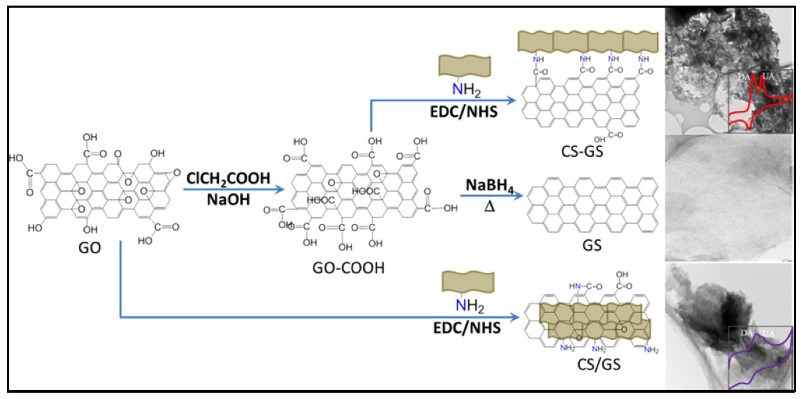
3.3.4. Graphene/Chitosan Nanocomposite

Among graphene/polymer nanocomposites, chitosan is an outstanding natural biopolymer for the development of electrochemical sensors thanks to its non-toxic, biocompatible, and biodegradable properties, and its film-forming ability [75]. In particular, the wealth of hydroxyl (OH) and amine (NH2) groups is another excellent property of chitosan that facilitates the modification of its surface with graphene sheets, nanoparticles and conductive polymers, producing composites with an enhanced electron transfer rate and a specific surface area for the development of electrochemical sensors [76]. In fact, amino groups of chitosan are highly reactive under mild conditions with carbonyl groups forming very strong imine or amide bonds, thus explaining the high stability and reproducibility of the electrochemical sensor [77]. In addition, the existence of amino groups can be protonated and create cationic chitosan which facilitates the electrostatic interaction between the chitosan matrix and the negatively charged groups of the desired biomolecule [78]. Due to its relatively poor conductivity, chitosan blended with highly conductive graphene sheet is often used in research for the design of highly sensitive electrochemical sensors. Chitosan can covalently link graphene through the amine groups or hydroxyl groups of chitosan and the carboxylic groups of graphene. It can also interact with graphene through electrostatic interaction through the amine and hydroxyl groups. The association of chitosan and graphene lead to high dispersion and improved chemical properties. In addition, due to the flexibility of chitosan membrane it can be used in association with graphene in sensors devices as biostrips or flexible patches [79]. The hydrophilic character of chitosan and presence of many functional groups also allows improving the charge transfer ability. In this context, we provide in this section an overview of recently published electrochemical DA sensors based on graphene/chitosan nanocomposite and seek to extract from each publication the advantages of chitosan over the graphene-based nanocomposite.
Abdul Kafi et al. [79] have successfully fabricated a bendable biostrip with a mesoporous-chitosan graphene oxide (m-Chit-GO) composite as the working electrode for label free electrochemical detection of dopamine (DA). In this work, the m-Chit-GO was prepared thanks to the amino and hydroxyl groups of chitosan that offered a chemical reaction with graphene oxide used as the cross linker. The m-Chit-GO based biostrip offers low LOD = 10 pM, good stability and high selectivity for DA detection against interfering agents like ascorbic acid (AA) and uric acid (UA). This analytical performance was attributed to the high surface area and the mesoporous morphology of the nanocomposite, which provided increased redox sites for DA molecules. The authors have also mentioned the advantages of the chitosan membrane that offered a biodegradable, bioresorbable and flexible m-Chit-GO electrochemical device due to the eco-friendly property and film forming ability of chitosan. Based on these properties, the ultra-thin flexible and eco-friendly m-Chit-GO electrochemical sensor is suited and ready for wearable and implantable applications where conformability on soft and curvy tissues is an important requirement.
Koyun et al. have developed a sensor based on a reduced graphene oxide/chitosan/chromium oxide nanocomposite modified glassy carbon electrode (GCE/rGO/CS/Cr2O3) for synchronous determination of Dopamine (DA), Uric acid (UA), Xanthine (XN), and Hypoxanthine (HXN) [80]. The GCE/rGO/CS/Cr2O3 nanohybrid demonstrated a synergistic effect, resulting in good conductivity and catalytic properties provided by the combination of the enlarged active surface area, the strong adsorptive capability of the nanomaterials and their specific interaction ability achieved by using a chitosan membrane. As a result, the modified electrode allowed successful detection of DA, UA, XN, and HXN in a ternary mixture with well-separated voltammetry peaks. Moreover, the applicability of the sensor for the analysis of DA, UA, XN, and HXN in fish meat has been successfully demonstrated.
Begum et al. have reported, for the first time, the enhanced amidation reaction between chitosan (CS) and graphene sheets (GS), denoted as CS−GS platform, as an electrochemical sensing matrix for the simultaneous determination of dopamine (DA) and uric acid (UA) [81]. In this work, the authors value the amide-functionalized materials as promising and precious catalysts for electrochemical sensing and catalysis. Hence, to achieve this goal, they have chosen CS which is well known for its amino and hydroxyl groups to achieve the amidation reaction with carboxylic acids of GS (Figure 7). As they have mentioned in their work, many researchers have made nanocomposites with GS and CS via amidation; however, the lower amount of the −COOH group in GO induces a lack of amide functions, resulting to a lower performance of the designed electrochemical sensor. Based on this, GO has been chemically pretreated to increase the amount of −COOH on its surface to increase the degree of amidation between GS and CS. The electrochemical results have revealed that the obtained CS−GS enhances the electrocatalytic performance of DA and UA in terms of the oxidation potential and peak current due to the higher degree of amide functionalization. Moreover, CS−GS provides selective determination toward DA and UA in various real samples.
Liu and coworkers fabricated an enzymatic biosensor based on tyrosinase, chitosan, and reduced graphene oxide (rGO) (tyrosinase/chitosan/rGO/SPCE) for the specific electrochemical detection of dopamine (DA) [82]. In this work, chitosan has enhanced the mechanical and electrical stabilities of graphene through electrostatic interaction between positively charged functions of chitosan and carboxyl functional groups in rGOs. Chitosan has also achieved the immobilization of the tyrosinase enzyme without affecting its stability and biological activity. The designed tyrosinase/chitosan/rGO/SPCE electrode showed high sensitivity and selectivity toward DA detection with a very low detection limit of 22 nM and broad linear ranges of 0.4–8 µM and 40–500 µM. In addition, the modified electrodes have achieved satisfactory results when applied to human urine samples, thus demonstrating their feasibility for analyzing DA in physiological samples.
It’s clear that the chemical properties of chitosan present various advantages for the development of sensors. However, when combined with graphene, chitosan still lacks conductivity and electroactivity. The current research in sensors aims to improve sensitivity and is oriented towards the combination of various materials with graphene where each component contributes its properties to obtain efficient electrodes for electroanalytical sensors. Thus, composite graphene and chitosan has been associated with various other polymers and nanomaterials to improve the conductivity and then electron transfer ability.
Shen et al. [83] have prepared a modified electrode with poly(3,4-ethylenedioxythiophene)-poly(styrenesulfonate) poly(PEDOT:PSS), chitosan (CS), and graphene (G) for the determination of DA (Figure 8). In this study, a screen-printed electrode (SPE) has been modified with the layer-by-layer method (LBL) with four layers. The electrode was immersed in a PEDOT: PSS suspension and then in a graphene/chitosan suspension (CS–G). The chitosan has the advantage of reducing the agglomeration between graphene sheet (G). In addition, the presence of amine groups in the chitosan structure leads to a self-assembly under electrostatic interaction between negatively charged PEDOT:PSS and positively charged CS. The obtained sensor demonstrated selective detection of DA in the presence of interfering substances such as ascorbic acid (AA) and uric acid (UA). In addition, the sensor showed high sensitivity to DA with a detection limit 0.29 µM and a linear range of DA concentrations from 0.05 to 70 µM.
As a ternary nanocomposite, RGO/CS was also associated with metal oxide semiconductor Cr2O3 for the simultaneous detection of DA, UA, xanthine (XN), and hypoxanthine (HXN), in which Cr2O3 was associated for its catalytic and electrochemical properties [80]. The sensors were made by electrodepositing chitosan as well as Cr2O3 nanoparticles. The LOD and dynamic range measured for DA are 0.65 µM and 5–160 µM, respectively.
By comparing the analytical performance of the sensors, it is easy to notice that the electrochemical sensors of DA based on graphene nanocomposites have a better analytical performance compared to DA sensors based on graphene and N-doped graphene-based materials. Moreover, it can be seen that graphene or doped graphene modified electrodes generally present a LOD that does not exceed the normal concentration range in brain fluid, which is 0.01–1 μM [84] (Table 1), which limits the use of these electrodes in vivo. The association of graphene with other nanomaterials creates a new class of nanohybrid with unique electrical and structural properties, resulting in a higher electrocatalytic activity. Thus, the graphene nanohybrid sensors have higher sensitivity for DA detection with a LOD reaching the nanomolar range. The analytical performance properties provided by some nanocomposites enable these sensors to diagnose persons affected by neurological disorders.

4. Conclusions

Electrochemical sensors of DA designed for real application need a recognized material with unique electrical and structural properties. Graphene offers these properties thanks to its atomically thin sheet-like architecture, high surface area, electron transfer capability, biocompatibility, and biomolecular affinity. Recently, the electronic and electrochemical characteristics of graphene have been improved by using new synthesized methods, by heteroatom doping, or by its incorporation with other nanomaterials to form a nanocomposite. This review focuses mainly on high-performing electrochemical sensors of DA neurotransmitter, which use graphene and its related advanced derivatives and nanocomposite-based materials as interfaces. It also highlights the recent advances of chitosan biopolymer incorporated in graphene nanocomposites and its attractive properties, offering a highly sensitive platform, as well as wearable and implantable DA electrochemical sensors that could be applied in diagnosis or the design of therapeutic agents.

Author Contributions

Study conception and design, Z.H., H.K.-Y. and R.M.; methodology, Z.H., H.K.-Y. and R.M.; data collection, Z.H.; validation, Z.H., H.K.-Y. and R.M.; formal analysis, Z.H.; investigation, Z.H.; data curation, Z.H.; writing—original draft preparation, Z.H.; writing—review and editing, H.K.-Y., R.M. and Z.H.; visualization, N.J.-R.; supervision, H.K.-Y. and R.M.; project administration, H.K.-Y., R.M. and N.J.-R. All authors have read and agreed to the published version of the manuscript.

Funding

This research received no external funding.

Conflicts of Interest

The authors declare no conflict of interest.

References

  1. Pandikumar, A.; How, G.T.S.; Peik See, T.; Saiha Omar, F.; Jayabal, S.; Zangeneh Kamali, K.; Yusoff, N.; Jamil, A.; Ramaraj, R.; Abraham John, S.; et al. Graphene and Its Nanocomposite Material Based Electrochemical Sensor Platform for Dopamine. RSC Adv. 2014, 4, 63296–63323. [Google Scholar] [CrossRef] [Green Version]
  2. Shafi, P.M.; Joseph, N.; Karthik, R.; Shim, J.-J.; Bose, A.C.; Ganesh, V. Lemon Juice-Assisted Synthesis of LaMnO3 Perovskite Nanoparticles for Electrochemical Detection of Dopamine. Microchem. J. 2021, 164, 105945. [Google Scholar] [CrossRef]
  3. Shi, Z.; Wu, X.; Zou, Z.; Yu, L.; Hu, F.; Li, Y.; Guo, C.; Li, C.M. Screen-Printed Analytical Strip Constructed with Bacteria-Templated Porous N-Doped Carbon Nanorods/Au Nanoparticles for Sensitive Electrochemical Detection of Dopamine Molecules. Biosens. Bioelectron. 2021, 186, 113303. [Google Scholar] [CrossRef] [PubMed]
  4. Li, Y.-Y.; Kang, P.; Wang, S.-Q.; Liu, Z.-G.; Li, Y.-X.; Guo, Z. Ag Nanoparticles Anchored onto Porous CuO Nanobelts for the Ultrasensitive Electrochemical Detection of Dopamine in Human Serum. Sens. Actuators B Chem. 2021, 327, 128878. [Google Scholar] [CrossRef]
  5. Wang, L.; Yang, H.; Xu, L.; Peng, C.; Song, Y. A Novel Popamine-Imprinted Chitosan/CuCo2O4@carbon/Three-Dimensional Macroporous Carbon Integrated Electrode. J. Alloys Compd. 2020, 817, 152771. [Google Scholar] [CrossRef]
  6. Ali, S.R.; Ma, Y.; Parajuli, R.R.; Balogun, Y.; Lai, W.Y.-C.; He, H. A Nonoxidative Sensor Based on a Self-Doped Polyaniline/Carbon Nanotube Composite for Sensitive and Selective Detection of the Neurotransmitter Dopamine. Anal. Chem. 2007, 79, 2583–2587. [Google Scholar] [CrossRef]
  7. Zhao, K.; Quan, X. Carbon-Based Materials for Electrochemical Reduction of CO2 to C2+ Oxygenates: Recent Progress and Remaining Challenges. ACS Catal. 2021, 11, 2076–2097. [Google Scholar] [CrossRef]
  8. Scotto, J.; Piccinini, E.; von Bilderling, C.; Coria-Oriundo, L.L.; Battaglini, F.; Knoll, W.; Marmisolle, W.A.; Azzaroni, O. Flexible Conducting Platforms Based on PEDOT and Graphite Nanosheets for Electrochemical Biosensing Applications. Appl. Surf. Sci. 2020, 525, 146440. [Google Scholar] [CrossRef]
  9. Ananda Murthy, H.C.; Gebremedhn Kelele, K.; Ravikumar, C.R.; Nagaswarupa, H.P.; Tadesse, A.; Desalegn, T. Graphene-Supported Nanomaterials as Electrochemical Sensors: A Mini Review. Results Chem. 2021, 3, 100131. [Google Scholar] [CrossRef]
  10. Lawal, A.T. Graphene-Based Nano Composites and Their Applications. A Review. Biosens. Bioelectron. 2019, 141, 111384. [Google Scholar] [CrossRef]
  11. Silva, A.O.; Cunha, R.S.; Hotza, D.; Machado, R.A.F. Chitosan as a Matrix of Nanocomposites: A Review on Nanostructures, Processes, Properties, and Applications. Carbohydr. Polym. 2021, 272, 118472. [Google Scholar] [CrossRef] [PubMed]
  12. da Silva Alves, D.C.; Healy, B.; Yu, T.; Breslin, C. Graphene-Based Materials Immobilized within Chitosan: Applications as Adsorbents for the Removal of Aquatic Pollutants. Materials 2021, 14, 3655. [Google Scholar] [CrossRef] [PubMed]
  13. Eddin, F.B.K.; Fen, Y.W. Recent Advances in Electrochemical and Optical Sensing of Dopamine. Sensors 2020, 20, 1039. [Google Scholar] [CrossRef] [PubMed] [Green Version]
  14. He, Q.; Liu, J.; Liu, X.; Li, G.; Chen, D.; Deng, P.; Liang, J. Fabrication of Amine-Modified Magnetite-Electrochemically Reduced Graphene Oxide Nanocomposite Modified Glassy Carbon Electrode for Sensitive Dopamine Determination. Nanomaterials 2018, 8, 194. [Google Scholar] [CrossRef] [Green Version]
  15. Prekodravac, J.R.; Kepić, D.P.; Colmenares, J.C.; Giannakoudakis, D.A.; Jovanović, S.P. A Comprehensive Review on Selected Graphene Synthesis Methods: From Electrochemical Exfoliation through Rapid Thermal Annealing towards Biomass Pyrolysis. J. Mater. Chem. C 2021, 9, 6722–6748. [Google Scholar] [CrossRef]
  16. Lee, J.-H.; Park, S.-J.; Choi, J.-W. Electrical Property of Graphene and Its Application to Electrochemical Biosensing. Nanomaterials 2019, 9, 297. [Google Scholar] [CrossRef] [Green Version]
  17. Liu, F.; Wang, C.; Sui, X.; Riaz, M.A.; Xu, M.; Wei, L.; Chen, Y. Synthesis of Graphene Materials by Electrochemical Exfoliation: Recent Progress and Future Potential. Carbon Energy 2019, 1, 173–199. [Google Scholar] [CrossRef] [Green Version]
  18. Olabi, A.G.; Abdelkareem, M.A.; Wilberforce, T.; Sayed, E.T. Application of Graphene in Energy Storage Device—A Review. Renew. Sustain. Energy Rev. 2021, 135, 110026. [Google Scholar] [CrossRef]
  19. Phan, D.-T.; Chung, G.-S. Effects of Rapid Thermal Annealing on Humidity Sensor Based on Graphene Oxide Thin Films. Sens. Actuators B Chem. 2015, 220, 1050–1055. [Google Scholar] [CrossRef]
  20. Bhuyan, M.S.A.; Uddin, M.N.; Islam, M.; Bipasha, F.A.; Hossain, S.S. Synthesis of Graphene. Int. Nano Lett. 2016, 6, 65–83. [Google Scholar] [CrossRef] [Green Version]
  21. Yue, H.Y.; Song, S.S.; Guo, X.R.; Huang, S.; Gao, X.; Wang, Z.; Wang, W.Q.; Zhang, H.J.; Wu, P.F. Three-Dimensional ZnO Nanosheet Spheres/Graphene Foam for Electrochemical Determination of Levodopa in the Presence of Uric Acid. J. Electroanal. Chem. 2019, 838, 142–147. [Google Scholar] [CrossRef]
  22. Yu, X.Z.; Hwang, C.G.; Jozwiak, C.M.; Köhl, A.; Schmid, A.K.; Lanzara, A. New Synthesis Method for the Growth of Epitaxial Graphene. J. Electron Spectrosc. Relat. Phenom. 2011, 184, 100–106. [Google Scholar] [CrossRef] [Green Version]
  23. Prekodravac, J.; Marković, Z.; Jovanović, S.; Budimir, M.; Peruško, D.; Holclajtner-Antunović, I.; Pavlović, V.; Syrgiannis, Z.; Bonasera, A.; Todorović-Marković, B. The Effect of Annealing Temperature and Time on Synthesis of Graphene Thin Films by Rapid Thermal Annealing. Synth. Met. 2015, 209, 461–467. [Google Scholar] [CrossRef]
  24. Wang, H.; Zhao, Z.; Liu, P.; Guo, X. Laser-Induced Graphene Based Flexible Electronic Devices. Biosensors 2022, 12, 55. [Google Scholar] [CrossRef] [PubMed]
  25. Korri-Youssoufi, H.; Zribi, B.; Miodek, A.; Haghiri-Gosnet, A.-M. Chapter 4-Carbon-Based Nanomaterials for Electrochemical DNA Sensing. In Nanotechnology and Biosensors; Nikolelis, D.P., Nikoleli, G.-P., Eds.; Advanced Nanomaterials; Elsevier: Amsterdam, The Netherlands, 2018; pp. 113–150. ISBN 978-0-12-813855-7. [Google Scholar]
  26. Brownson, D.A.C.; Kampouris, D.K.; Banks, C.E. Graphene Electrochemistry: Fundamental Concepts through to Prominent Applications. Chem. Soc. Rev. 2012, 41, 6944–6976. [Google Scholar] [CrossRef]
  27. Hsine, Z.; Bizid, S.; Mlika, R.; Sauriat-Dorizon, H.; Haj Said, A.; Korri-Youssoufi, H. Nanocomposite Based on Poly (Para-Phenylene)/Chemical Reduced Graphene Oxide as a Platform for Simultaneous Detection of Ascorbic Acid, Dopamine and Uric Acid. Sensors 2020, 20, 1256. [Google Scholar] [CrossRef] [Green Version]
  28. Chu, K.; Wang, J.; Liu, Y.; Li, Y.; Jia, C.; Zhang, H. Creating Defects on Graphene Basal-Plane toward Interface Optimization of Graphene/CuCr Composites. Carbon 2019, 143, 85–96. [Google Scholar] [CrossRef]
  29. Pavlov, S.V.; Kislenko, S.A. Graphene Electrochemistry: Edge vs. Basal Plane Sites. J. Phys. Conf. Ser. 2018, 1092, 012112. [Google Scholar] [CrossRef]
  30. Pavlov, S.V.; Nazmutdinov, R.R.; Fedorov, M.V.; Kislenko, S.A. Role of Graphene Edges in the Electron Transfer Kinetics: Insight from Theory and Molecular Modeling. J. Phys. Chem. C 2019, 123, 6627–6634. [Google Scholar] [CrossRef]
  31. Burke, M.; Larrigy, C.; Vaughan, E.; Paterakis, G.; Sygellou, L.; Quinn, A.J.; Herzog, G.; Galiotis, C.; Iacopino, D. Fabrication and Electrochemical Properties of Three-Dimensional (3D) Porous Graphitic and Graphene like Electrodes Obtained by Low-Cost Direct Laser Writing Methods. ACS Omega 2020, 5, 1540–1548. [Google Scholar] [CrossRef]
  32. Zhang, G.; Cuharuc, A.S.; Güell, A.G.; Unwin, P.R. Electrochemistry at Highly Oriented Pyrolytic Graphite (HOPG): Lower Limit for the Kinetics of Outer-Sphere Redox Processes and General Implications for Electron Transfer Models. Phys. Chem. Chem. Phys. 2015, 17, 11827–11838. [Google Scholar] [CrossRef] [Green Version]
  33. Webb, M.J.; Palmgren, P.; Pal, P.; Karis, O.; Grennberg, H. A Simple Method to Produce Almost Perfect Graphene on Highly Oriented Pyrolytic Graphite. Carbon 2011, 49, 3242–3249. [Google Scholar] [CrossRef] [Green Version]
  34. Sumdani, M.G.; Islam, M.R.; Yahaya AN, A.; Safie, S.I. Recent Advances of the Graphite Exfoliation Processes and Structural Modification of Graphene: A Review. J. Nanoparticle Res. 2021, 23, 253. [Google Scholar] [CrossRef]
  35. Ashwini, R.; Mohanta, Z.; Kumar, M.K.P.; Santosh, M.S.; Srivastava, C. Enhanced Heterogeneous Electron Transfer Kinetics in Graphene Oxide Produced from Mechanically Milled Graphite. Carbon Trends 2021, 5, 100095. [Google Scholar] [CrossRef]
  36. Kislenko, V.A.; Pavlov, S.V.; Kislenko, S.A. Influence of Defects in Graphene on Electron Transfer Kinetics: The Role of the Surface Electronic Structure. Electrochim. Acta 2020, 341, 136011. [Google Scholar] [CrossRef]
  37. Yadav, A.; Wehrhold, M.; Neubert, T.J.; Iost, R.M.; Balasubramanian, K. Fast Electron Transfer Kinetics at an Isolated Graphene Edge Nanoelectrode with and without Nanoparticles: Implications for Sensing Electroactive Species. ACS Appl. Nano Mater. 2020, 3, 11725–11735. [Google Scholar] [CrossRef]
  38. Govindasamy, M.; Wang, S.-F.; Pan, W.C.; Subramanian, B.; Ramalingam, R.J.; Al-lohedan, H. Facile Sonochemical Synthesis of Perovskite-Type SrTiO3 Nanocubes with Reduced Graphene Oxide Nanocatalyst for an Enhanced Electrochemical Detection of α-Amino Acid (Tryptophan). Ultrason. Sonochemistry 2019, 56, 193–199. [Google Scholar] [CrossRef]
  39. Roy, I.; Sarkar, G.; Mondal, S.; Rana, D.; Bhattacharyya, A.; Saha, N.R.; Adhikari, A.; Khastgir, D.; Chattopadhyay, S.; Chattopadhyay, D. Synthesis and Characterization of Graphene from Waste Dry Cell Battery for Electronic Applications. RSC Adv. 2016, 6, 10557–10564. [Google Scholar] [CrossRef]
  40. Singh, M.; Sahu, A.; Mahata, S.; Singh, P.K.; Rai, V.K.; Rai, A. Efficient Electrochemical Determination of P-Aminophenol Using a Novel Tricomponent Graphene-Based Nanocomposite. New J. Chem. 2019, 43, 14972–14978. [Google Scholar] [CrossRef]
  41. Beitollahi, H.; Safaei, M.; Tajik, S. Different Electrochemical Sensors for Determination of Dopamine as Neurotransmitter in Mixed and Clinical Samples: A Review. Anal. Bioanal. Chem. Res. 2019, 6, 81–96. [Google Scholar] [CrossRef]
  42. dos Santos, P.L.; Katic, V.; Toledo, K.C.F.; Bonacin, J.A. Photochemical One-Pot Synthesis of Reduced Graphene Oxide/Prussian Blue Nanocomposite for Simultaneous Electrochemical Detection of Ascorbic Acid, Dopamine, and Uric Acid. Sens. Actuators B Chem. 2018, 255, 2437–2447. [Google Scholar] [CrossRef]
  43. Vinodhkumar, G.; Ramya, R.; Vimalan, M.; Potheher, I.; Cyrac Peter, A. Reduced Graphene Oxide Based on Simultaneous Detection of Neurotransmitters. Prog. Chem. Biochem. Res. 2018, 1, 40–49. [Google Scholar] [CrossRef] [Green Version]
  44. Ahammad, A.J.S.; Islam, T.; Hasan, M.M.; Mozumder, M.N.I.; Karim, R.; Odhikari, N.; Pal, P.R.; Sarker, S.; Kim, D.M. Reduced Graphene Oxide Screen-Printed FTO as Highly Sensitive Electrodes for Simultaneous Determination of Dopamine and Uric Acid. J. Electrochem. Soc. 2018, 165, B174. [Google Scholar] [CrossRef]
  45. Xu, G.; Jarjes, Z.A.; Wang, H.-W.; Phillips, A.R.J.; Kilmartin, P.A.; Travas-Sejdic, J. Detection of Neurotransmitters by Three-Dimensional Laser-Scribed Graphene Grass Electrodes. ACS Appl. Mater. Interfaces 2018, 10, 42136–42145. [Google Scholar] [CrossRef]
  46. Yu, J.; Kim, T.H. A Facile Electrochemical Fabrication of Reduced Graphene Oxide-Modified Glassy Carbon Electrode for Simultaneous Detection of Dopamine, Ascorbic Acid, and Uric Acid. J. Electrochem. Sci. Technol 2017, 8, 274–281. [Google Scholar] [CrossRef] [Green Version]
  47. Mohammadi, S.; Taher, M.A.; Beitollahi, H. Treated Screen Printed Electrodes Based on Electrochemically Reduced Graphene Nanoribbons for the Sensitive Voltammetric Determination of Dopamine in the Presence of Uric Acid. Electroanalysis 2020, 32, 2036–2044. [Google Scholar] [CrossRef]
  48. Gong, J.; Tang, H.; Wang, M.; Lin, X.; Wang, K.; Liu, J. Novel Three-Dimensional Graphene Nanomesh Prepared by Facile Electro-Etching for Improved Electroanalytical Performance for Small Biomolecules. Mater. Des. 2022, 215, 110506. [Google Scholar] [CrossRef]
  49. Baig, N.; Kawde, A.-N.; Ibrahim, M. Efficient Ionic Medium Supported Reduced Graphene Oxide-Based Sensor for Selective Sensing of Dopamine. Mater. Adv. 2020, 1, 783–793. [Google Scholar] [CrossRef]
  50. Butler, D.; Moore, D.; Glavin, N.R.; Robinson, J.A.; Ebrahimi, A. Facile Post-Deposition Annealing of Graphene Ink Enables Ultrasensitive Electrochemical Detection of Dopamine. ACS Appl. Mater. Interfaces 2021, 13, 11185–11194. [Google Scholar] [CrossRef]
  51. Anuar, N.S.; Basirun, W.J.; Ladan, M.; Shalauddin, M.D.; Mehmood, M.S. Fabrication of Platinum Nitrogen-Doped Graphene Nanocomposite Modified Electrode for the Electrochemical Detection of Acetaminophen. Sens. Actuators B Chem. 2018, 266, 375–383. [Google Scholar] [CrossRef]
  52. Zhang, R.; Zhang, C.; Zheng, F.; Li, X.; Sun, C.-L.; Chen, W. Nitrogen and Sulfur Co-Doped Graphene Nanoribbons: A Novel Metal-Free Catalyst for High Performance Electrochemical Detection of 2, 4, 6-Trinitrotoluene (TNT). Carbon 2018, 126, 328–337. [Google Scholar] [CrossRef]
  53. Yang, Z.; Zheng, X.; Zheng, J. A Facile One-Step Synthesis of Fe2O3/Nitrogen-Doped Reduced Graphene Oxide Nanocomposite for Enhanced Electrochemical Determination of Dopamine. J. Alloys Compd. 2017, 709, 581–587. [Google Scholar] [CrossRef] [Green Version]
  54. Kaushal, S.; Kaur, M.; Kaur, N.; Kumari, V.; Pal Singh, P. Heteroatom-Doped Graphene as Sensing Materials: A Mini Review. RSC Adv. 2020, 10, 28608–28629. [Google Scholar] [CrossRef]
  55. Bian, S.; Shen, C.; Qian, Y.; Liu, J.; Xi, F.; Dong, X. Facile Synthesis of Sulfur-Doped Graphene Quantum Dots as Fluorescent Sensing Probes for Ag+ Ions Detection. Sens. Actuators B Chem. 2017, 242, 231–237. [Google Scholar] [CrossRef]
  56. Wiench, P.; González, Z.; Gryglewicz, S.; Menéndez, R.; Gryglewicz, G. Enhanced Performance of Pyrrolic N-Doped Reduced Graphene Oxide-Modified Glassy Carbon Electrodes for Dopamine Sensing. J. Electroanal. Chem. 2019, 852, 113547. [Google Scholar] [CrossRef]
  57. Zhang, H.; Liu, S. Electrochemical Sensors Based on Nitrogen-Doped Reduced Graphene Oxide for the Simultaneous Detection of Ascorbic Acid, Dopamine and Uric Acid. J. Alloys Compd. 2020, 842, 155873. [Google Scholar] [CrossRef]
  58. Soni, R.; Palit, K.; Soni, M.; Kumar, R.; Sharma, S.K. Highly Sensitive Electrochemical Sensing of Neurotransmitter Dopamine from Scalable UV Irradiation-Based Nitrogen-Doped Reduced Graphene Oxide-Modified Electrode. Bull. Mater. Sci. 2020, 43, 175. [Google Scholar] [CrossRef]
  59. Feng, S.; Yu, L.; Yan, M.; Ye, J.; Huang, J.; Yang, X. Holey Nitrogen-Doped Graphene Aerogel for Simultaneously Electrochemical Determination of Ascorbic Acid, Dopamine and Uric Acid. Talanta 2021, 224, 121851. [Google Scholar] [CrossRef]
  60. Jiang, J.; Ding, D.; Wang, J.; Lin, X.; Diao, G. Three-Dimensional Nitrogen-Doped Graphene-Based Metal-Free Electrochemical Sensors for Simultaneous Determination of Ascorbic Acid, Dopamine, Uric Acid, and Acetaminophen. Analyst 2021, 146, 964–970. [Google Scholar] [CrossRef]
  61. Li, G.; Xia, Y.; Tian, Y.; Wu, Y.; Liu, J.; He, Q.; Chen, D. Review—Recent Developments on Graphene-Based Electrochemical Sensors toward Nitrite. J. Electrochem. Soc. 2019, 166, B881. [Google Scholar] [CrossRef]
  62. Ravi, A.; Punith Kumar, M.K.; Rekha, M.Y.; Santosh, M.S.; Srivastava, C. Chapter One-Graphene Based Nanocomposites: Synthesis, Properties and Application as Electrochemical Sensors. In Comprehensive Analytical Chemistry; Hussain, C.M., Ed.; Analytical Applications of Graphene for Comprehensive Analytical Chemistry; Elsevier: Amsterdam, The Netherlands, 2020; Volume 91, pp. 1–20. [Google Scholar]
  63. Hira, S.A.; Yusuf, M.; Annas, D.; Nagappan, S.; Song, S.; Park, S.; Park, K.H. Recent Advances on Conducting Polymer-Supported Nanocomposites for Nonenzymatic Electrochemical Sensing. Ind. Eng. Chem. Res. 2021, 60, 13425–13437. [Google Scholar] [CrossRef]
  64. Subramaniam, T.; Kesavan, G.; Venkatachalam, G. Development of CuAlO2-Encapsulated Reduced Graphene Oxide Nanocomposites: An Efficient and Selective Electrocatalyst for Detection of Neurodegenerative Disorders. ACS Appl. Bio. Mater. 2020, 3, 7769–7778. [Google Scholar] [CrossRef]
  65. Wang, W.; Wei, F.; Han, B. Preparation of Electrochemical Sensor Based on Magnetic Graphene Nanocomposite for Determination of Dopamine. Int. J. Electrochem. Sci. 2022, 17, 220232. [Google Scholar] [CrossRef]
  66. Vinodhkumar, G.; Jose, S.P.; Lokeswarareddy, S.; Sekar, C.; Potheher, I.V.; Peter, A.C. Sensitivity Enhancement in RGO/Mn3O4 Hybrid Nanocomposites: A Modified Glassy Carbon Electrode for the Simultaneous Detection of Dopamine and Uric Acid. Synth. Met. 2021, 280, 116859. [Google Scholar] [CrossRef]
  67. Li, Q.; Xia, Y.; Wan, X.; Yang, S.; Cai, Z.; Ye, Y.; Li, G. Morphology-Dependent MnO2/Nitrogen-Doped Graphene Nanocomposites for Simultaneous Detection of Trace Dopamine and Uric Acid. Mater. Sci. Eng. C 2020, 109, 110615. [Google Scholar] [CrossRef]
  68. Minta, D.; González, Z.; Wiench, P.; Gryglewicz, S.; Gryglewicz, G. N-Doped Reduced Graphene Oxide/Gold Nanoparticles Composite as an Improved Sensing Platform for Simultaneous Detection of Dopamine, Ascorbic Acid, and Uric Acid. Sensors 2020, 20, 4427. [Google Scholar] [CrossRef]
  69. Hsine, Z.; Blili, S.; Milka, R.; Dorizon, H.; Said, A.H.; Korri-Youssoufi, H. Sensor Based on Redox Conjugated Poly(Para-Phenylene) for the Simultaneous Detection of Dopamine, Ascorbic Acid, and Uric Acid in Human Serum Sample. Anal. Bioanal. Chem. 2020, 412, 4433–4446. [Google Scholar] [CrossRef]
  70. Teng, H.; Song, J.; Xu, G.; Gao, F.; Luo, X. Nitrogen-Doped Graphene and Conducting Polymer PEDOT Hybrids for Flexible Supercapacitor and Electrochemical Sensor. Electrochim. Acta 2020, 355, 136772. [Google Scholar] [CrossRef]
  71. Kadir, A.; Jamal, R.; Abdiryim, T.; Sawut, N.; Che, Y.; Helil, Z.; Zhang, H. Electrochemical Sensor Formed from Poly(3,4-Ethylenedioxyselenophene) and Nitrogen-Doped Graphene Composite for Dopamine Detection. RSC Adv. 2021, 11, 37544–37551. [Google Scholar] [CrossRef]
  72. Minta, D.; González, Z.; Melendi-Espina, S.; Gryglewicz, G. Highly Efficient and Stable PANI/TRGO Nanocomposites as Active Materials for Electrochemical Detection of Dopamine. Surf. Interfaces 2022, 28, 101606. [Google Scholar] [CrossRef]
  73. Tan, C.; Zhao, J.; Sun, P.; Zheng, W.; Cui, G. Gold Nanoparticle Decorated Polypyrrole/Graphene Oxide Nanosheets as a Modified Electrode for Simultaneous Determination of Ascorbic Acid, Dopamine and Uric Acid. New J. Chem. 2020, 44, 4916–4926. [Google Scholar] [CrossRef]
  74. Santhosh Kumar, R.; Govindan, K.; Ramakrishnan, S.; Kim, A.R.; Kim, J.-S.; Yoo, D.J. Fe3O4 Nanorods Decorated on Polypyrrole/Reduced Graphene Oxide for Electrochemical Detection of Dopamine and Photocatalytic Degradation of Acetaminophen. Appl. Surf. Sci. 2021, 556, 149765. [Google Scholar] [CrossRef]
  75. El Khamlichi, R.; Bouchta, D.; Ben Atia, M.; Choukairi, M.; Khalid, R.T.; Raissouni, I.; Tazi, S.; Mohammadi, A.; Soussi, A.; Draoui, K.; et al. A Novel Carbon/Chitosan Paste Electrode for Electrochemical Detection of Normetanephrine in the Urine. J. Solid State Electrochem. 2018, 22, 1983–1994. [Google Scholar] [CrossRef]
  76. Annu; Raja, A.N. Recent Development in Chitosan-Based Electrochemical Sensors and Its Sensing Application. Int. J. Biol. Macromol. 2020, 164, 4231–4244. [Google Scholar] [CrossRef]
  77. Selvarajan, S.; Suganthi, A.; Rajarajan, M. A Facile Approach to Synthesis of Mesoporous SnO2/Chitosan Nanocomposite Modified Electrode for Simultaneous Determination of Ascorbic Acid, Dopamine and Uric Acid. Surf. Interfaces 2017, 7, 146–156. [Google Scholar] [CrossRef]
  78. Tashkhourian, J.; Nami-Ana, S.F.; Shamsipur, M. Designing a Modified Electrode Based on Graphene Quantum Dot-Chitosan Application to Electrochemical Detection of Epinephrine. J. Mol. Liq. 2018, 266, 548–556. [Google Scholar] [CrossRef]
  79. Kafi, M.A.; Paul, A.; Vilouras, A.; Dahiya, R. Mesoporous Chitosan Based Conformable and Resorbable Biostrip for Dopamine Detection. Biosens. Bioelectron. 2020, 147, 111781. [Google Scholar] [CrossRef]
  80. Ghanbari, K.; Nejabati, F. Ternary Nanocomposite-Based Reduced Graphene Oxide/Chitosan/Cr2O3 for the Simultaneous Determination of Dopamine, Uric Acid, Xanthine, and Hypoxanthine in Fish Meat. Anal. Methods 2020, 12, 1650–1661. [Google Scholar] [CrossRef]
  81. New Approach for Porous Chitosan–Graphene Matrix Preparation through Enhanced Amidation for Synergic Detection of Dopamine and Uric Acid|ACS Omega. Available online: https://pubs.acs.org/doi/10.1021/acsomega.7b00331 (accessed on 29 December 2021).
  82. Liu, C.-Y.; Chou, Y.-C.; Tsai, J.-H.; Huang, T.-M.; Chen, J.-Z.; Yeh, Y.-C. Tyrosinase/Chitosan/Reduced Graphene Oxide Modified Screen-Printed Carbon Electrode for Sensitive and Interference-Free Detection of Dopamine. Appl. Sci. 2019, 9, 622. [Google Scholar] [CrossRef] [Green Version]
  83. Shen, X.; Ju, F.; Li, G.; Ma, L. Smartphone-Based Electrochemical Potentiostat Detection System Using PEDOT: PSS/Chitosan/Graphene Modified Screen-Printed Electrodes for Dopamine Detection. Sensors 2020, 20, 2781. [Google Scholar] [CrossRef]
  84. Yang, L.; Liu, D.; Huang, J.; You, T. Simultaneous Determination of Dopamine, Ascorbic Acid and Uric Acid at Electrochemically Reduced Graphene Oxide Modified Electrode. Sens. Actuators B Chem. 2014, 193, 166–172. [Google Scholar] [CrossRef]
Figure 1. Schematic illustration of graphene−based electrodes for electrochemical sensing of AA, DA, and UA.
Chemosensors 10 00249 g001
Figure 2. Description of the electrochemical method for measuring the dopamine redox properties, (A) cyclic voltammetry method, (B) differential pulse voltammetry, and (C) amperometry.
Chemosensors 10 00249 g002
Figure 3. Schematic outlining the fabrication and electrochemical testing process of the graphene ink DA sensor. Reproduced from ref. [50] with permission of ACS.
Chemosensors 10 00249 g003
Figure 4. Differential pulse voltammetries (DPVs) of a bare GCE and N-rGO/GCE in 0.2 M PBS at pH 7.0 in the presence of 5 mM AA, 0.5 mM DA, and 0.5 mM UA. Reproduced from ref. [57] with permission of Elsevier publisher.
Chemosensors 10 00249 g004
Figure 5. DPV curves of HNGA/GCE, HGA/GCE, GO/GCE, and bare GCE for the mixture solution of 1 mM AA, 0.06 mM DA, and 0.08 mM UA, electrolyte: 0.1 M PBS (pH = 7.4). Reproduced from ref. [59] with permission of Elsevier publisher.
Chemosensors 10 00249 g005
Figure 6. Schematic representation of the formation of CRGO/Fc-ac-PPP nanocomposite (left). DPV of the improved response of CRGO/Fc-ac-PPP nanocomposite toward AA, DA, and UA detection (red curve) compared to that obtained with Fc-ac-PPP (black curve). Reprinted from ref. [27] (MDPI publisher).
Chemosensors 10 00249 g006
Figure 7. Schematic presentation of the various way of covalent attachment of graphene oxide and chitosan. Reprinted from ref. [81] (ACS publisher).
Chemosensors 10 00249 g007
Figure 8. The process of layer-by-layer electrostatic self-assembly produced with from ref. [83] (MDPI Publisher).
Chemosensors 10 00249 g008
Publisher’s Note: MDPI stays neutral with regard to jurisdictional claims in published maps and institutional affiliations.

