Epidermoid Cyst of the Uterine Cervix, an Unusual Location: Literature Review and Case Report

 , , , , and
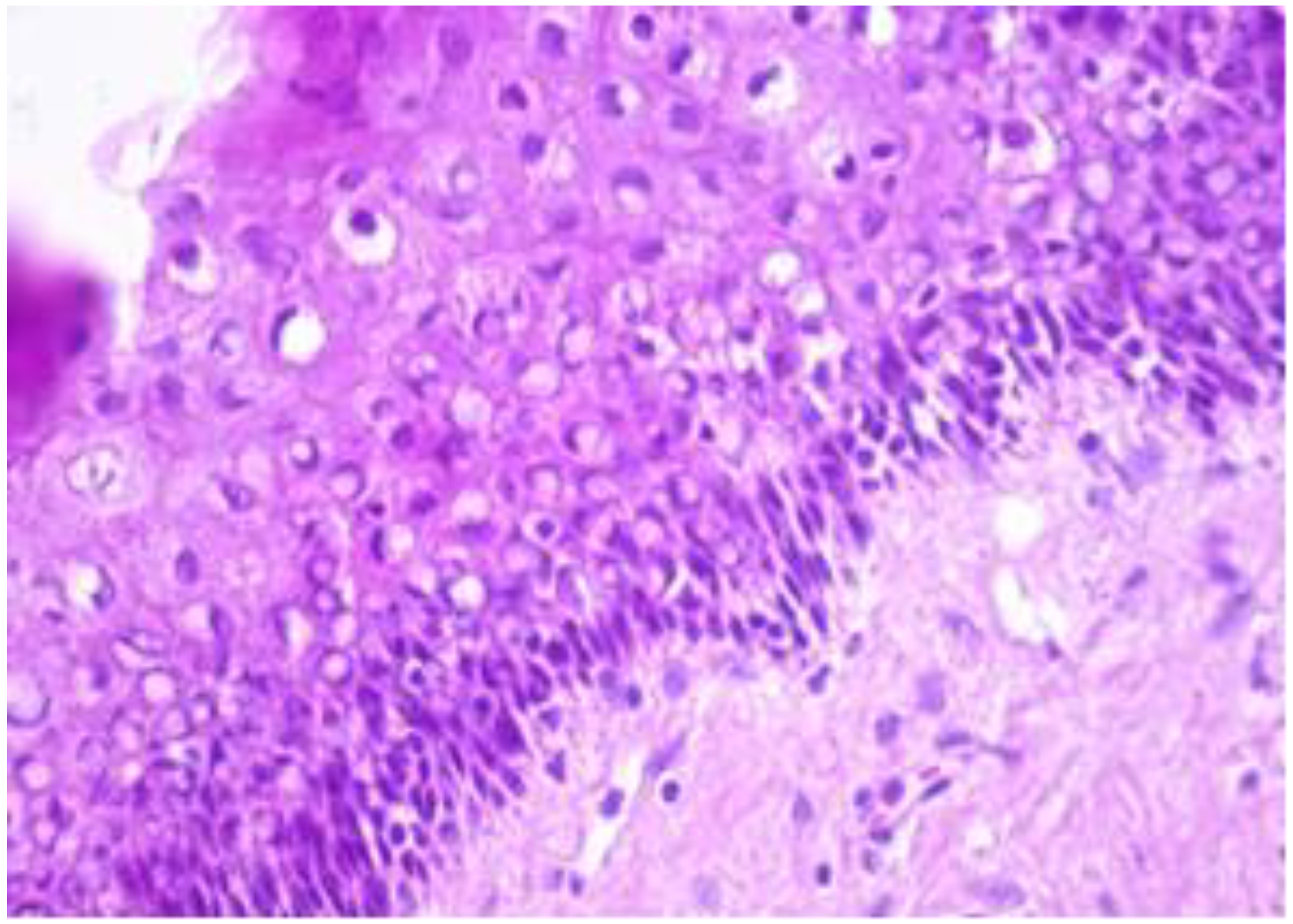
Abstract
:1. Introduction
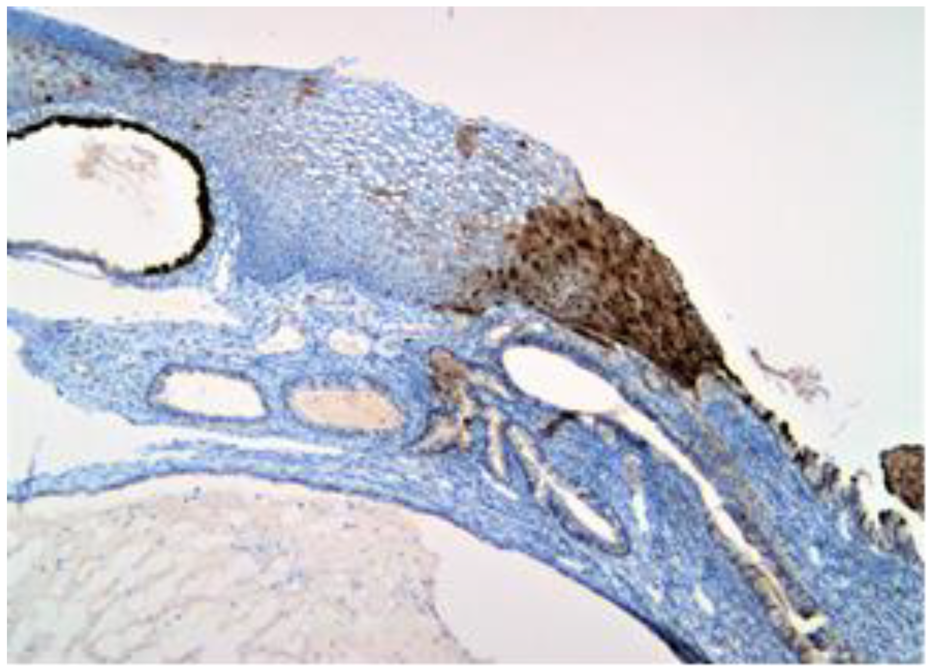
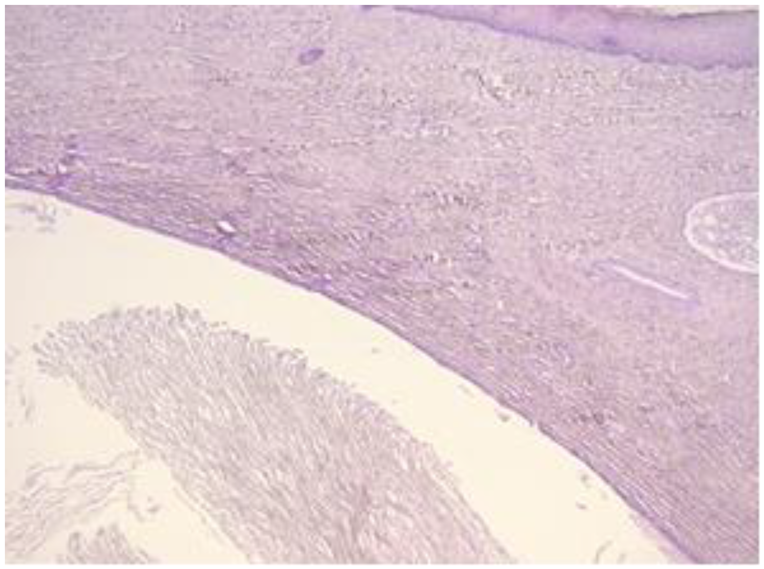
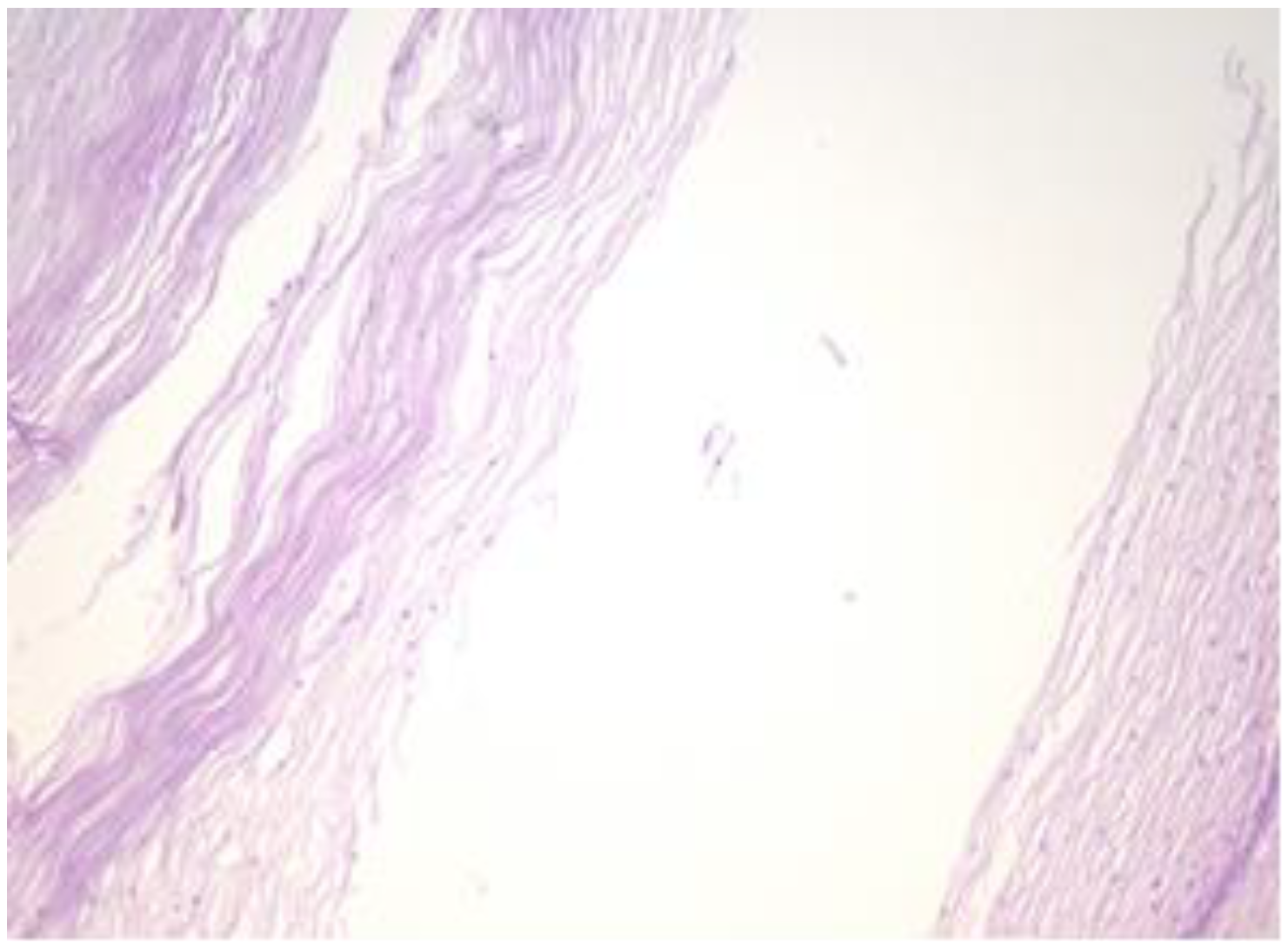
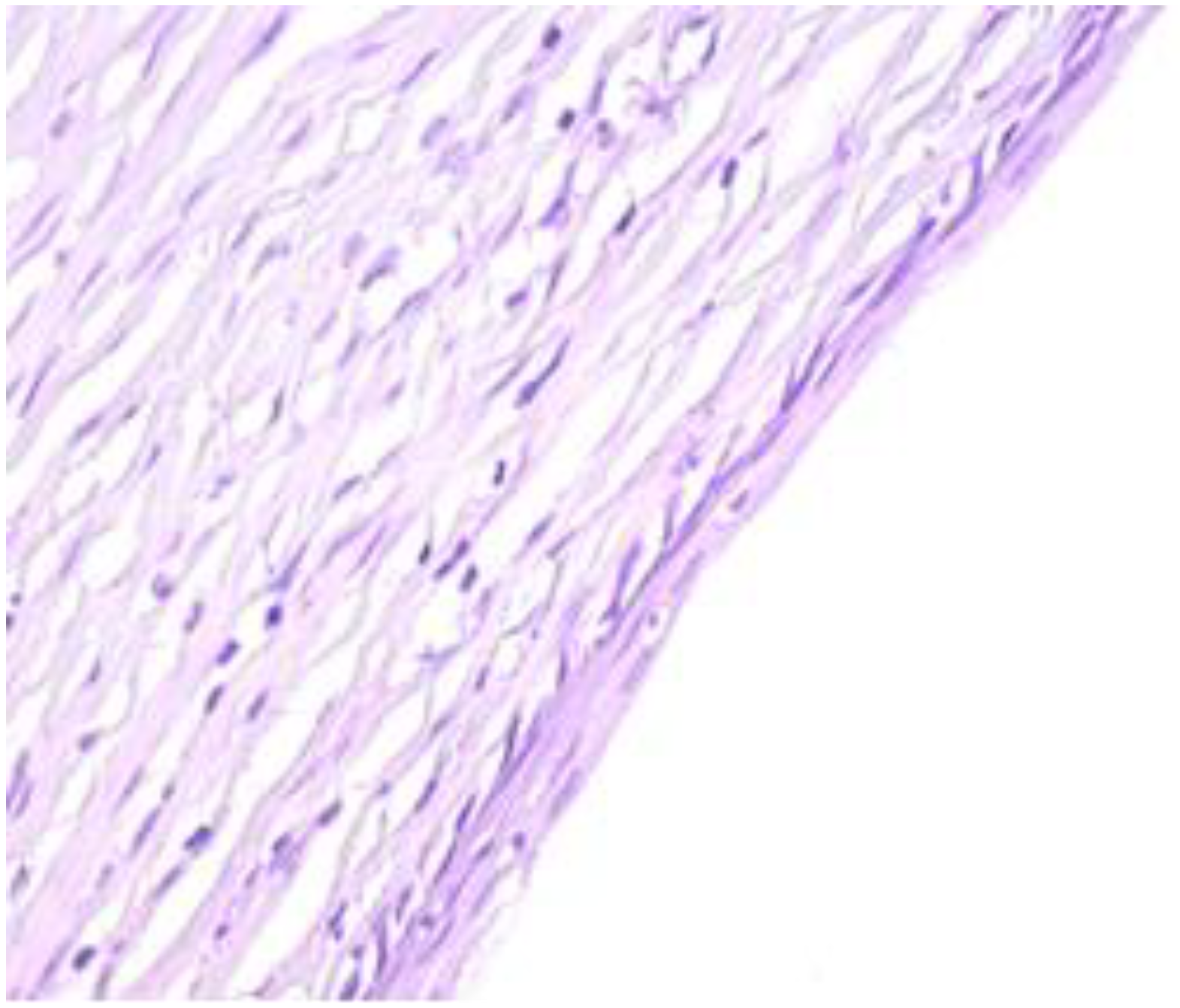
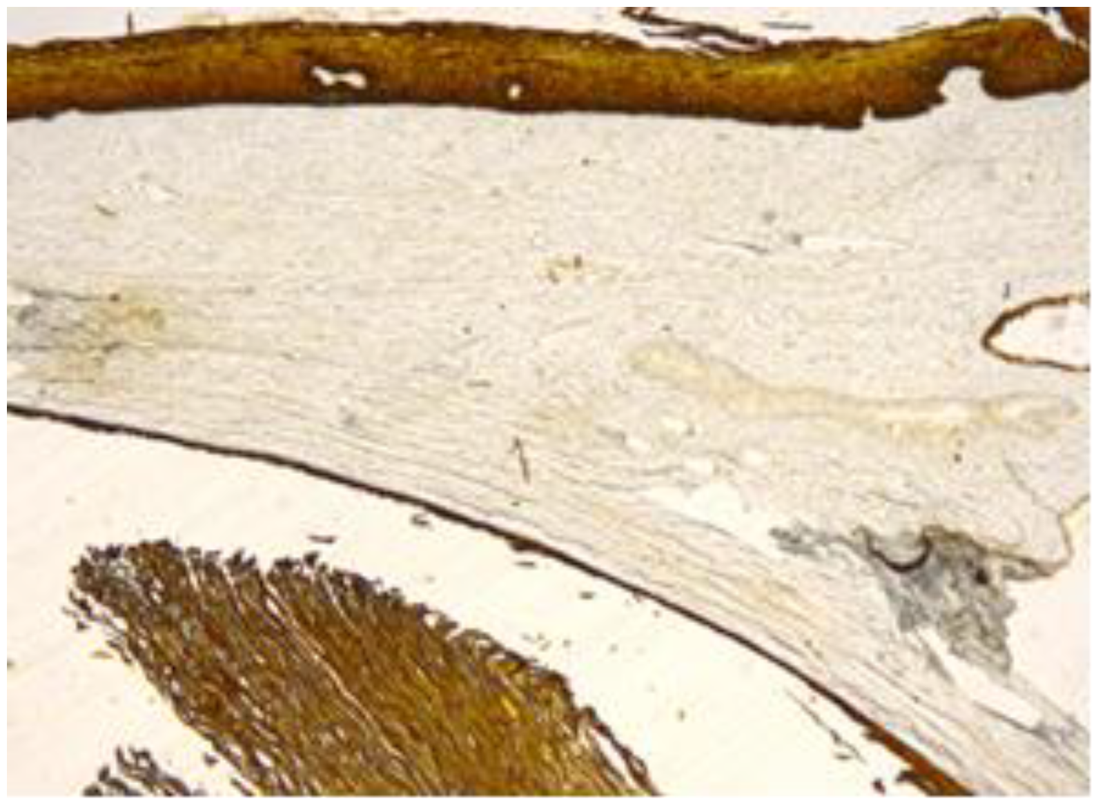
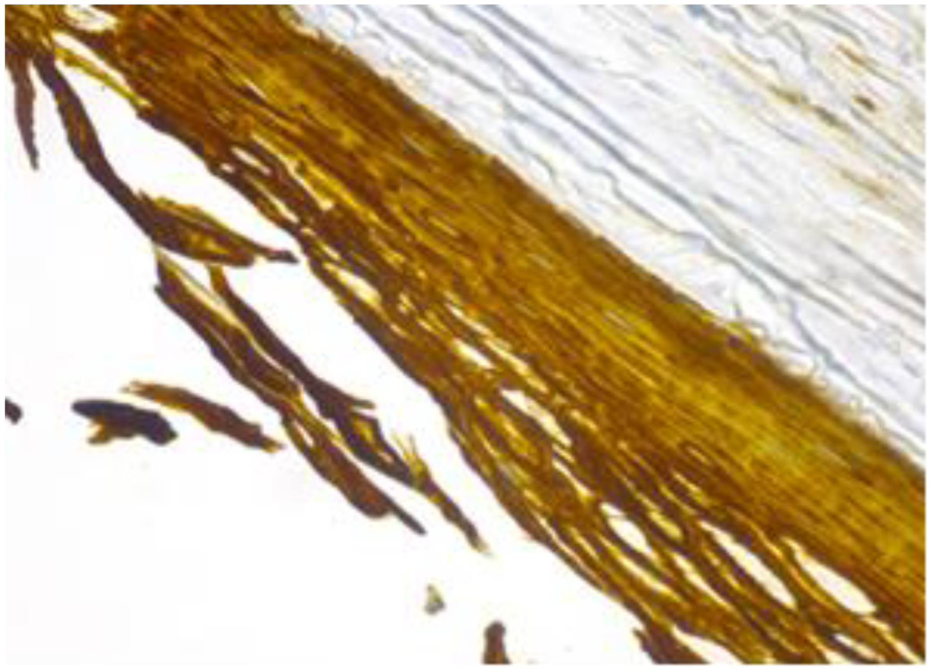
2. Case Report
3. Discussion
4. Conclusions
Author Contributions
Funding
Institutional Review Board Statement
Informed Consent Statement
Data Availability Statement
Conflicts of Interest
References
- Rosai, J.; Ackerman, L.V.; Rosai, J. Rosai and Ackerman’s Surgical Pathology, 10th ed.; Mosby: Edinburgh, UK, 2011. [Google Scholar]
- Ramakrishnaiah, S.B.; Rajput, S.S.; Gopinathan, N.S. Epidermoid Cyst of the Sole—A Case Report. J. Clin. Diagn. Res. 2016, 10, PD06–PD07. [Google Scholar] [CrossRef] [PubMed]
- Schmidt, A.; Lang, U.; Kiess, W. Epidermal cyst of the clitoris: A rare cause of clitorimegaly. Eur. J. Obstet. Gynecol. Reprod. Biol. 1999, 87, 163–165. [Google Scholar] [CrossRef] [PubMed]
- Karaman, E.; Çim, N.; Akdemir, Z.; Elçi, E.; Akdeniz, H. Giant vulvar epidermoid cyst in an adolescent girl. Case Rep. Obstet. Gynecol. 2015, 2015, 942190. [Google Scholar] [CrossRef] [PubMed] [Green Version]
- Kirkham, N. Tumors and cysts of the epidermis. In Lever’s Histopathology of the Skin, 9th ed.; Elder, D.E., Elenitsas, R., Johnson, B.L., Murphy, G.F., Jr., Eds.; Lippincott Williams & Wilkins: Philadelphia, PA, USA, 2005; pp. 805–866. [Google Scholar]
- Choi, J.E.; Kwon, I.H.; Hong Seo, S.; Kye, Y.C.; Ahn, H.H. Pathogenesis of Plantar Epidermal Cyst: Three-Dimensional Reconstruction Analysis. Ann. Dermatol. 2016, 28, 133–135. [Google Scholar] [CrossRef] [Green Version]
- Kimura, S.; Sato, N.; Shigemasu, H. Epidermal cyst of sole having a peculiar inclusion body and showing positive reaction to papillomavirus antigen. Jpn. J. Dermatol. 1986, 96, 864. [Google Scholar]
- Marquart, J.D.; Trakimas, C.A.; Sawchuk, W.S.; Nuovo, G.J.; De Villiers, E.M.; Turiansky, G.W. Human papillomavirus 57-induced extensive, recalcitrant cutaneous verrucae. J. Am. Acad. Dermatol. 2006, 55, 907–908. [Google Scholar] [CrossRef]
- Park, H.S.; Kim, W.S.; Lee, J.H.; Yang, J.M.; Lee, E.S.; Jang, K.T.; Kim, D.S.; Lee, D.Y. Association of Human Papillomavirus Infection with Palmoplantar Epidermal Cysts in Korean Patients. Acta Derm. Venereol. 2005, 85, 404–408. [Google Scholar] [CrossRef] [Green Version]
- Yoo, C.; Choi, Y.J.; Lee, K.; Kye, Y.C.; Ahn, H.H. Epidermoid Cyst in the Kidney with Nephrolithiasis: A Case Report. Korean J. Pathol. 2005, 39, 348–350. [Google Scholar]
- Pehlivan, M.; Özbay, P.O.; Temur, M.; Yılmaz, Ö.; Gümüş, Z.; Güzeld, A. Epidermal cyst in an unusual site: A case report. Int. J. Surg. Case Rep. 2015, 8, 114–116. [Google Scholar] [CrossRef] [Green Version]
- Osifo, O.D. Post genital mutilation giant clitoral epidermoid inclusion cyst in Benin City, Nigeria. J. Pediatr. Adolesc. Gynecol. 2010, 23, 336–340. [Google Scholar] [CrossRef]
- Onuigbo, W.I.B. Vulval Epidermoid Cyst in Igbos of Nigeria. Arch. Dermatol. 1976, 112, 1405–1406. [Google Scholar] [CrossRef] [PubMed]
- Lambert, B. Epidermoid cyst of the clitoris: A case report. J. Low Genit. Tract Dis. 2011, 15, 161–162. [Google Scholar] [CrossRef] [PubMed]
- Yang, W.C.; Huang, W.C.; Yang, J.M.; Lee, F.M. Successful management of a giant primary epidermoid cyst arising in the labia majora. Taiwan J. Obstet. Gynecol. 2012, 51, 112–114. [Google Scholar] [CrossRef] [PubMed] [Green Version]
- Bacon, J.B. Epidermoid cyst of cervix uteri. JAMA 1925, 84, 1632–1633. [Google Scholar] [CrossRef]
- Leininger, J.R.; Jokinen, M.P. Oviduct, uterus and vagina. In Pathology of the Fischer Rat; Boorman, G.A., Eustis, S.L., Elwell, M.R., Montgomery, C.A., MacKenzie, W.F., Eds.; Academic Press: San Diego, CA, USA, 1990; pp. 443–459. [Google Scholar]
- Greaves, P. Female genital tract. In Histopathology of Preclinical Toxicity Studies: Interpretation and Relevance in Drug Safety Evaluation, 4th ed.; Elsevier: Amsterdam, The Netherlands, 2012; pp. 667–724. [Google Scholar]
- Yoshitomi, K. Cystic dilatation of the vaginal fornix in aged female Crj: F344/Du rats. Vet. Pathol. 1990, 27, 282–284. [Google Scholar] [CrossRef] [Green Version]
- Ventana. Available online: http://www.ventana.com/product/catalog (accessed on 18 May 2019).
- Pop, O.; Bembea, M.; Pusta, C.; Pascalau, A.N. Immune response in squamous cell carcinoma (SCC): Tumor microenvironmental study. Virchows Arch. 2017, 471 (Suppl. 1), s118. [Google Scholar]
- Pusta, C.T.; Mihalache, G.; Buhas, C.; Pop, O. A rare case of cardiac fibroma in a dead truck driver. Rom. J. Leg. Med. 2015, 23, 247–250. [Google Scholar] [CrossRef]
- Anderson, J.; Genadry, R. Anatomy and Embriology. In Novak’s Gynecology, 12th ed.; Callisto Medical Press: Bucharest, Romania, 1999; pp. 94–95. [Google Scholar]
- Judea-Pusta, C.; Judea, A.; Negrutiu, B.M. Uterus didelphys at the limit between Fertility and Infertility: Case report. In Proceedings of the 35th Balkan Medical Week on Healthy Ageing—An Endless Challenge, Athens, Greece, 25–27 September 2018; Diaconu, C., Ed.; Filodiritto Publisher: Bologna, Italy, 2018; pp. 566–571. [Google Scholar]
- Egawa, K.; Egawa, N.; Honda, Y. Human papillomavirus-associated plantar epidermoid cyst related to epidermoid metaplasia of the eccrine duct epithelium: A combined histological, immunohistochemical, DNA-DNA in situ hybridization and three-dimensional reconstruction analysis. Br. J. Dermatol. 2005, 152, 961–967. [Google Scholar] [CrossRef]
- Endres, L.; Uivarosan, D.; Tit, D.M.; Pop, O.; Bungau, S.; Buhas, C. Demographic and pathologic characteristics of malignant melanoma in west part of Romania. Iran. J. Public Health 2018, 47, 606–607. [Google Scholar]
- Chiang, A.J.; Chen, D.R.; Cheng, J.T.; Chang, T.H. Detection of human papillomavirus in squamous cell carcinoma arising from dermoid cysts. Taiwan J. Obstet. Gynecol. 2015, 54, 559–566. [Google Scholar] [CrossRef] [Green Version]
- Burd, E.M. Human Papillomavirus and Cervical Cancer. Clin. Microbiol. Rev. 2003, 16, 1–17. [Google Scholar] [CrossRef] [PubMed] [Green Version]
- Kitasato, H.; Egawa, K.; Honda, Y.; Ono, T.; Mizushima, Y.; Kawai, S. A putative human papillomavirus type 57 new subtype isolated from plantar epidermoid cysts without intracytoplasmic inclusion bodies. J. Gen. Virol. 1998, 79, 1977–1981. [Google Scholar] [CrossRef] [PubMed] [Green Version]
- Egawa, K.; Kitasato, H.; Honda, Y.; Kawai, S.; Mizushima, Y.; Ono, T. Human papillomavirus 57 identified in a plantar epidermoid cyst. Br. J. Dermatol. 1998, 138, 510–514. [Google Scholar] [CrossRef] [PubMed]
- Geißler, C.; Tahtali, A.; Diensthuber, M.; Gassner, D.; Stöver, T.; Wagenblast, J. The role of p16 expression as a predictive marker in HPV-positive oral SCCHN--a retrospective single-center study. Anticancer Res. 2013, 33, 913–916. [Google Scholar]
- Sze, S.; Richmond, I.; Bickers, A.; Saha, A. Squamous cell carcinoma arising from a vulval epidermal cyst. J. Obstet. Gynaecol. Res. 2016, 42, 1623–1626. [Google Scholar] [CrossRef]
- Cirstoiu, M.M.; Antoniac, I.; Ples, L.; Bratila, E.; Munteanu, O. Adverse reactions due to use of two intrauterine devices with different action mechanism in a rare clinical case. Mater. Plast. 2016, 53, 666–669. [Google Scholar]
- Gradinaru, S.; Stoicea, M.C.; Mocanu, L.; Antoniac, I.; Gheorghita, D.; Grigore, A.G.M. Rare Breast Carcinoma with Paradoxical Plasma Cell Immunoprofile: A Case Report. Medicina 2020, 56, 62. [Google Scholar] [CrossRef] [Green Version]
- Liau, J.L.; Altamura, D.; Ratynska, M.; Verdolini, R. Basal cell carcinoma arising from an epidermal cyst: When a cyst is not a cyst. Case Rep. Dermatol. 2015, 7, 75–78. [Google Scholar] [CrossRef]
- Cirstoiu, M.; Cirstoiu, C.; Antoniac, I.; Munteanu, O. Levonorgestrel-releasing intrauterine systems: Device design, biomaterials, mechanism of action and surgical technique. Mater. Plast. 2016, 52, 258–262. [Google Scholar]
- Endres, L.; Tit, D.M.; Bungau, S.; Cioca, G.; Abdel- Daim, M.; Buhas, C.; Pop, O.; Sava, C. Markers usefulness in the melanic metastatic celular epitops identification in the sentinel lymph node. Rev. Chim. 2018, 69, 3675–3679. [Google Scholar] [CrossRef]
- Sarbu, I.; Vassu, T.; Chifiriuc, M.C.; Bucur, M.; Stoica, I.; Stefana, P.; Rusu, E.; Moldovan, H.; Pelinescu, D. Assessment the Activity of Some Enzymes and Antibiotic Substances Sensitivity on Pathogenic Bacteria Species. Rev. Chim. 2018, 68, 3015–3021. [Google Scholar] [CrossRef]
- Prisada, R.M. Perspectives to describe surface properties of raw pharmaceutical materials. A fractal approach on the wetting of powders. Farmacia 2020, 68, 354–361. [Google Scholar] [CrossRef]
- Stolnicu, S.; Szekely, E.; Molnar, C.; Molnar, C.V.; Barsan, I.; D’Alfonso, V.; Moldovan, C.; Zheng, G.; Ronnett, B.M.; Soslow, R.A. Mature and immature solid teratomas involving uterine corpus, cervix, and ovary. Int. J. Gyn. Pathol. 2017, 36, 222–227. [Google Scholar] [CrossRef] [PubMed] [Green Version]
- Tataru, A.-L.D.; Furau, G.; Afilon, J.; Ionescu, C.; Dimitriu, M.; Bratu, O.G.; Tit, D.M.; Bungau, S.; Furau, C. The situation of cervical cancers in the context of female genital cancers clustering and burden of disease in Arad County, Romania. J. Clin. Med. 2019, 8, 96. [Google Scholar] [CrossRef] [PubMed]
- Stoicescu, M.; Bungau, S.; Tit, D.M.; Mutiu, G.; Purza, L.; Iovan, C.; Pop, O.L. Carcinogenic uterine risk of repeated abortions: Hormonal receptors tumoral expression. Rom. J. Morphol. Embriol. 2017, 58, 1429–1434. [Google Scholar]


Disclaimer/Publisher’s Note: The statements, opinions and data contained in all publications are solely those of the individual author(s) and contributor(s) and not of MDPI and/or the editor(s). MDPI and/or the editor(s) disclaim responsibility for any injury to people or property resulting from any ideas, methods, instructions or products referred to in the content. |
© 2023 by the authors. Licensee MDPI, Basel, Switzerland. This article is an open access article distributed under the terms and conditions of the Creative Commons Attribution (CC BY) license (https://creativecommons.org/licenses/by/4.0/).
Share and Cite
Buhas, C.L.; Pascalau, A.; Judea-Pusta, C.T.; Pop, O.L.; Judea, A.S.; Negrutiu, B.-M.; Marcut, L.; Buhas, B.A.; Gheorghita, D.; Bodog, A.D. Epidermoid Cyst of the Uterine Cervix, an Unusual Location: Literature Review and Case Report. Healthcare 2023, 11, 257. https://doi.org/10.3390/healthcare11020257
Buhas CL, Pascalau A, Judea-Pusta CT, Pop OL, Judea AS, Negrutiu B-M, Marcut L, Buhas BA, Gheorghita D, Bodog AD. Epidermoid Cyst of the Uterine Cervix, an Unusual Location: Literature Review and Case Report. Healthcare. 2023; 11(2):257. https://doi.org/10.3390/healthcare11020257
Chicago/Turabian StyleBuhas, Camelia Liana, Andrei Pascalau, Claudia Teodora Judea-Pusta, Ovidiu Laurean Pop, Adrian Sorin Judea, Bianca-Maria Negrutiu, Lavinia Marcut, Bogdan Adrian Buhas, Daniela Gheorghita, and Alin Danut Bodog. 2023. "Epidermoid Cyst of the Uterine Cervix, an Unusual Location: Literature Review and Case Report" Healthcare 11, no. 2: 257. https://doi.org/10.3390/healthcare11020257