
Workers with Suspected Diagnosis of Silicosis: A Case Study of Sarcoidosis Versus Siderosis
 , , and
Abstract
:1. Introduction
2. Materials and Methods
3. Results
3.1. Presentation of Cases
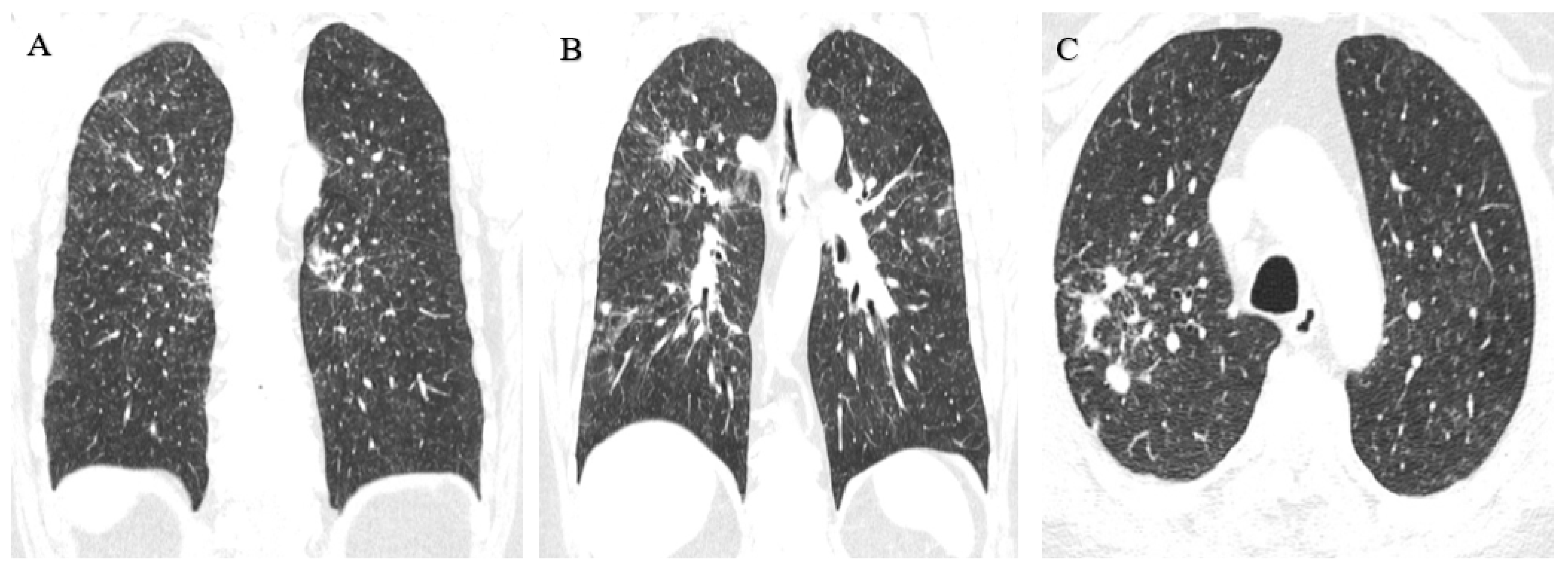
3.1.1. Case 1
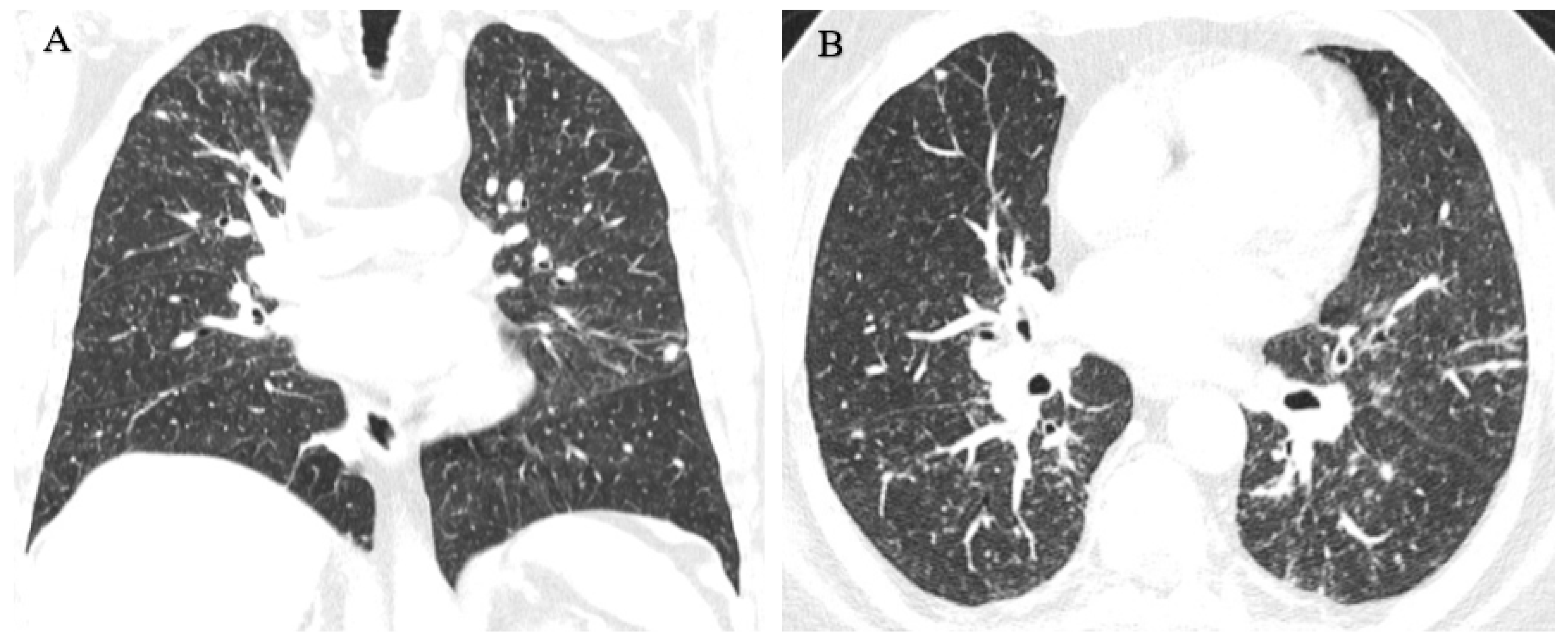
3.1.2. Case 2
4. Discussion
5. Conclusions
Author Contributions
Funding
Institutional Review Board Statement
Informed Consent Statement
Data Availability Statement
Conflicts of Interest
References
- Hoy, R.F.; Jeebhay, M.F.; Cavalin, C.; Chen, W.; Cohen, R.A.; Fireman, E.; Go, L.H.T.; León-Jiménez, A.; Menéndez-Navarro, A.; Ribeiro, M.; et al. Current global perspectives on silicosis—Convergence of old and newly emergent hazards. Respirology 2022, 27, 387–398. [Google Scholar] [CrossRef] [PubMed]
- Pavan, C.; Piane, M.D.; Gullo, M.; Filippi, F.; Fubini, B.; Hoet, P.; Horwell, C.J.; Huaux, F.; Lison, D.; Giudice, C.L.; et al. The puzzling issue of silica toxicity: Are silanols bridging the gaps between surface states and pathogenicity? Part. Fibre Toxicol. 2019, 16, 32. [Google Scholar] [CrossRef] [PubMed]
- Turci, F.; Pavan, C.; Leinardi, R.; Tomatis, M.; Pastero, L.; Garry, D.; Anguissola, S.; Lison, D.; Fubini, B. Revisiting the paradigm of silica pathogenicity with synthetic quartz crystals: The role of crystallinity and surface disorder. Part. Fibre Toxicol. 2016, 13, 32. [Google Scholar] [CrossRef] [PubMed] [Green Version]
- Barnes, H.; Goh, N.S.; Leong, T.L.; Hoy, R. Silica-associated lung disease: An old-world exposure in modern industries. Respirology 2019, 24, 1165–1175. [Google Scholar] [CrossRef] [Green Version]
- Levin, K.; McLean, C.; Hoy, R. Artificial stone-associated silicosis: Clinical-pathological-radiological correlates of disease. Respirol. Case Rep. 2019, 7, e00470. [Google Scholar] [CrossRef]
- Khan, S.N.S.; Stirling, R.G.; Mclean, C.A.; Russell, P.A.; Hoy, R.F. GM-CSF antibodies in artificial stone associated silicoproteinosis: A case report and literature review. Respirol. Case Rep. 2022, 10, e01021. [Google Scholar] [CrossRef]
- Nakládalová, M.; Stepanek, L.; Kolek, V.; Žurková, M.; Tichý, T. A case of accelerated silicosis. Occup. Med. 2018, 68, 482–484. [Google Scholar] [CrossRef] [Green Version]
- Parker, J.; Wagner, G.R. Silicosis. In International Labour Office. Encyclopedia of Occupational Health and Safety, 4th ed.; Stellman, J.M., Ed.; International Labour Office: Geneva, Switzerland, 2011; Available online: https://iloencyclopaedia.org/part-i-47946/respiratory-system/item/418-silicosis (accessed on 13 June 2023).
- International Labour Organization. Guidelines for the Use of the ILO International Classification of Radiographs of Pneumo-Conioses. Revised edition 2022; Occupational Safety and Health Series No. 22; International Labour Organization: Geneva, Switzerland, 2022. [Google Scholar]
- Şener, M.U.; Şimşek, C.; Özkara, Ş.; Evran, H.; Bursali, I.; Gökçek, A. Comparison of the International Classification of High-resolution Computed Tomography for occupational and environmental respiratory diseases with the International Labor Organization International Classification of Radiographs of Pneumoconiosis. Ind. Health 2019, 57, 495–502. [Google Scholar] [CrossRef] [Green Version]
- Nandi, S.S.; Dhatrak, S.V.; Sarkar, K. Silicosis, progressive massive fibrosis and silico-tuberculosis among workers with occupational exposure to silica dusts in sandstone mines of Rajasthan state: An urgent need for initiating national silicosis control programme in India. J. Fam. Med. Prim. Care 2021, 10, 686–691. [Google Scholar] [CrossRef]
- Jehangir, M. “Ladakh lung” a non-occupational pneu computed tomo. Int. J. Curr. Res. 2022, 14, 21506–21510. [Google Scholar] [CrossRef]
- Siribaddana, A.; Pathirathne, H.; Dassanayke, D. Three cases of progressive massive fibrosis due to silicosis in Sri Lanka. Chest 2019, 155, 226A. [Google Scholar] [CrossRef]
- Sarı, G.; Gökçek, A.; Koyuncu, A.; Şimşek, C. Computed tomography findings in progressive massive fibrosis: Analyses of 90 cases. Med. Lav. 2022, 113, e2022002. [Google Scholar] [CrossRef]
- Madan, M.; Mittal, R.; Gupta, P.; Chhabra, S.K. Eggshell calcification. Lung India 2017, 34, 200–201. [Google Scholar] [CrossRef]
- Li, T.; Yang, X.; Xu, H.; Liu, H. Early Identification, Accurate Diagnosis, and Treatment of Silicosis. Can. Respir. J. 2022, 2022, 3769134. [Google Scholar] [CrossRef]
- Young, C.; Barker, S.; Ehrlich, R.; Kistnasamy, B.; Yassi, A. Computer-aided detection for tuberculosis and silicosis in chest radiographs of gold miners of South Africa. Int. J. Tuberc. Lung Dis. 2020, 24, 444–451. [Google Scholar] [CrossRef]
- León-Jiménez, A.; Hidalgo-Molina, A.; Conde-Sánchez, M.; Pérez-Alonso, A.; Morales-Morales, J.M.; García-Gámez, E.M.; Córdoba-Doña, J.A. Artificial Stone Silicosis: Rapid progression following exposure cessation. Chest 2020, 158, 1060–1068. [Google Scholar] [CrossRef]
- Bukovitz, B.; Meiman, J.; Anderson, H.; Brooks, E.G. Silicosis: Diagnosis and Medicolegal Implications. J. Forensic Sci. 2019, 64, 1389–1398. [Google Scholar] [CrossRef]
- Sasi, A.; Ray, A.; Bhalla, A.S.; Arava, S.; Agarwal, S.; Jadon, R.S.; Vikram, N.K. Chylothorax in a case of accelerated silicosis with pulmonary silicoproteinosis: A unique association. Indian J. Occup. Environ. Med. 2020, 24, 39–41. [Google Scholar] [CrossRef]
- Djatioetomo, Y.C.E.D.; Marhana, I.A. Deadly dust: Silicotuberculosis as a downplayed and overlooked fatal disease in Indonesia. Ann. Med. Surg. 2022, 78, 103794. [Google Scholar] [CrossRef]
- Hoy, R.F.; Chambers, D.C. Silica-related diseases in the modern world. Allergy 2020, 75, 2805–2817. [Google Scholar] [CrossRef] [Green Version]
- Khan, D.; Danjuma, M.; Saddique, M.U.; Murshed, K.A.; Yassin, M.A. Adenocarcinoma de pulmón que simula tuberculosis miliar. Kompass. Neumol. 2020, 2, 76–80. [Google Scholar] [CrossRef]
- Bernardinello, N.; Petrarulo, S.; Balestro, E.; Cocconcelli, E.; Veltkamp, M.; Spagnolo, P. Pulmonary Sarcoidosis: Diagnosis and Differential Diagnosis. Diagnostics 2021, 11, 1558. [Google Scholar] [CrossRef] [PubMed]
- Mochizuka, Y.; Kono, M.; Katsumata, M.; Hirama, R.; Watanuki, M.; Oshima, Y.; Takeda, K.; Tsutsumi, A.; Miwa, H.; Miki, Y.; et al. Sarcoid-like Granulomatous Lung Disease with Subacute Progression in Silicosis. Intern. Med. 2022, 61, 395–400. [Google Scholar] [CrossRef] [PubMed]
- Oh, S.-J.; Hwang, K.-E.; Jeong, E.-T.; Kim, H.-R. A case of pulmonary siderosis misdiagnosed as pneumonia. Respir. Med. Case Rep. 2018, 25, 58–60. [Google Scholar] [CrossRef] [PubMed]
- Arun-Prasath, R.; Krishnapriya, R.; Srinivasan, R. Symptomatic Pulmonary Siderosis—A case report. IAIM 2017, 4, 191–194. [Google Scholar]
- Plaza, C.; Alvarez-Sala, W.; Villmañán, E.; Herrero, A. Neumoconiosis. Medicine 2018, 12, 3929–3935. [Google Scholar] [CrossRef]
- Ehrlich, R.; Akugizibwe, P.; Siegfried, N.; Rees, D. The association between silica exposure, silicosis and tuberculosis: A systematic review and meta-analysis. BMC Public Health 2021, 21, 953. [Google Scholar] [CrossRef]
- Mccrary, M.L.; Meland, J.; Kim, S.M.; Barrios, C. Progressive pulmonary fibrosis: Use of transbronchial biopsy to differentiate silicosis from sarcoidosis. Chest 2022, 162, A1190. [Google Scholar] [CrossRef]
- Stylemans, D.; Vekens, K.; Vincken, S.; Hanon, S.; Vanderhelst, E. A woman and her breathtaking jewelry. Acta Clin. Belg. 2021, 76, 304–306. [Google Scholar] [CrossRef]
- Tenes, J.; Laorden, D.; Cabanillas, J.; Prados, C. Enfermedad intersticial pulmonar. Medicine 2018, 12, 3909–3916. [Google Scholar] [CrossRef]
- Beijer, E.; Meek, B.; Kromhout, H.; van Es, H.W.; Seldenrijk, K.; Drent, M.; Rooijackers, J.M.; Veltkamp, M. Sarcoidosis in a patient clinically diagnosed with silicosis; is silica associated sarcoidosis a new phenotype? Respir. Med. Case Rep. 2019, 28, 100906. [Google Scholar] [CrossRef]
- Lucas, D.; Frachon, I.; Barnier, A.; Edy, P.; Tissot, V.; Dewitte, J.-D.; Lodde, B. Pulmonary siderosis in a welder, diagnosis on unusual pneumopathy. Rev. Des Mal. Respir. 2022, 39, 170–174. [Google Scholar] [CrossRef]
- Barber, C.M.; Fishwick, D.; Carder, M.; Van Tongeren, M. Epidemiology of silicosis: Reports from the SWORD scheme in the UK from 1996 to 2017. Occup. Environ. Med. 2019, 76, 17–21. [Google Scholar] [CrossRef] [Green Version]
- Akar, E.; Yildiz, T.; Atahan, S. Pulmonary siderosis cases diagnosed with minimally invasive surgical technique: A retrospective analysis of 7 cases. Ann. Thorac. Med. 2018, 13, 163–167. [Google Scholar] [CrossRef]
- Guarnieri, G.; Bizzotto, R.; Gottardo, O.; Velo, E.; Cassaro, M.; Vio, S.; Putzu, M.G.; Rossi, F.; Zuliani, P.; Liviero, F.; et al. Multiorgan accelerated silicosis misdiagnosed as sarcoidosis in two workers exposed to quartz conglomerate dust. Occup. Environ. Med. 2019, 76, 178–180. [Google Scholar] [CrossRef]
- Yavuz, M.Y.; Demiral, Y. Coexistence of Sarcoidosis and Silicosis: Case Series. Respir. Case Rep. 2022, 11, 59–64. [Google Scholar] [CrossRef]
- Spalgais, S.; Kumar, R.; Mrgipuri, P. Symptomatic pulmonary siderosis in scissors/knife sharpening worker: A case report. Indian J. Occup. Environ. Med. 2020, 24, 42–44. [Google Scholar] [CrossRef]
- Weissman, D.N. Progressive massive fibrosis: An overview of the recent literature. Pharmacol. Ther. 2022, 240, 108232. [Google Scholar] [CrossRef]
- Rees, D.; Murray, J. Silica, silicosis and tuberculosis. Occup. Health South. Afr. 2020, 26, 266–276. Available online: https://hdl.handle.net/10520/ejc-ohsa-v26-n5-a17 (accessed on 13 June 2023).
- Barac, A.; Vukicevic, T.A.; Ilic, A.D.; Rubino, S.; Zugic, V.; Stevanovic, G. Complications of chronic necrotizing pulmonary aspergillosis: Review of published case reports. Rev. Do Inst. De Med. Trop. De São Paulo 2017, 59, e19. [Google Scholar] [CrossRef] [Green Version]
- Moua, T.; Petnak, T.; Charokopos, A.; Baqir, M.; Ryu, J.H. Challenges in the Diagnosis and Management of Fibrotic Hypersensitivity Pneumonitis: A Practical Review of Current Approaches. J. Clin. Med. 2022, 11, 1473. [Google Scholar] [CrossRef] [PubMed]
- Lu, C.; Dasgupta, P.; Cameron, J.; Fritschi, L.; Baade, P. A systematic review and meta-analysis on international studies of prevalence, mortality and survival due to coal mine dust lung disease. PLoS ONE 2021, 16, e0255617. [Google Scholar] [CrossRef]
- Yao, Y.; Wei, T.; Zhang, H.; Xie, Y.; Gu, P.; Yao, Y.; Xiong, X.; Peng, Z.; Zhen, Z.; Liu, S.; et al. Characteristics of Diagnosed and Death Cases of Pneumoconiosis in Hubei Province, China, 1949–2019. Int. J. Environ. Res. Public Health 2022, 19, 15799. [Google Scholar] [CrossRef] [PubMed]
- Cheva, A.; Kilmpasani, M.; Stathakis, E.; Dimitriadis, I.; Mpikos, V.; Papaemmanouil, S. AB042. Sarcoidosis or Berylliosis? Two diseases with similar histologic findings. Ann. Transl. Med. 2016, 4, AB042. [Google Scholar] [CrossRef] [Green Version]
- Nordberg, G. Metals: Chemical properties and toxicity of chemicals. In International Labour Office. Encyclopedia of Occupational Health and Safety, 3rd ed.; Stellman, J.M., Ed.; International Labour Office: Geneva, Switzerland, 1998; Available online: https://www.ilo.org/safework/info/publications/WCMS_162039/lang--es/index.htm (accessed on 13 June 2023).


Disclaimer/Publisher’s Note: The statements, opinions and data contained in all publications are solely those of the individual author(s) and contributor(s) and not of MDPI and/or the editor(s). MDPI and/or the editor(s) disclaim responsibility for any injury to people or property resulting from any ideas, methods, instructions or products referred to in the content. |
© 2023 by the authors. Licensee MDPI, Basel, Switzerland. This article is an open access article distributed under the terms and conditions of the Creative Commons Attribution (CC BY) license (https://creativecommons.org/licenses/by/4.0/).
Share and Cite
Delgado-García, D.; Miranda-Astorga, P.; Delgado-Cano, A.; Gómez-Salgado, J.; Ruiz-Frutos, C. Workers with Suspected Diagnosis of Silicosis: A Case Study of Sarcoidosis Versus Siderosis. Healthcare 2023, 11, 1782. https://doi.org/10.3390/healthcare11121782
Delgado-García D, Miranda-Astorga P, Delgado-Cano A, Gómez-Salgado J, Ruiz-Frutos C. Workers with Suspected Diagnosis of Silicosis: A Case Study of Sarcoidosis Versus Siderosis. Healthcare. 2023; 11(12):1782. https://doi.org/10.3390/healthcare11121782
Chicago/Turabian StyleDelgado-García, Diemen, Patricio Miranda-Astorga, Ashley Delgado-Cano, Juan Gómez-Salgado, and Carlos Ruiz-Frutos. 2023. "Workers with Suspected Diagnosis of Silicosis: A Case Study of Sarcoidosis Versus Siderosis" Healthcare 11, no. 12: 1782. https://doi.org/10.3390/healthcare11121782