
Age and Gender-Specific Pattern of Cardiovascular Disease Risk Factors in Saudi Arabia: A Subgroup Analysis from the Heart Health Promotion Study


Abstract
:1. Introduction
2. Materials and Methods
2.1. Consent and Ethics
2.2. Study Setting
2.3. Study Population and Sampling Technique
2.4. Data Collection and Physical Measurements
2.5. Study Variables
2.5.1. Obesity
2.5.2. Central Obesity
2.5.3. Current Smokers
2.5.4. Physical Inactivity
2.5.5. Low Fruit and Vegetable Intake
2.5.6. Hypertension
2.5.7. Diabetes Mellitus
2.5.8. Cardiovascular Risk (CVR) Scores
2.5.9. Metabolic Syndrome (MetS)
2.5.10. Dyslipidaemia
2.6. Statistical Analysis
3. Results
4. Discussion
4.1. Strength and Limitations
4.2. Implication to Practice
- Preventive interventions should target school children and university students to stop tobacco smoking initiation and to implement active programs for tobacco smoking cessation.
- Programs for screening and treatment of dyslipidaemia, especially for men, should be an integral part of any preventive program for CVD.
- Obesity prevention and management for women should be targeted at the reproductive age group to prevent excessive weight gain during pregnancy and postpartum weight retention.
- Programs for primary and secondary prevention of CVD in old men and women should include the prevention and management of obesity in postmenopausal women.
- Saudi men and women should be at the center of preventive and curative care for CVD.
4.3. Implication to Research
- Extensive research should aim at investigating the effective means of improving the physical activity of individuals in the Saudi community as part of lifestyle modification.
- Surveillance systems for CVD and all the risk factors should be established to monitor the effectiveness of interventions.
5. Conclusions
Author Contributions
Funding
Institutional Review Board Statement
Informed Consent Statement
Data Availability Statement
Acknowledgments
Conflicts of Interest
References
- Global, regional, and national comparative risk assessment of 84 behavioural, environmental and occupational, and metabolic risks or clusters of risks for 195 countries and territories, 1990–2017: A systematic analysis for the Global Burden of Disease Study 2017. Lancet 2018, 392, 1923–1994.
- Yusuf, S.; Hawken, S.; Ounpuu, S.; Dans, T.; Avezum, A.; Lanas, F.; McQueen, M.; Budaj, A.; Pais, P.; Varigos, J.; et al. Effect of potentially modifiable risk factors associated with myocardial infarction in 52 countries (the INTERHEART study): Case-control study. Lancet 2004, 364, 937–952. [Google Scholar] [CrossRef] [PubMed]
- Vogel, B.; Acevedo, M.; Appelman, Y.; Bairey Merz, C.N.; Chieffo, A.; Figtree, G.A.; Guerrero, M.; Kunadian, V.; Lam, C.S.P.; Maas, A.H.E.M.; et al. The Lancet women and cardiovascular disease commission: Reducing the global burden by 2030. Lancet 2021, 397, 2385–2438. [Google Scholar] [CrossRef] [PubMed]
- Karlson, B.W.; Herlitz, J.; Hartford, M. Prognosis in myocardial infarction in relation to gender. Am. Heart J. 1994, 128, 477–483. [Google Scholar] [CrossRef] [PubMed]
- Honigberg, M.C.; Zekavat, S.M.; Aragam, K.; Finneran, P.; Klarin, D.; Bhatt, D.L.; Januzzi, J.L.; Scott, N.S.; Natarajan, P. Association of premature natural and surgical menopause with incident cardiovascular disease. Jama 2019, 322, 2411–2421. [Google Scholar] [CrossRef] [PubMed]
- Mendelsohn, M.E.; Karas, R.H. Molecular and cellular basis of cardiovascular gender differences. Science 2005, 308, 1583–1587. [Google Scholar] [CrossRef] [Green Version]
- Leon, L.J.; McCarthy, F.P.; Direk, K.; Gonzalez-Izquierdo, A.; Prieto-Merino, D.; Casas, J.P.; Chappell, L. Preeclampsia and cardiovascular disease in a large UK pregnancy cohort of linked electronic health records: A CALIBER study. Circulation 2019, 140, 1050–1060. [Google Scholar] [CrossRef] [PubMed] [Green Version]
- Kramer, C.K.; Campbell, S.; Retnakaran, R. Gestational diabetes and the risk of cardiovascular disease in women: A systematic review and meta-analysis. Diabetologia 2019, 62, 905–914. [Google Scholar] [CrossRef] [Green Version]
- Fayed, A.; Wahabi, H.A.; Esmaeil, S.; Elkouny, R.; Elmorshedy, H.; Bakhsh, H. Independent effect of gestational weight gain and prepregnancy obesity on pregnancy outcomes among Saudi women: A sub-cohort analysis from Riyadh mother and baby cohort study (RAHMA). PLoS ONE 2022, 17, e0262437. [Google Scholar] [CrossRef]
- Wahabi, H.A.; Fayed, A.A.; Tharkar, S.; Esmaeil, S.A.; Bakhsh, H. Postpartum weight retention and cardiometabolic risk among saudi women: A follow-up study of RAHMA subcohort. Biomed. Res. Int. 2019, 2019, 2957429. [Google Scholar] [CrossRef]
- Janssen, I.; Powell, L.H.; Crawford, S.; Lasley, B.; Sutton-Tyrrell, K. Menopause and the metabolic syndrome: The study of women’s health across the nation. Arch. Intern. Med. 2008, 168, 1568–1575. [Google Scholar] [CrossRef] [Green Version]
- Matthews, K.A.; Crawford, S.L.; Chae, C.U.; Everson-Rose, S.A.; Sowers, M.F.; Sternfeld, B.; Sutton-Tyrrell, K. Are changes in cardiovascular disease risk factors in midlife women due to chronological aging or to the menopausal transition? J. Am. Coll. Cardiol. 2009, 54, 2366–2373. [Google Scholar] [CrossRef] [PubMed] [Green Version]
- Matthews, K.A.; El Khoudary, S.R.; Brooks, M.M.; Derby, C.A.; Harlow, S.D.; Barinas-Mitchell, E.J.; Thurston, R.C. Lipid changes around the final menstrual period predict carotid subclinical disease in postmenopausal women. Stroke 2017, 48, 70–76. [Google Scholar] [CrossRef] [Green Version]
- Zhao, D.; Guallar, E.; Ouyang, P.; Subramanya, V.; Vaidya, D.; Ndumele, C.E.; Lima, J.A.; Allison, M.A.; Shah, S.J.; Bertoni, A.G.; et al. Endogenous sex hormones and incident cardiovascular disease in post-menopausal women. J. Am. Coll. Cardiol. 2018, 71, 2555–2566. [Google Scholar] [CrossRef] [PubMed]
- Tyrovolas, S.; El Bcheraoui, C.; Alghnam, S.A.; Alhabib, K.F.; Almadi MA, H.; Al-Raddadi, R.M.; Bedi, N.; El Tantawi, M.; Krish, V.S.; Memish, Z.A.; et al. The burden of disease in Saudi Arabia 1990–2017: Results from the Global Burden of Disease Study 2017. Lancet Planet Health 2020, 4, e195–e208. [Google Scholar] [CrossRef]
- Alshaikh, M.K.; Filippidis, F.T.; Baldove, J.P.; Majeed, A.; Rawaf, S. Women in Saudi Arabia and the prevalence of cardiovascular risk factors: A systematic review. J. Environ. Public. Health 2016, 2016, 7479357. [Google Scholar] [CrossRef] [PubMed] [Green Version]
- Walli-Attaei, M.; Joseph, P.; Rosengren, A.; Chow, C.K.; Rangarajan, S.; Lear, S.A.; AlHabib, K.F.; Davletov, K.; Dans, A.; Lanas, F.; et al. Variations between women and men in risk factors, treatments, cardiovascular disease incidence, and death in 27 high-income, middle-income, and low-income countries (PURE): A prospective cohort study. Lancet 2020, 396, 97–109. [Google Scholar] [CrossRef] [PubMed]
- Gupta, R.; Wood, D.A. Primary prevention of ischaemic heart disease: Populations, individuals, and health professionals. Lancet 2019, 394, 685–696. [Google Scholar] [CrossRef] [PubMed]
- Vundavalli, S.; Alfawzan, A.A.; Ramaiah, V.V.; Alruwaithi, M.; AlMogbel, A.; Mathew, M. Exposure to parental and sibling smoking and future intentions to smoke among 13–15 years old school age children in Saudi Arabia. Pan Afr. Med. J. 2021, 38, 158. [Google Scholar] [CrossRef]
- Al Shaikh, A.; Farahat, F.; Abaalkhail, B.; Kaddam, I.; Aseri, K.; Al Saleh, Y.; Al Qarni, A.; Al Shuaibi, A.; Tamimi, W. Prevalence of obesity and overweight among school-aged children in Saudi Arabia and its association with vitamin D status. Acta Biomed. 2020, 91, e2020133. [Google Scholar] [PubMed]
- Alzeidan, R.R.F.; Mandil, A.; Hersi, A.; Fayed, A. Non-communicable disease risk factors among employees and their families of a Saudi University: An epidemiological study. PLoS ONE 2016, 11, e0165036. [Google Scholar] [CrossRef] [PubMed] [Green Version]
- Disease, W.-N. Surveillance, Monitoring and Reporting. STEPwise Approach to NCD Risk Factor Surveillance (STEPS). Available online: https://www.who.int/teams/noncommunicable-diseases/surveillance/systems-tools/steps (accessed on 10 May 2023).
- WHO. Global Database on Body Mass Index: BMI Classification. 2015. Available online: http://apps.who.int/bmi/index.jsp?introPage=intro_3.html (accessed on 1 March 2023).
- WHO Expert Consultation. Waist Circumference and Waist-Hip Ratio, Report of a WHO Expert Consultation, Geneva. 2008. Available online: http://apps.who.int/iris/bitstream/10665/44583/1/9789241501491_eng (accessed on 1 March 2023).
- Eriksen, M.P.; Mackay, J.; Ross, H. The Tobacco Atlas: Citeseer; American Cancer Society: Atlanta, GA, USA, 2012. [Google Scholar]
- WHO. Physical Activity. Available online: https://www.who.int/news-room/fact-sheets/detail/physical-activity (accessed on 1 March 2023).
- World Health Organization. Diet, Nutrition, and the Prevention of Chronic Diseases: Report of a Joint WHO/FAO Expert Consultation; World Health Organization: Geneva, Switzerland, 2003.
- WHO. Global Strategy on Diet, Physical Activity and Health; World Health Organization: Geneva, Switzerland, 2004.
- Chobanian, A.V.; Bakris, G.L.; Black, H.R.; Cushman, W.C.; Green, L.A.; Izzo, J.L., Jr.; Jones, D.W.; Materson, B.J.; Oparil, S.; Wright, J.T., Jr.; et al. Seventh report of the joint national committee on prevention, detection, evaluation, and treatment of high blood pressure. Hypertension 2003, 42, 1206–1252. [Google Scholar] [CrossRef] [PubMed] [Green Version]
- WHO. HBA1C Use of Glycated Haemoglobin (HbA1c) in the Diagnosis of Diabetes Mellitus. 2011. Available online: http://www.who.int/diabetes/publications/report-hba1c_2011.pdf (accessed on 1 March 2023).
- Lee, H.M.; Le, H.; Lee, B.T.; Lopez, V.A.; Wong, N.D. Forced vital capacity paired with Framingham Risk Score for prediction of all-cause mortality. Eur. Respir. J. 2010, 36, 1002–1006. [Google Scholar] [CrossRef] [PubMed] [Green Version]
- Grundy, S.M. Third report of the National Cholesterol Education Program (NCEP) expert panel on detection, evaluation, and treatment of high blood cholesterol in adults (adult treatment panel III) final report. Circulation 2002, 106, 3143–3421. [Google Scholar]
- Trautwein, E.A.; McKay, S. The role of specific components of a plant-based diet in management of dyslipidemia and the impact on cardiovascular risk. Nutrients 2020, 12, 2671. [Google Scholar] [CrossRef] [PubMed]
- Wojda, A.; Janczy, A.; Małgorzewicz, S. Mediterranean, vegetarian and vegan diets as practical outtakes of EAS and ACC/AHA recommendations for lowering lipid profile. Acta Biochim. Pol. 2021, 68, 41–48. [Google Scholar] [CrossRef] [PubMed]
- Vega, G.L.; Grundy, S.M. Hypoalphalipoproteinemia (low high density lipoprotein) as a risk factor for coronary heart disease. Curr. Opin. Lipidol. 1996, 7, 209–216. [Google Scholar] [CrossRef]
- Springmann, M.; Wiebe, K.; Mason-D’Croz, D.; Sulser, T.B.; Rayner, M.; Scarborough, P. Health and nutritional aspects of sustainable diet strategies and their association with environmental impacts: A global modelling analysis with country-level detail. Lancet Planet. Health 2018, 2, e451–e461. [Google Scholar] [CrossRef] [Green Version]
- Alruwaili, H.; Ahmed, A.; Fatani, A.; Al-Otaibi, K.; Al-Jahdali, S.; Ali, Y.; Al-Harbi, A.; Baharoon, S.; Khan, M.; Al-Jahdali, H. Symptoms and risk for obstructive sleep apnea among sample of Saudi Arabian adults. Sleep Biol. Rhythm. 2015, 13, 332–341. [Google Scholar] [CrossRef]
- Al-Nozha, M.M.; Al-Hazzaa, H.M.; Arafah, M.R.; Al-Khadra, A.; Al-Mazrou, Y.Y.; Al-Maatouq, M.A.; Khan, N.B.; Al-Marzouki, K.; Al-Harthi, S.S.; Abdullah, M.; et al. Prevalence of physical activity and inactivity among Saudis aged 30–70 years. A population-based cross-sectional study. Saudi Med. J. 2007, 28, 559–568. [Google Scholar]
- Al-Hazzaa, H.M. Prevalence of physical inactivity in Saudi Arabia: A brief review. East. Mediterr. Health J. 2004, 10, 663–670. [Google Scholar] [CrossRef] [PubMed]
- Bellettiere, J.; LaMonte, M.J.; Evenson, K.R.; Rillamas-Sun, E.; Kerr, J.; Lee, I.M.; Di, C.; Rosenberg, D.E.; Stefanick, M.L.; Buchner, D.M.; et al. Sedentary behavior and cardiovascular disease in older women: The Objective Physical Activity and Cardiovascular Health (OPACH) study. Circulation 2019, 139, 1036–1046. [Google Scholar] [CrossRef] [PubMed]
- Elagizi, A.; Kachur, S.; Carbone, S.; Lavie, C.J.; Blair, S.N. A review of obesity, physical activity, and cardiovascular disease. Curr. Obes. Rep. 2020, 9, 571–581. [Google Scholar] [CrossRef] [PubMed]
- Khaing, W.; Vallibhakara, S.A.; Attia, J.; McEvoy, M.; Thakkinstian, A. Effects of education and income on cardiovascular outcomes: A systematic review and meta-analysis. Eur. J. Prev. Cardiol. 2017, 24, 1032–1042. [Google Scholar] [CrossRef]
- Manrique-Garcia, E.; Sidorchuk, A.; Hallqvist, J.; Moradi, T. Socioeconomic position and incidence of acute myocardial infarction: A meta-analysis. J. Epidemiol. Community Health 2011, 65, 301–309. [Google Scholar] [CrossRef] [Green Version]
- Bostock, S.; Steptoe, A. Association between low functional health literacy and mortality in older adults: Longitudinal cohort study. BMJ 2012, 344, e1602. [Google Scholar] [CrossRef] [PubMed] [Green Version]
- Nielsen, K.M.; Faergeman, O.; Foldspang, A.; Larsen, M.L. Cardiac rehabilitation: Health characteristics and socio-economic status among those who do not attend. Eur. J. Public Health 2008, 18, 479–483. [Google Scholar] [CrossRef] [Green Version]
- Gurka, M.J.; Vishnu, A.; Santen, R.J.; DeBoer, M.D. Progression of metabolic syndrome severity during the menopausal transition. J. Am. Heart Assoc. 2016, 5, e003609. [Google Scholar] [CrossRef] [PubMed] [Green Version]
- O’Kelly, A.C.; Michos, E.D.; Shufelt, C.L.; Vermunt, J.V.; Minissian, M.B.; Quesada, O.; Smith, G.N.; Rich-Edwards, J.W.; Garovic, V.D.; El Khoudary, S.R.; et al. Pregnancy and reproductive risk factors for cardiovascular disease in women. Circ. Res. 2022, 130, 652–672. [Google Scholar] [CrossRef] [PubMed]
- Nappi, R.E.; Chedraui, P.; Lambrinoudaki, I.; Simoncini, T. Menopause: A cardiometabolic transition. Lancet Diabetes Endocrinol. 2022, 10, 442–456. [Google Scholar] [CrossRef]
- Kodoth, V.; Scaccia, S.; Aggarwal, B. Adverse changes in body composition during the menopausal transition and relation to cardiovascular risk: A contemporary review. Womens Health Rep. 2022, 3, 573–581. [Google Scholar] [CrossRef] [PubMed]
- Srichaiveth, B.; Ruengsakulrach, P.; Visudharom, K.; Sanguanwong, S.; Tangsubutr, W.; Insamian, P. Impact of gender on treatment and clinical outcomes in acute ST elevation myocardial infarction patients in Thailand. J. Med. Assoc. Thai. 2007, 90 (Suppl. S1), 65–73. [Google Scholar] [PubMed]
- Anand, S.S.; Islam, S.; Rosengren, A.; Franzosi, M.G.; Steyn, K.; Yusufali, A.H.; Keltai, M.; Diaz, R.; Rangarajan, S.; Yusuf, S. Risk factors for myocardial infarction in women and men: Insights from the INTERHEART study. Eur. Heart J. 2008, 29, 932–940. [Google Scholar] [CrossRef] [PubMed] [Green Version]
- Walli-Attaei, M.; Rosengren, A.; Rangarajan, S.; Breet, Y.; Abdul-Razak, S.; Sharief, W.A.; Alhabib, K.F.; Avezum, A.; Chifamba, J.; Diaz, R.; et al. Metabolic, behavioural, and psychosocial risk factors and cardiovascular disease in women compared with men in 21 high-income, middle-income, and low-income countries: An analysis of the PURE study. Lancet 2022, 400, 811–821. [Google Scholar] [CrossRef]
- Roth, G.A.; Mensah, G.A.; Johnson, C.O.; Addolorato, G.; Ammirati, E.; Baddour, L.M.; Barengo, N.C.; Beaton, A.Z.; Benjamin, E.J.; Benziger, C.P.; et al. Global burden of cardiovascular diseases and risk factors, 1990–2019: Update from the GBD 2019 study. J. Am. Coll. Cardiol. 2020, 76, 2982–3021. [Google Scholar] [CrossRef] [PubMed]
- Naito, T.S.M.; Arai, T.; Chida, K.; Hamamatsu, A.; Harada, K.; Ozawa, T.; Murayama, S.; Muramatsu, M. Dyslipidemia is a major determinant of systemic atherosclerosis in the elderly: An autopsy study. Geriatr. Gerontol. Int. 2007, 7, 229–237. [Google Scholar] [CrossRef]
- Yusuf, S.; Joseph, P.; Rangarajan, S.; Islam, S.; Mente, A.; Hystad, P.; Brauer, M.; Kutty, V.R.; Gupta, R.; Wielgosz, A.; et al. Modifiable risk factors, cardiovascular disease, and mortality in 155,722 individuals from 21 high-income, middle-income, and low-income countries (PURE): A prospective cohort study. Lancet 2020, 395, 795–808. [Google Scholar] [CrossRef] [Green Version]
- Fanshawe, T.R.; Halliwell, W.; Lindson, N.; Aveyard, P.; Livingstone-Banks, J.; Hartmann-Boyce, J. Tobacco cessation interventions for young people. Cochrane Database Syst. Rev. 2017, 11, Cd003289. [Google Scholar] [CrossRef]
- Patnode, C.D.; Henderson, J.T.; Thompson, J.H.; Senger, C.A.; Fortmann, S.P.; Whitlock, E.P. Behavioral counseling and pharmacotherapy interventions for tobacco cessation in adults, including pregnant women: A review of reviews for the U.S. preventive services task force. Ann. Intern. Med. 2015, 163, 608–621. [Google Scholar] [CrossRef] [PubMed] [Green Version]
- Al Rasadi, K.; Almahmeed, W.; AlHabib, K.F.; Abifadel, M.; Farhan, H.A.; AlSifri, S.; Jambart, S.; Zubaid, M.; Awan, Z.; Al-Waili, K.; et al. Dyslipidaemia in the Middle East: Current status and a call for action. Atherosclerosis 2016, 252, 182–187. [Google Scholar] [CrossRef] [PubMed] [Green Version]
- Ortega, F.B.; Lavie, C.J.; Blair, S.N. Obesity and cardiovascular disease. Circ. Res. 2016, 118, 1752–1770. [Google Scholar] [CrossRef] [PubMed] [Green Version]
- Flegal, K.M.; Kit, B.K.; Orpana, H.; Graubard, B.I. Association of all-cause mortality with overweight and obesity using standard body mass index categories: A systematic review and meta-analysis. JAMA 2013, 309, 71–82. [Google Scholar] [CrossRef] [PubMed] [Green Version]
- Wahabi, H.; Fayed, A.A.; Shata, Z.; Esmaeil, S.; Alzeidan, R.; Saeed, E.; Amer, Y.; Titi, M.; Bahkali, K.; Hneiny, L. The impact of age, gender, temporality, and geographical region on the prevalence of obesity and overweight in Saudi Arabia: Scope of evidence. Healthcare 2023, 11, 1143. [Google Scholar] [CrossRef] [PubMed]
- Wahabi, H.; Esmaeil, S.; Zeidan, R.; Fayed, A. Effects of age, metabolic and socioeconomic factors on cardiovascular risk among Saudi women: A subgroup analysis from the heart health promotion study. Medicina 2023, 59, 623. [Google Scholar] [CrossRef] [PubMed]
- Hooper, L.; Martin, N.; Jimoh, O.F.; Kirk, C.; Foster, E.; Abdelhamid, A.S. Reduction in saturated fat intake for cardiovascular disease. Cochrane Database Syst. Rev. 2020, 5, Cd011737. [Google Scholar] [CrossRef] [PubMed]
- Alanazi, M.; Reddy, N.P.; Shaik, J.P.; Ajaj, S.A.; Jafari, A.A.; Saeed, H.; Khan, Z.; Khan, A. Association of BRCA2 variants with cardiovascular disease in Saudi Arabia. Genet. Mol. Res. 2014, 13, 3876–3884. [Google Scholar] [CrossRef]
- Warren, J.M.; Smith, N.; Ashwell, M. A structured literature review on the role of mindfulness, mindful eating and intuitive eating in changing eating behaviours: Effectiveness and associated potential mechanisms. Nutr. Res. Rev. 2017, 30, 272–283. [Google Scholar] [CrossRef]
- Dunn, C.; Haubenreiser, M.; Johnson, M.; Nordby, K.; Aggarwal, S.; Myer, S.; Thomas, C. Mindfulness approaches and weight loss, weight maintenance, and weight regain. Curr. Obes. Rep. 2018, 7, 37–49. [Google Scholar] [CrossRef]


| Ages Groups | |||||||||||||||||||||
|---|---|---|---|---|---|---|---|---|---|---|---|---|---|---|---|---|---|---|---|---|---|
| 18–29 Years Old | 30–39 Years Old | 40–49 Years Old | 50–59 Years Old | >60 Years Old | |||||||||||||||||
| Gender | Gender | Gender | Gender | Gender | |||||||||||||||||
| Male | Female | Male | Female | Male | Female | Male | Female | Male | Female | ||||||||||||
| N | % | N | % | N | % | N | % | N | % | N | % | N | % | N | % | N | % | N | % | ||
| Education levels | University | 225 | (54.3) | 350 | (55.4) | 234 | (84.8) | 297 | (76.7) | 147 | (84.0) | 203 | (54.9) | 136 | (81.4) | 157 | (41.9) | 91 | (73.4) | 30 | (21.0) |
| school | 189 | (45.7) | 276 | (43.7) | 42 | (15.2) | 86 | (22.2) | 27 | (15.4) | 147 | (39.7) | 30 | (18.0) | 168 | (44.8) | 29 | (23.4) | 41 | (28.7) | |
| Illiterate | 0 | (0.0) | 6 | (0.9) | 0 | (0.0) | 4 | (1.0) | 1 | (0.6) | 20 | (5.4) | 1 | (0.6) | 50 | (13.3) | 4 | (3.2) | 72 | (50.3) | |
| p-value | 0.12 | 0.02 | <0.01 | <0.01 | <0.01 | ||||||||||||||||
| Marital status | Married | 163 | (39.4) | 211 | (33.4) | 259 | (93.8) | 293 | (75.7) | 170 | (97.1) | 339 | (91.6) | 167 | (100.0) | 347 | (92.5) | 123 | (99.2) | 115 | (80.4) |
| Single | 250 | (60.4) | 404 | (63.9) | 16 | (5.8) | 70 | (18.1) | 2 | (1.1) | 10 | (2.7) | 0 | (0.0) | 2 | (0.5) | 0 | (0.0) | 3 | (2.1) | |
| Widowed and divorced | 1 | (0.2) | 17 | (2.7) | 1 | (0.4) | 24 | (6.2) | 3 | (1.7) | 21 | (5.7) | 0 | (0.0) | 26 | (6.9) | 1 | (0.8) | 25 | (17.5) | |
| p-value | <0.01 | <0.01 | 0.05 | <0.01 | <0.01 | ||||||||||||||||
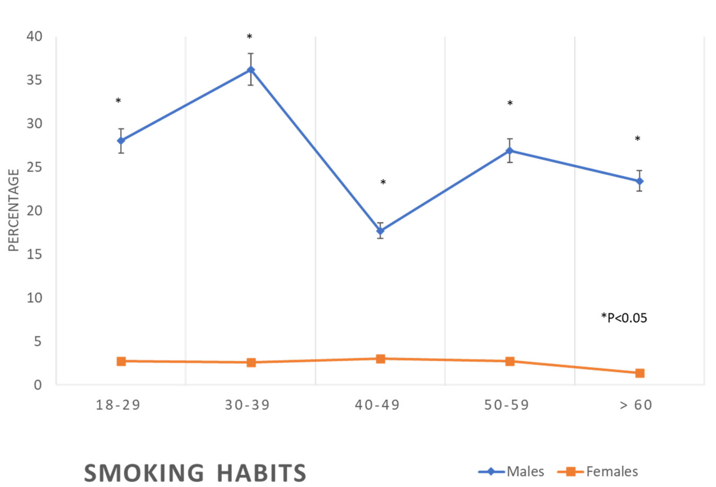
| Smoking | No | 298 | (72.0) | 615 | (97.3) | 176 | (63.8) | 377 | (97.4) | 144 | (82.3) | 359 | (97.0) | 122 | (73.1) | 365 | (97.3) | 95 | (76.6) | 141 | (98.6) |
| Yes | 116 | (28.0) | 17 | (2.7) | 100 | (36.2) | 10 | (2.6) | 31 | (17.7) | 11 | (3.0) | 45 | (26.9) | 10 | (2.7) | 29 | (23.4) | 2 | (1.4) | |
| p-value | <0.01 | <0.01 | <0.01 | <0.01 | <0.01 | ||||||||||||||||
| Dietary Habits | Poor | 375 | (90.6) | 583 | (92.2) | 233 | (84.4) | 356 | (92.0) | 142 | (81.1) | 311 | (84.1) | 137 | (82.0) | 328 | (87.5) | 98 | (79.0) | 120 | (83.9) |
| Good | 39 | (9.4) | 49 | (7.8) | 43 | (15.6) | 31 | (8.0) | 33 | (18.9) | 59 | (15.9) | 30 | (18.0) | 47 | (12.5) | 26 | (21.0) | 23 | (16.1) | |
| p-value | 0.34 | <0.01 | 0.39 | 0.09 | 0.30 | ||||||||||||||||
| Physical activity | Inactive | 240 | (58.0) | 517 | (81.8) | 177 | (64.1) | 340 | (87.9) | 125 | (71.4) | 334 | (90.3) | 117 | (70.1) | 344 | (91.7) | 91 | (73.4) | 137 | (95.8) |
| Active | 174 | (42.0) | 115 | (18.2) | 99 | (35.9) | 47 | (12.1) | 50 | (28.6) | 36 | (9.7) | 50 | (29.9) | 31 | (8.3) | 33 | (26.6) | 6 | (4.2) | |
| p-value | <0.01 | <0.01 | <0.01 | <0.01 | <0.01 | ||||||||||||||||
| Ages Groups | |||||||||||||||||||||
|---|---|---|---|---|---|---|---|---|---|---|---|---|---|---|---|---|---|---|---|---|---|
| 18–29 Years Old | 30–39 Years Old | 40–49 Years Old | 50–59 Years Old | >60 Years Old | |||||||||||||||||
| Gender | Gender | Gender | Gender | Gender | |||||||||||||||||
| Male | Female | Male | Female | Male | Female | Male | Female | Male | Female | ||||||||||||
| N | % | N | % | N | % | N | % | N | % | N | % | N | % | N | % | N | % | N | % | ||
| DM | Normal | 409 | (98.8) | 620 | (98.1) | 260 | (94.2) | 368 | (95.1) | 147 | (84.0) | 309 | (83.5) | 108 | (64.7) | 261 | (69.6) | 76 | (61.3) | 76 | (53.1) |
| Diabetes | 5 | (1.2) | 12 | (1.9) | 16 | (5.8) | 19 | (4.9) | 28 | (16.0) | 61 | (16.5) | 59 | (35.3) | 114 | (30.4) | 48 | (38.7) | 67 | (46.9) | |
| p-value | 0.39 | 0.61 | 0.89 | 0.26 | 0.18 | ||||||||||||||||
| HTN | Normal | 387 | (93.5) | 617 | (97.6) | 238 | (86.2) | 366 | (94.6) | 132 | (75.4) | 292 | (78.9) | 100 | (59.9) | 225 | (60.0) | 47 | (37.9) | 46 | (32.2) |
| HTN | 27 | (6.5) | 15 | (2.4) | 38 | (13.8) | 21 | (5.4) | 43 | (24.6) | 78 | (21.1) | 67 | (40.1) | 150 | (40.0) | 77 | (62.1) | 97 | (67.8) | |
| p-value | <0.01 | <0.01 | 0.36 | 0.98 | 0.33 | ||||||||||||||||
| Obesity | BMI < 30 | 309 | (74.6) | 528 | (83.5) | 176 | (63.8) | 262 | (67.7) | 114 | (65.1) | 162 | (43.8) | 97 | (58.1) | 160 | (42.7) | 77 | (62.1) | 53 | (37.1) |
| BMI ≥30 | 105 | (25.4) | 104 | (16.5) | 100 | (36.2) | 125 | (32.3) | 61 | (34.9) | 208 | (56.2) | 70 | (41.9) | 215 | (57.3) | 47 | (37.9) | 90 | (62.9) | |
| p-value | <0.01 | 0.29 | <0.01 | <0.01 | <0.01 | ||||||||||||||||
| Central Obesity | No | 364 | (87.9) | 582 | (92.1) | 214 | (77.5) | 312 | (80.6) | 135 | (77.1) | 230 | (62.2) | 116 | (69.5) | 215 | (57.3) | 94 | (75.8) | 69 | (48.3) |
| Yes | 50 | (12.1) | 50 | (7.9) | 62 | (22.5) | 75 | (19.4) | 40 | (22.9) | 140 | (37.8) | 51 | (30.5) | 160 | (42.7) | 30 | (24.2) | 74 | (51.7) | |
| p-value | 0.03 | 0.33 | <0.01 | <0.01 | <0.01 | ||||||||||||||||
| TG | normal | 357 | (86.2) | 607 | (96.0) | 199 | (72.1) | 344 | (88.9) | 122 | (69.7) | 294 | (79.5) | 102 | (61.1) | 277 | (73.9) | 93 | (75.0) | 115 | (80.4) |
| High TG | 57 | (13.8) | 25 | (4.0) | 77 | (27.9) | 43 | (11.1) | 53 | (30.3) | 76 | (20.5) | 65 | (38.9) | 98 | (26.1) | 31 | (25.0) | 28 | (19.6) | |
| p-value | <0.01 | <0.01 | 0.01 | <0.01 | 0.29 | ||||||||||||||||
| TC/HDL-c ratio | <5 | 320 | (77.3) | 602 | (95.3) | 165 | (59.8) | 345 | (89.1) | 100 | (57.1) | 305 | (82.4) | 95 | (56.9) | 304 | (81.1) | 92 | (74.2) | 125 | (87.4) |
| 5+ | 94 | (22.7) | 30 | (4.7) | 111 | (40.2) | 42 | (10.9) | 75 | (42.9) | 65 | (17.6) | 72 | (43.1) | 71 | (18.9) | 32 | (25.8) | 18 | (12.6) | |
| p-value | <0.01 | <0.01 | <0.01 | <0.01 | <0.01 | ||||||||||||||||
| HDL-c | Normal | 250 | (60.4) | 567 | (89.7) | 145 | (52.5) | 326 | (84.2) | 108 | (61.7) | 302 | (81.6) | 82 | (49.1) | 314 | (83.7) | 71 | (57.3) | 113 | (79.0) |
| Low HDL | 164 | (39.6) | 65 | (10.3) | 131 | (47.5) | 61 | (15.8) | 67 | (38.3) | 68 | (18.4) | 85 | (50.9) | 61 | (16.3) | 53 | (42.7) | 30 | (21.0) | |
| p-value | <0.01 | <0.01 | <0.01 | <0.01 | <0.01 | ||||||||||||||||
| LDL-c | Normal | 314 | (75.8) | 516 | (81.6) | 157 | (56.9) | 263 | (68.0) | 83 | (47.4) | 210 | (56.8) | 85 | (50.9) | 220 | (58.7) | 84 | (67.7) | 95 | (66.4) |
| High LDL | 100 | (24.2) | 116 | (18.4) | 119 | (43.1) | 124 | (32.0) | 92 | (52.6) | 160 | (43.2) | 82 | (49.1) | 155 | (41.3) | 40 | (32.3) | 48 | (33.6) | |
| p-value | 0.02 | <0.01 | 0.04 | 0.09 | 0.80 | ||||||||||||||||
| Total Cholesterol | Normal | 331 | (80.0) | 483 | (76.4) | 168 | (60.9) | 246 | (63.6) | 80 | (45.7) | 185 | (50.0) | 79 | (47.3) | 191 | (50.9) | 84 | (67.7) | 89 | (62.2) |
| High TC | 83 | (20.0) | 149 | (23.6) | 108 | (39.1) | 141 | (36.4) | 95 | (54.3) | 185 | (50.0) | 88 | (52.7) | 184 | (49.1) | 40 | (32.3) | 54 | (37.8) | |
| p-value | 0.18 | 0.48 | 0.35 | 0.44 | 0.35 | ||||||||||||||||
| MetS | No | 382 | (92.3) | 613 | (97.0) | 226 | (81.9) | 346 | (89.4) | 135 | (77.1) | 276 | (74.6) | 94 | (56.3) | 225 | (60.0) | 70 | (56.5) | 63 | (44.1) |
| Yes | 32 | (7.7) | 19 | (3.0) | 50 | (18.1) | 41 | (10.6) | 40 | (22.9) | 94 | (25.4) | 73 | (43.7) | 150 | (40.0) | 54 | (43.5) | 80 | (55.9) | |
| p-value | <0.01 | <0.01 | 0.52 | 0.42 | 0.04 | ||||||||||||||||
| FRS | Low risk | 414 | (100.0) | 632 | (100.0) | 266 | (96.4) | 387 | (100.0) | 141 | (80.6) | 349 | (94.3) | 34 | (20.4) | 277 | (73.9) | 7 | (5.6) | 52 | (36.4) |
| Intermediate risk | 0 | (0.0) | 0 | (0.0) | 9 | (3.3) | 0 | (0.0) | 31 | (17.7) | 20 | (5.4) | 82 | (49.1) | 84 | (22.4) | 33 | (26.6) | 50 | (35.0) | |
| High risk | 0 | (0.0) | 0 | (0.0) | 1 | (0.4) | 0 | (0.0) | 3 | (1.7) | 1 | (0.3) | 51 | (30.5) | 14 | (3.7) | 84 | (67.7) | 41 | (28.7) | |
| p-value | . | <0.01 | <0.01 | <0.01 | <0.01 | ||||||||||||||||
Disclaimer/Publisher’s Note: The statements, opinions and data contained in all publications are solely those of the individual author(s) and contributor(s) and not of MDPI and/or the editor(s). MDPI and/or the editor(s) disclaim responsibility for any injury to people or property resulting from any ideas, methods, instructions or products referred to in the content. |
© 2023 by the authors. Licensee MDPI, Basel, Switzerland. This article is an open access article distributed under the terms and conditions of the Creative Commons Attribution (CC BY) license (https://creativecommons.org/licenses/by/4.0/).
Share and Cite
Wahabi, H.; Esmaeil, S.; Zeidan, R.; Jamal, A.; Fayed, A.A. Age and Gender-Specific Pattern of Cardiovascular Disease Risk Factors in Saudi Arabia: A Subgroup Analysis from the Heart Health Promotion Study. Healthcare 2023, 11, 1737. https://doi.org/10.3390/healthcare11121737
Wahabi H, Esmaeil S, Zeidan R, Jamal A, Fayed AA. Age and Gender-Specific Pattern of Cardiovascular Disease Risk Factors in Saudi Arabia: A Subgroup Analysis from the Heart Health Promotion Study. Healthcare. 2023; 11(12):1737. https://doi.org/10.3390/healthcare11121737
Chicago/Turabian StyleWahabi, Hayfaa, Samia Esmaeil, Rasmieh Zeidan, Amr Jamal, and Amel A. Fayed. 2023. "Age and Gender-Specific Pattern of Cardiovascular Disease Risk Factors in Saudi Arabia: A Subgroup Analysis from the Heart Health Promotion Study" Healthcare 11, no. 12: 1737. https://doi.org/10.3390/healthcare11121737