
Where Is the Artificial Intelligence Applied in Dentistry? Systematic Review and Literature Analysis

 ,
,  , ,
, ,  and
and
Abstract
:
1. Introduction
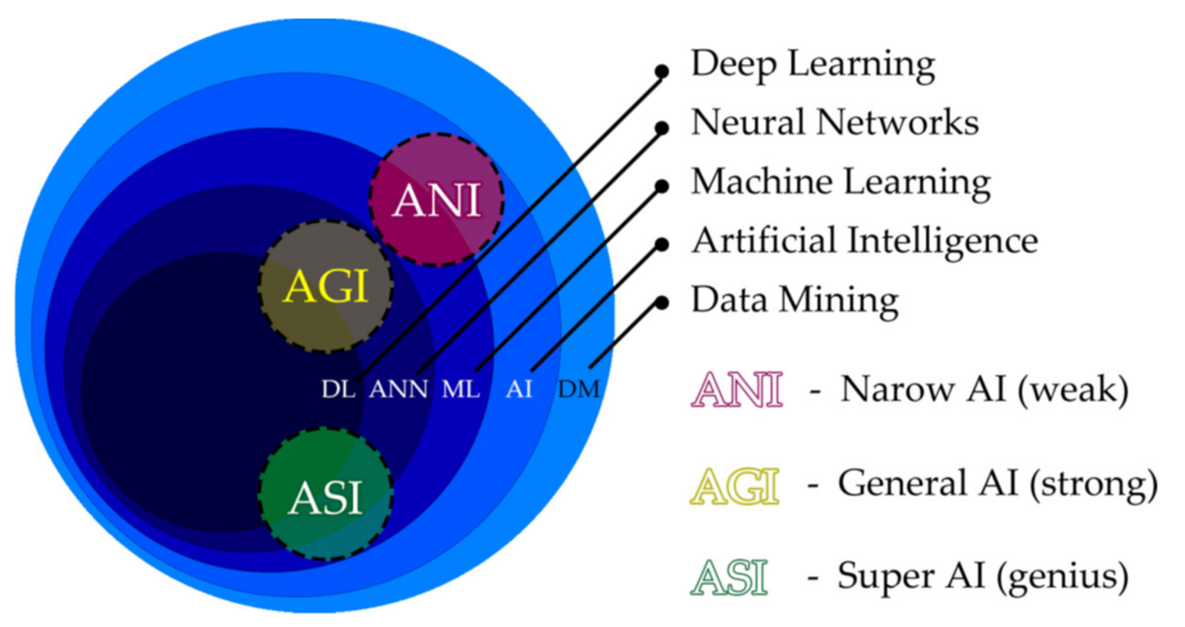
- Machine Learning (ML)
- Neural Networks (ANN)
- Computation
- Vision, Robotics
- Expert Systems
- Speech Processing
- Natural Language Processing
- To quantify how frequently artificial intelligence (AI) was utilized in dental literature for every year from 2011 until 2021.
- To identify the focus of such publications with regard to particular dental fields and topics from January 2021 until the present.
- Orthodontics
- Oral and maxillofacial surgery
- Therapeutic Dentistry
- Periodontology
- Endodontics
- Craniofacial disorders including cleft management
- Prosthodontics and smile design
- Airway management
2. Materials and Methods
2.1. The Framework, the Protocol and Research Questions
- What is the quantity per year of publications focused on dental AI utilization? Is it increasing? Identify the annual publication count. (2011–2021)
- What are the qualitative characteristics focused on and the proportion of focus devoted to particular dental fields and specialized topics in recent dental AI publications since 2021?
- Quantitative assessment of dental AI publications in the past decade.
- Qualitative assessment of the current literature from 2021 until the present.
2.2. Evaluating the Past Publications 2011–2021
2.3. Evaluating the Present Publications 2011–2021
3. Results
3.1. The Past Publications 2011–2021
3.2. The Current Publications 2011–2021 Analysis
3.3. Literature Analysis of Current AI Applications in Orthodontics
3.4. Literature Analysis of Current AI Applications in Oral and Maxillofacial Surgery
3.5. Literature Analysis of Current AI Applications in Therapeutic Dentistry
3.6. Literature Analysis of Current AI Applications in Periodontology
3.7. Literature Analysis of Current AI Applications in Endodontics
3.8. Literature Analysis of Current AI Applications in Patients with Cleft Lip and Palate and in Genetic Syndromes with Orofacial Features
3.9. Literature Analysis of Current AI Applications in Prosthodontics & Smile Design
3.10. Literature Analysis of Current AI Applications in Airway Management
4. Discussion
5. Conclusions
Supplementary Materials
Author Contributions
Funding
Institutional Review Board Statement
Informed Consent Statement
Data Availability Statement
Acknowledgments
Conflicts of Interest
References
- Obermeyer, Z.; Emanuel, E.J. Predicting the Future—Big Data, Machine Learning, and Clinical Medicine. N. Engl. J. Med. 2016, 375, 1216. [Google Scholar] [CrossRef] [PubMed] [Green Version]
- Gharavi, S.M.H.; Faghihimehr, A. Clinical Application of Artificial Intelligence in PET Imaging of Head and Neck Cancer. PET Clin. 2022, 17, 65–76. [Google Scholar] [CrossRef] [PubMed]
- Patil, S.; Albogami, S.; Hosmani, J.; Mujoo, S.; Kamil, M.A.; Mansour, M.A.; Abdul, H.N.; Bhandi, S.; Ahmed, S.S.S.J. Artificial Intelligence in the Diagnosis of Oral Diseases: Applications and Pitfalls. Diagnostics 2022, 12, 1029. [Google Scholar] [CrossRef] [PubMed]
- Rasteau, S.; Ernenwein, D.; Savoldelli, C.; Bouletreau, P. Artificial Intelligence for Oral and Maxillo-Facial Surgery: A Narrative Review. J. Stomatol. Oral Maxillofac. Surg. 2022, 123, 276–282. [Google Scholar] [CrossRef] [PubMed]
- Monterubbianesi, R.; Tosco, V.; Vitiello, F.; Orilisi, G.; Fraccastoro, F.; Putignano, A.; Orsini, G. Augmented, Virtual and Mixed Reality in Dentistry: A Narrative Review on the Existing Platforms and Future Challenges. Appl. Sci. 2022, 12, 877. [Google Scholar] [CrossRef]
- Oshida, Y. Artificial Intelligence for Medicine. Artif. Intell. Med. 2021. [Google Scholar] [CrossRef]
- Carrillo-Perez, F.; Pecho, O.E.; Morales, J.C.; Paravina, R.D.; della Bona, A.; Ghinea, R.; Pulgar, R.; del Mar Pérez, M.; Herrera, L.J. Applications of Artificial Intelligence in Dentistry: A Comprehensive Review. J. Esthet. Restor. Dent. 2022, 34, 259–280. [Google Scholar] [CrossRef]
- Pethani, F. Promises and Perils of Artificial Intelligence in Dentistry. Aust. Dent. J. 2021, 66, 124–135. [Google Scholar] [CrossRef]
- Nguyen, T.T. Use of Artificial Intelligence in Dentistry: Current Clinical Trends and Research Advances. J. Can. Dent. Assoc. 2021. [Google Scholar]
- Thurzo, A.; Jančovičová, V.; Hain, M.; Thurzo, M.; Novák, B.; Kosnáčová, H.; Lehotská, V.; Moravanský, N.; Varga, I. Human Remains Identification Using Micro-CT, Spectroscopic and A.I. Methods in Forensic Experimental Reconstruction of Dental Patterns After Concentrated Acid Significant Impact. Molecules 2022, 27, 4035. [Google Scholar] [CrossRef]
- Thurzo, A.; Kosnáčová, H.S.; Kurilová, V.; Kosmeľ, S.; Beňuš, R.; Moravanský, N.; Kováč, P.; Kuracinová, K.M.; Palkovič, M.; Varga, I. Use of Advanced Artificial Intelligence in Forensic Medicine, Forensic Anthropology and Clinical Anatomy. Healthcare 2021, 9, 1545. [Google Scholar] [CrossRef]
- Obermeyer, Z.; Powers, B.; Vogeli, C.; Mullainathan, S. Dissecting Racial Bias in an Algorithm Used to Manage the Health of Populations. Science (1979) 2019, 366, 447–453. [Google Scholar] [CrossRef] [Green Version]
- Zhang, Y.; Weng, Y.; Lund, J.; Faust, O.; Su, L.; Acharya, R. Applications of Explainable Artificial Intelligence in Diagnosis and Surgery. Diagnostics 2022, 12, 237. [Google Scholar] [CrossRef]
- Kavya, R.; Christopher, J.; Panda, S.; Lazarus, Y.B. Machine Learning and XAI Approaches for Allergy Diagnosis. Biomed. Signal Process. Control 2021, 69, 102681. [Google Scholar] [CrossRef]
- Barredo Arrieta, A.; Díaz-Rodríguez, N.; del Ser, J.; Bennetot, A.; Tabik, S.; Barbado, A.; Garcia, S.; Gil-Lopez, S.; Molina, D.; Benjamins, R.; et al. Explainable Artificial Intelligence (XAI): Concepts, Taxonomies, Opportunities and Challenges toward Responsible AI. Inf. Fusion 2020, 58, 82–115. [Google Scholar] [CrossRef] [Green Version]
- Wolf, T.G.; Campus, G. Changing Dental Profession—Modern Forms and Challenges in Dental Practice. Int. J. Environ. Res. Public Health 2021, 18, 1945. [Google Scholar] [CrossRef]
- Neville, P.; van der Zande, M. Dentistry, e-Health and Digitalisation: A Critical Narrative Review of the Dental Literature on Digital Technologies with Insights from Health and Technology Studies. Community Dent. Health 2020, 37, 51–58. [Google Scholar] [CrossRef]
- Tele-Orthodontics and the Future of Dental Digitalization|Dentistry IQ. Available online: https://www.dentistryiq.com/practice-management/practice-management-software/article/14035741/teleorthodontics-and-the-future-of-dental-digitalization (accessed on 1 November 2021).
- Putra, R.H.; Doi, C.; Yoda, N.; Astuti, E.R.; Sasaki, K. Current Applications and Development of Artificial Intelligence for Digital Dental Radiography. Dentomaxillofac. Radiol. 2022, 51, 51. [Google Scholar] [CrossRef]
- Alauddin, M.S.; Baharuddin, A.S.; Ghazali, M.I.M. The Modern and Digital Transformation of Oral Health Care: A Mini Review. Healthcare 2021, 9, 118. [Google Scholar] [CrossRef]
- Joda, T.; Bornstein, M.M.; Jung, R.E.; Ferrari, M.; Waltimo, T.; Zitzmann, N.U. Recent Trends and Future Direction of Dental Research in the Digital Era. Int. J. Environ. Res. Public Health 2020, 17, 1987. [Google Scholar] [CrossRef] [Green Version]
- Revilla-León, M.; Gómez-Polo, M.; Barmak, A.B.; Inam, W.; Kan, J.Y.K.; Kois, J.C.; Akal, O. Artificial Intelligence Models for Diagnosing Gingivitis and Periodontal Disease: A Systematic Review. J. Prosthet. Dent. 2022. [Google Scholar] [CrossRef]
- Baniulyte, G.; Ali, K. Artificial Intelligence—Can It Be Used to Outsmart Oral Cancer? Evid. Based Dent. 2022, 23, 12–13. [Google Scholar] [CrossRef]
- Heo, M.S.; Kim, J.E.; Hwang, J.J.; Han, S.S.; Kim, J.S.; Yi, W.J.; Park, I.W. Dmfr 50th Anniversary: Review Article Artificial Intelligence in Oral and Maxillofacial Radiology: What Is Currently Possible? Dentomaxillofac. Radiol. 2020, 50, 50. [Google Scholar] [CrossRef]
- Schwendicke, F.; Samek, W.; Krois, J. Artificial Intelligence in Dentistry: Chances and Challenges. J. Dent. Res. 2020, 99, 769–774. [Google Scholar] [CrossRef]
- Ferro, A.S.; Nicholson, K.; Koka, S. Innovative Trends in Implant Dentistry Training and Education: A Narrative Review. J. Clin. Med. 2019, 8, 1618. [Google Scholar] [CrossRef] [Green Version]
- Prados-Privado, M.; Villalón, J.G.; Martínez-Martínez, C.H.; Ivorra, C. Dental Images Recognition Technology and Applications: A Literature Review. Appl. Sci. 2020, 10, 2856. [Google Scholar] [CrossRef] [Green Version]
- Hung, M.; Hon, E.S.; Ruiz-Negron, B.; Lauren, E.; Moffat, R.; Su, W.; Xu, J.; Park, J.; Prince, D.; Cheever, J.; et al. Exploring the Intersection between Social Determinants of Health and Unmet Dental Care Needs Using Deep Learning. Int. J. Environ. Res. Public Health 2020, 17, 7286. [Google Scholar] [CrossRef]
- Prados-Privado, M.; Villalón, J.G.; Martínez-Martínez, C.H.; Ivorra, C.; Prados-Frutos, J.C. Dental Caries Diagnosis and Detection Using Neural Networks: A Systematic Review. J. Clin. Med. 2020, 9, 3579. [Google Scholar] [CrossRef] [PubMed]
- Müller, A.; Mertens, S.M.; Göstemeyer, G.; Krois, J.; Schwendicke, F. Barriers and Enablers for Artificial Intelligence in Dental Diagnostics: A Qualitative Study. J. Clin. Med. 2021, 10, 1612. [Google Scholar] [CrossRef] [PubMed]
- Moran, M.; Faria, M.; Giraldi, G.; Bastos, L.; Oliveira, L.; Conci, A. Classification of Approximal Caries in Bitewing Radiographs Using Convolutional Neural Networks. Sensors 2021, 21, 5192. [Google Scholar] [CrossRef] [PubMed]
- Hassani, H.; Andi, P.A.; Ghodsi, A.; Norouzi, K.; Komendantova, N.; Unger, S. Shaping the Future of Smart Dentistry: From Artificial Intelligence (AI) to Intelligence Augmentation (IA). IoT 2021, 2, 510–523. [Google Scholar] [CrossRef]
- Roongruangsilp, P.; Khongkhunthian, P. The Learning Curve of Artificial Intelligence for Dental Implant Treatment Planning: A Descriptive Study. Appl. Sci. 2021, 11, 10159. [Google Scholar] [CrossRef]
- Gajic, M.; Vojinovic, J.; Kalevski, K.; Pavlovic, M.; Kolak, V.; Vukovic, B.; Mladenovic, R.; Aleksic, E. Analysis of the Impact of Oral Health on Adolescent Quality of Life Using Standard Statistical Methods and Artificial Intelligence Algorithms. Children 2021, 8, 1156. [Google Scholar] [CrossRef]
- Zaborowicz, M.; Zaborowicz, K.; Biedziak, B.; Garbowski, T. Deep Learning Neural Modelling as a Precise Method in the Assessment of the Chronological Age of Children and Adolescents Using Tooth and Bone Parameters. Sensors 2022, 22, 637. [Google Scholar] [CrossRef]
- Zadrożny, Ł.; Regulski, P.; Brus-Sawczuk, K.; Czajkowska, M.; Parkanyi, L.; Ganz, S.; Mijiritsky, E. Artificial Intelligence Application in Assessment of Panoramic Radiographs. Diagnostics 2022, 12, 224. [Google Scholar] [CrossRef]
- De Angelis, F.; Pranno, N.; Franchina, A.; di Carlo, S.; Brauner, E.; Ferri, A.; Pellegrino, G.; Grecchi, E.; Goker, F.; Stefanelli, L.V. Artificial Intelligence: A New Diagnostic Software in Dentistry: A Preliminary Performance Diagnostic Study. Int. J. Environ. Res. Public Health 2022, 19, 1728. [Google Scholar] [CrossRef]
- Mudrak, J. Artificial Intelligence and Deep Learning in Dental Radiology. Available online: https://www.oralhealthgroup.com/features/artificial-intelligence-and-deep-learning-in-dental-radiology-a-way-forward-in-point-of-care-radiology/ (accessed on 28 December 2021).
- Ezhov, M.; Gusarev, M.; Golitsyna, M.; Yates, J.M.; Kushnerev, E.; Tamimi, D.; Aksoy, S.; Shumilov, E.; Sanders, A.; Orhan, K. Clinically Applicable Artificial Intelligence System for Dental Diagnosis with CBCT. Sci. Rep. 2021, 11, 15006. [Google Scholar] [CrossRef]
- Van Rijn, R.R.; de Luca, A. Three Reasons Why Artificial Intelligence Might Be the Radiologist’s Best Friend. Radiology 2020, 296, 159–160. [Google Scholar] [CrossRef]
- Bose, S.; Sur, J.; Khan, F.; Tuzoff, D. The Scope of Artificial Intelligence in Oral Radiology- A Review. Int. J. Med. Health Sci. 2020, 9, 67–72. [Google Scholar]
- Orhan, K.; Bayrakdar, I.S.; Ezhov, M.; Kravtsov, A.; Özyürek, T. Evaluation of Artificial Intelligence for Detecting Periapical Pathosis on Cone-Beam Computed Tomography Scans. Int. Endod. J. 2020, 53, 680–689. [Google Scholar] [CrossRef]
- Makaremi, M.; Lacaule, C.; Mohammad-Djafari, A. Deep Learning and Artificial Intelligence for the Determination of the Cervical Vertebra Maturation Degree from Lateral Radiography. Entropy 2019, 21, 1222. [Google Scholar] [CrossRef] [Green Version]
- Tanikawa, C.; Yamashiro, T. Development of Novel Artificial Intelligence Systems to Predict Facial Morphology after Orthognathic Surgery and Orthodontic Treatment in Japanese Patients. Sci. Rep. 2021, 11, 15853. [Google Scholar] [CrossRef]
- Zhang, C.; Chen, Z.; Liu, J.; Wu, M.; Yang, J.; Zhu, Y.; Lu, W.W.; Ruan, C. 3D-Printed Pre-Tapped-Hole Scaffolds Facilitate One-Step Surgery of Predictable Alveolar Bone Augmentation and Simultaneous Dental Implantation. Compos. Part B Eng. 2021, 229, 109461. [Google Scholar] [CrossRef]
- Bayrakdar, S.K.; Orhan, K.; Bayrakdar, I.S.; Bilgir, E.; Ezhov, M.; Gusarev, M.; Shumilov, E. A Deep Learning Approach for Dental Implant Planning in Cone-Beam Computed Tomography Images. BMC Med. Imaging 2021, 21, 1–9. [Google Scholar] [CrossRef]
- Mashouri, P.; Skreta, M.; Phillips, J.; McAllister, D.; Roy, M.; Senkaiahliyan, S.; Brudno, M.; Singh, D. 3D Photography Based Neural Network Craniosynostosis Triaging System. Proc. Mach. Learn. Res. 2020, 136, 226–237. [Google Scholar]
- Ajmera, D.H.; Singh, P.; Leung, Y.Y.; Gu, M. Three-Dimensional Evaluation of Soft-Tissue Response to Osseous Movement after Orthognathic Surgery in Patients with Facial Asymmetry: A Systematic Review. J. Craniomaxillofac. Surg. 2021, 49, 763–774. [Google Scholar] [CrossRef]
- Thurzo, A.; Kurilová, V.; Varga, I. Artificial Intelligence in Orthodontic Smart Application for Treatment Coaching and Its Impact on Clinical Performance of Patients Monitored with AI-TeleHealth System. Healthcare 2021, 9, 1695. [Google Scholar] [CrossRef]
- Mucchi, L.; Jayousi, S.; Gant, A.; Paoletti, E.; Zoppi, P. Tele-Monitoring System for Chronic Diseases Management: Requirements and Architecture. Int. J. Environ. Res. Public Health 2021, 18, 7459. [Google Scholar] [CrossRef]
- Kravitz, N.; Burris, B.; Butler, D.; Dabney, C. Teledentistry, Do-It-Yourself Orthodontics, and Remote Treatment Monitoring. J. Clin. Orthod. 2016, 50, 718–726. [Google Scholar]
- Morris, R.S.; Hoye, L.N.; Elnagar, M.H.; Atsawasuwan, P.; Galang-Boquiren, M.T.; Caplin, J.; Viana, G.C.; Obrez, A.; Kusnoto, B. Accuracy of Dental Monitoring 3D Digital Dental Models Using Photograph and Video Mode. Am. J. Orthod. Dentofac. Orthop. 2019, 156, 420–428. [Google Scholar] [CrossRef]
- Park, J.H.; Rogowski, L.; Kim, J.H.; al Shami, S.; Howell, S.E.I. Teledentistry Platforms for Orthodontics. J. Clin. Pediatr. Dent. 2021, 45, 48–53. [Google Scholar] [CrossRef] [PubMed]
- Mandall, N.; O’Brien, K.; Brady, J.; Worthington, H.; Harvey, L. Teledentistry for Screening New Patient Orthodontic Referrals. Part 1: A Randomised Controlled Trial. Br. Dent. J. 2005, 199, 659–662. [Google Scholar] [CrossRef] [PubMed] [Green Version]
- Turchetta, B.; Fishman, L.; Subtelny, J. Facial Growth Prediction: A Comparison of Methodologies. Am. J. Orthod. Dentofac. Orthop. 2007, 132, 439–449. [Google Scholar] [CrossRef] [PubMed]
- Cao, K.; Choi, K.; Jung, H.; Duan, L. Deep Learning for Facial Beauty Prediction. Information 2020, 11, 391. [Google Scholar] [CrossRef]
- Sajedi, H.; Pardakhti, N. Age Prediction Based on Brain MRI Image: A Survey. J. Med. Syst. 2019, 43, 279. [Google Scholar] [CrossRef] [PubMed]
- Iyer, T.J.; Rahul, K.; Nersisson, R.; Zhuang, Z.; Joseph Raj, A.N.; Refayee, I. Machine Learning-Based Facial Beauty Prediction and Analysis of Frontal Facial Images Using Facial Landmarks and Traditional Image Descriptors. Comput. Intell. Neurosci. 2021, 2021, 4423407. [Google Scholar] [CrossRef]
- Artificial Intelligence in Orthodontics: Prediction and Planning|International Journal of Advances in Scientific Research. Available online: https://ssjournals.com/index.php/ijasr/article/view/5672 (accessed on 7 April 2022).
- Kishimoto, T.; Goto, T.; Matsuda, T.; Iwawaki, Y.; Ichikawa, T. Application of Artificial Intelligence in the Dental Field: A Literature Review. J. Prosthodont. Res. 2022, 66, 19–28. [Google Scholar] [CrossRef]
- Shan, T.; Tay, F.R.; Gu, L. Application of Artificial Intelligence in Dentistry. J. Dent. Res. 2021, 100, 232–244. [Google Scholar] [CrossRef]
- Nissan, J.; Kosan, E.; Krois, J.; Wingenfeld, K.; Deuter, C.E.; Gaudin, R.; Schwendicke, F. Patients’ Perspectives on Artificial Intelligence in Dentistry: A Controlled Study. J. Clin. Med. 2022, 11, 2143. [Google Scholar] [CrossRef]
- Kim, S.-H.; Kim, K.B.; Choo, H. New Frontier in Advanced Dentistry: CBCT, Intraoral Scanner, Sensors, and Artificial Intelligence in Dentistry. Sensors 2022, 22, 2942. [Google Scholar] [CrossRef]
- Ossowska, A.; Kusiak, A.; Świetlik, D. Artificial Intelligence in Dentistry—Narrative Review. Int. J. Environ. Res. Public Health 2022, 19, 3449. [Google Scholar] [CrossRef]
- Ibrahim, H.; Liu, X.; Rivera, S.C.; Moher, D.; Chan, A.W.; Sydes, M.R.; Calvert, M.J.; Denniston, A.K. Reporting Guidelines for Clinical Trials of Artificial Intelligence Interventions: The SPIRIT-AI and CONSORT-AI Guidelines. Trials 2021, 22, 11. [Google Scholar] [CrossRef]
- Campbell, J.P.; Lee, A.Y.; Abràmoff, M.; Keane, P.A.; Ting, D.S.W.; Lum, F.; Chiang, M.F. Reporting Guidelines for Artificial Intelligence in Medical Research. Ophthalmology 2020, 127, 1596–1599. [Google Scholar] [CrossRef]
- Ibrahim, H.; Liu, X.; Denniston, A.K. Reporting Guidelines for Artificial Intelligence in Healthcare Research. Clin. Exp. Ophthalmol 2021, 49, 470–476. [Google Scholar] [CrossRef]
- Sounderajah, V.; Ashrafian, H.; Golub, R.M.; Shetty, S.; de Fauw, J.; Hooft, L.; Moons, K.; Collins, G.; Moher, D.; Bossuyt, P.M.; et al. Developing a Reporting Guideline for Artificial Intelligence-Centred Diagnostic Test Accuracy Studies: The STARD-AI Protocol. BMJ Open 2021, 11, e047709. [Google Scholar] [CrossRef]
- Page, M.J.; McKenzie, J.E.; Bossuyt, P.M.; Boutron, I.; Hoffmann, T.C.; Mulrow, C.D.; Shamseer, L.; Tetzlaff, J.M.; Akl, E.A.; Brennan, S.E.; et al. The PRISMA 2020 Statement: An Updated Guideline for Reporting Systematic Reviews. BMJ 2021, 372, n71. [Google Scholar] [CrossRef]
- Shamseer, L.; Moher, D.; Clarke, M.; Ghersi, D.; Liberati, A.; Petticrew, M.; Shekelle, P.; Stewart, L.A.; Altman, D.G.; Booth, A.; et al. Preferred Reporting Items for Systematic Review and Meta-Analysis Protocols (PRISMA-P) 2015: Elaboration and Explanation. BMJ 2015, 349, 7647. [Google Scholar] [CrossRef] [Green Version]
- Moher, D.; Shamseer, L.; Clarke, M.; Ghersi, D.; Liberati, A.; Petticrew, M.; Shekelle, P.; Stewart, L.A.; Estarli, M.; Barrera, E.S.A.; et al. Preferred Reporting Items for Systematic Review and Meta-Analysis Protocols (PRISMA-P) 2015 Statement. Rev. Esp. De Nutr. Hum. Y Diet. 2016, 20, 148–160. [Google Scholar] [CrossRef] [Green Version]
- Alonso-Coello, P.; Schünemann, H.J.; Moberg, J.; Brignardello-Petersen, R.; Akl, E.A.; Davoli, M.; Treweek, S.; Mustafa, R.A.; Rada, G.; Rosenbaum, S.; et al. GRADE Evidence to Decision (EtD) Frameworks: A Systematic and Transparent Approach to Making Well Informed Healthcare Choices. 1: Introduction. BMJ 2016, 353. [Google Scholar] [CrossRef] [Green Version]
- Harzing, A.W. Publish or Perish. Available online: https://harzing.com/resources/publish-or-perish (accessed on 9 April 2022).
- The, A.; Chaushu, G.; Wai Kan Yeung, A. The Diagnostic Relevance and Interfaces Covered by Mach Band Effect in Dentistry: An Analysis of the Literature. Healthcare 2022, 10, 632. [Google Scholar] [CrossRef]
- Cantrell, A.; Booth, A.; Chambers, D. A Systematic Review Case Study of Urgent and Emergency Care Configuration Found Citation Searching of Web of Science and Google Scholar of Similar Value. Health Info. Libr. J. 2022, 1–16. [Google Scholar] [CrossRef]
- Lee, J.H.; Kim, D.H.; Jeong, S.N.; Choi, S.H. Detection and Diagnosis of Dental Caries Using a Deep Learning-Based Convolutional Neural Network Algorithm. J. Dent. 2018, 77, 106–111. [Google Scholar] [CrossRef]
- Lee, J.H.; Kim, D.H.; Jeong, S.N.; Choi, S.H. Diagnosis and Prediction of Periodontally Compromised Teeth Using a Deep Learning-Based Convolutional Neural Network Algorithm. J. Periodontal Implant Sci. 2018, 48, 114–123. [Google Scholar] [CrossRef] [Green Version]
- Ekert, T.; Krois, J.; Meinhold, L.; Elhennawy, K.; Emara, R.; Golla, T.; Schwendicke, F. Deep Learning for the Radiographic Detection of Apical Lesions. J. Endod. 2019, 45, 917–922.e5. [Google Scholar] [CrossRef]
- Schwendicke, F.; Golla, T.; Dreher, M.; Krois, J. Convolutional Neural Networks for Dental Image Diagnostics: A Scoping Review. J. Dent. 2019, 91, 103226. [Google Scholar] [CrossRef]
- Hwang, J.J.; Jung, Y.H.; Cho, B.H.; Heo, M.S. An Overview of Deep Learning in the Field of Dentistry. Imaging Sci. Dent. 2019, 49, 1–7. [Google Scholar] [CrossRef]
- Fourcade, A.; Khonsari, R.H. Deep Learning in Medical Image Analysis: A Third Eye for Doctors. J. Stomatol. Oral Maxillofac. Surg. 2019, 120, 279–288. [Google Scholar] [CrossRef]
- Hiraiwa, T.; Ariji, Y.; Fukuda, M.; Kise, Y.; Nakata, K.; Katsumata, A.; Fujita, H.; Ariji, E. A Deep-Learning Artificial Intelligence System for Assessment of Root Morphology of the Mandibular First Molar on Panoramic Radiography. Dentomaxillofac. Radiol. 2019, 48, 20180218. [Google Scholar] [CrossRef] [PubMed]
- Murata, M.; Ariji, Y.; Ohashi, Y.; Kawai, T.; Fukuda, M.; Funakoshi, T.; Kise, Y.; Nozawa, M.; Katsumata, A.; Fujita, H.; et al. Deep-Learning Classification Using Convolutional Neural Network for Evaluation of Maxillary Sinusitis on Panoramic Radiography. Oral Radiol. 2019, 35, 301–307. [Google Scholar] [CrossRef] [PubMed]
- Almangush, A.; Mäkitie, A.A.; Triantafyllou, A.; de Bree, R.; Strojan, P.; Rinaldo, A.; Hernandez-Prera, J.C.; Suárez, C.; Kowalski, L.P.; Ferlito, A.; et al. Staging and Grading of Oral Squamous Cell Carcinoma: An Update. Oral Oncol. 2020, 107, 104799. [Google Scholar] [CrossRef] [PubMed]
- Faber, J.; Faber, C.; Faber, P. Artificial Intelligence in Orthodontics. APOS Trends Orthod. 2019, 9, 201–205. [Google Scholar] [CrossRef]
- Rousseau, M.; Retrouvey, J.M. Machine Learning in Orthodontics: Automated Facial Analysis of Vertical Dimension for Increased Precision and Efficiency. Am. J. Orthod. Dentofac. Orthop. 2022, 161, 445–450. [Google Scholar] [CrossRef]
- Monill-González, A.; Rovira-Calatayud, L.; d’Oliveira, N.G.; Ustrell-Torrent, J.M. Artificial Intelligence in Orthodontics: Where Are We Now? A Scoping Review. Orthod. Craniofacial Res. 2021, 24, 6–15. [Google Scholar] [CrossRef]
- Thrall, J.H.; Li, X.; Li, Q.; Cruz, C.; Do, S.; Dreyer, K.; Brink, J. Artificial Intelligence and Machine Learning in Radiology: Opportunities, Challenges, Pitfalls, and Criteria for Success. J. Am. Coll. Radiol 2018, 15, 504–508. [Google Scholar] [CrossRef]
- Hirschfeld, W.J.; Moyers, R.E.; Ann, D.D.S. Prediction of Craniofacial Growth: The State of the Art. Am. J. Orthod. 1971, 60, 435–444. [Google Scholar] [CrossRef] [Green Version]
- Kaźmierczak, S.; Juszka, Z.; Vandevska-Radunovic, V.; Maal, T.J.J.; Fudalej, P.; Mańdziuk, J. Prediction of the Facial Growth Direction Is Challenging; Springer: Cham, Switzerland, 2021; pp. 665–673. [Google Scholar]
- Hansa, I.; Katyal, V.; Ferguson, D.J.; Vaid, N. Outcomes of Clear Aligner Treatment with and without Dental Monitoring: A Retrospective Cohort Study. Am. J. Orthod. Dentofac. Orthop. 2021, 159, 453–459. [Google Scholar] [CrossRef]
- Caruso, S.; Caruso, S.; Pellegrino, M.; Skafi, R.; Nota, A.; Tecco, S. A Knowledge-Based Algorithm for Automatic Monitoring of Orthodontic Treatment: The Dental Monitoring System. Two Cases. Sensors 2021, 21, 1856. [Google Scholar] [CrossRef]
- Impellizzeri, A.; Horodinsky, M.; Barbato, E.; Polimeni, A.; Salah, P.; Galluccio, G. Dental Monitoring Application: It Is a Valid Innovation in the Orthodontics Practice? Clin. Ter. 2020, 171, 260–267. [Google Scholar] [CrossRef]
- Roisin, L.-C.; Brézulier, D.; Sorel, O. Remotely-Controlled Orthodontics: Fundamentals and Description of the Dental Monitoring System. J. Dentofac. Anom. Orthod. 2016, 19, 408. [Google Scholar] [CrossRef]
- Hansa, I.; Semaan, S.J.; Vaid, N.R. Clinical Outcomes and Patient Perspectives of Dental Monitoring® GoLive® with Invisalign®—a Retrospective Cohort Study. Prog. Orthod. 2020, 21, 16. [Google Scholar] [CrossRef]
- Saccomanno, S.; Quinzi, V.; Albani, A.; D’Andrea, N.; Marzo, G.; Macchiarelli, G. Utility of Teleorthodontics in Orthodontic Emergencies during the COVID-19 Pandemic: A Systematic Review. Healthcare 2022, 10, 1108. [Google Scholar] [CrossRef]
- Thurzo, A.; Kočiš, F.; Novák, B.; Czako, L.; Varga, I. Three-Dimensional Modeling and 3D Printing of Biocompatible Orthodontic Power-Arm Design with Clinical Application. Appl. Sci. 2021, 11, 9693. [Google Scholar] [CrossRef]
- Thurzo, A.; Urbanová, W.; Novák, B.; Waczulíková, I.; Varga, I. Utilization of a 3D Printed Orthodontic Distalizer for Tooth-Borne Hybrid Treatment in Class II Unilateral Malocclusions. Materials 2022, 15, 1740. [Google Scholar] [CrossRef]
- Fatima, A.; Shahid, A.R.; Raza, B.; Madni, T.M.; Janjua, U.I. State-of-the-Art Traditional to the Machine- and Deep-Learning-Based Skull Stripping Techniques, Models, and Algorithms. J. Digit. Imaging 2020, 33, 1443–1464. [Google Scholar] [CrossRef]
- Herath, B.; Suresh, S.; Downing, D.; Cometta, S.; Tino, R.; Castro, N.J.; Leary, M.; Schmutz, B.; Wille, M.-L.; Hutmacher, D.W. Mechanical and Geometrical Study of 3D Printed Voronoi Scaffold Design for Large Bone Defects. Mater. Des. 2021, 212, 110224. [Google Scholar] [CrossRef]
- Thurzo, A.; Urbanová, W.; Waczulíková, I.; Kurilová, V.; Mriňáková, B.; Kosnáčová, H.; Gális, B.; Varga, I.; Matajs, M.; Novák, B. Dental Care and Education Facing Highly Transmissible SARS-CoV-2 Variants: Prospective Biosafety Setting: Prospective, Single-Arm, Single-Center Study. Int. J. Environ. Res. Public Health 2022, 19, 7693. [Google Scholar] [CrossRef]
- MacHoy, M.E.; Szyszka-Sommerfeld, L.; Vegh, A.; Gedrange, T.; Woźniak, K. The Ways of Using Machine Learning in Dentistry. Adv. Clin. Exp. Med. 2020, 29, 375–384. [Google Scholar] [CrossRef]
- Choi, J.W.; Park, H.; In-Hwan Kim, B.S.; Kim, N.; Kwon, S.-M.; Lee, J.Y. Surgery-First Orthognathic Approach to Correct Facial Asymmetry: Artificial Intelligence–Based Cephalometric Analysis. Plast. Reconstr. Surg. 2022, 149, 496e–499e. [Google Scholar] [CrossRef]
- Joshi, V.K. Dental Treatment Planning and Management for the Mouth Cancer Patient. Oral Oncol. 2010, 46, 475–479. [Google Scholar] [CrossRef]
- Chang, S.W.; Abdul-Kareem, S.; Merican, A.F.; Zain, R.B. Oral Cancer Prognosis Based on Clinicopathologic and Genomic Markers Using a Hybrid of Feature Selection and Machine Learning Methods. BMC Bioinform. 2013, 14, 170. [Google Scholar] [CrossRef] [PubMed] [Green Version]
- Kumar, V.; Gu, Y.; Basu, S.; Berglund, A.; Eschrich, S.A.; Schabath, M.B.; Forster, K.; Aerts, H.J.W.L.; Dekker, A.; Fenstermacher, D.; et al. Radiomics: The Process and the Challenges. Magn. Reson. Imaging 2012, 30, 1234–1248. [Google Scholar] [CrossRef] [PubMed] [Green Version]
- Patcas, R.; Bernini, D.A.J.; Volokitin, A.; Agustsson, E.; Rothe, R.; Timofte, R. Applying Artificial Intelligence to Assess the Impact of Orthognathic Treatment on Facial Attractiveness and Estimated Age. Int. J. Oral Maxillofac. Surg. 2019, 48, 77–83. [Google Scholar] [CrossRef] [PubMed] [Green Version]
- Bouletreau, P.; Makaremi, M.; Ibrahim, B.; Louvrier, A.; Sigaux, N. Artificial Intelligence: Applications in Orthognathic Surgery. J. Stomatol. Oral Maxillofac. Surg. 2019, 120, 347–354. [Google Scholar] [CrossRef] [PubMed]
- Arık, S.; Ibragimov, B.; Xing, L. Fully Automated Quantitative Cephalometry Using Convolutional Neural Networks. J. Med. Imaging 2017, 4, 014501. [Google Scholar] [CrossRef]
- Kang, S.H.; Jeon, K.; Kim, H.-J.; Seo, J.K.; Lee, S.-H. Automatic Three-Dimensional Cephalometric Annotation System Using Three-Dimensional Convolutional Neural Networks: A Developmental Trial. Comput. Methods Biomech. Biomed. Eng. Imaging Vis. 2019, 8, 210–218. [Google Scholar] [CrossRef]
- Yang, C.; Rangarajan, A.; Ranka, S. Visual Explanations From Deep 3D Convolutional Neural Networks for Alzheimer’s Disease Classification. AMIA Annu. Symp. Proc. 2018, 2018, 1571. [Google Scholar]
- Schwendicke, F.; Rossi, J.G.; Göstemeyer, G.; Elhennawy, K.; Cantu, A.G.; Gaudin, R.; Chaurasia, A.; Gehrung, S.; Krois, J. Cost-Effectiveness of Artificial Intelligence for Proximal Caries Detection. J. Dent. Res. 2021, 100, 369–376. [Google Scholar] [CrossRef]
- Shimada, Y.; Bakhsh, T.A.; Schlenz, M.A.; Schupp, B.; Schmidt, A.; Wöstmann, B.; Baresel, I.; Krämer, N.; Schulz-Weidner, N. New Caries Diagnostic Tools in Intraoral Scanners: A Comparative In Vitro Study to Established Methods in Permanent and Primary Teeth. Sensors 2022, 22, 2156. [Google Scholar] [CrossRef]
- Bayrakdar, I.S.; Orhan, K.; Akarsu, S.; Çelik, Ö.; Atasoy, S.; Pekince, A.; Yasa, Y.; Bilgir, E.; Sağlam, H.; Aslan, A.F.; et al. Deep-Learning Approach for Caries Detection and Segmentation on Dental Bitewing Radiographs. Oral Radiol. 2021. [Google Scholar] [CrossRef]
- Yasa, Y.; Çelik, Ö.; Bayrakdar, I.S.; Pekince, A.; Orhan, K.; Akarsu, S.; Atasoy, S.; Bilgir, E.; Odabaş, A.; Aslan, A.F. An Artificial Intelligence Proposal to Automatic Teeth Detection and Numbering in Dental Bite-Wing Radiographs. Acta Odontol. Scand. 2021, 79, 275–281. [Google Scholar] [CrossRef]
- Kelleher, M.; Bishop, K. Tooth Surface Loss: An. Overview. British. Dental. 1999, 186, 61–66. [Google Scholar] [CrossRef]
- Al Haidan, A.; Abu-Hammad, O.; Dar-Odeh, N. Predicting Tooth Surface Loss Using Genetic Algorithms-Optimized Artificial Neural Networks. Comput. Math. Methods Med. 2014, 2014, 106236. [Google Scholar] [CrossRef] [Green Version]
- Meghil, M.M.; Rajpurohit, P.; Awad, M.E.; McKee, J.; Shahoumi, L.A.; Ghaly, M. Artificial Intelligence in Dentistry. Dent. Rev. 2022, 2, 100009. [Google Scholar] [CrossRef]
- Ahmed, N.; Abbasi, M.S.; Zuberi, F.; Qamar, W.; bin Halim, M.S.; Maqsood, A.; Alam, M.K. Artificial Intelligence Techniques: Analysis, Application, and Outcome in Dentistry—A Systematic Review. BioMed Res. Int. 2021, 2021, 9751564. [Google Scholar] [CrossRef]
- Farhadian, M.; Shokouhi, P.; Torkzaban, P. A Decision Support System Based on Support Vector Machine for Diagnosis of Periodontal Disease. BMC Res. Notes 2020, 13, 337. [Google Scholar] [CrossRef]
- Chen, W.P.; Chang, S.H.; Tang, C.Y.; Liou, M.L.; Tsai, S.J.J.; Lin, Y.L. Composition Analysis and Feature Selection of the Oral Microbiota Associated with Periodontal Disease. BioMed Res. Int. 2018, 2018 , 3130607. [Google Scholar] [CrossRef] [Green Version]
- Li, W.; Chen, Y.; Sun, W.; Brown, M.; Zhang, X.; Wang, S.; Miao, L. A Gingivitis Identification Method Based on Contrast-Limited Adaptive Histogram Equalization, Gray-Level Co-Occurrence Matrix, and Extreme Learning Machine. Int. J. Imaging Syst. Technol. 2019, 29, 77–82. [Google Scholar] [CrossRef] [Green Version]
- Akther, S.; Saleheen, N.; Samiei, S.A.; Shetty, V.; Ertin, E.; Kumar, S. MORAL: An mHealth Model for Inferring Oral Hygiene Behaviors in-the-wild Using Wrist-worn Inertial Sensors. Proc. ACM Interact. Mob. Wearable Ubiquitous Technol. 2019, 3, 1–25. [Google Scholar] [CrossRef]
- Boreak, N. Effectiveness of Artifcial Intelligence Applications Designed for Endodontic Diagnosis, Decision-Making, and Prediction of Prognosis: A Systematic Review. J. Contemp. Dent. Pract. 2020, 21, 926–934. [Google Scholar] [CrossRef]
- Keskin, C.; Keles, A. Digital Applications in Endodontics: An Update and Review. J. Exp. Clin. Med. 2021, 38, 168–174. [Google Scholar] [CrossRef]
- Aminoshariae, A.; Kulild, J.; Nagendrababu, V. Artificial Intelligence in Endodontics: Current Applications and Future Directions. J. Endod. 2021, 47, 1352–1357. [Google Scholar] [CrossRef]
- Li, C.W.; Lin, S.Y.; Chou, H.S.; Chen, T.Y.; Chen, Y.A.; Liu, S.Y.; Liu, Y.L.; Chen, C.A.; Huang, Y.C.; Chen, S.L.; et al. Detection of Dental Apical Lesions Using Cnns on Periapical Radiograph. Sensors 2021, 21, 7049. [Google Scholar] [CrossRef]
- Pauwels, R.; Brasil, D.M.; Yamasaki, M.C.; Jacobs, R.; Bosmans, H.; Freitas, D.Q.; Haiter-Neto, F. Artificial Intelligence for Detection of Periapical Lesions on Intraoral Radiographs: Comparison between Convolutional Neural Networks and Human Observers. Oral Surg. Oral Med. Oral Pathol. Oral Radiol. 2021, 131, 610–616. [Google Scholar] [CrossRef]
- Setzer, F.C.; Shi, K.J.; Zhang, Z.; Yan, H.; Yoon, H.; Mupparapu, M.; Li, J. Artificial Intelligence for the Computer-Aided Detection of Periapical Lesions in Cone-Beam Computed Tomographic Images. J. Endod. 2020, 46, 987–993. [Google Scholar] [CrossRef]
- Fukuda, M.; Inamoto, K.; Shibata, N.; Ariji, Y.; Yanashita, Y.; Kutsuna, S.; Nakata, K.; Katsumata, A.; Fujita, H.; Ariji, E. Evaluation of an Artificial Intelligence System for Detecting Vertical Root Fracture on Panoramic Radiography. Oral Radiol. 2020, 36, 337–343. [Google Scholar] [CrossRef]
- Liao, W.C.; Chen, C.H.; Pan, Y.H.; Chang, M.C.; Jeng, J.H. Vertical Root Fracture in Non-Endodontically and Endodontically Treated Teeth: Current Understanding and Future Challenge. J. Pers. Med. 2021, 11, 1375. [Google Scholar] [CrossRef]
- Shafi, N.; Bukhari, F.; Iqbal, W.; Almustafa, K.M.; Asif, M.; Nawaz, Z. Cleft Prediction before Birth Using Deep Neural Network. Health Inform. J. 2020, 26, 2568–2585. [Google Scholar] [CrossRef] [Green Version]
- Zhang, S.J.; Meng, P.; Zhang, J.; Jia, P.; Lin, J.; Wang, X.; Chen, F.; Wei, X. Machine Learning Models for Genetic Risk Assessment of Infants with Non-Syndromic Orofacial Cleft. Genom. Proteom. Bioinform. 2018, 16, 354–364. [Google Scholar] [CrossRef] [PubMed]
- Baker, N.C.; Sipes, N.S.; Franzosa, J.; Belair, D.G.; Abbott, B.D.; Judson, R.S.; Knudsen, T.B. Characterizing Cleft Palate Toxicants Using ToxCast Data, Chemical Structure, and the Biomedical Literature. Birth Defects Res. 2020, 112, 19–39. [Google Scholar] [CrossRef] [PubMed]
- Jurek, J.; Wójtowicz, W.; Wójtowicz, A. Syntactic Pattern Recognition-Based Diagnostics of Fetal Palates. Pattern Recognit. Lett. 2020, 133, 144–150. [Google Scholar] [CrossRef]
- Kuwada, C.; Ariji, Y.; Kise, Y.; Funakoshi, T.; Fukuda, M.; Kuwada, T.; Gotoh, K.; Ariji, E. Detection and Classification of Unilateral Cleft Alveolus with and without Cleft Palate on Panoramic Radiographs Using a Deep Learning System. Sci. Rep. 2021, 11, 16044. [Google Scholar] [CrossRef]
- Zhang, Y.; Pei, Y.; Chen, S.; Guo, Y.; Ma, G.; Xu, T.; Zha, H. Volumetric Registration-Based Cleft Volume Estimation of Alveolar Cleft Grafting Procedures. In Proceedings of the International Symposium on Biomedical Imaging, Iowa City, IA, USA, 3–7 April 2020; pp. 99–103. [Google Scholar]
- Ortiz-Posadas, M.R.; Vega-Alvarado, L.; Toni, B. A Similarity Function to Evaluate the Orthodontic Condition in Patients with Cleft Lip and Palate. Med. Hypotheses 2004, 63, 35–41. [Google Scholar] [CrossRef]
- Tanikawa, C.; Lee, C.; Lim, J.; Oka, A.; Yamashiro, T. Clinical Applicability of Automated Cephalometric Landmark Identification: Part I-Patient-Related Identification Errors. Orthod. Craniofacial Res. 2021, 24, 43–52. [Google Scholar] [CrossRef]
- Alam, M.K.; Alfawzan, A.A. Dental Characteristics of Different Types of Cleft and Non-Cleft Individuals. Front. Cell Dev. Biol. 2020, 8, 789. [Google Scholar] [CrossRef]
- Lim, J.; Tanikawa, C.; Kogo, M.; Yamashiro, C. Determination of Prognostic Factors for Orthognathic Surgery in Children with Cleft Lip and or Palate. Orthod. Craniofacial Res. 2021, 24, 153–162. [Google Scholar] [CrossRef]
- Jihee, S.; Yang, I.-H.; Choi, J.-Y.; Lee, J.-H.; Baek, S.-H. Three-Dimensional Facial Soft Tissue Changes After Orthognathic Surgery in Cleft Patients Using Artificial Intelligence-Assisted Landmark Autodigitization. J. Craniofacial Surg. 2021, 32, 2695–2700. [Google Scholar]
- Pietruski, P.; Majak, M.; Debski, T.; Antoszewski, B. A Novel Computer System for the Evaluation of Nasolabial Morphology, Symmetry and Aesthetics after Cleft Lip and Palate Treatment. Part 1: General Concept and Validation. J. Cranio-Maxillofac. Surg. 2017, 45, 491–504. [Google Scholar] [CrossRef]
- Patcas, R.; Timofte, R.; Volokitin, A.; Agustsson, E.; Eliades, T.; Eichenberger, M.; Bornstein, M.M. Facial Attractiveness of Cleft Patients: A Direct Comparison between Artificial-Intelligence-Based Scoring and Conventional Rater Groups. Eur. J. Orthod. 2019, 41, 428–433. [Google Scholar] [CrossRef]
- Wang, X.; Yang, S.; Tang, M.; Yin, H.; Huang, H.; He, L. HypernasalityNet: Deep Recurrent Neural Network for Automatic Hypernasality Detection. Int. J. Med. Inform. 2019, 129, 1–12. [Google Scholar] [CrossRef]
- Gugsch, C.; Dannhauer, K.H.; Fuchs, M. Stimmanalytische Beurteilung Des Therapieverlaufs Bei Patienten Mit Lippen-Kiefer-Gaumen-Spalten—Eine Pilotstudie. J. Orofac. Orthop. 2008, 69, 257–267. [Google Scholar] [CrossRef]
- Bernauer, S.A.; Zitzmann, N.U.; Joda, T. The Use and Performance of Artificial Intelligence in Prosthodontics: A Systematic Review. Sensors 2021, 21, 6628. [Google Scholar] [CrossRef]
- Sacher, M.; Schulz, G.; Deyhle, H.; Jäger, K.; Müller, B. Accuracy of Commercial Intraoral Scanners. J. Med. Imaging 2021, 8, 035501. [Google Scholar] [CrossRef]
- Amornvit, P.; Rokaya, D.; Sanohkan, S. Comparison of Accuracy of Current Ten Intraoral Scanners. BioMed Res. Int. 2021. Available online: https://www.hindawi.com/journals/bmri/2021/2673040/ (accessed on 6 March 2022).
- Celeghin, G.; Franceschetti, G.; Mobilio, N.; Fasiol, A.; Catapano, S.; Corsalini, M.; Grande, F. Complete-Arch Accuracy of Four Intraoral Scanners: An In Vitro Study. Healthcare 2021, 9, 246. [Google Scholar] [CrossRef]
- ITero Element|ITero Intraoral Scanner. Available online: https://global.itero.com/products/itero_element (accessed on 6 March 2022).
- 3Shape® Introduces TRIOS® AI Scanning! CADpro Academy. Available online: https://cadproacademy.com/news/3shape%C2%AE-introduces-trios%C2%AE-ai-scanning (accessed on 6 March 2022).
- Cabanes-Gumbau, G.; Palma, J.C.; Kois, J.C.; Revilla-León, M. Transferring the Tooth Preparation Finish Line on Intraoral Digital Scans to Dental Software Programs: A Dental Technique. J. Prosthet. Dent. 2022. [Google Scholar] [CrossRef]
- Logozzo, S.; Franceschini, G.; Kilpela, A.; Caponi, M.; Governi, L.; Blois, L. A Comparative Analysis of Intraoral 3d Digital Scanners for Restorative Dentistry. Internet J. Med. Technol. 2011, 5, 1–18. [Google Scholar] [CrossRef]
- Oğuz, E.İ.; Kılıçarslan, M.A.; Ocak, M.; Bilecenoğlu, B.; Ekici, Z. Marginal and Internal Fit of Feldspathic Ceramic CAD/CAM Crowns Fabricated via Different Extraoral Digitization Methods: A Micro-Computed Tomography Analysis. Odontology 2021, 109, 440–447. [Google Scholar] [CrossRef]
- Leeson, D. The Digital Factory in Both the Modern Dental Lab and Clinic. Dent. Mater. 2020, 36, 43–52. [Google Scholar] [CrossRef] [PubMed]
- AI Isn’t Science Fiction: It’s Digital Design’s Reality|July 2021|Inside Dental Technology. Available online: https://www.aegisdentalnetwork.com/idt/2021/07/ai-isnt-science-fiction-its-digital-designs-reality (accessed on 6 March 2022).
- Lerner, H.; Mouhyi, J.; Admakin, O.; Mangano, F. Artificial Intelligence in Fixed Implant Prosthodontics: A Retrospective Study of 106 Implant-Supported Monolithic Zirconia Crowns Inserted in the Posterior Jaws of 90 Patients. BMC Oral Health 2020, 20, 80. [Google Scholar] [CrossRef] [PubMed]
- Yamaguchi, S.; Lee, C.; Karaer, O.; Ban, S.; Mine, A.; Imazato, S. Predicting the Debonding of CAD/CAM Composite Resin Crowns with AI. J. Dent. Res. 2019, 98, 1234–1238. [Google Scholar] [CrossRef] [PubMed]
- Takahashi, T.; Nozaki, K.; Gonda, T.; Ikebe, K. A System for Designing Removable Partial Dentures Using Artificial Intelligence. Part 1. Classification of Partially Edentulous Arches Using a Convolutional Neural Network. J. Prosthodont. Res. 2020, 65, 115–118. [Google Scholar] [CrossRef] [PubMed]
- Takaichi, A.; Fueki, K.; Murakami, N.; Ueno, T.; Inamochi, Y.; Wada, J.; Arai, Y.; Wakabayashi, N. A Systematic Review of Digital Removable Partial Dentures. Part II: CAD/CAM Framework, Artificial Teeth, and Denture Base. J. Prosthodont. Res. 2021, 66, 53–67. [Google Scholar] [CrossRef]
- Marinello, C.; Brugger, R. Digital Removable Complete Denture—An Overview. Curr. Oral Health Rep. 2021, 8, 117–131. [Google Scholar] [CrossRef]
- Hein, S.; Modrić, D.; Westland, S.; Tomeček, M. Objective Shade Matching, Communication, and Reproduction by Combining Dental Photography and Numeric Shade Quantification. J. Esthet. Restor. Dent. 2020, 33, 107–117. [Google Scholar] [CrossRef]
- Medit I500’s Artificial Intelligence Custom Implant Identification Using The Analog Impression Abutment As A Scanbody|CAD-Ray.Com. Available online: https://www.cad-ray.com/medit-i500s-artificial-intellegence-custom-implant-identification-using-the-analog-impression-abutment-as-a-scanbdoy/ (accessed on 6 March 2022).
- Revilla-León, M.; Gomez-Polo, M.; Vyas, S.; Barmak, A.B.; Gallucci, G.; Att, W.; Krishnamurthy, V. Artificial Intelligence Applications in Implant Dentistry: A Systematic Review. J. Prosthet. Dent. 2021. [Google Scholar] [CrossRef]
- Lee, S.; Kim, J.E. Evaluating the Precision of Automatic Segmentation of Teeth, Gingiva and Facial Landmarks for 2D Digital Smile Design Using Real-Time Instance Segmentation Network. J. Clin. Med. 2022, 11, 852. [Google Scholar] [CrossRef]
- Alvari, G.; Furlanello, C.; Venuti, P. Is Smiling the Key? Machine Learning Analytics Detect Subtle Patterns in Micro-Expressions of Infants with ASD. J. Clin. Med. 2021, 10, 1776. [Google Scholar] [CrossRef]
- Spagnuolo, G.; Sorrentino, R. The Role of Digital Devices in Dentistry: Clinical Trends and Scientific Evidences. J. Clin. Med. 2020, 9, 1692. [Google Scholar] [CrossRef]
- Scattarelli, P.; Smaniotto, P.; Leuci, S.; Cervino, G.; Gisotti, M. The Digital Integrated Workflow in the Aesthetic Management of the Smile: A Case Report. Prosthesis 2020, 2, 196–210. [Google Scholar] [CrossRef]
- Coachman, C.; Calamita, M.; Sesma, N. Dynamic Documentation of the Smile and the 2D/3D Digital Smile Design Process. Int. J. Periodontics Restor. Dent. 2017, 37, 183–193. [Google Scholar] [CrossRef] [Green Version]
- Mendoza, G.; Cornejo, H.; Villanueva, M.; Alva, R.; Souza, A. Periodontal Plastic Surgery for Esthetic Crown Lengthening by Using Data Merging and a CAD-CAM Surgical Guide. J. Prosthet. Dent. 2020. [Google Scholar] [CrossRef]
- Jafri, Z.; Ahmad, N.; Sawai, M.; Sultan, N.; Bhardwaj, A. Digital Smile Design-An Innovative Tool in Aesthetic Dentistry. J. Oral Biol. Craniofacial Res. 2020, 10, 194–198. [Google Scholar] [CrossRef]
- Jreige, C.; Kimura, R.; Segundo, Â.; Coachman, C.; Sesma, N. Esthetic Treatment Planning with Digital Animation of the Smile Dynamics: A Technique to Create a 4-Dimensional Virtual Patient. J. Prosthet. Dent. 2021. [Google Scholar] [CrossRef]
- Rajkomar, A.; Dean, J.; Kohane, I. Machine Learning in Medicine. N. Engl. J. Med. 2019, 380, 1347–1358. [Google Scholar] [CrossRef]
- Kaplan, A.; Cao, H.; FitzGerald, J.M.; Iannotti, N.; Yang, E.; Kocks, J.W.H.; Kostikas, K.; Price, D.; Reddel, H.K.; Tsiligianni, I.; et al. Artificial Intelligence/Machine Learning in Respiratory Medicine and Potential Role in Asthma and COPD Diagnosis. J. Allergy Clin. Immunol. Pract. 2021, 9, 2255–2261. [Google Scholar] [CrossRef]
- Messinger, A.I.; Luo, G.; Deterding, R.R. The Doctor Will See You Now: How Machine Learning and Artificial Intelligence Can Extend Our Understanding and Treatment of Asthma. J. Allergy Clin. Immunol. 2020, 145, 476–478. [Google Scholar] [CrossRef] [Green Version]
- Chen, Y.; Wang, W.; Guo, Y.; Zhang, H.; Chen, Y.; Xie, L. A Single-Center Validation of the Accuracy of a Photoplethysmography-Based Smartwatch for Screening Obstructive Sleep Apnea. Nat. Sci. Sleep 2021, 13, 1533–1544. [Google Scholar] [CrossRef]
- Chen, X.; Xiao, Y.; Tang, Y.; Fernandez-Mendoza, J.; Cao, G. ApneaDetector: Detecting Sleep Apnea with Smartwatches. Proc. ACM Interact. Mob. Wearable Ubiquitous Technol. 2021, 5, 1–22. [Google Scholar] [CrossRef]
- Matava, C.; Pankiv, E.; Ahumada, L.; Weingarten, B.; Simpao, A. Artificial Intelligence, Machine Learning and the Pediatric Airway. Paediatr. Anaesth. 2020, 30, 264–268. [Google Scholar] [CrossRef]
- Topalovic, M.; Das, N.; Burgel, P.R.; Daenen, M.; Derom, E.; Haenebalcke, C.; Janssen, R.; Kerstjens, H.A.M.; Liistro, G.; Louis, R.; et al. Artificial Intelligence Outperforms Pulmonologists in the Interpretation of Pulmonary Function Tests. Eur. Respir. J. 2019, 53, 1801660. [Google Scholar] [CrossRef]
- Lu, M.T.; Ivanov, A.; Mayrhofer, T.; Hosny, A.; Aerts, H.J.W.L.; Hoffmann, U. Deep Learning to Assess Long-Term Mortality from Chest Radiographs. JAMA Netw. Open 2019, 2, e197416. [Google Scholar] [CrossRef] [PubMed]
- Nam, J.G.; Park, S.; Hwang, E.J.; Lee, J.H.; Jin, K.N.; Lim, K.Y.; Vu, T.H.; Sohn, J.H.; Hwang, S.; Goo, J.M.; et al. Development and Validation of Deep Learning-Based Automatic Detection Algorithm for Malignant Pulmonary Nodules on Chest Radiographs. Radiology 2019, 290, 218–228. [Google Scholar] [CrossRef] [PubMed] [Green Version]
- Hwang, E.J.; Park, S.; Jin, K.N.; Kim, J.I.; Choi, S.Y.; Lee, J.H.; Goo, J.M.; Aum, J.; Yim, J.J.; Park, C.M.; et al. Development and Validation of a Deep Learning-Based Automatic Detection Algorithm for Active Pulmonary Tuberculosis on Chest Radiographs. Clin. Infect. Dis. 2019, 69, 739–747. [Google Scholar] [CrossRef]
- Das, N.; Topalovic, M.; Janssens, W. Artificial Intelligence in Diagnosis of Obstructive Lung Disease: Current Status and Future Potential. Curr. Opin. Pulm. Med. 2018, 24, 117–123. [Google Scholar] [CrossRef] [PubMed]
- Lovejoy, C.A.; Phillips, E.; Maruthappu, M. Application of Artificial Intelligence in Respiratory Medicine: Has the Time Arrived? Respirology 2019, 24, 1136–1137. [Google Scholar] [CrossRef] [PubMed] [Green Version]
- Ardila, D.; Kiraly, A.P.; Bharadwaj, S.; Choi, B.; Reicher, J.J.; Peng, L.; Tse, D.; Etemadi, M.; Ye, W.; Corrado, G.; et al. End-to-End Lung Cancer Screening with Three-Dimensional Deep Learning on Low-Dose Chest Computed Tomography. Nat. Med. 2019, 25, 954–961. [Google Scholar] [CrossRef] [PubMed]
- Gonem, S.; Janssens, W.; Das, N.; Topalovic, M. Applications of Artificial Intelligence and Machine Learning in Respiratory Medicine. Thorax 2020, 75, 695–701. [Google Scholar] [CrossRef]
- Exarchos, K.P.; Beltsiou, M.; Votti, C.A.; Kostikas, K. REVIEW ASTHMA Artificial Intelligence Techniques in Asthma: A Systematic Review and Critical Appraisal of the Existing Literature. Eur. Respir. J. 2020, 56, 2000521. [Google Scholar] [CrossRef]
- Abdelkarim, A. Cone-Beam Computed Tomography in Orthodontics. Dent. J. 2019, 7, 89. [Google Scholar] [CrossRef] [Green Version]
- Aboudara, C.; Nielsen, I.; Huang, J.C.; Maki, K.; Miller, A.J.; Hatcher, D. Comparison of Airway Space with Conventional Lateral Headfilms and 3-Dimensional Reconstruction from Cone-Beam Computed Tomography. Am. J. Orthod. Dentofac. Orthop. 2009, 135, 468–479. [Google Scholar] [CrossRef]
- Richert, R.; Ducret, M.; Alliot-Licht, B.; Bekhouche, M.; Gobert, S.; Farges, J.C. A Critical Analysis of Research Methods and Experimental Models to Study Pulpitis. Int. Endod. J. 2022, 55, 14–36. [Google Scholar] [CrossRef]
- Joda, T.; Zitzmann, N.U. Personalized Workflows in Reconstructive Dentistry—Current Possibilities and Future Opportunities. Clin. Oral Investig. 2022, 26, 4283–4290. [Google Scholar] [CrossRef]
- Umer, F.; Habib, S. Critical Analysis of Artificial Intelligence in Endodontics: A Scoping Review. J. Endod. 2022, 48, 152–160. [Google Scholar] [CrossRef]
- Khanagar, S.B.; Al-ehaideb, A.; Maganur, P.C.; Vishwanathaiah, S.; Patil, S.; Baeshen, H.A.; Sarode, S.C.; Bhandi, S. Developments, Application, and Performance of Artificial Intelligence in Dentistry—A Systematic Review. J. Dent. Sci. 2021, 16, 508–522. [Google Scholar] [CrossRef]
- Le, V.N.T.; Kang, J.; Oh, I.-S.; Kim, J.-G.; Yang, Y.-M.; Lee, D.-W. Effectiveness of Human–Artificial Intelligence Collaboration in Cephalometric Landmark Detection. J. Pers. Med. 2022, 12, 387. [Google Scholar] [CrossRef]
- Shimizu, Y.; Tanikawa, C.; Kajiwara, T.; Nagahara, H.; Yamashiro, T. The Validation of Orthodontic Artificial Intelligence Systems That Perform Orthodontic Diagnoses and Treatment Planning. Eur. J. Orthod. 2022, cjab083. [Google Scholar] [CrossRef]
- Del Real, A.; del Real, O.; Sardina, S.; Oyonarte, R. Use of Automated Artificial Intelligence to Predict the Need for Orthodontic Extractions. Korean J. Orthod. 2022, 52, 102. [Google Scholar] [CrossRef]
- Mohammad-Rahimi, H.; Motamedian, S.R.; Rohban, M.H.; Krois, J.; Uribe, S.; Nia, E.M.; Rokhshad, R.; Nadimi, M.; Schwendicke, F. Deep Learning for Caries Detection: A Systematic Review: DL for Caries Detection. J. Dent. 2022, 122, 104115. [Google Scholar] [CrossRef]
- Futyma-Gabka, K.; Rózylo-Kalinowska, I. The Use of Artificial Intelligence in Radiological Diagnosis and Detection of Dental Caries: A Systematic Review. J. Stomatol. 2021, 74, 262–266. [Google Scholar] [CrossRef]
- Singh, N.K.; Raza, K. Progress in Deep Learning-Based Dental and Maxillofacial Image Analysis: A Systematic Review. Expert Syst. Appl. 2022, 199, 116968. [Google Scholar] [CrossRef]
- Farook, T.H.; bin Jamayet, N.; Abdullah, J.Y.; Alam, M.K. Machine Learning and Intelligent Diagnostics in Dental and Orofacial Pain Management: A Systematic Review. Pain Res. Manag. 2021, 2021, 6659133. [Google Scholar] [CrossRef]
- Devlin, H.; Williams, T.; Graham, J.; Ashley, M. The ADEPT Study: A Comparative Study of Dentists’ Ability to Detect Enamel-Only Proximal Caries in Bitewing Radiographs with and without the Use of AssistDent Artificial Intelligence Software. Br. Dent. J. 2021, 231, 481–485. [Google Scholar] [CrossRef]
- NCT05096624 Impact of Complete Removable Prosthetic Rehabilitations Performed by an Innovative DDTENS Protocol, on the Quality of Masticatory Function and the Management of Completely Edentulous Patients. 2021. Available online: https://clinicaltrials.gov/show/NCT05096624 (accessed on 6 March 2022).


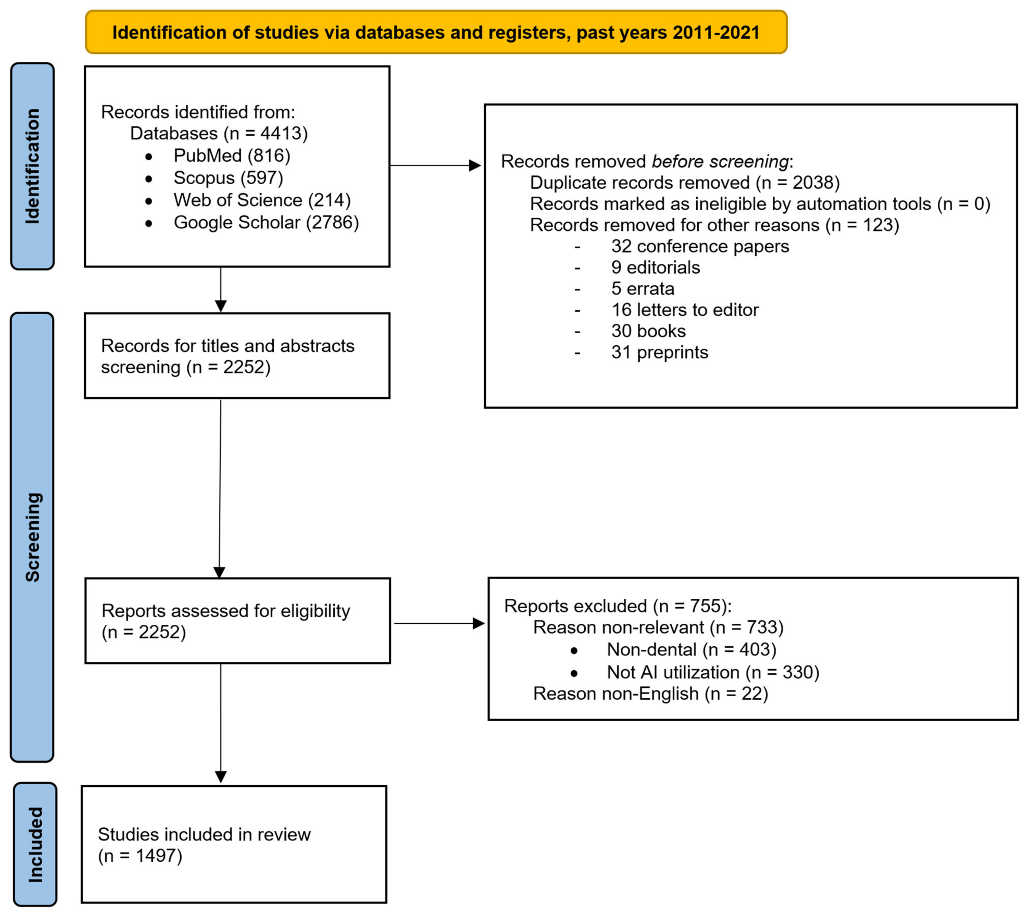
| Methods | Item No. | Checklist Item |
|---|---|---|
| Eligibility criteria | 8 | Only original articles and reviews in English covering dental topics with objective AI utilization were eligible. On 19 April 2022, PubMed, Scopus, Web of Science—Core Collection, and Google Scholar were queried. The focus for the 1st objective was on publications in the years from 1 January 2011 until 31 December 2021. The focus for the 2nd objective was on publications in the years from January 2021 until 19 April 2022. Only published publications were considered. Only publications truly implementing AI (including subfields) in the context of a dental topic or dental specialization were eligible. Respected subfields of AI were: (#1) Machine Learning, (#2) Deep learning, (#3) Neural Networks, (#4) Cognitive Computing, (#5) Natural Language Processing, (#6) Computer Vision. Study design analysis:
|
| Information sources | 9 | Only the following four electronic databases were queried on 19 April 2022:
|
| Search strategy | 10 | PubMed, web search: https://pubmed.ncbi.nlm.nih.gov/advanced/ software: Harzing’s Publish or Perish for batch export, with API key Query: “artificial intelligence”AND(dental OR dentistry OR tooth OR teeth OR dentofacial OR maxillofacial OR orofacial OR orthodontic OR endodontic OR periodontal OR prosthodontic) Type: Articles and reviews in English Relevant publication dates for the 1st objective: 1 January 2011–31 December 2021. Relevant publication dates for the 2nd objective: 2021–19 April 2022, Scopus, web search: https://www.scopus.com/search/form.uri?display=advanced software: Harzing’s Publish or Perish for batch export, with API key Query: “artificial intelligence”AND(dental OR dentistry OR tooth OR teeth OR dentofacial OR maxillofacial OR orofacial OR orthodontic OR endodontic OR periodontal OR prosthodontic) Limited to subject area: Dentistry Type: Articles and reviews in English Relevant publication dates for the 1st objective: 1 January 2011–31 December 2021. Relevant publication dates for the 2nd objective: 2021–19 April 2022 Web of Science—Core Collection web search: https://www.webofscience.com/wos/woscc/advanced-search software: Publish or Perish for batch export, IP based access with login/password Query: “artificial intelligence”AND(dental OR dentistry OR tooth OR teeth OR dentofacial OR maxillofacial OR orofacial OR orthodontic OR endodontic OR periodontal OR prosthodontic) Limited to subject area: Dentistry Oral Surgery Medicine Type: Articles or review articles in English Relevant publication dates for the 1st objective: 1 January 2011–31 December 2021. Relevant publication dates for the 2nd objective 2021–19 April 2022 Google Scholar, web advanced search: https://scholar.google.com/#d=gs_asd&t=1650796298842 software: Harzing’s Publish or Perish for batch export Query: “artificial intelligence” AND (dental OR dentistry OR tooth OR teeth OR dentofacial OR maxillofacial OR orofacial OR orthodontics OR endodontics OR periodontics OR prosthodontics) -motor -rotors -gears -”amelogenesis Imperfecta” Defined Custom year range Type: All, excluding patents and citations Relevant publication dates for the 1st objective: 1 January 2011–31 December 2021. Relevant publication dates for the 2nd objective: 2021–19 April 2022 |
| Study records: | ||
| Data management | 11a | Search and batch export with software: Harzing’s “Publish or Perish” (https://harzing.com/resources/publish-or-perish (accessed on 23 March 2022)). Version for Microsoft Windows: 8.2.3944 (from 23 March 2022), (Windows 11 pro; Microsoft Corp., Redmond, WA, USA) and processed in Microsoft Excel (Excel 365; Microsoft Corp., Redmond, WA, USA). For export and data manipulation, Google Sheets (Alphabet Inc., Mountain View, CA, USA) were also used. This is an online spreadsheet program included as part of the free, web-based Google Docs Editors suite offered by Google. |
| Selection process | 11b | Three independent reviewers conducted each phase of the review. |
| Data collection process | 11c | As the first objective was solely quantitative, the need to extract data from reports was valid only for the second qualitative objective. Publication title and abstract analysis defined the focus of each paper; this was performed separately by three reviewers. Upon the preliminary evaluation of registered papers, 22 dental topics/specializations were defined. Only in cases where an eligible publication was not clearly classifiable from the title and abstract, was a full text analyzed by three independent reviewers. After the reviewers reached a consensus, either the paper’s focus was recorded, or (more frequently) the paper was removed as irrelevant. Only one qualitative marker was assigned to each selected publication for the 2nd objective. In cases of discrepancy between independent reviewers, a consensus had to be reached. The dominant dental topic regarding AI utilization was evaluated, unless the article was equally balanced between two specializations. There were 5 areas that covered all undividable interdisciplinary intersections, which were mostly found between dental radiology, orthodontics, and surgery. For these cases, five border groups had to be defined. |
| Data items | 12 | All 22 qualitative variables included five interdisciplinary groups of papers (# 4,5,6,8,9). |
| Outcomes and prioritization | 13 | The primary outcome of the 1st objective was the list of selected publications for each year. The primary outcome of the 2nd objective was the list of qualitatively evaluated publications distributed into 22 focus groups/topics. Papers focused on more than two qualitative groups were assigned to “general scope” group. Papers addressing dental education in at least one of the defined topics/fields were assigned separately to the “dental education” group. |
| Risk of bias in individual studies | 14 | Each eligible study was evaluated independently by three reviewers. To minimize risk of bias of individual studies, only the studies truly dealing with AI implementation were included. As a number of discrepancies occurred between the three independent reviewers, further tools used to assess the risk of bias in the systematic review were considered. Each initial disagreement was noted. |
| Data synthesis | 15a | The 1st objective provided a list of selected papers. These were grouped according to their publication year and their topics summarized for each year. Percentual increments per year were calculated. The 2nd objective provided a list of selected papers to be distributed into defined focus groups. |
| 15b | For simplicity, no combining of data from other studies was planned. | |
| 15c | No other additional analyses were proposed (such as sensitivity or subgroup analyses, or meta-regression). | |
| 15d | If quantitative synthesis was not appropriate, a systematic narrative synthesis was provided with information presented in the text and tables to summarize and explain the impact of included studies. | |
| Meta-bias(es) | 16 | The weight of each study included was equivalent. Overall, the selective reporting of study results (and the failure to publish small or nonsignificant results) leads to the overestimation of intervention effects in systematic reviews, a phenomenon called meta-bias. |
| Confidence in cumulative evidence | 17 | For the simplicity of this review design, only a “summary of findings” tables are included with a summary of the amount of evidence, in accordance with the GRADE framework (GRADE Working Group, 2004; Guyatt et al. [72]) which combines considerations of risk of bias, directness, heterogeneity, precision, and publication bias [72]. |
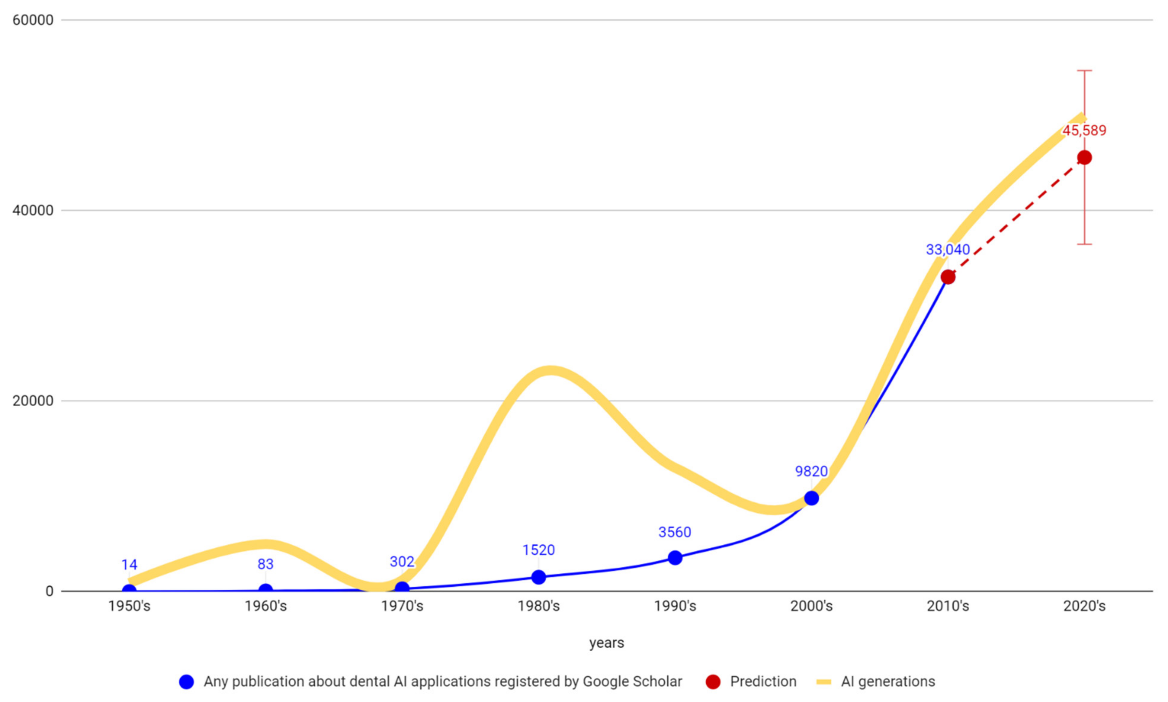
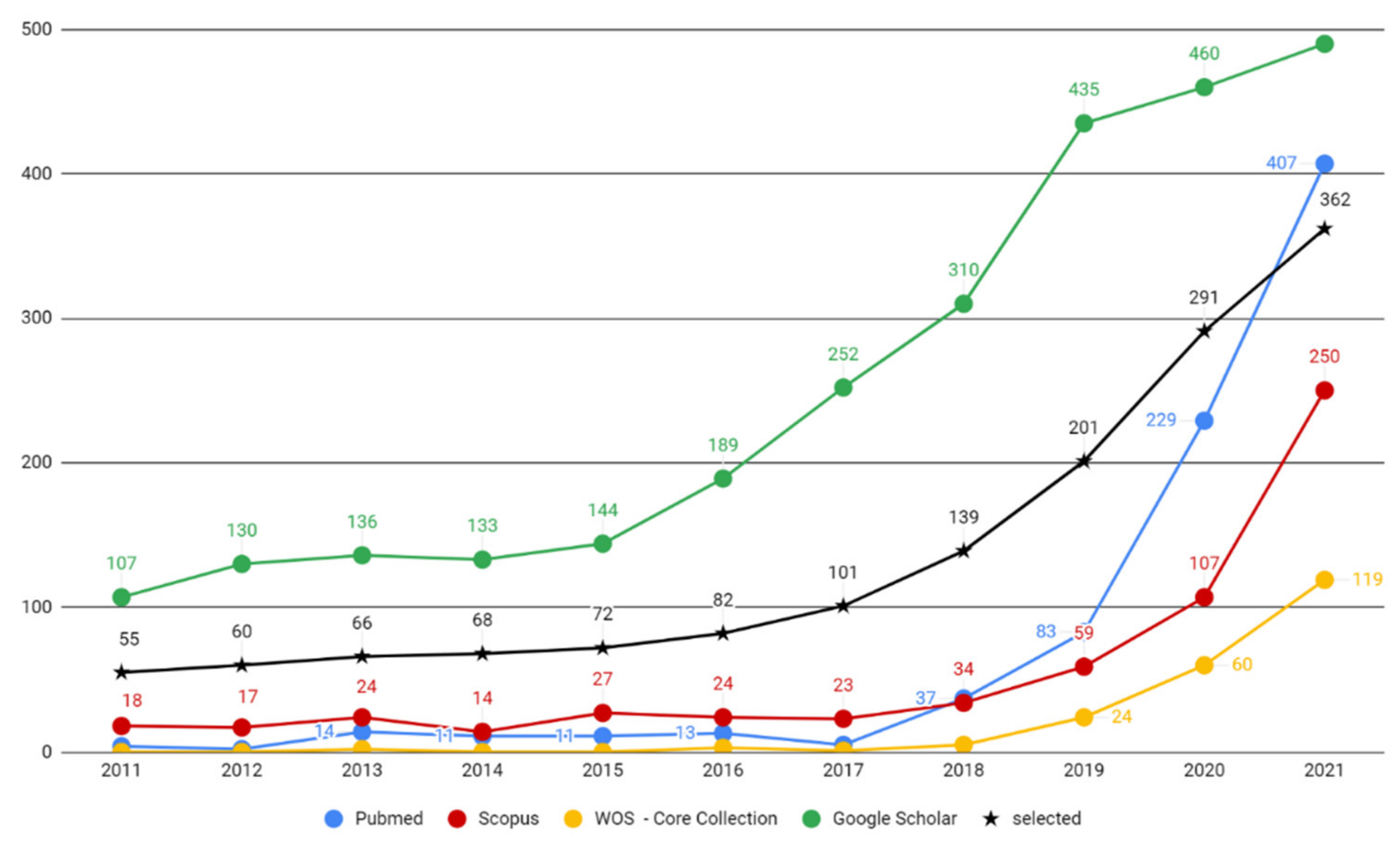
| Year | PubMed | Scopus 1 | WOS 1 | Google Scholar | Registered | Included | Increase |
|---|---|---|---|---|---|---|---|
| 2011 | 4 | 18 | 0 | 107 | 129 | 55 | |
| 2012 | 2 | 17 | 0 | 130 | 149 | 60 | +9.09% |
| 2013 | 14 | 24 | 2 | 136 | 166 | 66 | +10.00% |
| 2014 | 11 | 14 | 0 | 133 | 168 | 68 | +3.03% |
| 2015 | 11 | 27 | 0 | 144 | 182 | 72 | +5.88% |
| 2016 | 13 | 24 | 3 | 189 | 229 | 82 | +13.89% |
| 2017 | 5 | 23 | 1 | 252 | 281 | 101 | +23.17% |
| 2018 | 37 | 34 | 5 | 310 | 386 | 139 | +37.62% |
| 2019 | 83 | 59 | 24 | 435 | 601 | 201 | +44.60% |
| 2020 | 229 | 107 | 60 | 460 | 856 | 291 | +44.78% |
| 2021 | 407 | 250 | 119 | 490 | 1266 | 362 | +24.40% |
| Year. | PubMed | Scopus 1 | WOS 1 | Google Scholar | Registered | Included |
|---|---|---|---|---|---|---|
| 2022 | 39 | 85 | 23 | 304 | 451 | 135 |
| 2021 | 407 | 250 | 119 | 490 | 1266 | 362 |
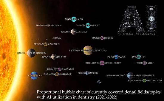
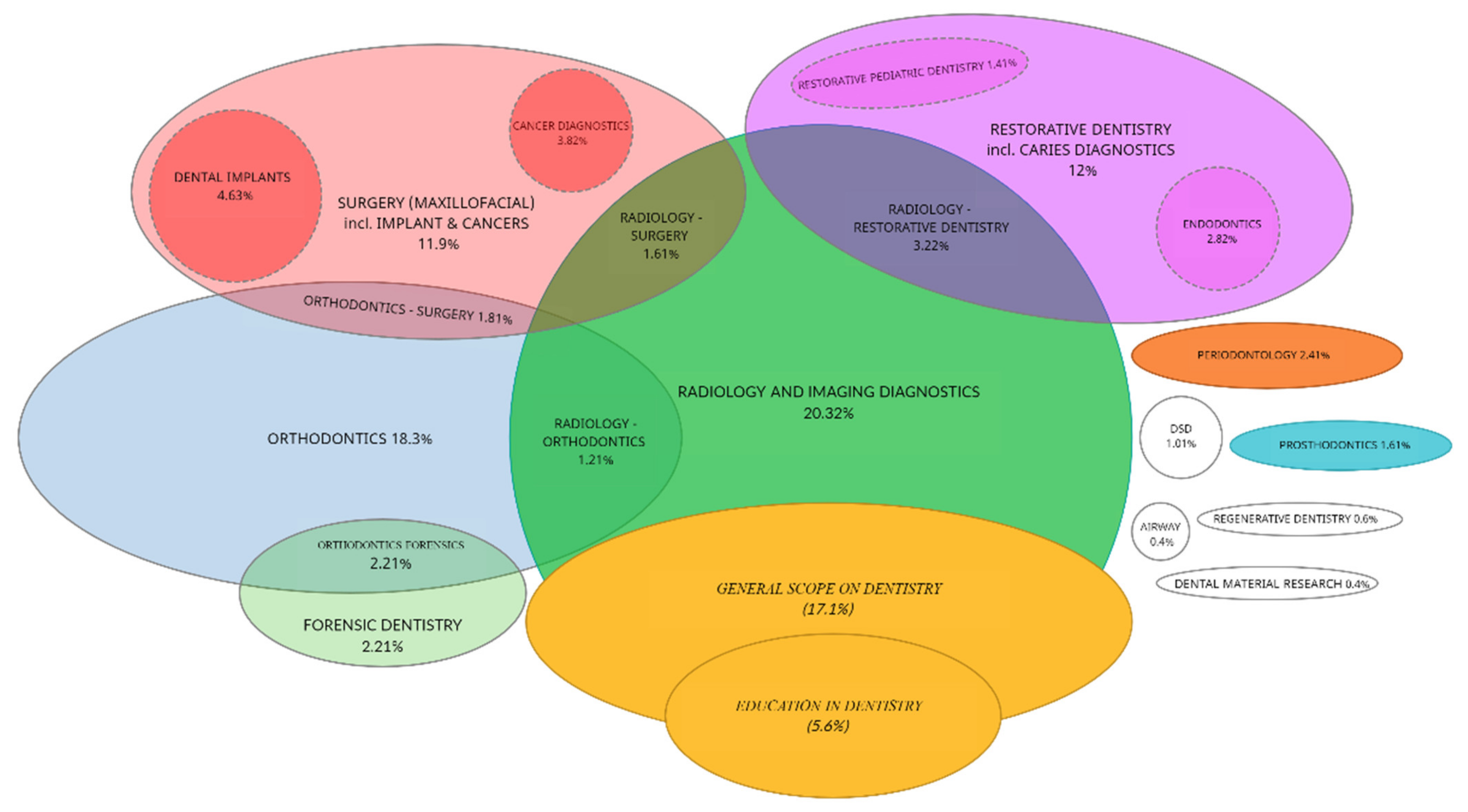
| # | Focus in (Topic/Specialty) | Amount | Percentage |
|---|---|---|---|
| 1 | GENERAL SCOPE ON DENTISTRY | 85 | 17.10% |
| 2 | EDUCATION IN DENTISTRY | 28 | 5.63% |
| 3 | RADIOLOGY AND IMAGING DIAGNOSTICS | 101 | 20.32% |
| 4 | RADIOLOGY—ORTHODONTICS 1 | 6 | 1.21% |
| 5 | RADIOLOGY—RESTORATIVE DENTISTRY 1 | 16 | 3.22% |
| 6 | RADIOLOGY—SURGERY 1 | 8 | 1.61% |
| 7 | ORTHODONTICS | 72 | 14.49% |
| 8 | ORTHODONTICS—FORENSICS 1 | 10 | 2.01% |
| 9 | ORTHODONTICS—SURGERY 1 | 9 | 1.81% |
| 10 | SURGERY (MAXILLOFACIAL) | 17 | 3.42% |
| 11 | CANCER DIAGNOSTICS | 19 | 3.82% |
| 12 | DENTAL IMPLANTS | 23 | 4.63% |
| 13 | RESTORATIVE DENTISTRY incl. CARIES DIAG. | 39 | 7.85% |
| 14 | RESTORATIVE PEDIATRIC DENTISTRY | 7 | 1.41% |
| 15 | PERIODONTOLOGY | 12 | 2.41% |
| 16 | ENDODONTICS | 14 | 2.82% |
| 17 | FORENSIC DENTISTRY | 11 | 2.21% |
| 18 | PROSTHODONTICS | 8 | 1.61% |
| 19 | DSD | 5 | 1.01% |
| 20 | REGENERATIVE DENTISTRY | 3 | 0.60% |
| 21 | AIRWAY | 2 | 0.40% |
| 22 | DENTAL MATERIAL RESEARCH | 2 | 0.40% |
| Title | Author | Journal | Year |
|---|---|---|---|
| Detection and diagnosis of dental caries using a deep learning-based convolutional neural network algorithm [76] | Lee, Jae-Hong; et al. | JOURNAL OF DENTISTRY | 2018 |
| Diagnosis and prediction of periodontally compromised teeth using a deep learning-based convolutional neural network algorithm [77] | Lee, Jae-Hong; et al. | JOURNAL OF PERIODONTAL AND IMPLANT SCIENCE | 2018 |
| Deep Learning for the Radiographic Detection of Apical Lesions [78] | Ekert, Thomas; et al. | JOURNAL OF ENDODONTICS | 2019 |
| Convolutional neural networks for dental image diagnostics: A scoping review [79] | Schwendicke, Falk; et al. | JOURNAL OF DENTISTRY | 2019 |
| Artificial Intelligence in Dentistry: Chances and Challenges [25] | Schwendicke, F.; et al. | JOURNAL OF DENTAL RESEARCH | 2020 |
| An overview of deep learning in the field of dentistry [80] | Hwang, Jae-Joon; et al. | IMAGING SCIENCE IN DENTISTRY | 2019 |
| Deep learning in medical image analysis: A third eye for doctors [81] | Fourcade, A.; Khonsari, R. H. | JOURNAL OF STOMATOLOGY ORAL AND MAXILLOFACIAL SURGERY | 2019 |
| A deep-learning artificial intelligence system for assessment of root morphology of the mandibular first molar on panoramic radiography [82] | Hiraiwa, Teruhiko; et al. | DENTOMAXILLOFACIAL RADIOLOGY | 2019 |
| Deep-learning classification using convolutional neural network for evaluation of maxillary sinusitis on panoramic radiography [83] | Murata, Makoto; et al. | ORAL RADIOLOGY | 2019 |
| Staging and grading of oral squamous cell carcinoma: An update [84] | Almangush, Alhadi; et al. | ORAL ONCOLOGY | 2020 |
Publisher’s Note: MDPI stays neutral with regard to jurisdictional claims in published maps and institutional affiliations. |
© 2022 by the authors. Licensee MDPI, Basel, Switzerland. This article is an open access article distributed under the terms and conditions of the Creative Commons Attribution (CC BY) license (https://creativecommons.org/licenses/by/4.0/).
Share and Cite
Thurzo, A.; Urbanová, W.; Novák, B.; Czako, L.; Siebert, T.; Stano, P.; Mareková, S.; Fountoulaki, G.; Kosnáčová, H.; Varga, I. Where Is the Artificial Intelligence Applied in Dentistry? Systematic Review and Literature Analysis. Healthcare 2022, 10, 1269. https://doi.org/10.3390/healthcare10071269
Thurzo A, Urbanová W, Novák B, Czako L, Siebert T, Stano P, Mareková S, Fountoulaki G, Kosnáčová H, Varga I. Where Is the Artificial Intelligence Applied in Dentistry? Systematic Review and Literature Analysis. Healthcare. 2022; 10(7):1269. https://doi.org/10.3390/healthcare10071269
Chicago/Turabian StyleThurzo, Andrej, Wanda Urbanová, Bohuslav Novák, Ladislav Czako, Tomáš Siebert, Peter Stano, Simona Mareková, Georgia Fountoulaki, Helena Kosnáčová, and Ivan Varga. 2022. "Where Is the Artificial Intelligence Applied in Dentistry? Systematic Review and Literature Analysis" Healthcare 10, no. 7: 1269. https://doi.org/10.3390/healthcare10071269