1. Introduction
Epithelioid hemangioma (EH, ISSVA classification 2018) [
1] is a rare reactive vasoproliferative disease presenting with painless vascular nodules in the dermal and subcutaneous tissues of the head and neck. It is also known as angiolymphoid hyperplasia with eosinophilia, histiocytoid hemangioma, inflammatory angiomatous nodule, atypical granuloma and pseudopyogenic granuloma. Typically, EH arises in the head and neck region of young women, presenting as raised colored itchy nodules. While it rarely regresses spontaneously, there are no reports of malignant transformation [
2,
3].
As EH is quite rare, it can be clinically misdiagnosed for a number of benign inflammatory conditions such as Kimura disease, IgG4-related skin disease, bacillary angiomatosis, cutaneous epithelioid angiomatous nodule and malignant epithelial vascular tumors such as epithelioid sarcoma-like hemangioendothelioma, epithelioid hemangioendothelioma and epithelioid angiosarcoma [
4,
5]. EH can also be confused with more common conditions such as lipomas, sebaceous cysts and skin neoplasms such as basal and squamous cell carcinoma [
6,
7].
Although the etiology of EH remains unclear, surgical excision with negative margins has proven to be the treatment of choice.
Thus far, few cases of EH localized to the nose have been described, rather incompletely, in the literature, but this is the first case reported of a nasal tip involvement. Herein, we present in detail a case of a 47-year-old woman with just such a lesion of the nose, focusing on its diagnostic iter, emphasizing the importance of a radical surgical excision with a one-year follow-up experience.
2. Case Report
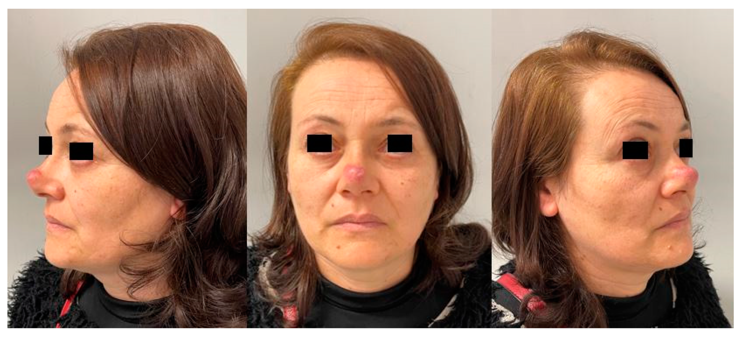
A 47-year-old woman was referred to our department with a solitary, painless but itchy nodule occupying the tip and part of the nasal dorsum. The lesion onset dated 2 years. The patient had no family history of any cutaneous diseases or vascular malformations and no history of trauma. A punch biopsy showed a rich amount of sebaceous adnexa, a periannesial and perivascular lymphoplasmacellular infiltrate with vascular ectasias and mild fibrosis, suggesting the possibility of discoid erythematous lupus.
The patient started topical treatment but with no benefits. The patient’s lesion progressively increased in size and after one year, on examination, it had developed into a fixed lump covered by thin whitish skin with small telangiectasias (
Figure 1).
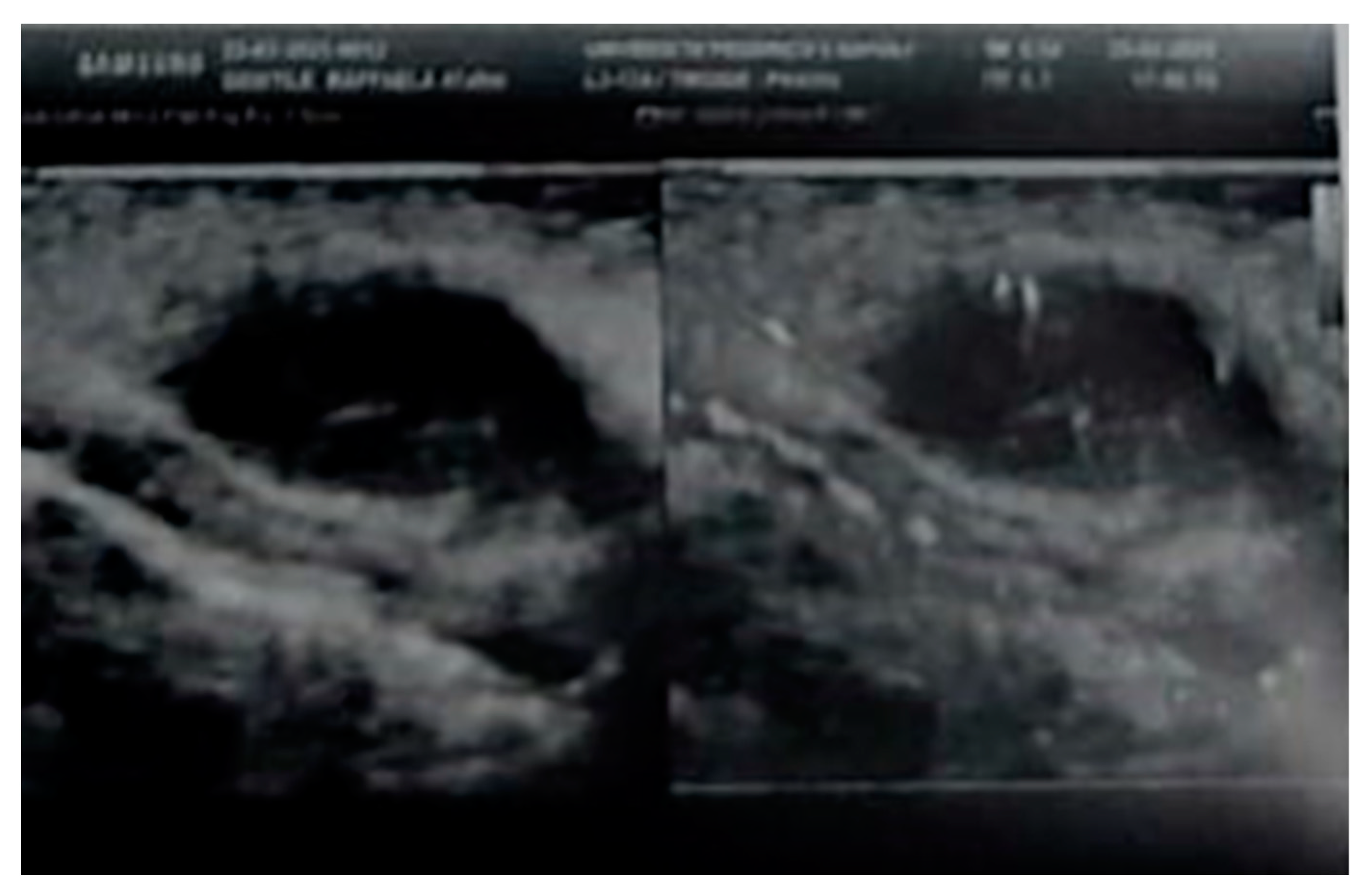
A Doppler ultrasonography using an 18 Mhz linear transducer revealed a highly vascularized nodular subcutaneous lesion of 23 mm × 16 mm (
Figure 2).
A biochemistry test, routine hematology and erythrocyte sedimentation rate were all within the normal reference ranges.
While refusing another biopsy, the patient requested the total excision of the lesion and, therefore, a definitive diagnosis was postponed until formal lesion excision.
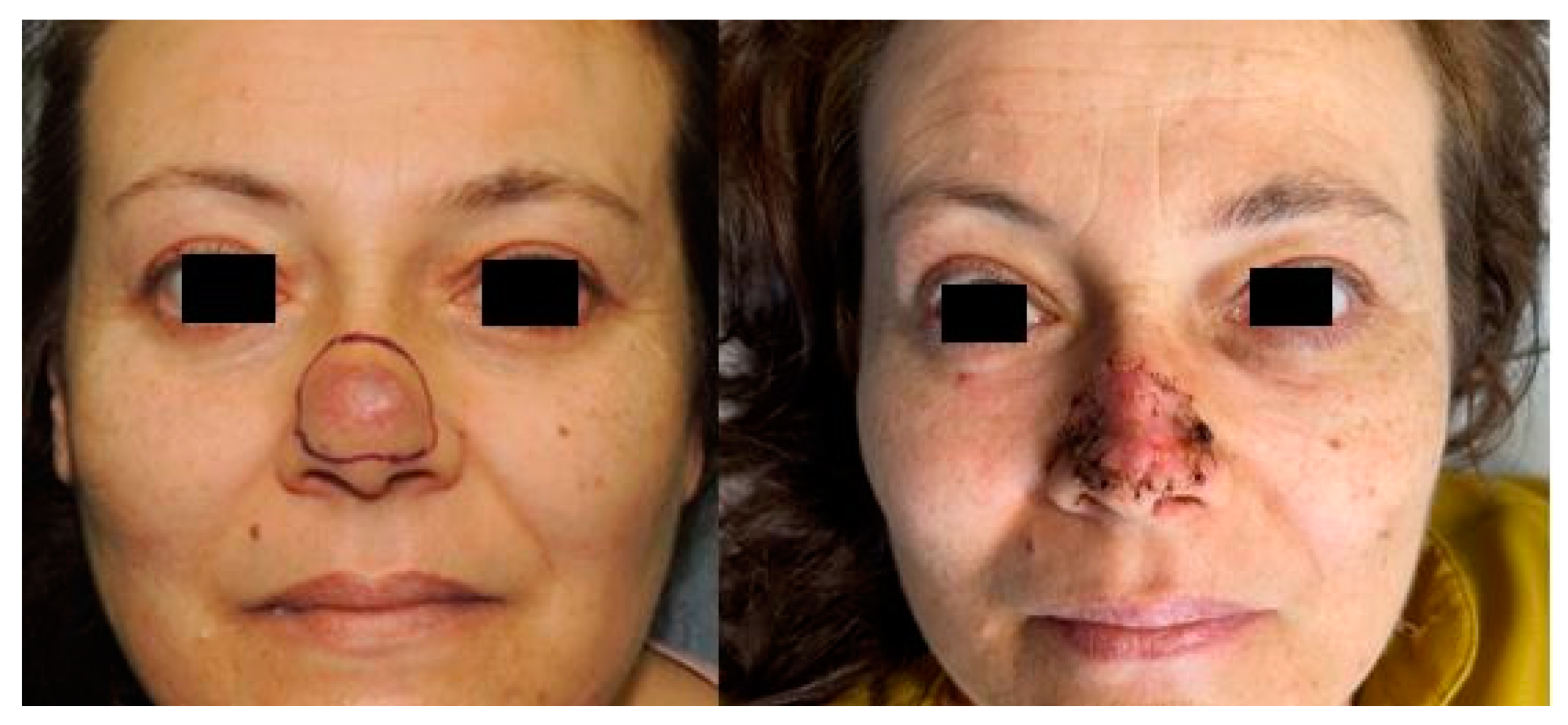
Subsequently, an excision under local anesthesia with sedation was performed. The residual defect resulting from the wide excision was covered using a full-thickness skin graft harvested from the preauricular region (
Figure 3).
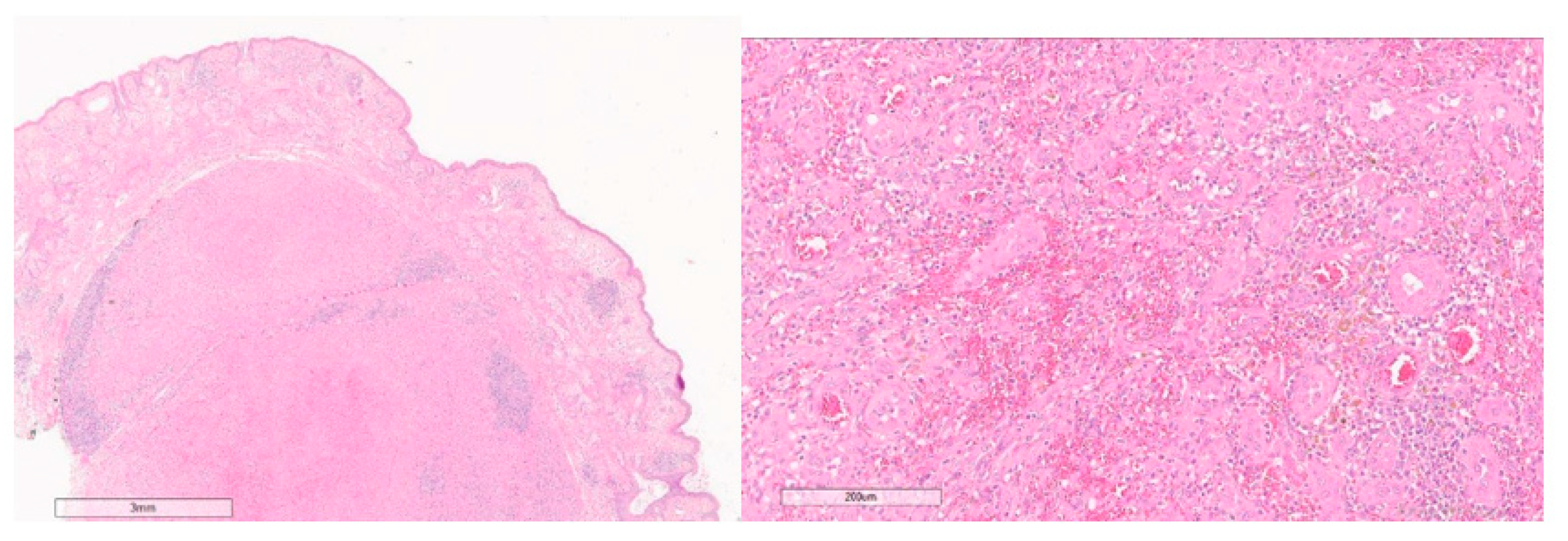
At histology, hematoxylin and eosin-stained sections showed redundant vascular proliferation with multilobular architecture occupying the medium and deep dermis consisting of small caliper vessels with mildly hyperplastic endothelium and epithelioid aspects (
Figure 4). A rich lymphocytic infiltration with a predominant eosinophilic quote was associated. An immunohistochemistry stain showed the presence of the vascular proliferation markers CD31+, CD34+ and ERG1+. In accordance with these findings, the lesion was diagnosed as epithelioid hemangioma.
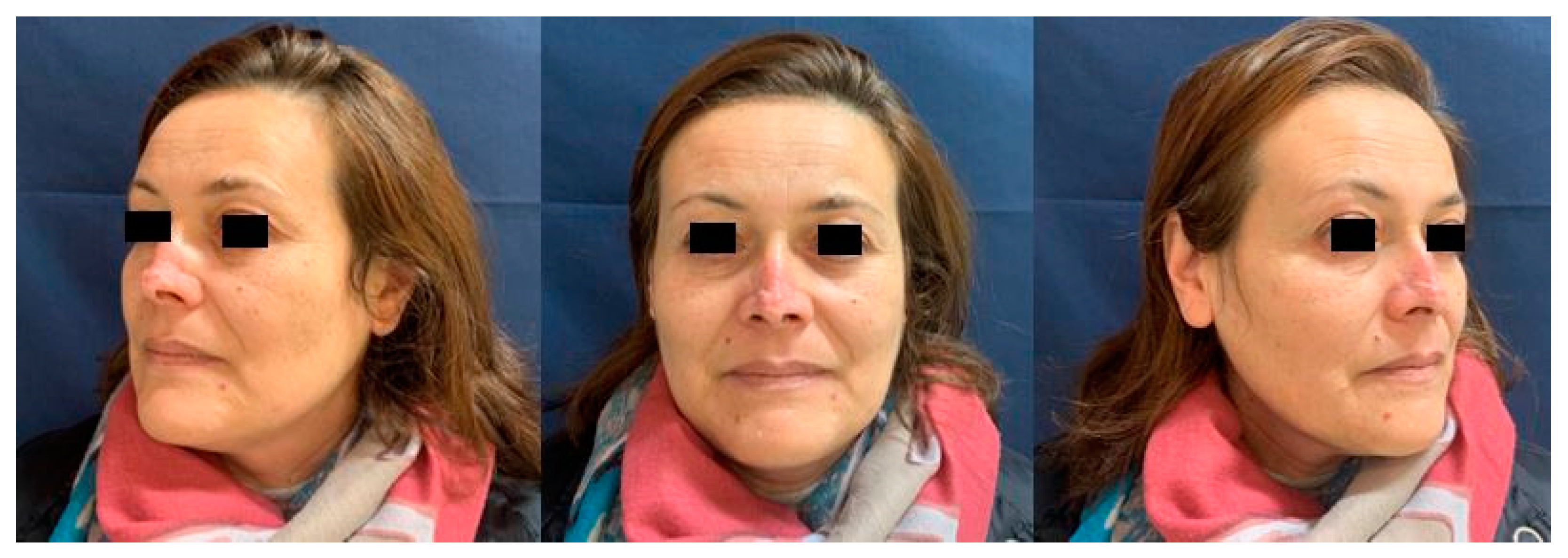
At the one-year follow-up, the patient had no sign of recurrence and was satisfied with the result (
Figure 5). A further nasal tip surgical revision was offered but she refused.
3. Discussion
EH is an uncommon benign vascular proliferative lesion of unknown origin with a good prognosis and no reported incidence of metastatic disease. Nevertheless, these lesions have high rates of local recurrence and in some cases can have a simultaneous multifocal presentation, even in different organs [
6]. Some authors suggest that traumatic events may trigger the onset of EH [
8]. EH is commonly found in the small vessels of the dermis or subcutis, but it can also affect larger muscular arteries such as the facial artery and the temporal artery [
4].
Clinical diagnosis can be difficult as, in most cases, the only symptom is a progressively tender swelling next to a vessel course. Skin discoloration may take place, especially in districts with a terminal circulation such as the nose [
8]. Pain and/or functional symptoms may rarely manifest because of tumor compression on adjacent structures (nerves, vessels or tendons). Imaging can be useful for studying tumor extension, vascular involvement and any other simultaneous presentations, but a histology is mandatory in order to obtain a definitive diagnosis. Microscopic features include lined vessels with atypical endothelial cells which appear plump/swollen and epithelioid, associated with an eosinophilic and lymphocytic infiltration in the perivascular tissue. In 20% of the cases, generalized eosinophilia can be present and, occasionally, accompanied by raised serum IgE levels [
4,
6]. Endothelial cell markers such as CD31 or CD34 can confirm diagnosis [
9].
The differential diagnosis of EH should include Kimura disease, pyogenic granuloma and Kaposi sarcoma [
10].
Surgical excision is the treatment of choice for EH. Due to its high risk of recurrence, a wide radical excision should be performed [
11]. When important facial areas with a high aesthetic impact such as the nose are involved, reconstruction can be difficult. Depending on the defect size left after a wide excision, different options can be considered [
12,
13,
14]. In this patient, because the punch biopsy result was not diagnostic and due to the uncertain nature of the lesion, we preferred to cover the defect with a full thickness skin graft. A local flap-based reconstruction was reserved if any further excision widening was necessary.
Once completely removed, EH local recurrence is rare; however, with an incomplete resection, almost 33% of patients have a recurrence, either at the same site or a distant site along the course of the blood vessel from where the EH originated [
5,
11].
The nasal localization of EH is uncommon and only six cases have been reported in the literature so far [
10,
15,
16,
17,
18,
19]. Cutaneous involvement has been reported in only two cases [
10,
17]. Surgical excision has been the treatment of choice in all cases, but no details on reconstruction techniques or possible complications have been provided. Furthermore, no follow-up data have been reported, with the exception of Sedran et al who followed a patient for seven years and Youssef et al. who reported a lesion recurrence, which was treated with intralesional prednisolone and a 0.1% Tacrolimus ointment.
To conclude, HE groupings include a variety of rare vascular lesions that can affect different tissues and organs. Diagnostic investigations should include imaging techniques to determine the extent of the lesion and to confirm vascular involvement. Once the diagnosis of HE is suspected, an early and complete surgical excision of the lesions with a thorough histological and immunohistochemical analysis should be undertaken.
Author Contributions
Conceptualization, F.S., A.C. (Armando Calogero) and A.B.; writing—original draft preparation, A.C. (Annachiara Cavaliereand), G.P. and L.D.; writing—review and editing, F.S. and A.C. (Annachiara Cavaliereand); supervision, F.S., A.S. and C.S. All authors have read and agreed to the published version of the manuscript.
Funding
This research received no external funding.
Informed Consent Statement
Written informed consent has been obtained from the patient(s) to publish this paper.
Data Availability Statement
Not applicable.
Acknowledgments
This paper has been English language edited by Juliet Ippolito, B.A.
Conflicts of Interest
The authors declare no conflict of interest.
References
- ISSVA Classification of Vascular Anomalies ©2018 International Society for the Study of Vascular Anomalies. Available online: Issva.org/classification (accessed on 19 January 2022).
- Rosai, J.; Gold, J.; Landy, R. The histiocytoid hemangiomas. A unifying concept embracing several previously described entities of skin, soft tissue, large vessels, bone, and heart. Hum. Pathol. 1979, 10, 707–730. [Google Scholar] [CrossRef]
- Allen, P.W.; Ramakrishna, B.; MacCormac, L.B. The histiocytoid hemangiomas and other controversies. Pathol. Annu. 1992, 27 Pt 2, 51–87. [Google Scholar] [PubMed]
- Arnander, M.W.; Anderson, N.G.; Schönauer, F. The ultrasound halo sign in angiolymphoid hyperplasia of the temporal artery. Br. J. Radiol. 2006, 79, e184–e186. [Google Scholar] [CrossRef] [PubMed]
- Wiggins, C.J.; Dibbs, R.P.; Bartlett, E.L.; Ashton, D.J.; Maricevich, R.S. Atypical presentation and management of an epithelioid hemangioma: A case report and review of the literature. Ann. Pediatr. Surg. 2020, 16, 53. [Google Scholar] [CrossRef]
- Fetsch, J.F.; Weiss, S.W. Observations concerning the pathogenesis of epithelioid hemangioma (angiolymphoid hyperplasia). Mod. Pathol. 1991, 4, 449–455. [Google Scholar] [PubMed]
- Sharp, J.F.; Rodgers, M.J.C.; MacGregor, F.B.; Meehan, C.J.; McLaren, K. Angiolymphoid hyperplasia with eosinophilia. Laryngol. Otol. 1990, 104, 977–979. [Google Scholar] [CrossRef] [PubMed]
- Accardo, G.; Nele, G.; Turrà, F.; La Rusca, I.; Schonauer, F. A rare case of ‘histiocytoid haemangioma’ of the hand. J. Plast. Reconstr. Aesthet. Surg. 2013, 66, e318–e320. [Google Scholar] [CrossRef] [PubMed] [Green Version]
- Mentzel, T.; Beham, A.; Calonje, E.; Katenkamp, D.; Fletcher, C.D. Epithelioid hemangioendothelioma of skin and soft tissues: Clinicopathologic and immunohistochemical study of 30 cases. Am. J. Surg. Pathol. 1997, 21, 363–374. [Google Scholar] [CrossRef] [PubMed]
- Kabir, S.; Teimoorian, M.; Mahdavi, M.; Meibodi, N.T.; Tajalli, M.; Goyonlo, V.M.; Sabourirad, S. A solitary erythematous papule on the nose. JAAD Case Rep. 2020, 6, 663–665. [Google Scholar] [CrossRef] [PubMed]
- Baghestani, S.; Firooz, A.; Ghazisaidi, M.R. A refractory case of angiolymphoid hyperplasia with eosinophilia successfully treated by surgery. J. Dermatol. Treat. 2011, 22, 49–51. [Google Scholar] [CrossRef] [PubMed]
- Cavaliere, A.; Maisto, B.; Zaporojan, T.; Giordano, L.; Sorbino, L.; Zaffiro, A.; Vozza, A.; La Padula, S.; D’Andrea, F.; Schonauer, F. Extended rotation flap for reconstruction of partial thickness defects of the tip and nasal ala region: In search of better aesthetic results. JPRAS Open 2021, 29, 82–88. [Google Scholar] [CrossRef] [PubMed]
- La Padula, S.; Abbate, V.; Di Monta, G.; Schonauer, F. Levator alae nasi muscle V-Y island flap for nasal tip reconstruction. J. Cranio-Maxillo-Facial Surg. 2017, 45, 432–435. [Google Scholar] [CrossRef] [PubMed]
- Schonauer, F.; Scafati, S.T.; Molea, G. Supratrochlear artery based V-Y flap for partial eyebrow reconstruction. J. Plast. Reconstr. Aesthet Surg. 2010, 63, 1391–1392. [Google Scholar] [CrossRef] [PubMed]
- Chiu, S.-C. An unusual case of angiolymphoid hyperplasia with eosinophilia of the nose. Ear Nose Throat J. 2013, 92, E10–E11. [Google Scholar] [PubMed]
- Panduranga Kamath, M.; Bhojwani, K.M.; Bhandarkar, A.M.; Pai, R.R.; Rent, N.H. Angiolymphoid hyperplasia with eosinophilia of root of nose: A rare phenomenon. J. Clin. Diagn. Res. 2014, 8, 144–145. [Google Scholar] [PubMed]
- Van Ratingen, A.R.; Linden, V.; Sillevis Smitt, J.H. Case Report: Angiolymphoid Hyperplasia with Eosinophilia of the Nose. Dermatol. Case Rep. 2016, 1, 105. [Google Scholar]
- Youssef, A.; Hasan, A.R.; Youssef, Y.; Al-Soufi, L.; Elshimali, Y.; Alshehabi, Z. Angiolymphoid hyperplasia with eosinophilia: A case report. J. Med. Case Rep. 2018, 12, 89. [Google Scholar] [CrossRef] [PubMed]
- Sedran, L.; Bonaso, M.; Mettus, A.; Roccia, F. Epithelioid Hemangioma of the Face. J. Craniofacial Surg. 2018, 29, e736–e739. [Google Scholar] [CrossRef] [PubMed]
| Publisher’s Note: MDPI stays neutral with regard to jurisdictional claims in published maps and institutional affiliations. |
© 2022 by the authors. Licensee MDPI, Basel, Switzerland. This article is an open access article distributed under the terms and conditions of the Creative Commons Attribution (CC BY) license (https://creativecommons.org/licenses/by/4.0/).


 ,
, 


