

Implementing Magnetic Resonance Imaging Brain Disorder Classification via AlexNet–Quantum Learning


Abstract
:1. Introduction
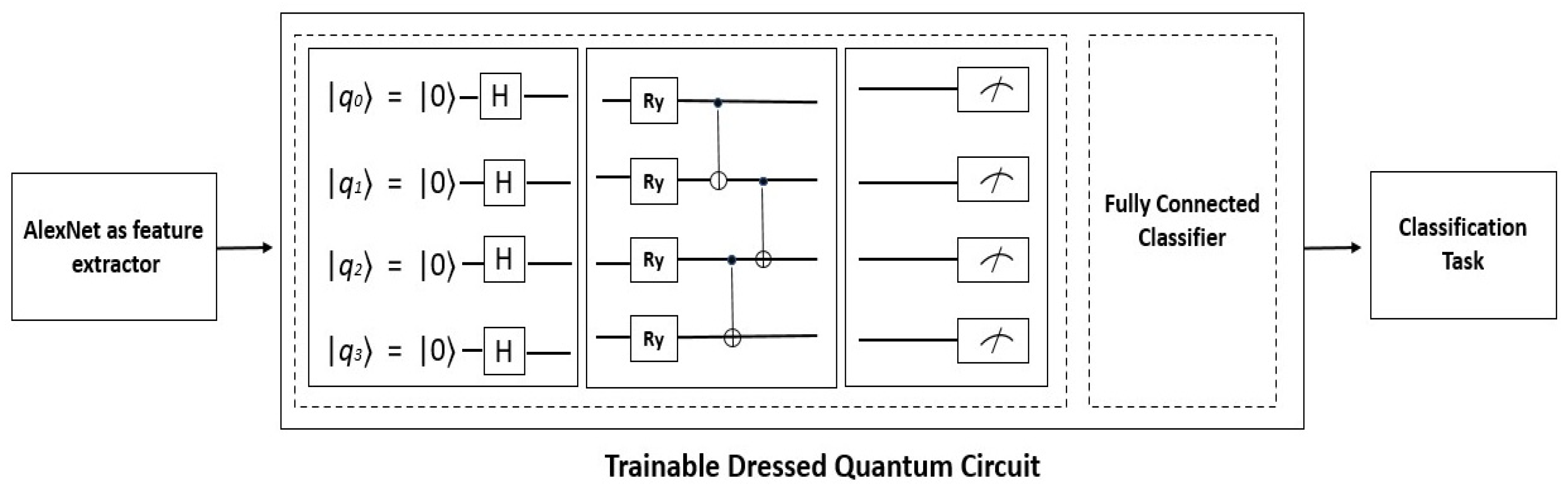
- A binary classification framework for brain disorders based on the AlexNet–quantum transfer learning network is proposed;
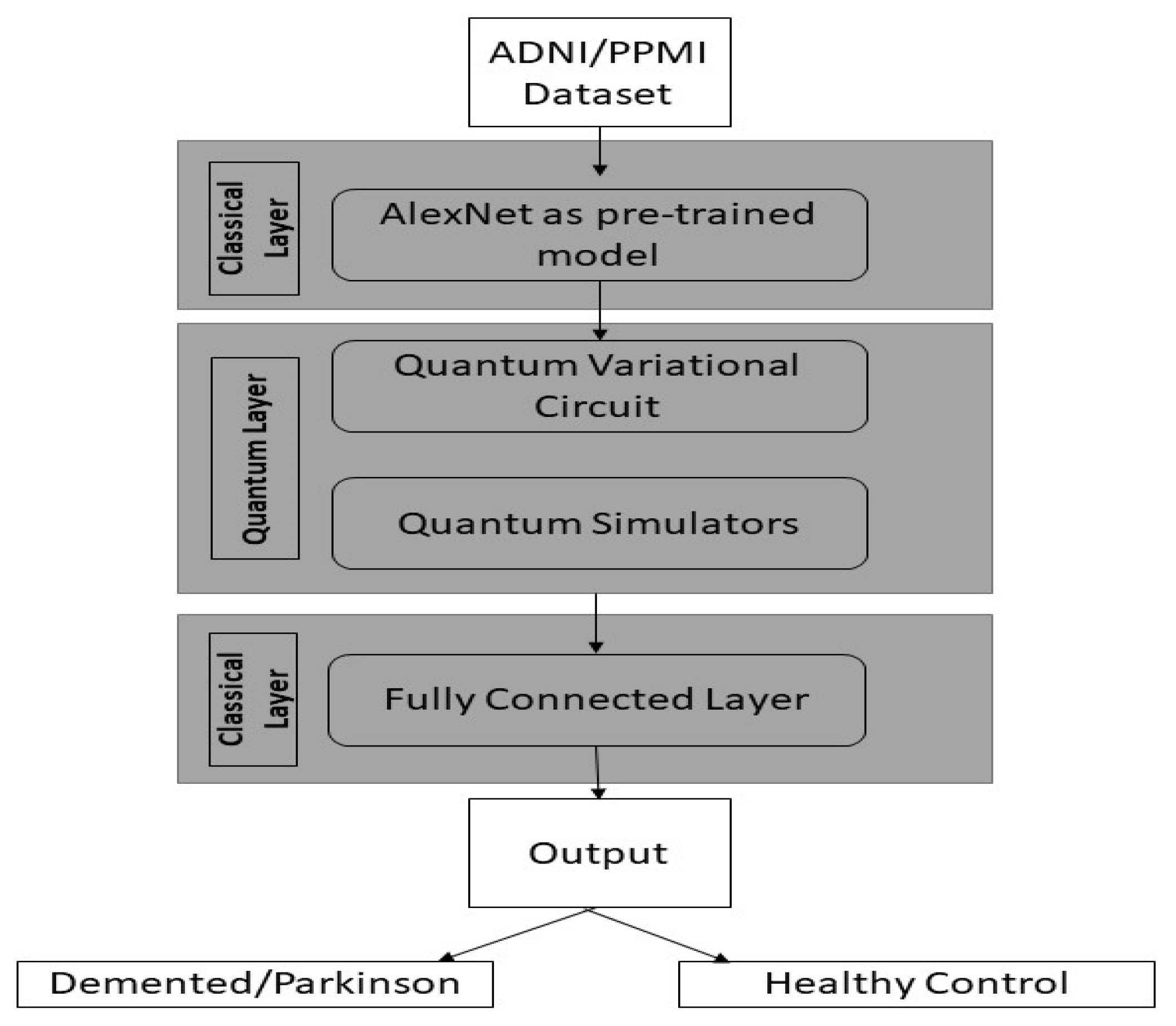
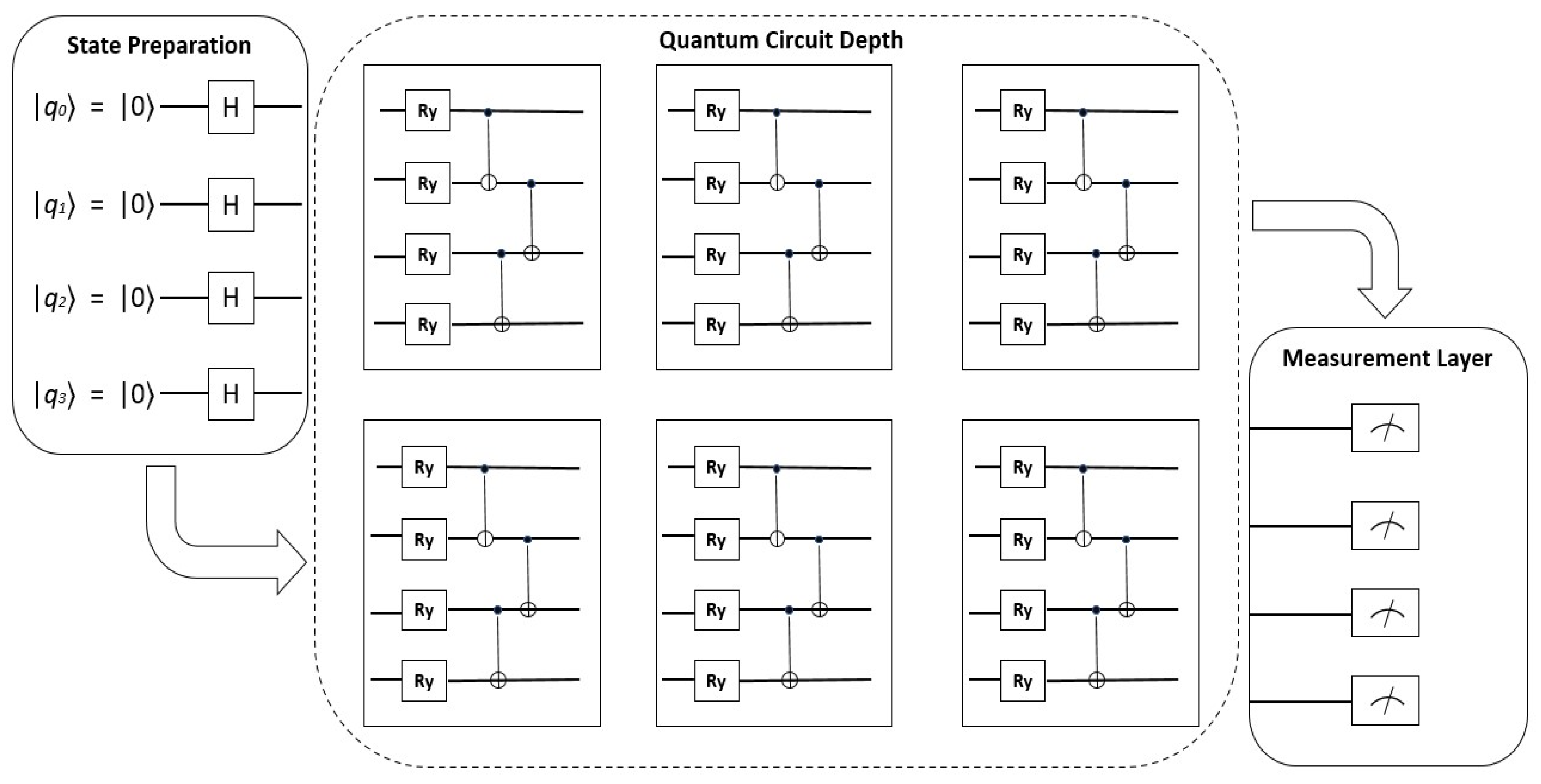
- The Quantum learning model is implemented with the depth of six quantum layers and this model leverages quantum simulator;
- To validate the robustness, and efficiency of the brain disease system in real-time, the PPMI dataset for PD classification and the ADNI dataset for AD classification was used for training and testing the model; and
- Lastly, the performance of the brain disease–quantum neural system is compared with other deep transfer learning models such as AlexNet, VGG-16, ResNet 50, and Inception v3 on the same brain disorder dataset.
2. Literature Review
3. Materials and Methods
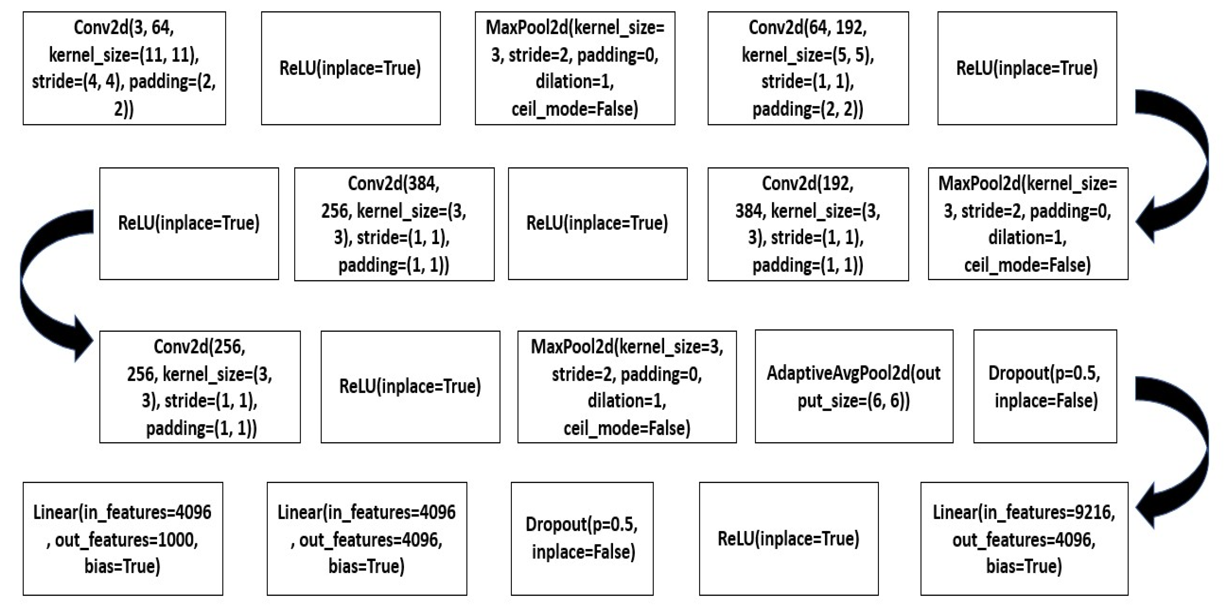
3.1. AlexNet Architecture Using Transfer Learning
3.2. Quantum Variational Circuit
| Algorithm 1: AlexNet–quantum deep network |
| Input: The brain disorder dataset consists of MRI images of brain disease and normal controls Output: Binary classification of Brain disease using MRI scans based on the AlexNet–quantum model |
| Steps: Organize the brain disorder dataset by downloading it from the PPMI and ADNI databases. Preprocess the MRI images. Using AlexNet, Extract features to give as input to quantum learning circuit whose steps are given as: |
| Quantum learning Circuit |
| V = (𝓃, 𝓃) Feature vector dataset Inserting the feature vector dataset into the quantum learning circuit Taking inner product by creating superposition and entanglement state |𝓜*⟩ = Measurement state for decoding the vector into a classical state Return value 𝓜* |
| Classifier: Using AlexNet fully connected layer to classify the vector into two classes. |
4. Results and Analysis
5. Conclusions
Author Contributions
Funding
Institutional Review Board Statement
Informed Consent Statement
Data Availability Statement
Acknowledgments
Conflicts of Interest
Abbreviations
| AD | Alzheimer’s disease |
| ADNI | Alzheimer’s disease neuroimaging initiative |
| ANN | artificial neural network |
| CAD | computer-assisted diagnostic systems |
| CNN | convolutional neural network |
| CPU | central processing unit |
| DNN | deep neural network |
| EEG | electroencephalography |
| GPU | graphics processing unit |
| MRI | magnetic resonance imaging |
| NC | normal control |
| NDD | neurodegenerative Disease |
| PD | Parkinson’s disease |
| PET | positron emission tomography |
| PPMI | Parkinson progression marker initiative |
| QML | quantum machine learning |
| QPU | quantum processing unit |
| TPU | tensor processing unit |
References
- Association, A. Alzheimer’s disease facts and figures. Alzheimer’s Dement. 2017, 13, 325–373. [Google Scholar]
- Beitz, J.M. Parkinson’s disease: A review. Front. Biosci.-Sch. 2014, 6, 65–74. [Google Scholar] [CrossRef] [PubMed]
- Aich, S.; Joo, M.I.; Hee-Cheol, K.; Park, J. Improvisation of classification performance based on feature optimization for differentiation of Parkinson’s disease from other neurological diseases using gait characteristics. Int. J. Electr. Comput. Eng. 2019, 9, 5176. [Google Scholar] [CrossRef]
- Younis, A.; Qiang, L.; Nyatega, C.O.; Adamu, M.J.; Kawuwa, H.B. Brain Tumor Analysis Using Deep Learning and VGG-16 Ensembling Learning Approaches. Appl. Sci. 2022, 12, 7282. [Google Scholar] [CrossRef]
- Rajinikanth, V.; Joseph Raj, A.N.; Thanaraj, K.P.; Naik, G.R. A Customized VGG19 Network with Concatenation of Deep and Handcrafted Features for Brain Tumor Detection. Appl. Sci. 2020, 10, 3429. [Google Scholar] [CrossRef]
- Mahmud, M.; Kaiser, M.S.; Hussain, A.; Vassanelli, S. Applications of deep learning and reinforcement learning to biological data. IEEE Trans. Neural Netw. Learn. Syst. 2018, 29, 2063–2079. [Google Scholar] [CrossRef] [PubMed] [Green Version]
- Nahar, N.; Hossain, M.S.; Andersson, K. A Machine Learning Based Fall Detection for Elderly People with Neurodegenerative Disorders. In BI 2020. LNCS (LNAI); Mahmud, M., Vassanelli, S., Kaiser, M.S., Zhong, N., Eds.; Springer Science + Business Media: Berlin, Germany, 2020; Volume 12241, pp. 194–203. [Google Scholar]
- Noor, M.B.T.; Zenia, N.Z.; Kaiser, M.S. Application of deep learning in detecting neurological disorders from magnetic resonance images: A survey on the detection of Alzheimer’s disease, Parkinson’s disease and schizophrenia. Brain Inf. 2020, 7, 11. [Google Scholar] [CrossRef]
- Li, M.; Qin, Y.; Gao, F.; Zhu, W.; He, X. Discriminative analysis of multivariate features from structural MRI and diffusion tensor images. Magn. Reson. Imaging 2014, 32, 1043–1051. [Google Scholar] [CrossRef]
- Li, M.; Oishi, K.; He, X.; Qin, Y.; Gao, F.; Mori, S.; Alzheimer’s Disease Neuroimaging Initiative. An efficient approach for differentiating Alzheimer’s disease from normal elderly based on multicentre MRI using gray-level invariant features. PLoS ONE 2014, 9, e105563. [Google Scholar]
- Dyrba, M.; Grothe, M.; Kirste, T.; Teipel, S.J. Multimodal analysis of functional and structural disconnection in Alzheimer’s disease using multiple kernels SVM. Hum. Brain Mapp. 2015, 36, 2118–2131. [Google Scholar] [CrossRef]
- Farzan, A.; Mashohor, S.; Ramli, A.R.; Mahmud, R. Boosting diagnosis accuracy of Alzheimer’s disease using high dimensional recognition of longitudinal brain atrophy patterns. Behav. Brain Res. 2015, 290, 124–130. [Google Scholar] [CrossRef]
- Ni, H.; Zhou, L.; Ning, X.; Wang, L. Alzheimer’s disease neuroimaging: Exploring multi fractal-based features for mild Alzheimer’s disease classification. Magn. Reason. Med. 2016, 76, 259–269. [Google Scholar] [CrossRef]
- Glozman, T.; Solomon, J.; Pestilli, F.; Guibas, L. Alzheimer’s disease neuroimaging: Shape-attributes of brain structures as biomarkers for Alzheimer’s disease. J. Alzheimer’s Dis. 2017, 56, 287–295. [Google Scholar] [CrossRef] [Green Version]
- Guo, H.; Zhang, F.; Hen, J.; Xu, Y.; Xiang, J. Machine learning classification combining multiple features of a hyper-network of fMRI data in Alzheimer’s disease. Front. Neurosci. 2017, 11, 615. [Google Scholar] [CrossRef] [Green Version]
- Garali, I.; Adel, M.; Bourennane, S.; Guedj, E. Histogram-based features selection and volume of interest ranking for brain PET image classification. IEEE J. Trans. Eng. Health Med. 2017, 6, 2100212. [Google Scholar] [CrossRef]
- Wang, S.H.; Phillips, P.; Sui, Y.; Liu, B.; Yang, M.; Cheng, H. Classification of Alzheimer’s disease based on eight-layer convolutional neural network with leaky rectified linear unit and max pooling. J. Med. Syst. 2018, 42, 85. [Google Scholar] [CrossRef]
- Choi, H.; Jin, K.H. Alzheimer’s disease neuroimaging predicting cognitive decline with deep learning of brain metabolism and amyloid imaging. Behav. Brain Reson. 2018, 344, 103–109. [Google Scholar] [CrossRef] [Green Version]
- Goceri, E. Diagnosis of Alzheimer’s disease with Sobolev gradient-based optimization and 3D convolutional neural network. Int. J. Numer. Methods Biomed. Eng. 2019, 35, e3225. [Google Scholar] [CrossRef]
- Fulton, L.V.; Dolezel, D.; Harrop, J.; Yan, Y.; Fulton, C.P. Classification of Alzheimer’s disease with and without Imagery using gradient boosted machines and ResNet-50. Brain Sci. 2019, 9, 212. [Google Scholar] [CrossRef] [Green Version]
- Talo, M.; Baloglu, U.B.; Yıldırım, O.; Acharya, R. Application of deep transfer learning for automated brain abnormality classification using MR images. Cogn. Syst. Res. 2019, 54, 176–188. [Google Scholar] [CrossRef]
- El-Sappagh, S.; Abuhmed, T.; Islam, S.R.; Kwak, K.S. Multimodal multitask deep learning model for Alzheimer’s disease progression detection based on time series data. Neurocomputing 2020, 418, 197–215. [Google Scholar] [CrossRef]
- Feng, W.; Halm-Lutterodt, N.V.; Tang, H.; Mecum, A.; Mesregah, M.K.; Ma, Y.; Guo, X. Automated MRI-based deep learning model for detection of Alzheimer’s disease process. Int. J. Neural Syst. 2020, 30, 2050032. [Google Scholar] [CrossRef] [PubMed]
- Ramzan, F.; Khan, M.U.G.; Rehmat, A.; Iqbal, S.; Saba, T.; Rehman, A.; Mehmood, Z. A deep learning approach for automated diagnosis and multi-class classification of Alzheimer’s disease stages using resting-state fMRI and residual neural networks. J. Med. Syst. 2020, 44, 37. [Google Scholar] [CrossRef] [PubMed]
- Naz, S.; Ashraf, A.; Zaib, A. Transfer learning using freeze features for Alzheimer neurological disorder detection using ADNI dataset. Multimed. Syst. 2021, 28, 85–94. [Google Scholar] [CrossRef]
- Adeli, E.; Shi, F.; An, L.; Wee, C.Y.; Wu, G.; Wang, T.; Shen, D. Joint feature-sample selection and robust diagnosis of Parkinson’s disease from MRI data. J. Neuroimaging 2016, 141, 206–219. [Google Scholar] [CrossRef] [Green Version]
- Sundas, A.; Badotra, S.; Bharany, S.; Almogren, A.; Tag-ElDin, E.M.; Rehman, A.U. HealthGuard: An Intelligent Healthcare System Security Framework Based on Machine Learning. Sustainability 2022, 14, 11934. [Google Scholar] [CrossRef]
- Amoroso, N.; La Rocca, M.; Monaco, A.; Bellotti, R.; Tangaro, S. Complex networks reveal early MRI markers of Parkinson’s disease. Med. Image Anal. 2021, 48, 12–24. [Google Scholar] [CrossRef]
- Oliveira, F.P.M.; Faria, D.B.; Costa, D.C.; Castelo-Branco, M.; Tavares, J.M.R.S. Extraction, selection and comparison of features for an effective automated computer-aided diagnosis of Parkinson’s disease based on FP-CIT SPECT images. Eur. J. Nucl. Med. Mol. Imaging 2017, 45, 1052–1062. [Google Scholar] [CrossRef] [Green Version]
- Yagis, E.; De Herrera, A.G.S.; Citi, L. Generalization performance of deep learning models in neurodegenerative disease classification. In Proceedings of the IEEE International Conference on Bioinformatics and Biomedicine (BIBM), San Diego, CA, USA, 18–21 November 2019; pp. 1692–1698. [Google Scholar]
- Chakraborty, S.; Aich, S.; Kim, H.C. Detection of Parkinson’s disease from 3T T1 Weighted MRI scans using 3D convolutional neural network. Diagnostics 2021, 10, 402–419. [Google Scholar] [CrossRef]
- Vyas, T.; Yadav, R.; Solanki, C.; Darji, R.; Desai, S.; Tanwar, S. Deep learning-based scheme to diagnose Parkinson’s disease. Expert Syst. 2021, 39, e12739. [Google Scholar] [CrossRef]
- Aliyah, G.; Lakshmi, P.; Rohit, D.; Virmani, T. Olfactory Deficits in the Freezing of Gait Phenotype of Parkinson’s Disease. Front. Neurol. 2021, 12, 656379. [Google Scholar]
- Saima, K.; Feng, T.; Almogren, A.; Rehman, A.; Taj, R.; Ayman, R. A Robust data hiding reversible technique for improving the security in e-health care system. Comput. Model. Eng. Sci. 2022, 134, 201–219. [Google Scholar] [CrossRef]
- Mozhdehfarahbakhsh, A.; Chitsazian, S.; Chakrabarti, P.; Chakrabarti, T.; Kateb, B.; Nami, M. An MRI-based deep learning model to predict Parkinson’s disease stages. medRxiv 2021. [Google Scholar] [CrossRef]
- Tsai, C.C.; Lin, Y.C.; Ng, S.H.; Chen, Y.L.; Cheng, J.S.; Lu, C.S.; Weng, Y.H.; Lin, S.H.; Chen, P.Y.; Wu, Y.M.; et al. A Method for the Prediction of Clinical Outcome Using Diffusion Magnetic Resonance Imaging: Application on Parkinson’s Disease. J. Clin. Med. 2020, 9, 647. [Google Scholar] [CrossRef] [Green Version]
- Magesh, P.R.; Myloth, R.D.; Tom, R.J. An Explainable Machine Learning Model for Early Detection of Parkinson’s Disease using LIME on DaTSCAN Imagery. Comput. Biol. Med. 2020, 126, 104041. [Google Scholar] [CrossRef]
- Hsu, S.Y.; Lin, H.C.; Chen, T.B.; Du, W.C.; Hsu, Y.H.; Wu, Y.C.; Tu, P.W.; Huang, Y.H.; Chen, H.Y. Feasible Classified Models for Parkinson Disease from 99mTc-TRODAT-1 SPECT Imaging. Sensors 2019, 19, 1740. [Google Scholar]
- Wenzel, M.; Milletari, F.; Krüger, J.; Lange, C.; Schenk, M.; Apostolova, I.; Klutmann, S.; Ehrenburg, M.; Buchert, R. Automatic classification of dopamine transporter SPECT: Deep convolutional neural networks can be trained to be robust with respect to variable image characteristics. Eur. J. Nucl. Med. Mol. Imaging 2019, 46, 2800–2811. [Google Scholar]
- Shinde, S.; Prasad, S.; Saboo, Y.; Kaushick, R.; Saini, J.; Pal, P.K.; Ingalhalikar, M. Predictive markers for Parkinson’s disease using deep neural nets on neuromelanin sensitive MRI. NeuroImage Clin. 2019, 22, 101748. [Google Scholar]
- Oh, S.L.; Hagiwara, Y.; Raghavendra, U.; Yuvaraj, R.; Arunkumar, N.; Murugappan, M.; Acharya, U.R. A deep learning approach for Parkinson’s disease diagnosis from EEG signals. Neural Comput. Appl. 2020, 32, 10927–10933. [Google Scholar]
- Sultana, F.; Sufian, A.; Dutta, P. Advancements in Image Classification using Convolutional Neural Network. arXiv 2019, arXiv:1905.03288. [Google Scholar]
- Shah, P.M.; Zeb, A.; Shafi, U.; Zaidi, S.F.A.; Shah, M.A. Detection of Parkinson Disease in Brain MRI using Convolutional Neural Network. In Proceedings of the 24th International Conference on Automation and Computing (ICAC), Newcastle upon Tyne, UK, 6–7 September 2018; pp. 1–6. [Google Scholar]
- Ortiz, A.; Munilla, J.; Martínez-Ibañez, M.; Górriz, J.M.; Ramírez, J.; Salas-Gonzalez, D. Parkinson’s Disease Detection Using Isosurfaces-Based Features and Convolutional Neural Networks. Front. Neuroinform. 2019, 2, 48. [Google Scholar] [CrossRef] [PubMed] [Green Version]
- Alzubaidi, M.S.; Shah, U.; Dhia Zubaydi, H.; Dolaat, K.; Abd-Alrazaq, A.A.; Ahmed, A.; Househ, M. The Role of Neural Network for the Detection of Parkinson’s Disease: A Scoping Review. Healthcare 2021, 9, 740. [Google Scholar] [CrossRef] [PubMed]
- Pagano, G.; Niccolini, F.; Politis, M. Imaging in Parkinson’s disease. Clin. Med. 2016, 16, 371–375. [Google Scholar] [CrossRef] [PubMed]
- Li, G.; Zhai, G.; Zhao, X.; An, H.; Spincemaille, P.; Gillen, K.M.; Ku, Y.; Wang, Y.; Huang, D.; Li, J. 3D texture analyses within the substantia nigra of Parkinson’s disease patients on quantitative susceptibility maps and R2∗ maps. NeuroImage 2019, 188, 465–472. [Google Scholar] [CrossRef] [PubMed]
- Betrouni, N.; Moreau, C.; Rolland, A.S.; Carrière, N.; Chupin, M.; Kuchcinski, G.; Lopes, R.; Viard, R.; Defebvre, L.; Devos, D. Texture-based markers from structural imaging correlate with motor handicap in Parkinson’s disease. Sci. Rep. 2021, 11, 2724. [Google Scholar] [CrossRef]
- Wolters, A.F.; van de Weijer, S.C.; Leentjens, A.F.; Duits, A.A.; Jacobs, H.I.; Kuijf, M.L. Resting-state fMRI in Parkinson’s disease patients with cognitive impairment: A meta-analysis. Park. Relat Disord. 2019, 62, 16–27. [Google Scholar] [CrossRef]
- Zeng, Y.; Wang, H.; He, J.; Huang, Q.; Chang, S. A Multi-Classification Hybrid Quantum Neural Network Using an All-Qubit Multi-Observable Measurement Strategy. Entropy 2022, 24, 394. [Google Scholar] [CrossRef]
- Piat, S.; Usher, N.; Severini, S.; Herbster, M.; Mansi, T.; Mountney, P. Image classification with quantum pre-training and auto-encoders. Int. J. Quantum Inf. 2018, 16, 1840009. [Google Scholar] [CrossRef]
- NguyeN, N.; Chen, K.C. Bayesian Quantum Neural Networks. IEEE Access 2022, 10, 54110. [Google Scholar] [CrossRef]
- Mari, A.; Thomas, R.B.; Izaac, J.; Schuld, M.; Killoran, N. Transfer learning in hybrid classical-quantum neural networks. Quantum. 2020, 4, 340. [Google Scholar] [CrossRef]
- Mir, A.; Yasin, U.; Khan, S.N.; Athar, A.; Jabeen, R.; Aslam, S. Diabetic retinopathy detection using classical-quantum transfer learning approach and probability model. Comput. Mater. Contin. 2022, 71, 3733–3746. [Google Scholar] [CrossRef]
- Kerenidis, I.; Landman, J.; Prakash, A. Quantum Algorithms for Deep Convolutional Neural Networks. arXiv 2019, arXiv:1911.01117. [Google Scholar]
- Shahwar, T.; Zafar, J.; Almogren, A.; Zafar, H.; Rehman, A.U.; Shafiq, M.; Hamam, H. Automated Detection of Alzheimer’s via Hybrid Classical Quantum Neural Networks. Electronics 2022, 11, 721. [Google Scholar] [CrossRef]
- Parkinson’s Progressive Markers Initiative 2021. Available online: https://www.ppmi-info.org/ (accessed on 6 June 2022).
- Alzheimer’s Disease Neuroimaging Initiative. Available online: www.adni-info.org (accessed on 6 June 2022).
- Peng, B.; Wang, S.; Zhou, Z.; Liu, Y.; Tong, B.; Zhang, T.; Dai, Y. A multilevel-ROI-features-based machine learning method for detection of morph metric biomarkers in Parkinson’s disease. Neurosci. Lett. 2017, 65, 88–94. [Google Scholar] [CrossRef]
- Sivaranjini, S.; Sujatha, C. Deep learning-based diagnosis of Parkinson’s disease using convolutional neural network. Multimed. Tools Appl. 2019, 79, 15467–15479. [Google Scholar] [CrossRef]
- Mazhar, M.S.; Saleem, Y.; Almogren, A.; Arshad, J.; Jaffery, M.H.; Rehman, A.U.; Shafiq, M.; Hamam, H. Forensic Analysis on Internet of Things (IoT) Device Using Machine-to-Machine (M2M) Framework. Electronics 2022, 11, 1126. [Google Scholar] [CrossRef]
- Martinez-Murcia, F.J.; Ortiz, A.; Górriz, J.M.; Ramírez, J.; Segovia, F.; Salas-Gonzalez, D.; Castillo-Barnes, D.; Illán, I.A. A 3D convolutional neural network approach for the diagnosis of Parkinson’s disease. In Proceedings of the International Work-Conference on the Interplay Between Natural and Artificial Computation, Corunna, Spain, 19–23 June 2017; pp. 324–333. [Google Scholar]
- Long, D.; Wang, J.; Xuan, M.; Gu, Q.; Xu, X.; Kong, D.; Zhang, M. Automatic classification of early Parkinson’s disease with multi-modal MR imaging. PLoS ONE 2012, 7, e47714. [Google Scholar] [CrossRef] [Green Version]
- Lu, D.; Popuri, K.; Ding, G.W.; Balachandar, R.; Beg, M.F.; Alzheimer’s Disease Neuroimaging Initiative. Multiscale deep neural network-based analysis of FDG-PET images for the early diagnosis of Alzheimer’s disease. Med. Image Anal. 2018, 46, 26–34. [Google Scholar] [CrossRef]
- Pan, Y.; Liu, M.; Lian, C.; Zhou, T.; Xia, Y.; Shen, D. Synthesizing missing PET from MRI with cycle-consistent generative adversarial networks for Alzheimer’s disease diagnosis. In Proceedings of the International Conference on Medical Image Computing and Computer-Assisted Intervention, Granada, Spain, 16–20 September 2018; pp. 455–463. [Google Scholar]
- Martinez-Murcia, F.J.; Ortiz, A.; Gorriz, J.M.; Ramirez, J.; Castillo-Barnes, D. Studying the manifold structure of Alzheimer’s disease: A deep learning approach using convolutional autoencoders. IEEE J. Biomed. Health Inf. 2019, 24, 17–26. [Google Scholar] [CrossRef]
- Murugan, S.; Venkatesa, N.C.; Sumithra, M.G.; Gao, X.Z.; Elakkiya, B.; Akila, M.; Manoharan, S. DEMNET: A deep learning model for early diagnosis of alzheimer diseases and dementia from MRI images. IEEE Access 2021, 9, 90319–90329. [Google Scholar] [CrossRef]
- Ommen, L.; Chandran, S.; Prathapan, V.L. Early detection of Alzheimer’s disease using deep learning techniques. Int. Res. J. Eng. Technol. 2020, 7, 3187–3198. [Google Scholar]
- Liu, M.; Cheng, D.; Wang, K.; Wang, Y. Multi-Modality Cascaded Convolutional Neural Networks for Alzheimer’s Disease Diagnosis. Neuroinformatics 2018, 16, 295–308. [Google Scholar] [CrossRef] [PubMed]


| Dataset | No. of Participants | Healthy Control | Disease | Male/Female | Age (Years) | Disease Type |
|---|---|---|---|---|---|---|
| PPMI MRI Images | 621 | 198 | 423 | 412M/209F | 33–70 | Parkinson |
| ADNI MRI Images | 787 | 229 | 358 | 423M/364F | 61–90 | Alzheimer’s |
| Hyperparameters | Qubits | Quantum Depth | Cost Function | Batch Size | Learning Rate | Epochs |
|---|---|---|---|---|---|---|
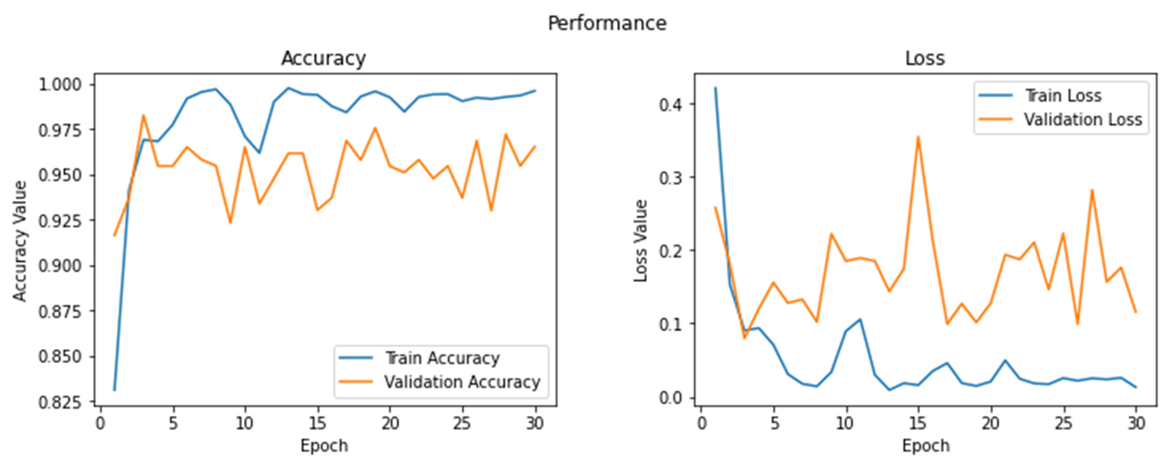
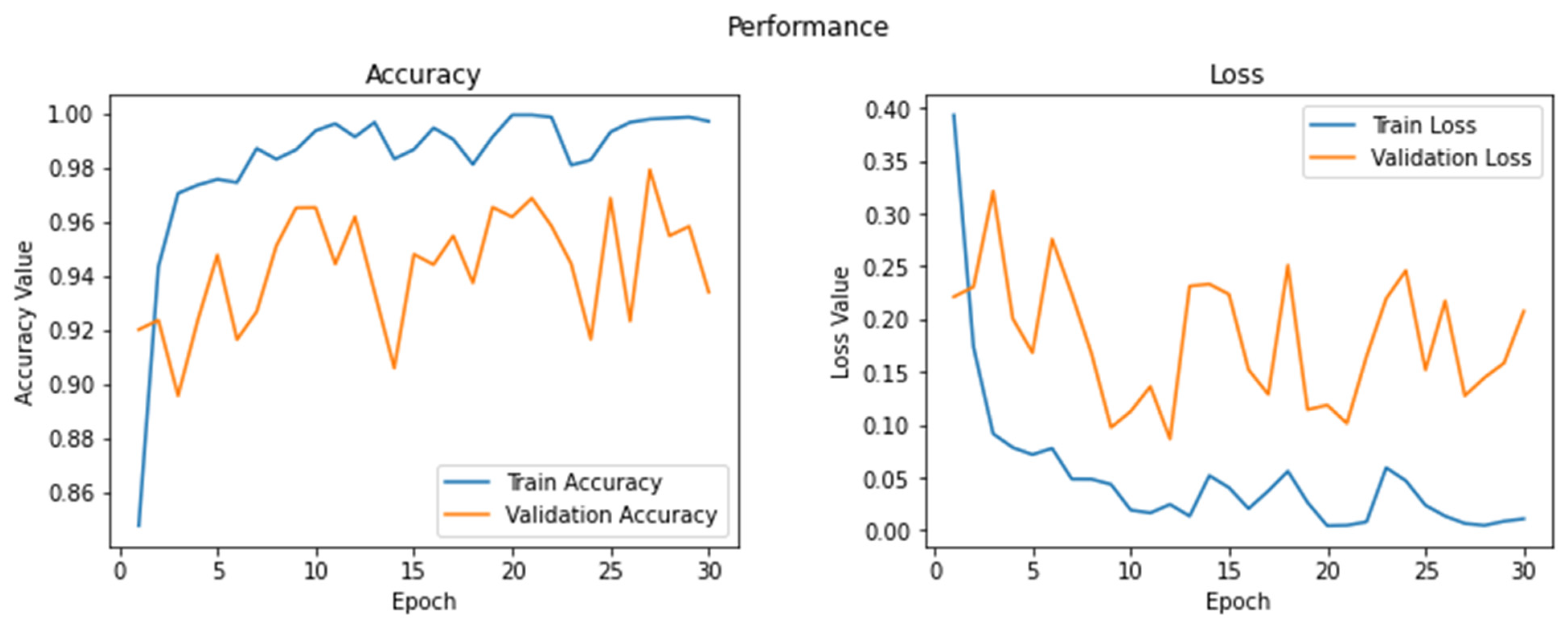
| Values | 4 | 6 | Cross-entropy | 32 | 10−2 | 30 |
| Model | MRI Database | Precision (%) | Recall (%) | F1-Score (%) | Test Accuracy (%) |
|---|---|---|---|---|---|
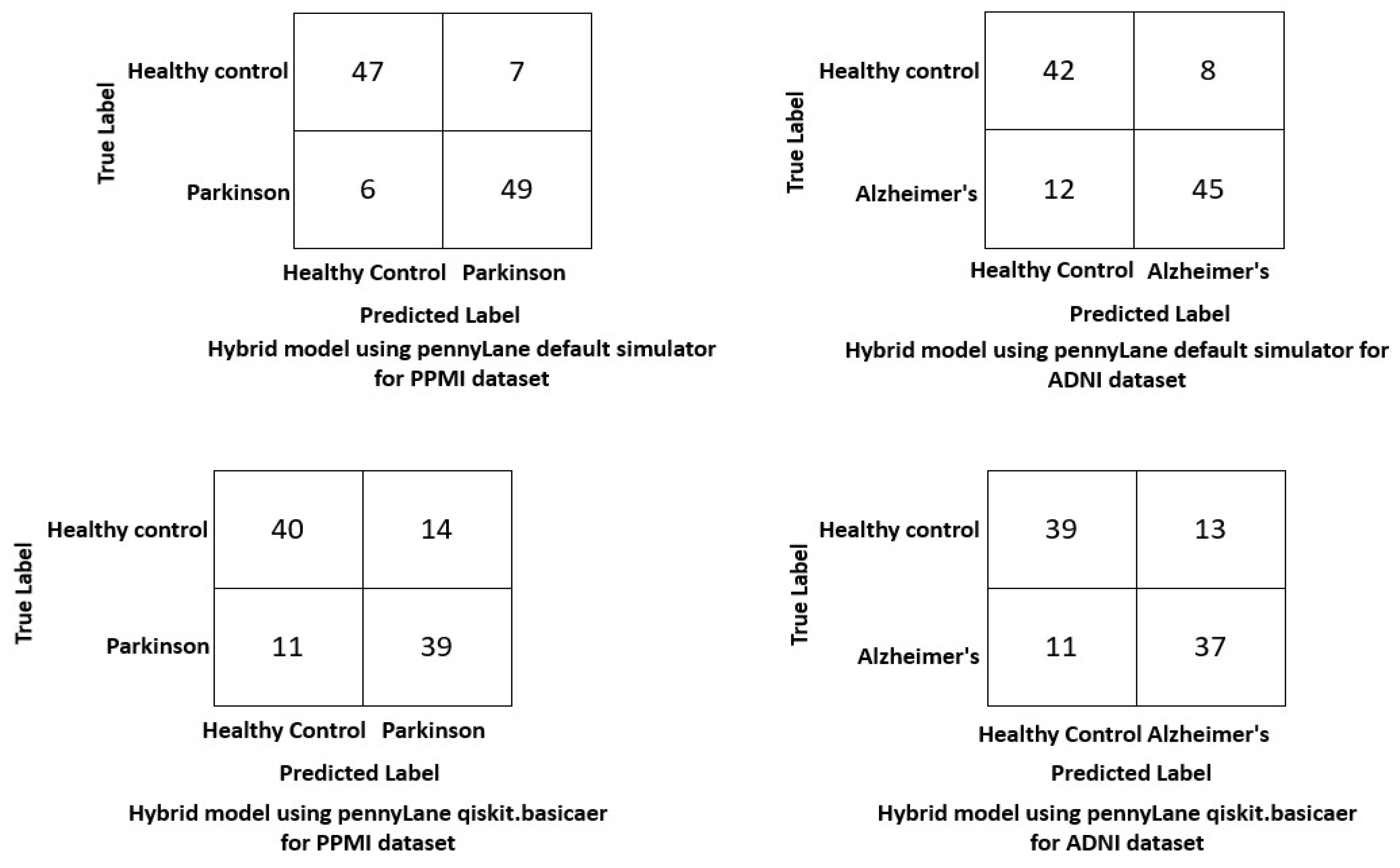
| Hybrid AlexNet–quantum learning model On default simulator | PPMI ADNI | 93 | 92 | 93 | 97 |
| 91.5 | 90 | 94 | 96 | ||
| Hybrid AlexNet–quantum learning model On qiskit basic.aer | PPMI | 91.5 | 86.9 | 91.4 | 95.5 |
| ADNI | 90 | 89.7 | 93.6 | 94 |
| Model | MRI Database | Precision (%) | Recall (%) | F1-Score (%) | Test Accuracy (%) |
|---|---|---|---|---|---|
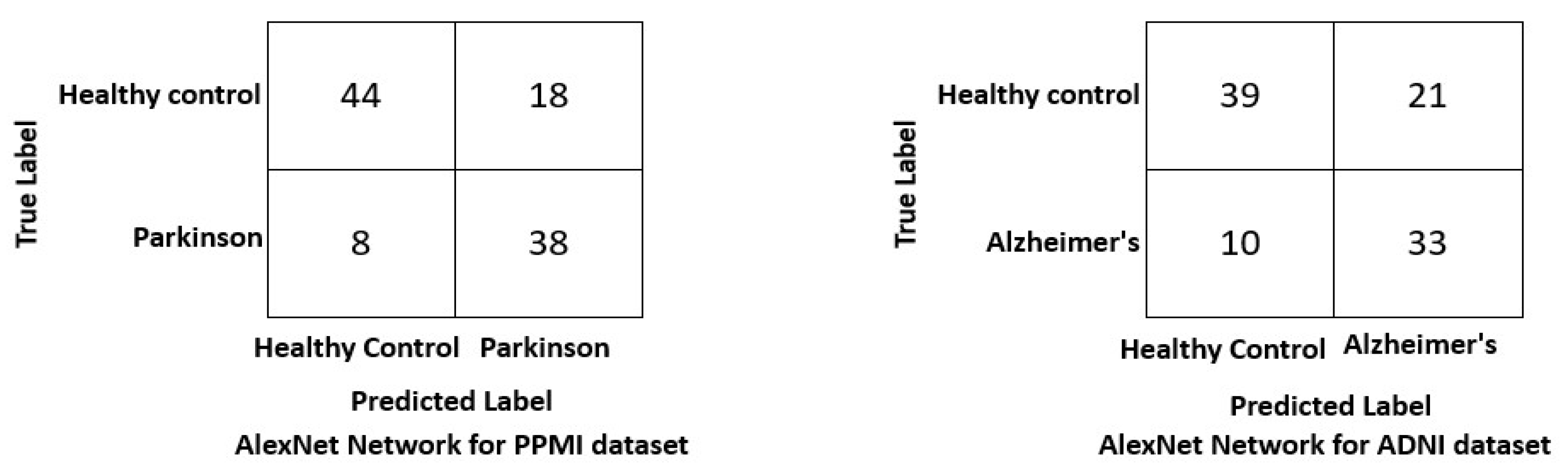
| AlexNet using classical neural network | PPMI | 91.5 | 92.5 | 90 | 93 |
| ADNI | 92 | 89 | 89.7 | 91.9 |
| Model | MRI Database | Precision (%) | Recall (%) | F1-Score (%) | Test Accuracy (%) |
|---|---|---|---|---|---|
| AlexNet | PPMI | 91.5 | 92.5 | 90 | 93 |
| ADNI | 92 | 89 | 89.7 | 91.9 | |
| Inceptionv3 | PPMI | 85 | 90 | 83 | 92 |
| ADNI | 91 | 87.4 | 85.9 | 89 | |
| ResNet18 | PPMI | 85.5 | 93.5 | 86 | 90.5 |
| ADNI | 91 | 89 | 90 | 91 | |
| VGG16 | PPMI | 88.8 | 91.7 | 85.4 | 92.5 |
| ADNI | 90 | 90.9 | 94.5 | 89 | |
| Proposed method | PPMI | 93 | 92 | 93 | 97 |
| ADNI | 91.5 | 90 | 94 | 96 |
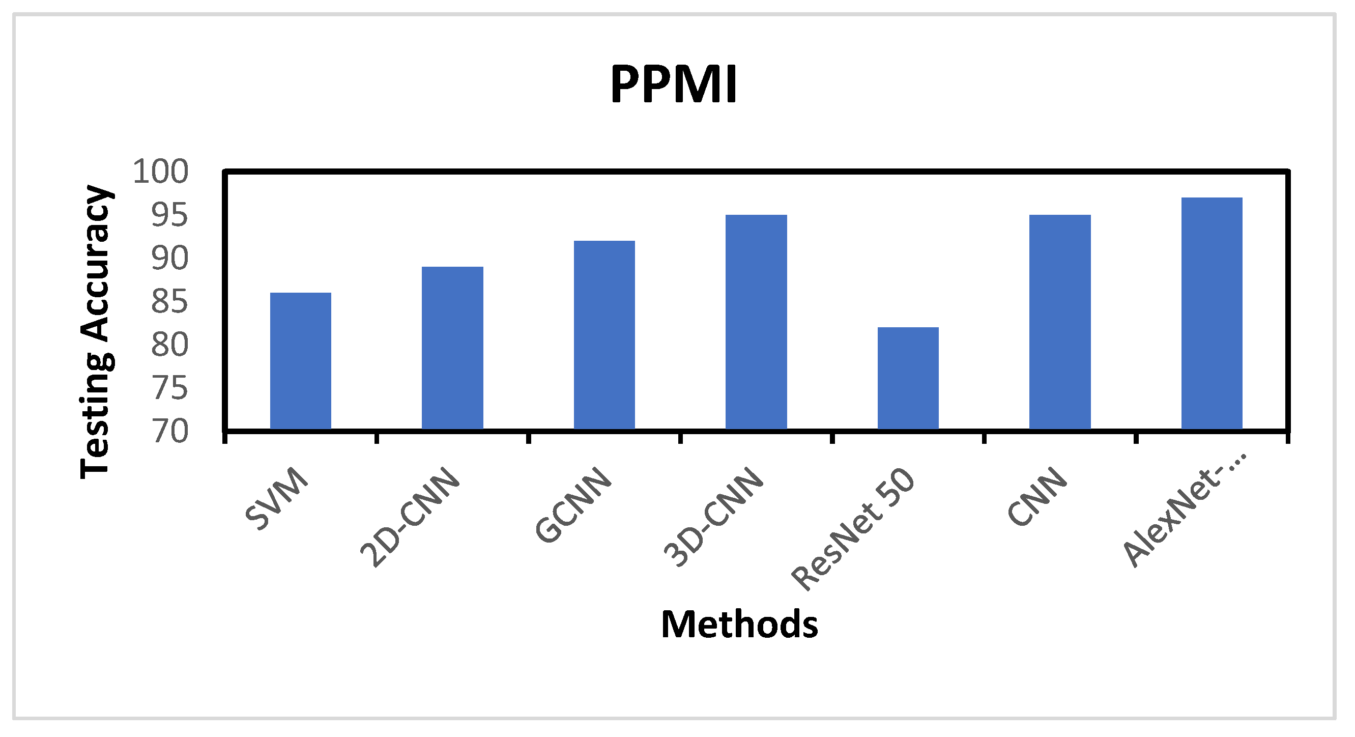
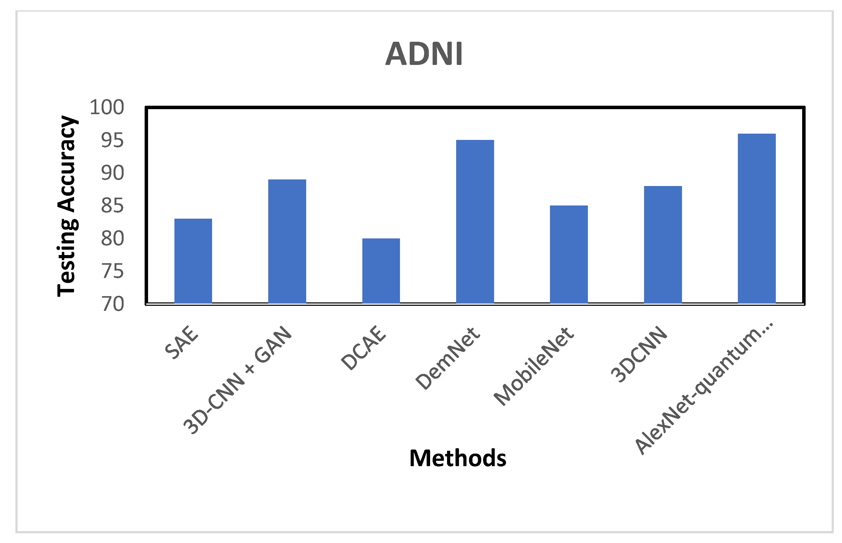
| MRI Database | Reference | Modality | Model | Test Accuracy |
|---|---|---|---|---|
| PPMI | [59] | MRI | support vector machine based on Muti Kernel (SVM) | 85.78 |
| [60] | SPECT | 2D-CNN | 89 | |
| [61] | sMRI | GCNN | 92 | |
| [62] | SPECT | 3D-CNN | 95 | |
| [30] | MRI | VGG16 and ResNet 50 | 82 | |
| [63] | T2-Weighted MRI | CNN | 95 | |
| Proposed Method | MRI | AlexNet–quantum transfer learning | 97 | |
| ADNI | [64] | PET | SAE | 82.5 |
| [65] | sMRI + PET | 3D-CNN + GAN | 89 | |
| [66] | rs-fMRI | DCAE | 80 | |
| [67] | MRI | DemNet | 95.23 | |
| [68] | MRI | MobileNet | 85 | |
| [69] | MRI | 3DCNN | 88 | |
| Proposed Method | MRI | AlexNet–quantum transfer learning | 96 |
Disclaimer/Publisher’s Note: The statements, opinions and data contained in all publications are solely those of the individual author(s) and contributor(s) and not of MDPI and/or the editor(s). MDPI and/or the editor(s) disclaim responsibility for any injury to people or property resulting from any ideas, methods, instructions or products referred to in the content. |
© 2023 by the authors. Licensee MDPI, Basel, Switzerland. This article is an open access article distributed under the terms and conditions of the Creative Commons Attribution (CC BY) license (https://creativecommons.org/licenses/by/4.0/).
Share and Cite
Alsharabi, N.; Shahwar, T.; Rehman, A.U.; Alharbi, Y. Implementing Magnetic Resonance Imaging Brain Disorder Classification via AlexNet–Quantum Learning. Mathematics 2023, 11, 376. https://doi.org/10.3390/math11020376
Alsharabi N, Shahwar T, Rehman AU, Alharbi Y. Implementing Magnetic Resonance Imaging Brain Disorder Classification via AlexNet–Quantum Learning. Mathematics. 2023; 11(2):376. https://doi.org/10.3390/math11020376
Chicago/Turabian StyleAlsharabi, Naif, Tayyaba Shahwar, Ateeq Ur Rehman, and Yasser Alharbi. 2023. "Implementing Magnetic Resonance Imaging Brain Disorder Classification via AlexNet–Quantum Learning" Mathematics 11, no. 2: 376. https://doi.org/10.3390/math11020376