

Unveiling the Dichotomy of Urinary Proteins: Diagnostic Insights into Breast and Prostate Cancer and Their Roles

Abstract
:1. Introduction
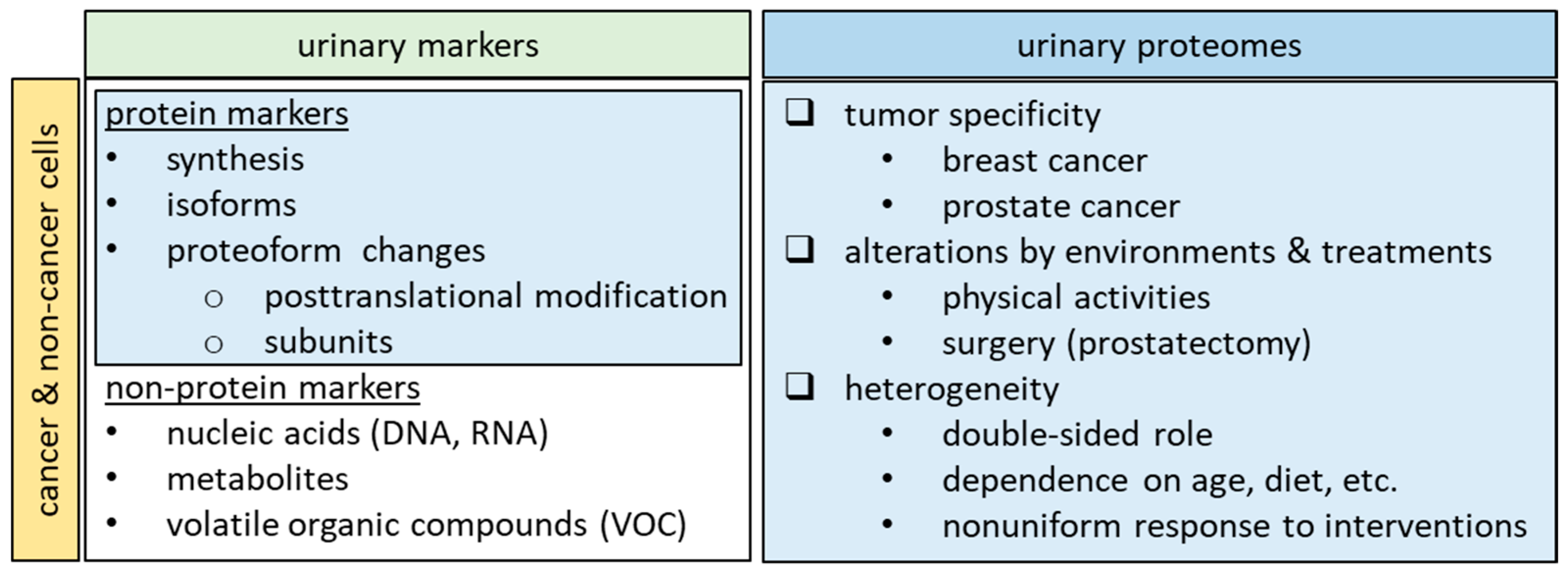
2. Urinary Biomarkers for Breast Cancer and Prostate Cancer

| Breast Cancer | ||||
| Gene | Name | Function | FDA Approval * | Ref. |
| MMP9 NGAL | matrix metalloproteinase 9 | facilitating angiogenesis and tumor growth | Yes | [44] |
| neutrophil gelatinase-associated lipocalin | Yes | |||
| MMP1 CD63 | matrix metalloproteinase 1 | involved in the cancer development of breast cancer | Yes | [46] |
| CD63 | Yes | |||
| MMP9 ADAM12 | matrix metalloproteinase 9 | urinary MMP9 and ADAM12 levels significantly increase with disease progression in breast cancer patients and correlate with the disease stage | Yes | [45] |
| a disintegrin and metalloprotease 12 | No | |||
| EG1 | endothelial-derived gene 1 | EG1 stimulates cellular proliferation | No | [70] |
| TFF1 | trefoil factor 1 | the tumor grade was correlated with TFF1 | No | [48] |
| ECM1 MAST4 Filaggrin | extracellular matrix protein 1 | expression is highly significantly correlated with survival in breast cancer patients | No | [49] |
| microtubule-associated serine/threonine kinase family member 4 | No | |||
| filaggrin | No | |||
| Prostate Cancer | ||||
| Gene | Name | Function | FDA Approval * | Ref. |
| PCA3 | prostate cancer-associated 3 | predict the tumor volume, extracapsular extension, and positive surgical margins in prostatectomy specimens | Yes | [58,71] |
| EN2 | engrailed 2 | pre-surgical urinary EN2 levels were associated with increasing tumor stage and closely reflected the volume of cancer in prostate cancer specimens | No | [62,63] |
| TMPRSS2-ERG SPDEF | transmembrane proteinase serine 2:v-ets erythroblastosis virus E26 oncogene homolog | predicting initial biopsy results in prostate cancer | No | [64,65] |
| SAM-pointed domain-containing Ets-like factor | No | |||
| β2M PGA3 MUC3 | β-2-microglobulin | distinguish between benign prostate hyperplasia (BPH) and localized prostate cancer | No | [66] |
| pepsinogen A3 | No | |||
| mucin 3 | No | |||
| CD105 | endoglin | urinary endoglin levels in men with prostate cancer correlated with radical prostatectomy tumor volume | No | [67] |
| IL18BP | interleukin-18 binding protein | IL18BP merits further study as a marker of aggressive prostate cancer and as a therapeutic target | No | [47] |
| GOLM1 | Golgi membrane protein 1 | GOLM1 is a resident cis-Golgi membrane protein of unknown function | No | [61] |
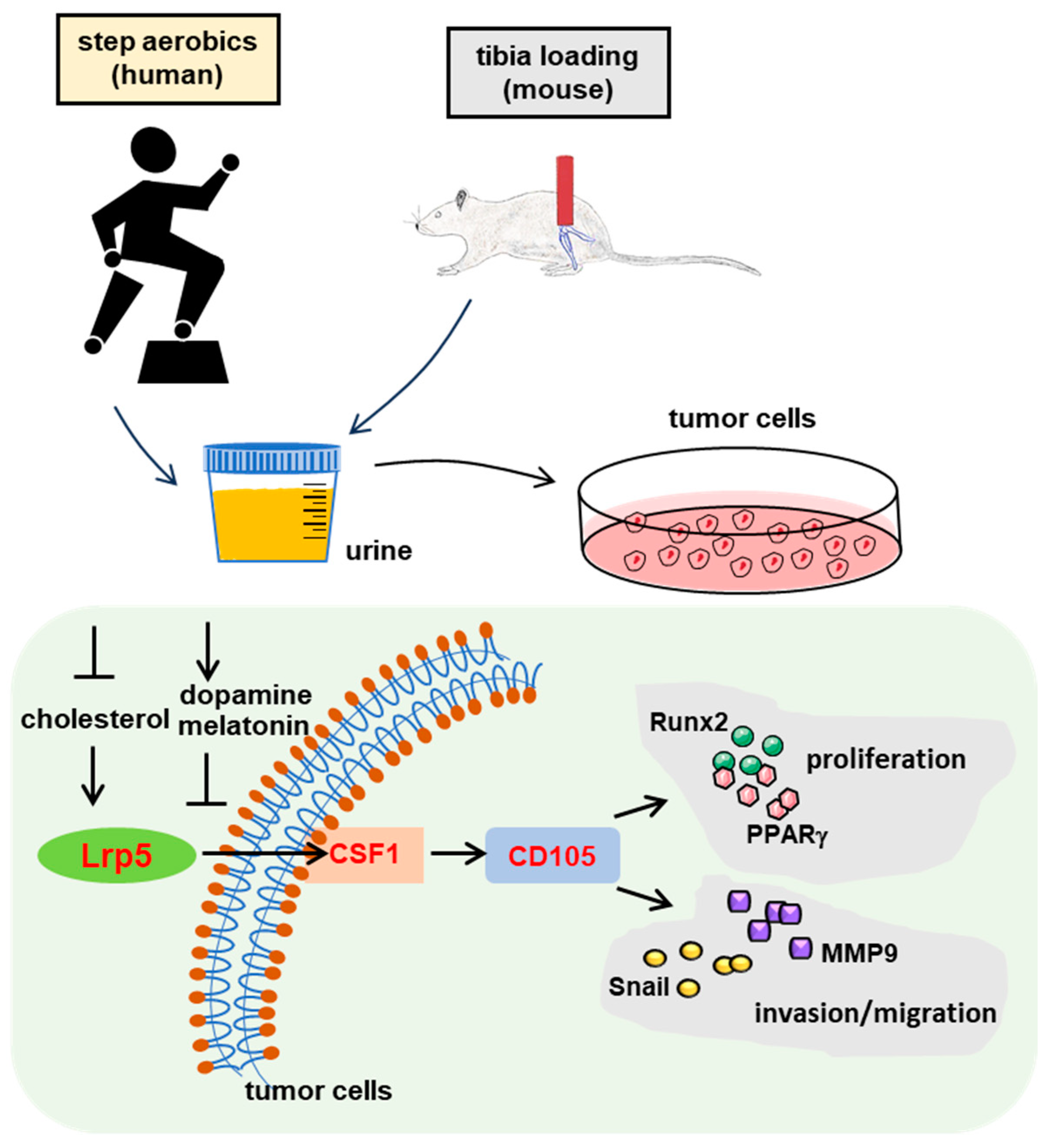
3. Effects of Physical Activities
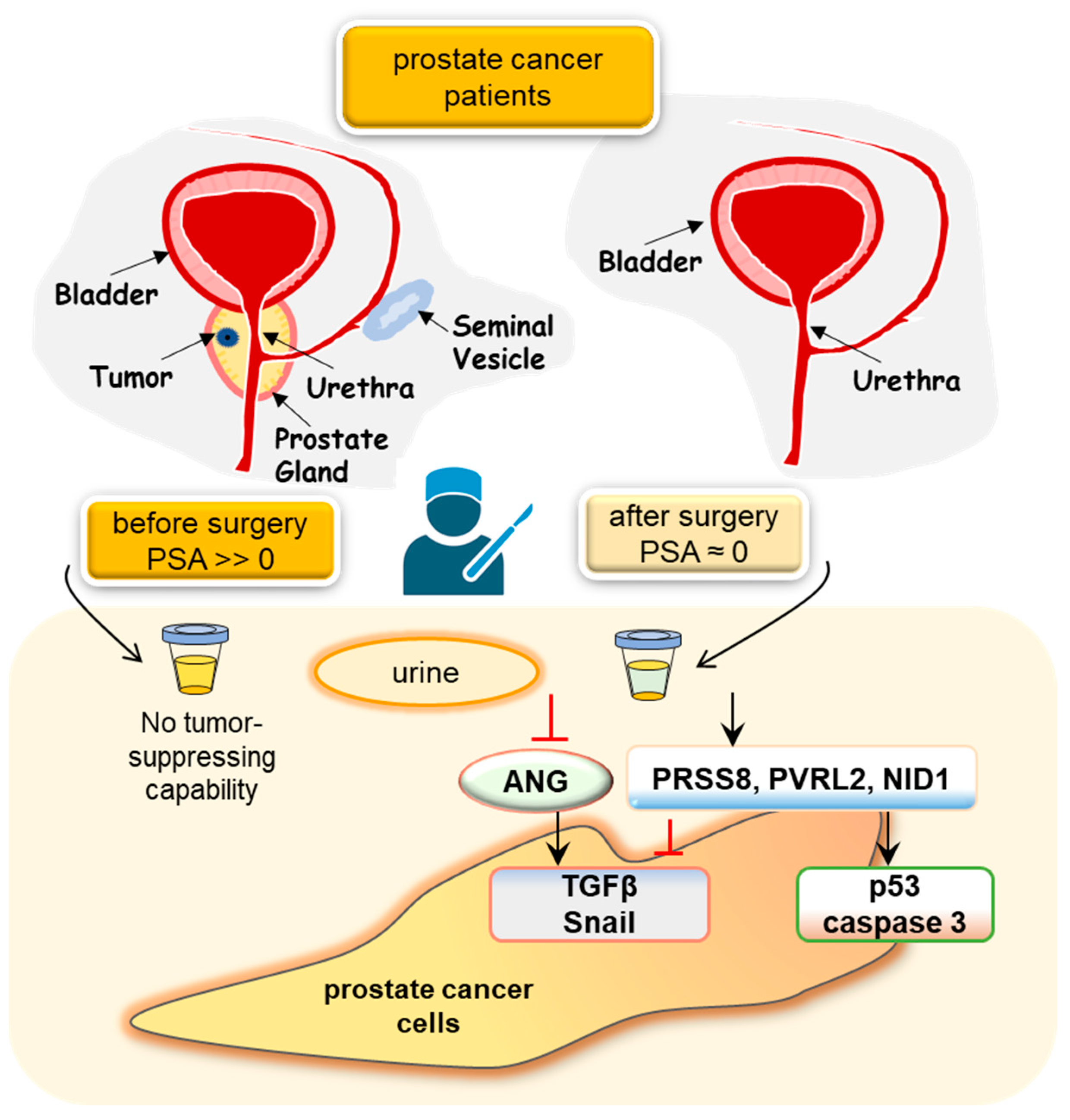
4. Treatment Effects
5. Double-Sided Role of Urinary Proteins
6. Future Perspective
7. Limitations of This Review
8. Conclusions
Author Contributions
Funding
Institutional Review Board Statement
Informed Consent Statement
Data Availability Statement
Acknowledgments
Conflicts of Interest
References
- Sarhadi, V.K.; Armengol, G. Molecular Biomarkers in Cancer. Biomolecules 2022, 12, 1021. [Google Scholar] [CrossRef] [PubMed]
- Pastore, A.L.; Palleschi, G.; Silvestri, L.; Moschese, D.; Ricci, S.; Petrozza, V.; Carbone, A.; Di Carlo, A. Serum and urine biomarkers for human renal cell carcinoma. Dis. Markers 2015, 2015, 251403. [Google Scholar] [CrossRef] [PubMed]
- Ain, Q.U.; Muhammad, S.; Hai, Y.; Peiling, L. The role of urine and serum biomarkers in the early detection of ovarian epithelial tumours. J. Obstet. Gynaecol. 2022, 42, 3441–3449. [Google Scholar] [CrossRef] [PubMed]
- Becerra, M.F.; Atluri, V.S.; Bhattu, A.S.; Punnen, S. Serum and urine biomarkers for detecting clinically significant prostate cancer. Urol. Oncol. 2021, 39, 686–690. [Google Scholar] [CrossRef] [PubMed]
- Chen, C.K.; Liao, J.; Li, M.S.; Khoo, B.L. Urine biopsy technologies: Cancer and beyond. Theranostics 2020, 10, 7872–7888. [Google Scholar] [CrossRef] [PubMed]
- Elsharkawi, F.; Elsabah, M.; Shabayek, M.; Khaled, H. Urine and Serum Exosomes as Novel Biomarkers in Detection of Bladder Cancer. Asian Pac. J. Cancer Prev. 2019, 20, 2219–2224. [Google Scholar] [CrossRef] [PubMed]
- Wood, S.L.; Knowles, M.A.; Thompson, D.; Selby, P.J.; Banks, R.E. Proteomic studies of urinary biomarkers for prostate, bladder and kidney cancers. Nat. Rev. Urol. 2013, 10, 206–218. [Google Scholar] [CrossRef]
- Wei, J.T. Urinary biomarkers for prostate cancer. Curr. Opin. Urol. 2015, 25, 77–82. [Google Scholar] [CrossRef]
- Owens, G.L.; Barr, C.E.; White, H.; Njoku, K.; Crosbie, E.J. Urinary biomarkers for the detection of ovarian cancer: A systematic review. Carcinogenesis 2022, 43, 311–320. [Google Scholar] [CrossRef]
- Grubmuller, B.; Roupret, M.; Briganti, A.; Shariat, S.F. The Use of Biomarkers for Bladder Cancer Diagnosis and Surveillance. Mini Rev. Med. Chem. 2016, 16, 1444–1449. [Google Scholar] [CrossRef]
- Harsanyi, S.; Novakova, Z.V.; Bevizova, K.; Danisovic, L.; Ziaran, S. Biomarkers of Bladder Cancer: Cell-Free DNA, Epigenetic Modifications and Non-Coding RNAs. Int. J. Mol. Sci. 2022, 23, 3206. [Google Scholar] [CrossRef] [PubMed]
- Chen, Z.; Kim, J. Urinary proteomics and metabolomics studies to monitor bladder health and urological diseases. BMC Urol. 2016, 16, 11. [Google Scholar] [CrossRef] [PubMed]
- Soorojebally, Y.; Neuzillet, Y.; Roumiguie, M.; Lamy, P.J.; Allory, Y.; Descotes, F.; Ferlicot, S.; Kassab-Chahmi, D.; Oudard, S.; Rebillard, X.; et al. Urinary biomarkers for bladder cancer diagnosis and NMIBC follow-up: A systematic review. World J. Urol. 2023, 41, 345–359. [Google Scholar] [CrossRef] [PubMed]
- Li, J.; Guan, X.; Fan, Z.; Ching, L.M.; Li, Y.; Wang, X.; Cao, W.M.; Liu, D.X. Non-Invasive Biomarkers for Early Detection of Breast Cancer. Cancers 2020, 12, 2767. [Google Scholar] [CrossRef] [PubMed]
- Ku, A.; Fredsoe, J.; Sorensen, K.D.; Borre, M.; Evander, M.; Laurell, T.; Lilja, H.; Ceder, Y. High-Throughput and Automated Acoustic Trapping of Extracellular Vesicles to Identify microRNAs With Diagnostic Potential for Prostate Cancer. Front. Oncol. 2021, 11, 631021. [Google Scholar] [CrossRef] [PubMed]
- Lin, J.; Li, J.; Huang, B.; Liu, J.; Chen, X.; Chen, X.M.; Xu, Y.M.; Huang, L.F.; Wang, X.Z. Exosomes: Novel biomarkers for clinical diagnosis. Sci. World J. 2015, 2015, 657086. [Google Scholar] [CrossRef] [PubMed]
- Hara, M.; Yanagihara, T.; Kihara, I.; Higashi, K.; Fujimoto, K.; Kajita, T. Apical cell membranes are shed into urine from injured podocytes: A novel phenomenon of podocyte injury. J. Am. Soc. Nephrol. 2005, 16, 408–416. [Google Scholar] [CrossRef]
- Janovicova, L.; Konecna, B.; Vlkova, B.; Celec, P. Isolation and Quantification of Extracellular DNA from Biofluids. Bio Protoc. 2020, 10, e3726. [Google Scholar] [CrossRef]
- Zhang, A.; Sun, H.; Wu, X.; Wang, X. Urine metabolomics. Clin. Chim. Acta 2012, 414, 65–69. [Google Scholar] [CrossRef]
- Sun, Y.S.; Zhao, Z.; Yang, Z.N.; Xu, F.; Lu, H.J.; Zhu, Z.Y.; Shi, W.; Jiang, J.; Yao, P.P.; Zhu, H.P. Risk Factors and Preventions of Breast Cancer. Int. J. Biol. Sci. 2017, 13, 1387–1397. [Google Scholar] [CrossRef]
- Ahlgren, M.; Melbye, M.; Wohlfahrt, J.; Sorensen, T.I. Growth patterns and the risk of breast cancer in women. N. Engl. J. Med. 2004, 351, 1619–1626. [Google Scholar] [CrossRef] [PubMed]
- Crawford, E.D. Epidemiology of prostate cancer. Urology 2003, 62, 3–12. [Google Scholar] [CrossRef] [PubMed]
- Rosenwald, I.B.; Chen, J.J.; Wang, S.; Savas, L.; London, I.M.; Pullman, J. Upregulation of protein synthesis initiation factor eIF-4E is an early event during colon carcinogenesis. Oncogene 1999, 18, 2507–2517. [Google Scholar] [CrossRef] [PubMed]
- Fernandez-Nogueira, P.; Fuster, G.; Gutierrez-Uzquiza, A.; Gascon, P.; Carbo, N.; Bragado, P. Cancer-Associated Fibroblasts in Breast Cancer Treatment Response and Metastasis. Cancers 2021, 13, 3146. [Google Scholar] [CrossRef] [PubMed]
- Maia, A.; Gu, Z.; Koch, A.; Berdiel-Acer, M.; Will, R.; Schlesner, M.; Wiemann, S. IFNbeta1 secreted by breast cancer cells undergoing chemotherapy reprograms stromal fibroblasts to support tumour growth after treatment. Mol. Oncol. 2021, 15, 1308–1329. [Google Scholar] [CrossRef]
- Hosseinalizadeh, H.; Mohamadzadeh, O.; Kahrizi, M.S.; Razaghi Bahabadi, Z.; Klionsky, D.J.; Mirzei, H. TRIM8: A double-edged sword in glioblastoma with the power to heal or hurt. Cell Mol. Biol. Lett. 2023, 28, 6. [Google Scholar] [CrossRef]
- Lone, S.N.; Nisar, S.; Masoodi, T.; Singh, M.; Rizwan, A.; Hashem, S.; El-Rifai, W.; Bedognetti, D.; Batra, S.K.; Haris, M.; et al. Liquid biopsy: A step closer to transform diagnosis, prognosis and future of cancer treatments. Mol. Cancer 2022, 21, 79. [Google Scholar] [CrossRef]
- Michaelsson, E.; Norberg, A.; Samuelsson, S.M. Assessment of thirst among severely demented patients in the terminal phase of life. Exploratory interviews with ward sisters and enrolled nurses. Int. J. Nurs. Stud. 1987, 24, 87–93. [Google Scholar] [CrossRef]
- Huang, C.K.; Sun, Y.; Lv, L.; Ping, Y. ENO1 and Cancer. Mol. Ther. Oncolytics 2022, 24, 288–298. [Google Scholar] [CrossRef]
- Qiao, G.; Wu, A.; Chen, X.; Tian, Y.; Lin, X. Enolase 1, a Moonlighting Protein, as a Potential Target for Cancer Treatment. Int. J. Biol. Sci. 2021, 17, 3981–3992. [Google Scholar] [CrossRef]
- Li, K.; Sun, X.; Minami, K.; Tamari, K.; Ogawa, K.; Li, H.; Ma, H.; Zhou, M.; Na, S.; Li, B.Y.; et al. Proteomes from AMPK-inhibited peripheral blood mononuclear cells suppress the progression of breast cancer and bone metastasis. Theranostics 2023, 13, 1247–1263. [Google Scholar] [CrossRef] [PubMed]
- Feng, Y.; Liu, S.; Zha, R.; Sun, X.; Li, K.; Wu, D.; Aryal, U.K.; Koch, M.; Li, B.Y.; Yokota, H. Prostate cancer-associated urinary proteomes differ before and after prostatectomy. Ther. Adv. Med. Oncol. 2022, 14, 17588359221131532. [Google Scholar] [CrossRef] [PubMed]
- Lee, B.; Mahmud, I.; Marchica, J.; Derezinski, P.; Qi, F.; Wang, F.; Joshi, P.; Valerio, F.; Rivera, I.; Patel, V.; et al. Integrated RNA and metabolite profiling of urine liquid biopsies for prostate cancer biomarker discovery. Sci. Rep. 2020, 10, 3716. [Google Scholar] [CrossRef] [PubMed]
- Sequeiros, T.; Rigau, M.; Chiva, C.; Montes, M.; Garcia-Grau, I.; Garcia, M.; Diaz, S.; Celma, A.; Bijnsdorp, I.; Campos, A.; et al. Targeted proteomics in urinary extracellular vesicles identifies biomarkers for diagnosis and prognosis of prostate cancer. Oncotarget 2017, 8, 4960–4976. [Google Scholar] [CrossRef] [PubMed]
- Adeola, H.A.; Calder, B.; Soares, N.C.; Kaestner, L.; Blackburn, J.M.; Zerbini, L.F. In silico verification and parallel reaction monitoring prevalidation of potential prostate cancer biomarkers. Future Oncol. 2016, 12, 43–57. [Google Scholar] [CrossRef] [PubMed]
- Independent, U.K.P.o.B.C.S. The benefits and harms of breast cancer screening: An independent review. Lancet 2012, 380, 1778–1786. [Google Scholar] [CrossRef]
- Muhlberger, N.; Boskovic, K.; Krahn, M.D.; Bremner, K.E.; Oberaigner, W.; Klocker, H.; Horninger, W.; Sroczynski, G.; Siebert, U. Benefits and harms of prostate cancer screening—Predictions of the ONCOTYROL prostate cancer outcome and policy model. BMC Public Health 2017, 17, 596. [Google Scholar] [CrossRef]
- Loud, J.T.; Murphy, J. Cancer Screening and Early Detection in the 21(st) Century. Semin. Oncol. Nurs. 2017, 33, 121–128. [Google Scholar] [CrossRef]
- Woollam, M.; Wang, L.; Grocki, P.; Liu, S.; Siegel, A.P.; Kalra, M.; Goodpaster, J.V.; Yokota, H.; Agarwal, M. Tracking the Progression of Triple Negative Mammary Tumors over Time by Chemometric Analysis of Urinary Volatile Organic Compounds. Cancers 2021, 13, 1462. [Google Scholar] [CrossRef]
- Owens, C.L.; Vandenbussche, C.J.; Burroughs, F.H.; Rosenthal, D.L. A review of reporting systems and terminology for urine cytology. Cancer Cytopathol. 2013, 121, 9–14. [Google Scholar] [CrossRef]
- Maas, M.; Bedke, J.; Stenzl, A.; Todenhofer, T. Can urinary biomarkers replace cystoscopy? World J. Urol. 2019, 37, 1741–1749. [Google Scholar] [CrossRef] [PubMed]
- Pansa, E.; Guzzardi, G.; Santocono, S.; Carriero, A. Vascular Complications following Vacuum-Assisted Breast Biopsy (VABB): A Case Report and Review of the Literature. Tomography 2023, 9, 1246–1253. [Google Scholar] [CrossRef]
- Montemezzi, S.; Cardano, G.; Storer, S.; Cardobi, N.; Cavedon, C.; Camera, L. MRI-guided breast biopsy based on diffusion-weighted imaging: A feasibility study. Eur. Radiol. 2021, 31, 2645–2656. [Google Scholar] [CrossRef]
- Fernandez, C.A.; Yan, L.; Louis, G.; Yang, J.; Kutok, J.L.; Moses, M.A. The matrix metalloproteinase-9/neutrophil gelatinase-associated lipocalin complex plays a role in breast tumor growth and is present in the urine of breast cancer patients. Clin. Cancer Res. 2005, 11, 5390–5395. [Google Scholar] [CrossRef]
- Pories, S.E.; Zurakowski, D.; Roy, R.; Lamb, C.C.; Raza, S.; Exarhopoulos, A.; Scheib, R.G.; Schumer, S.; Lenahan, C.; Borges, V.; et al. Urinary metalloproteinases: Noninvasive biomarkers for breast cancer risk assessment. Cancer Epidemiol. Biomark. Prev. 2008, 17, 1034–1042. [Google Scholar] [CrossRef] [PubMed]
- Ando, W.; Kikuchi, K.; Uematsu, T.; Yokomori, H.; Takaki, T.; Sogabe, M.; Kohgo, Y.; Otori, K.; Ishikawa, S.; Okazaki, I. Novel breast cancer screening: Combined expression of miR-21 and MMP-1 in urinary exosomes detects 95% of breast cancer without metastasis. Sci. Rep. 2019, 9, 13595. [Google Scholar] [CrossRef] [PubMed]
- Fujita, K.; Ewing, C.M.; Isaacs, W.B.; Pavlovich, C.P. Immunomodulatory IL-18 binding protein is produced by prostate cancer cells and its levels in urine and serum correlate with tumor status. Int. J. Cancer 2011, 129, 424–432. [Google Scholar] [CrossRef]
- Chenard, M.P.; Tomasetto, C.; Bellocq, J.P.; Rio, M.C. Urinary pS2/TFF1 levels in the management of hormonodependent breast carcinomas. Peptides 2004, 25, 737–743. [Google Scholar] [CrossRef]
- Beretov, J.; Wasinger, V.C.; Millar, E.K.; Schwartz, P.; Graham, P.H.; Li, Y. Proteomic Analysis of Urine to Identify Breast Cancer Biomarker Candidates Using a Label-Free LC-MS/MS Approach. PLoS ONE 2015, 10, e0141876. [Google Scholar] [CrossRef]
- Bax, C.; Lotesoriere, B.J.; Sironi, S.; Capelli, L. Review and Comparison of Cancer Biomarker Trends in Urine as a Basis for New Diagnostic Pathways. Cancers 2019, 11, 1244. [Google Scholar] [CrossRef]
- Carlsson, S.V.; Vickers, A.J. Screening for Prostate Cancer. Med. Clin. N. Am. 2020, 104, 1051–1062. [Google Scholar] [CrossRef] [PubMed]
- Kohaar, I.; Petrovics, G.; Srivastava, S. A Rich Array of Prostate Cancer Molecular Biomarkers: Opportunities and Challenges. Int. J. Mol. Sci. 2019, 20, 1813. [Google Scholar] [CrossRef] [PubMed]
- Albertsen, P.C. Prostate cancer screening and treatment: Where have we come from and where are we going? BJU Int. 2020, 126, 218–224. [Google Scholar] [CrossRef] [PubMed]
- Loeb, S.; Vellekoop, A.; Ahmed, H.U.; Catto, J.; Emberton, M.; Nam, R.; Rosario, D.J.; Scattoni, V.; Lotan, Y. Systematic review of complications of prostate biopsy. Eur. Urol. 2013, 64, 876–892. [Google Scholar] [CrossRef] [PubMed]
- Loeb, S.; Bjurlin, M.A.; Nicholson, J.; Tammela, T.L.; Penson, D.F.; Carter, H.B.; Carroll, P.; Etzioni, R. Overdiagnosis and overtreatment of prostate cancer. Eur. Urol. 2014, 65, 1046–1055. [Google Scholar] [CrossRef] [PubMed]
- Joshi, S.S.; Filson, C.P. Long-term consequences of the USPSTF Grade D recommendation for prostate-specific antigen screening. Cancer 2020, 126, 694–696. [Google Scholar] [CrossRef] [PubMed]
- Crulhas, B.P.; Basso, C.R.; Castro, G.R.; Pedrosa, V.A. Detection of Prostate Cancer Biomarker PCA3 by Using Aptasensors. Curr. Med. Chem. 2022, 29, 5895–5902. [Google Scholar] [CrossRef] [PubMed]
- Ploussard, G.; de la Taille, A. The role of prostate cancer antigen 3 (PCA3) in prostate cancer detection. Expert. Rev. Anticancer Ther. 2018, 18, 1013–1020. [Google Scholar] [CrossRef]
- Nakanishi, H.; Groskopf, J.; Fritsche, H.A.; Bhadkamkar, V.; Blase, A.; Kumar, S.V.; Davis, J.W.; Troncoso, P.; Rittenhouse, H.; Babaian, R.J. PCA3 molecular urine assay correlates with prostate cancer tumor volume: Implication in selecting candidates for active surveillance. J. Urol. 2008, 179, 1804–1809; discussion 1809–1810. [Google Scholar] [CrossRef]
- van Gils, M.P.; Hessels, D.; van Hooij, O.; Jannink, S.A.; Peelen, W.P.; Hanssen, S.L.; Witjes, J.A.; Cornel, E.B.; Karthaus, H.F.; Smits, G.A.; et al. The time-resolved fluorescence-based PCA3 test on urinary sediments after digital rectal examination; a Dutch multicenter validation of the diagnostic performance. Clin. Cancer Res. 2007, 13, 939–943. [Google Scholar] [CrossRef]
- Varambally, S.; Laxman, B.; Mehra, R.; Cao, Q.; Dhanasekaran, S.M.; Tomlins, S.A.; Granger, J.; Vellaichamy, A.; Sreekumar, A.; Yu, J.; et al. Golgi protein GOLM1 is a tissue and urine biomarker of prostate cancer. Neoplasia 2008, 10, 1285–1294. [Google Scholar] [CrossRef] [PubMed]
- McGrath, S.E.; Michael, A.; Morgan, R.; Pandha, H. EN2 in Prostate Cancer. Adv. Clin. Chem. 2015, 71, 47–76. [Google Scholar] [CrossRef] [PubMed]
- Pandha, H.; Sorensen, K.D.; Orntoft, T.F.; Langley, S.; Hoyer, S.; Borre, M.; Morgan, R. Urinary engrailed-2 (EN2) levels predict tumour volume in men undergoing radical prostatectomy for prostate cancer. BJU Int. 2012, 110, E287–E292. [Google Scholar] [CrossRef] [PubMed]
- Leyten, G.H.; Hessels, D.; Jannink, S.A.; Smit, F.P.; de Jong, H.; Cornel, E.B.; de Reijke, T.M.; Vergunst, H.; Kil, P.; Knipscheer, B.C.; et al. Prospective multicentre evaluation of PCA3 and TMPRSS2-ERG gene fusions as diagnostic and prognostic urinary biomarkers for prostate cancer. Eur. Urol. 2014, 65, 534–542. [Google Scholar] [CrossRef] [PubMed]
- McKiernan, J.; Donovan, M.J.; O’Neill, V.; Bentink, S.; Noerholm, M.; Belzer, S.; Skog, J.; Kattan, M.W.; Partin, A.; Andriole, G.; et al. A Novel Urine Exosome Gene Expression Assay to Predict High-grade Prostate Cancer at Initial Biopsy. JAMA Oncol. 2016, 2, 882–889. [Google Scholar] [CrossRef]
- Fujita, K.; Nonomura, N. Urinary biomarkers of prostate cancer. Int. J. Urol. 2018, 25, 770–779. [Google Scholar] [CrossRef]
- Fujita, K.; Ewing, C.M.; Chan, D.Y.; Mangold, L.A.; Partin, A.W.; Isaacs, W.B.; Pavlovich, C.P. Endoglin (CD105) as a urinary and serum marker of prostate cancer. Int. J. Cancer 2009, 124, 664–669. [Google Scholar] [CrossRef]
- Shahinian, V.B.; Bahl, A.; Niepel, D.; Lorusso, V. Considering renal risk while managing cancer. Cancer Manag. Res. 2017, 9, 167–178. [Google Scholar] [CrossRef]
- Swensen, A.C.; He, J.; Fang, A.C.; Ye, Y.; Nicora, C.D.; Shi, T.; Liu, A.Y.; Sigdel, T.K.; Sarwal, M.M.; Qian, W.J. A Comprehensive Urine Proteome Database Generated From Patients With Various Renal Conditions and Prostate Cancer. Front. Med. 2021, 8, 548212. [Google Scholar] [CrossRef]
- Lu, M.; Sartippour, M.R.; Zhang, L.; Norris, A.J.; Brooks, M.N. Targeted inhibition of EG-1 blocks breast tumor growth. Cancer Biol. Ther. 2007, 6, 936–941. [Google Scholar] [CrossRef]
- Yamkamon, V.; Htoo, K.P.P.; Yainoy, S.; Suksrichavalit, T.; Tangchaikeeree, T.; Eiamphungporn, W. Urinary PCA3 detection in prostate cancer by magnetic nanoparticles coupled with colorimetric enzyme-linked oligonucleotide assay. EXCLI J. 2020, 19, 501–513. [Google Scholar] [CrossRef] [PubMed]
- Available online: https://fda.gov/drugs/biomarker-qualification-program/list-qualified-biomarkers (accessed on 20 December 2023).
- Valenti, M.T.; Deiana, M.; Cheri, S.; Dotta, M.; Zamboni, F.; Gabbiani, D.; Schena, F.; Dalle Carbonare, L.; Mottes, M. Physical Exercise Modulates miR-21-5p, miR-129-5p, miR-378-5p, and miR-188-5p Expression in Progenitor Cells Promoting Osteogenesis. Cells 2019, 8, 742. [Google Scholar] [CrossRef] [PubMed]
- Dalle Carbonare, L.; Mottes, M.; Cheri, S.; Deiana, M.; Zamboni, F.; Gabbiani, D.; Schena, F.; Salvagno, G.L.; Lippi, G.; Valenti, M.T. Increased Gene Expression of RUNX2 and SOX9 in Mesenchymal Circulating Progenitors Is Associated with Autophagy during Physical Activity. Oxid. Med. Cell Longev. 2019, 2019, 8426259. [Google Scholar] [CrossRef] [PubMed]
- Deshmukh, A.S.; Hawley, J.A.; Zierath, J.R. Exercise-induced phospho-proteins in skeletal muscle. Int. J. Obes. 2008, 32 (Suppl. S4), S18–S23. [Google Scholar] [CrossRef] [PubMed]
- Langfort, J.; Viese, M.; Ploug, T.; Dela, F. Time course of GLUT4 and AMPK protein expression in human skeletal muscle during one month of physical training. Scand. J. Med. Sci. Sports 2003, 13, 169–174. [Google Scholar] [CrossRef] [PubMed]
- Rockl, K.S.; Witczak, C.A.; Goodyear, L.J. Signaling mechanisms in skeletal muscle: Acute responses and chronic adaptations to exercise. IUBMB Life 2008, 60, 145–153. [Google Scholar] [CrossRef] [PubMed]
- Lynch, B.M.; Neilson, H.K.; Friedenreich, C.M. Physical activity and breast cancer prevention. Recent. Results Cancer Res. 2011, 186, 13–42. [Google Scholar] [CrossRef]
- Kim, J.; Choi, W.J.; Jeong, S.H. The effects of physical activity on breast cancer survivors after diagnosis. J. Cancer Prev. 2013, 18, 193–200. [Google Scholar] [CrossRef]
- Xu, Y.; Rogers, C.J. Physical Activity and Breast Cancer Prevention: Possible Role of Immune Mediators. Front. Nutr. 2020, 7, 557997. [Google Scholar] [CrossRef]
- Bertram, L.A.; Stefanick, M.L.; Saquib, N.; Natarajan, L.; Patterson, R.E.; Bardwell, W.; Flatt, S.W.; Newman, V.A.; Rock, C.L.; Thomson, C.A.; et al. Physical activity, additional breast cancer events, and mortality among early-stage breast cancer survivors: Findings from the WHEL Study. Cancer Causes Control 2011, 22, 427–435. [Google Scholar] [CrossRef]
- Sternfeld, B.; Weltzien, E.; Quesenberry, C.P., Jr.; Castillo, A.L.; Kwan, M.; Slattery, M.L.; Caan, B.J. Physical activity and risk of recurrence and mortality in breast cancer survivors: Findings from the LACE study. Cancer Epidemiol. Biomark. Prev. 2009, 18, 87–95. [Google Scholar] [CrossRef] [PubMed]
- Segal, R.; Zwaal, C.; Green, E.; Tomasone, J.R.; Loblaw, A.; Petrella, T.; Exercise for People with Cancer Guideline Development Group. Exercise for people with cancer: A systematic review. Curr. Oncol. 2017, 24, e290–e315. [Google Scholar] [CrossRef] [PubMed]
- Rundqvist, H.; Augsten, M.; Stromberg, A.; Rullman, E.; Mijwel, S.; Kharaziha, P.; Panaretakis, T.; Gustafsson, T.; Ostman, A. Effect of acute exercise on prostate cancer cell growth. PLoS ONE 2013, 8, e67579. [Google Scholar] [CrossRef] [PubMed]
- Ji, L.L.; Gomez-Cabrera, M.C.; Vina, J. Exercise and hormesis: Activation of cellular antioxidant signaling pathway. Ann. N. Y Acad. Sci. 2006, 1067, 425–435. [Google Scholar] [CrossRef] [PubMed]
- Zheng, A.; Zhang, L.; Yang, J.; Yin, X.; Zhang, T.; Wu, X.; Ma, X. Physical activity prevents tumor metastasis through modulation of immune function. Front. Pharmacol. 2022, 13, 1034129. [Google Scholar] [CrossRef] [PubMed]
- Jee, H.; Park, E.; Hur, K.; Kang, M.; Kim, Y. High-Intensity Aerobic Exercise Suppresses Cancer Growth by Regulating Skeletal Muscle-Derived Oncogenes and Tumor Suppressors. Front. Mol. Biosci. 2022, 9, 818470. [Google Scholar] [CrossRef] [PubMed]
- Martinez, D.A.; Patterson-Buckendahl, P.E.; Lust, A.; Shea-Rangel, K.M.; Hoban-Higgins, T.M.; Fuller, C.A.; Vailas, A.C. A noninvasive analysis of urinary musculoskeletal collagen metabolism markers from rhesus monkeys subject to chronic hypergravity. J. Appl. Physiol. 2008, 105, 1255–1261. [Google Scholar] [CrossRef] [PubMed]
- Simsek, B.; Karacaer, O.; Karaca, I. Urine products of bone breakdown as markers of bone resorption and clinical usefulness of urinary hydroxyproline: An overview. Chin. Med. J. 2004, 117, 291–295. [Google Scholar]
- Gambella, G.R.; Zunino, M. The role of the urinary hydroxyproline level in the screening for collagen diseases. Minerva Med. 1986, 77, 607–612. [Google Scholar]
- Vittori, L.N.; Romasco, J.; Tarozzi, A.; Latessa, P.M. Urinary Markers and Chronic Effect of Physical Exercise. Methods Mol. Biol. 2021, 2292, 193–200. [Google Scholar] [CrossRef]
- Kosaki, K.; Kamijo-Ikemori, A.; Sugaya, T.; Tanahashi, K.; Sawano, Y.; Akazawa, N.; Ra, S.G.; Kimura, K.; Shibagaki, Y.; Maeda, S. Effect of habitual exercise on urinary liver-type fatty acid-binding protein levels in middle-aged and older adults. Scand. J. Med. Sci. Sports 2018, 28, 152–160. [Google Scholar] [CrossRef] [PubMed]
- Kosaki, K.; Kamijo-Ikemori, A.; Sugaya, T.; Tanahashi, K.; Kumagai, H.; Sawano, Y.; Akazawa, N.; Ra, S.G.; Kimura, K.; Shibagaki, Y.; et al. Relationship between exercise capacity and urinary liver-type fatty acid-binding protein in middle-aged and older individuals. Clin. Exp. Nephrol. 2017, 21, 810–817. [Google Scholar] [CrossRef] [PubMed]
- Dethlefsen, C.; Hansen, L.S.; Lillelund, C.; Andersen, C.; Gehl, J.; Christensen, J.F.; Pedersen, B.K.; Hojman, P. Exercise-Induced Catecholamines Activate the Hippo Tumor Suppressor Pathway to Reduce Risks of Breast Cancer Development. Cancer Res. 2017, 77, 4894–4904. [Google Scholar] [CrossRef] [PubMed]
- Lee, J.J.; Beak, S.; Ahn, S.H.; Moon, B.S.; Kim, J.; Lee, K.P. Suppressing breast cancer by exercise: Consideration to animal models and exercise protocols. Phys. Act. Nutr. 2020, 24, 22–29. [Google Scholar] [CrossRef] [PubMed]
- Smeda, M.; Przyborowski, K.; Proniewski, B.; Zakrzewska, A.; Kaczor, D.; Stojak, M.; Buczek, E.; Nieckarz, Z.; Zoladz, J.A.; Wietrzyk, J.; et al. Breast cancer pulmonary metastasis is increased in mice undertaking spontaneous physical training in the running wheel; a call for revising beneficial effects of exercise on cancer progression. Am. J. Cancer Res. 2017, 7, 1926–1936. [Google Scholar] [PubMed]
- Liu, S.; Wu, D.; Sun, X.; Fan, Y.; Zha, R.; Jalali, A.; Teli, M.; Sano, T.; Siegel, A.; Sudo, A.; et al. Mechanical stimulations can inhibit local and remote tumor progression by downregulating WISP1. FASEB J. 2020, 34, 12847–12859. [Google Scholar] [CrossRef] [PubMed]
- Wu, D.; Fan, Y.; Liu, S.; Woollam, M.D.; Sun, X.; Murao, E.; Zha, R.; Prakash, R.; Park, C.; Siegel, A.P.; et al. Loading-induced antitumor capability of murine and human urine. FASEB J. 2020, 34, 7578–7592. [Google Scholar] [CrossRef]
- Oshi, M.; Murthy, V.; Takahashi, H.; Huyser, M.; Okano, M.; Tokumaru, Y.; Rashid, O.M.; Matsuyama, R.; Endo, I.; Takabe, K. Urine as a Source of Liquid Biopsy for Cancer. Cancers 2021, 13, 2652. [Google Scholar] [CrossRef]
- Garcia-Baquero, R.; Puerta, P.; Beltran, M.; Alvarez, M.; Sacristan, R.; Alvarez-Ossorio, J.L.; Sanchez-Carbayo, M. Methylation of a novel panel of tumor suppressor genes in urine moves forward noninvasive diagnosis and prognosis of bladder cancer: A 2-center prospective study. J. Urol. 2013, 190, 723–730. [Google Scholar] [CrossRef]
- Sato, T.; Sugimoto, H.; Yan, W.X.; Endo, K.; Yamamoto, M. Alkaline phosphatase isozymes of serum and urine and urinary protein in young men before and after running 3 km. Eur. J. Appl. Physiol. Occup. Physiol. 1994, 69, 355–360. [Google Scholar] [CrossRef]
- Meng, W.; Xu, D.; Meng, Y.; Zhang, W.; Xue, Y.; Zhen, Z.; Gao, Y. Changes in the urinary proteome in rats with regular swimming exercise. PeerJ 2021, 9, e12406. [Google Scholar] [CrossRef] [PubMed]
- Heger, Z.; Gumulec, J.; Ondrak, A.; Skoda, J.; Zitka, Z.; Cernei, N.; Masarik, M.; Zitka, O.; Adam, V. Influence of Long-Distance Bicycle Riding on Serum/Urinary Biomarkers of Prostate Cancer. Int. J. Mol. Sci. 2016, 17, 377. [Google Scholar] [CrossRef] [PubMed]
- Kayabali, I.; Gokcora, I.H.; Kayabali, M. A contemporary evaluation of enteric perforations in typhoid fever: Analysis of 257 cases. Int. Surg. 1990, 75, 96–100. [Google Scholar] [PubMed]
- Swift, D.L.; Johannsen, N.M.; Earnest, C.P.; Blair, S.N.; Church, T.S. Effect of different doses of aerobic exercise training on total bilirubin levels. Med. Sci. Sports Exerc. 2012, 44, 569–574. [Google Scholar] [CrossRef] [PubMed]
- Nyren-Erickson, E.K.; Bouton, M.; Raval, M.; Totzauer, J.; Mallik, S.; Alberto, N. Urinary concentrations of ADAM 12 from breast cancer patients pre- and post-surgery vs. cancer-free controls: A clinical study for biomarker validation. J. Negat. Results Biomed. 2014, 13, 5. [Google Scholar] [CrossRef] [PubMed]
- Fowke, J.H.; Qi, D.; Bradlow, H.L.; Shu, X.O.; Gao, Y.T.; Cheng, J.R.; Jin, F.; Zheng, W. Urinary estrogen metabolites and breast cancer: Differential pattern of risk found with pre- versus post-treatment collection. Steroids 2003, 68, 65–72. [Google Scholar] [CrossRef]
- Heger, Z.; Michalek, P.; Guran, R.; Cernei, N.; Duskova, K.; Vesely, S.; Anyz, J.; Stepankova, O.; Zitka, O.; Adam, V.; et al. Differences in urinary proteins related to surgical margin status after radical prostatectomy. Oncol. Rep. 2015, 34, 3247–3255. [Google Scholar] [CrossRef]
- Chan, L.W.; Moses, M.A.; Goley, E.; Sproull, M.; Muanza, T.; Coleman, C.N.; Figg, W.D.; Albert, P.S.; Menard, C.; Camphausen, K. Urinary VEGF and MMP levels as predictive markers of 1-year progression-free survival in cancer patients treated with radiation therapy: A longitudinal study of protein kinetics throughout tumor progression and therapy. J. Clin. Oncol. 2004, 22, 499–506. [Google Scholar] [CrossRef]
- Quintero-Fabian, S.; Arreola, R.; Becerril-Villanueva, E.; Torres-Romero, J.C.; Arana-Argaez, V.; Lara-Riegos, J.; Ramirez-Camacho, M.A.; Alvarez-Sanchez, M.E. Role of Matrix Metalloproteinases in Angiogenesis and Cancer. Front. Oncol. 2019, 9, 1370. [Google Scholar] [CrossRef]
- Li, S.; Ibaragi, S.; Hu, G.F. Angiogenin as a molecular target for the treatment of prostate cancer. Curr. Cancer Ther. Rev. 2011, 7, 83–90. [Google Scholar] [CrossRef]
- Miana, G.A.; Riaz, M.; Shahzad-ul-Hussan, S.; Paracha, R.Z.; Paracha, U.Z. Prostratin: An Overview. Mini Rev. Med. Chem. 2015, 15, 1122–1130. [Google Scholar] [CrossRef] [PubMed]
- Oshima, T.; Sato, S.; Kato, J.; Ito, Y.; Watanabe, T.; Tsuji, I.; Hori, A.; Kurokawa, T.; Kokubo, T. Nectin-2 is a potential target for antibody therapy of breast and ovarian cancers. Mol. Cancer 2013, 12, 60. [Google Scholar] [CrossRef] [PubMed]
- Zhou, Y.; Zhu, Y.; Fan, X.; Zhang, C.; Wang, Y.; Zhang, L.; Zhang, H.; Wen, T.; Zhang, K.; Huo, X.; et al. NID1, a new regulator of EMT required for metastasis and chemoresistance of ovarian cancer cells. Oncotarget 2017, 8, 33110–33121. [Google Scholar] [CrossRef] [PubMed]
- Ferraro, D.A.; Patella, F.; Zanivan, S.; Donato, C.; Aceto, N.; Giannotta, M.; Dejana, E.; Diepenbruck, M.; Christofori, G.; Buess, M. Endothelial cell-derived nidogen-1 inhibits migration of SK-BR-3 breast cancer cells. BMC Cancer 2019, 19, 312. [Google Scholar] [CrossRef] [PubMed]
- Jagroop, R.; Martin, C.J.; Moorehead, R.A. Nidogen 1 regulates proliferation and migration/invasion in murine claudin-low mammary tumor cells. Oncol. Lett. 2021, 21, 52. [Google Scholar] [CrossRef] [PubMed]
- Zhang, B.; Xu, C.; Liu, J.; Yang, J.; Gao, Q.; Ye, F. Nidogen-1 expression is associated with overall survival and temozolomide sensitivity in low-grade glioma patients. Aging 2021, 13, 9085–9107. [Google Scholar] [CrossRef] [PubMed]
- Bao, Y.; Guo, Y.; Yang, Y.; Wei, X.; Zhang, S.; Zhang, Y.; Li, K.; Yuan, M.; Guo, D.; Macias, V.; et al. PRSS8 suppresses colorectal carcinogenesis and metastasis. Oncogene 2019, 38, 497–517. [Google Scholar] [CrossRef]
- Martin, C.E.; List, K. Cell surface-anchored serine proteases in cancer progression and metastasis. Cancer Metastasis Rev. 2019, 38, 357–387. [Google Scholar] [CrossRef]
- Chen, L.M.; Hodge, G.B.; Guarda, L.A.; Welch, J.L.; Greenberg, N.M.; Chai, K.X. Down-regulation of prostasin serine protease: A potential invasion suppressor in prostate cancer. Prostate 2001, 48, 93–103. [Google Scholar] [CrossRef]
- Ma, C.; Ma, W.; Zhou, N.; Chen, N.; An, L.; Zhang, Y. Protease Serine S1 Family Member 8 (PRSS8) Inhibits Tumor Growth In Vitro and In Vivo in Human Non-Small Cell Lung Cancer. Oncol. Res. 2017, 25, 781–787. [Google Scholar] [CrossRef]
- Rehman, I.; Azzouzi, A.R.; Catto, J.W.; Allen, S.; Cross, S.S.; Feeley, K.; Meuth, M.; Hamdy, F.C. Proteomic analysis of voided urine after prostatic massage from patients with prostate cancer: A pilot study. Urology 2004, 64, 1238–1243. [Google Scholar] [CrossRef] [PubMed]
- Li, K.; Sun, X.; Zha, R.; Liu, S.; Feng, Y.; Sano, T.; Aryal, U.K.; Sudo, A.; Li, B.Y.; Yokota, H. Counterintuitive production of tumor-suppressive secretomes from Oct4- and c-Myc-overexpressing tumor cells and MSCs. Theranostics 2022, 12, 3084–3103. [Google Scholar] [CrossRef] [PubMed]
- Li, K.; Huo, Q.; Li, B.Y.; Yokota, H. The Double-Edged Proteins in Cancer Proteomes and the Generation of Induced Tumor-Suppressing Cells (iTSCs). Proteomes 2023, 11, 5. [Google Scholar] [CrossRef] [PubMed]
- Li, K.; Dan, Z.; Hu, X.; Gesang, L.; Ze, Y.; Bianba, Z. CD14 regulates gastric cancer cell epithelial-mesenchymal transition and invasion in vitro. Oncol. Rep. 2013, 30, 2725–2732. [Google Scholar] [CrossRef] [PubMed]
- Gajbhiye, A.; Dabhi, R.; Taunk, K.; Vannuruswamy, G.; RoyChoudhury, S.; Adhav, R.; Seal, S.; Mane, A.; Bayatigeri, S.; Santra, M.K.; et al. Urinary proteome alterations in HER2 enriched breast cancer revealed by multipronged quantitative proteomics. Proteomics 2016, 16, 2403–2418. [Google Scholar] [CrossRef] [PubMed]
- Krstic, D.; Tomic, N.; Radosavljevic, B.; Avramovic, N.; Dragutinovic, V.; Skodric, S.R.; Colovic, M. Biochemical Markers of Renal Function. Curr. Med. Chem. 2016, 23, 2018–2040. [Google Scholar] [CrossRef]
- Tang, C.; Han, H.; Liu, Z.; Liu, Y.; Yin, L.; Cai, J.; He, L.; Liu, Y.; Chen, G.; Zhang, Z.; et al. Activation of BNIP3-mediated mitophagy protects against renal ischemia-reperfusion injury. Cell Death Dis. 2019, 10, 677. [Google Scholar] [CrossRef]
- Pascovici, D.; Wu, J.X.; McKay, M.J.; Joseph, C.; Noor, Z.; Kamath, K.; Wu, Y.; Ranganathan, S.; Gupta, V.; Mirzaei, M. Clinically Relevant Post-Translational Modification Analyses-Maturing Workflows and Bioinformatics Tools. Int. J. Mol. Sci. 2018, 20, 16. [Google Scholar] [CrossRef]
- Khadjavi, A.; Barbero, G.; Destefanis, P.; Mandili, G.; Giribaldi, G.; Mannu, F.; Pantaleo, A.; Ceruti, C.; Bosio, A.; Rolle, L.; et al. Evidence of abnormal tyrosine phosphorylated proteins in the urine of patients with bladder cancer: The road toward a new diagnostic tool? J. Urol. 2011, 185, 1922–1929. [Google Scholar] [CrossRef]
- Xu, M.; Yang, A.; Xia, J.; Jiang, J.; Liu, C.F.; Ye, Z.; Ma, J.; Yang, S. Protein glycosylation in urine as a biomarker of diseases. Transl. Res. J. Lab. Clin. Med. 2023, 253, 95–107. [Google Scholar] [CrossRef]
- Kammeijer, G.S.M.; Nouta, J.; de la Rosette, J.; de Reijke, T.M.; Wuhrer, M. An In-Depth Glycosylation Assay for Urinary Prostate-Specific Antigen. Anal. Chem. 2018, 90, 4414–4421. [Google Scholar] [CrossRef] [PubMed]
- Han, Z.J.; Feng, Y.H.; Gu, B.H.; Li, Y.M.; Chen, H. The post-translational modification, SUMOylation, and cancer (Review). Int. J. Oncol. 2018, 52, 1081–1094. [Google Scholar] [CrossRef] [PubMed]
- Schmidt, W.; Buschle, M.; Zauner, W.; Kirlappos, H.; Mechtler, K.; Trska, B.; Birnstiel, M.L. Cell-free tumor antigen peptide-based cancer vaccines. Proc. Natl. Acad. Sci. USA 1997, 94, 3262–3267. [Google Scholar] [CrossRef] [PubMed]
- Okarvi, S.M.; AlJammaz, I. Development of the Tumor-Specific Antigen-Derived Synthetic Peptides as Potential Candidates for Targeting Breast and Other Possible Human Carcinomas. Molecules 2019, 24, 3142. [Google Scholar] [CrossRef]
- Cui, C.P.; Huo, Q.J.; Xiong, X.; Li, K.X.; Ma, P.; Qiang, G.F.; Pandya, P.H.; Saadatzadeh, M.R.; Bijangi Vishehsaraei, K.; Kacena, M.A.; et al. Anticancer peptides from induced tumor-suppressing cells for inhibiting osteosarcoma cells. Am. J. Cancer Res. 2023, 13, 4057–4072. [Google Scholar] [PubMed]
- Cui, C.; Huo, Q.; Xiong, X.; Li, K.; Fishel, M.L.; Li, B.; Yokota, H. Anticancer Peptides Derived from Aldolase A and Induced Tumor-Suppressing Cells Inhibit Pancreatic Ductal Adenocarcinoma Cells. Pharmaceutics 2023, 15, 2447. [Google Scholar] [CrossRef] [PubMed]
- Bazzell, B.G.; Rainey, W.E.; Auchus, R.J.; Zocco, D.; Bruttini, M.; Hummel, S.L.; Byrd, J.B. Human Urinary mRNA as a Biomarker of Cardiovascular Disease. Circ. Genom. Precis. Med. 2018, 11, e002213. [Google Scholar] [CrossRef]
- Urquidi, V.; Netherton, M.; Gomes-Giacoia, E.; Serie, D.; Eckel-Passow, J.; Rosser, C.J.; Goodison, S. Urinary mRNA biomarker panel for the detection of urothelial carcinoma. Oncotarget 2016, 7, 38731–38740. [Google Scholar] [CrossRef]
- Gao, Q.; Lee, W.Y. Urinary metabolites for urological cancer detection: A review on the application of volatile organic compounds for cancers. Am. J. Clin. Exp. Urol. 2019, 7, 232–248. [Google Scholar]
- Kim, K.; Taylor, S.L.; Ganti, S.; Guo, L.; Osier, M.V.; Weiss, R.H. Urine metabolomic analysis identifies potential biomarkers and pathogenic pathways in kidney cancer. OMICS 2011, 15, 293–303. [Google Scholar] [CrossRef]
- Wang, L.; Wang, Y.; Chen, A.; Teli, M.; Kondo, R.; Jalali, A.; Fan, Y.; Liu, S.; Zhao, X.; Siegel, A.; et al. Pitavastatin slows tumor progression and alters urine-derived volatile organic compounds through the mevalonate pathway. FASEB J. 2019, 33, 13710–13721. [Google Scholar] [CrossRef] [PubMed]
- Woollam, M.; Siegel, A.P.; Munshi, A.; Liu, S.; Tholpady, S.; Gardner, T.; Li, B.Y.; Yokota, H.; Agarwal, M. Canine-Inspired Chemometric Analysis of Volatile Organic Compounds in Urine Headspace to Distinguish Prostate Cancer in Mice and Men. Cancers 2023, 15, 1352. [Google Scholar] [CrossRef] [PubMed]
- Yang, L.; Sun, J.; Li, M.; Long, Y.; Zhang, D.; Guo, H.; Huang, R.; Yan, J. Oxidized Low-Density Lipoprotein Links Hypercholesterolemia and Bladder Cancer Aggressiveness by Promoting Cancer Stemness. Cancer Res. 2021, 81, 5720–5732. [Google Scholar] [CrossRef] [PubMed]
- Katsumata, H.; Matsumoto, K.; Yanagita, K.; Shimizu, Y.; Hirano, S.; Kitajima, K.; Koguchi, D.; Ikeda, M.; Sato, Y.; Iwamura, M. Expression of S100A16 Is Associated with Biological Aggressiveness and Poor Prognosis in Patients with Bladder Cancer Who Underwent Radical Cystectomy. Int. J. Mol. Sci. 2023, 24, 14536. [Google Scholar] [CrossRef]
- Maas, M.; Todenhofer, T.; Black, P.C. Urine biomarkers in bladder cancer—Current status and future perspectives. Nat. Rev. Urol. 2023, 20, 597–614. [Google Scholar] [CrossRef]


Disclaimer/Publisher’s Note: The statements, opinions and data contained in all publications are solely those of the individual author(s) and contributor(s) and not of MDPI and/or the editor(s). MDPI and/or the editor(s) disclaim responsibility for any injury to people or property resulting from any ideas, methods, instructions or products referred to in the content. |
© 2023 by the authors. Licensee MDPI, Basel, Switzerland. This article is an open access article distributed under the terms and conditions of the Creative Commons Attribution (CC BY) license (https://creativecommons.org/licenses/by/4.0/).
Share and Cite
Feng, Y.; Huo, Q.; Li, B.-Y.; Yokota, H. Unveiling the Dichotomy of Urinary Proteins: Diagnostic Insights into Breast and Prostate Cancer and Their Roles. Proteomes 2024, 12, 1. https://doi.org/10.3390/proteomes12010001
Feng Y, Huo Q, Li B-Y, Yokota H. Unveiling the Dichotomy of Urinary Proteins: Diagnostic Insights into Breast and Prostate Cancer and Their Roles. Proteomes. 2024; 12(1):1. https://doi.org/10.3390/proteomes12010001
Chicago/Turabian StyleFeng, Yan, Qingji Huo, Bai-Yan Li, and Hiroki Yokota. 2024. "Unveiling the Dichotomy of Urinary Proteins: Diagnostic Insights into Breast and Prostate Cancer and Their Roles" Proteomes 12, no. 1: 1. https://doi.org/10.3390/proteomes12010001