Article

Foldscope as an Innovative Teaching Tool

by
Carlos Hernández-Pérez
1 and
María Nieto-Sobrino
2,*
1
Laboratory of Neuronal Plasticity and Neurorepair, Institute for Neuroscience of Castile and Leon, Universidad de Salamanca, 37007 Salamanca, Spain
2
Department of Education, Catholic University of Ávila, 05005 Ávila, Spain
*
Author to whom correspondence should be addressed.
Educ. Sci. 2022, 12(12), 927; https://doi.org/10.3390/educsci12120927
Submission received: 14 November 2022 / Revised: 27 November 2022 / Accepted: 13 December 2022 / Published: 15 December 2022
(This article belongs to the Special Issue New Online Technical Applications for Non-Face-to-Face Learning)

Abstract

:
This study deals with the descriptive analysis of the opinion of a pilot group of students at the University of Salamanca about the use of an innovative origami microscope: the Foldscope. Data were collected from an online satisfaction survey of 92 students of Plant Histology (Degree in Biology) during their laboratory practices. These surveys collected their opinions about the educational effectiveness of an innovative and economic foldable microscope made of paper. The foldscope is easy to use and can be used individually or collectively, thanks to its projection capability. The results show that the participants have a positive impression regarding the use of this tool to study plant structures, and they recommend its use in the years thereafter. In this regard, it is demonstrated that the price of microscopes is no longer a problem for the teaching of some subjects, such as Plant Histology.

1. Introduction

The preventive measure of confining the population to their homes, taken by several governments during the pandemic situation of COVID-19, caused a huge change in education, particularly at the university level [1]. In Spain, the house lockdown period took more than 2 months, while the academic season had to continue, resulting in drastic changes to teaching and student practices [1,2,3].
These changes meant teaching staff had to think about innovative alternatives to counter the deficiencies presented by virtual education compared to face-to-face education, such as the creation of digital learning environments (DLE), which demanded a certain level of digital competence on the part of the teachers [4]. For some qualifications, especially for the university degrees of biomedical sciences [5,6], this work was more difficult due to the use of some essential tools, such as microscopes [7].
These devices allow the visualization of objects that would be impossible to distinguish with the naked eye [7,8]. From its most primitive version to that of the modern day, the microscope has evolved extensively. However, simple microscopes are typically comprised of: a pair of ocular lenses, a height regulator, some objective lenses, a condenser, and a light source. In this iteration, the interior bulb emits a light that crosses the sample of interest. Then, the image generated is brought into focus by the lenses to finally reach the eyes (Figure 1).
Despite its simplicity, modern microscopes have become very costly and can require professional handling to operate or maintain. Hence, the necessary expertise or tools are not usually available in education or health centers with limited budgets [9]. Furthermore, they are unusable outside of these laboratories or research centers because they need a power supply to work [9,10]. This becomes a problem when it is necessary to study the samples at the places from which they were extracted, for example, in a river or a field hospital. It is precisely those places in which it can be harder to bring a microscope or make it work where some infections, such as schistosomiasis or malaria, prevail [11,12]. The microscopic examination of some of these pathogens is a quick method that can accelerate the treatment of these diseases, and it can be done easily with a simple microscope.
On the other hand, the use of microscopes is very interesting as a teaching tool. In fact, it is mandatory in some university studies. Nevertheless, to have a successful learning process, students must use microscopes by themselves, which implies a risk of damage [13,14]. Usually, schools and high schools cannot afford to purchase several microscopes, especially if the risk of them breaking is high.
Fortunately, modern technology has permitted the manufacture of small plastic lenses that can be attached to a paper or plastic folded structure to create an ultra-low-cost microscope [15,16,17].
The need for usable microscopes under a wide variety of circumstances led to the creation of the Foldscope (Figure 2) at a Stanford University (California) laboratory. This “origami microscope” simplifies all the parts of a conventional microscope, which allows it to be affordable and accessible outside of hospitals and research centers [11,12,18]. The Foldscope was first presented in 2014, and its use in biomedical sciences has become so popular that nowadays, more than 1 million Foldscopes have been sold. The cost of the materials is estimated to be less than one dollar and it can be assembled in 10 min. Conventional microscopes use a light bulb to illuminate the sample; therefore, they need a power supply. The Foldscope does not need to be plugged in because it includes an LED and a small battery [15,19]. To view the sample, the Foldscope must be held with both hands and placed close to the eye until the eyebrow touches the paper. To focus the image, a piece of the Foldscope can be moved with the thumbs to change the Z axis [20]. Moreover, a mobile phone camera can be attached to the Foldscope to take pictures of the samples.
Thanks to the advantages of the Foldscope, several clinical studies have arisen. One of them implied the examination of urine and dental plaque samples with conventional microscopes and Foldscopes operated by 12-year-old children. This study demonstrated that Foldscopes were as effective as conventional microscopes for the detection of the studied infections [21]. Moreover, it showed that the Foldscope was simple enough to be handled by children. In addition, the group that studied their own samples with the Foldscope became more concerned about their own dental hygiene than did the group that could not use the Foldscope. Hence, this confirms that the Foldscope is a valuable motivational teaching tool [21].
To summarize, Foldscopes showed several advantages over conventional microscopes: (i) these new microscopes are so easy to handle that even children can use them; (ii) even though the resolution they have is lower than that of conventional microscopes, they are good enough to distinguish tissular structures; (iii) their creators claim that the Foldscope can be thrown from a 3-story building or be stepped on by a person; (iv) it has a good acceptance between the students; and finally, (v) this microscope is so cheap that it can be bulk bought, and there is no problem if it breaks, although it is unlikely to happen. Given this scenario, the objective of this article is to analyse the effectiveness of the Foldscope in a pilot trial done by the University of Salamanca during the COVID-19 pandemic. In this project, the students were given each a Foldscope so they could study plant samples in their own houses. Furthermore, this study aims to examine the possibilities that these microscopes present in university education, and more specifically, to evaluate student acceptance of this innovative microscope.

2. Materials and Methods

2.1. Objective

The objective of this paper is to propose the Foldscope as an innovative teaching tool that compensates for the lack of microscopes in non-face-to-face education for subjects that require their use. This way, the students can be the protagonists of their own learning process, and can explore the tissular organization of the organic objects that surround them. To support this proposal, a series of data from a satisfaction survey will be shown.

2.2. Participants of the Project
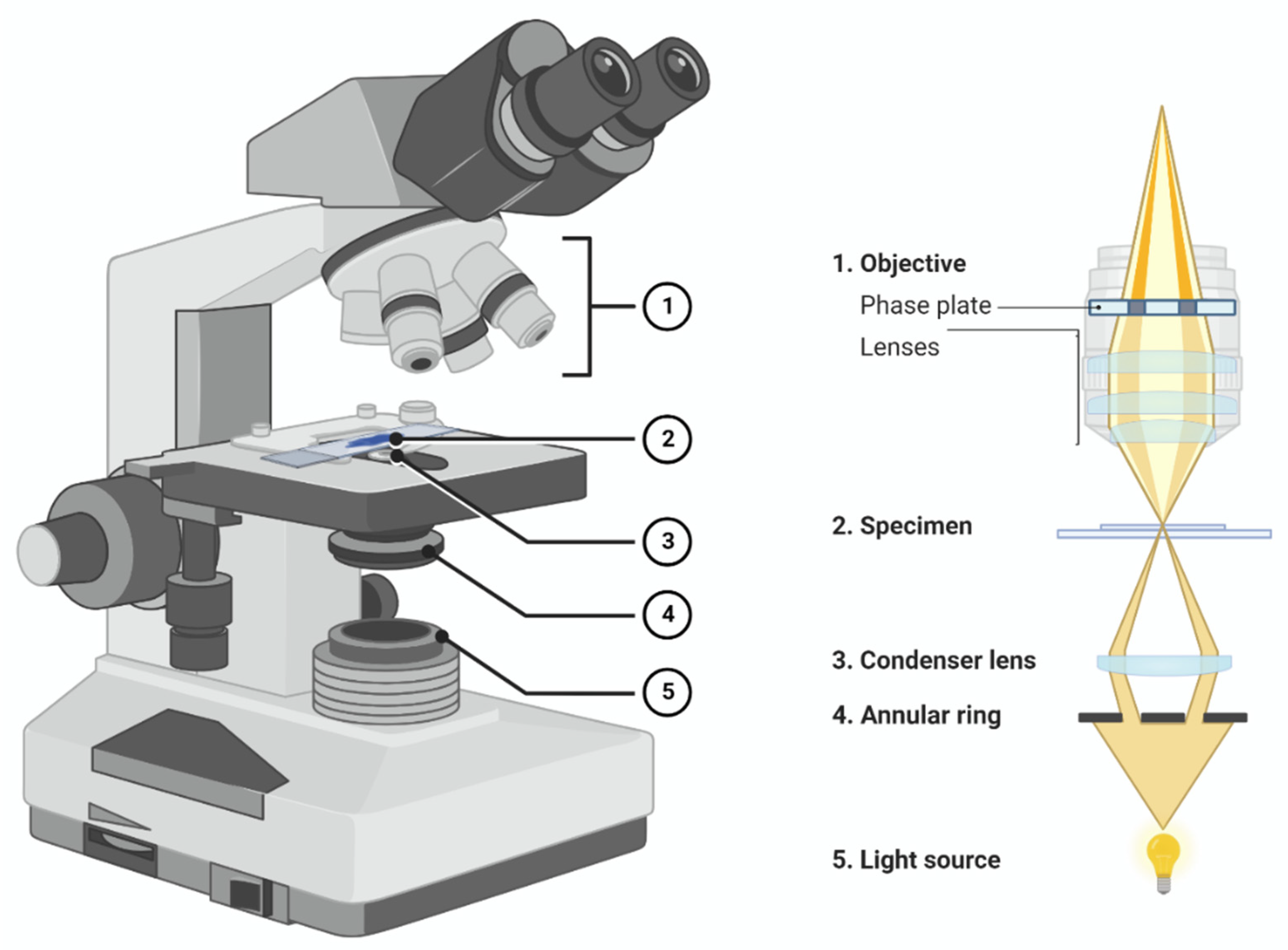
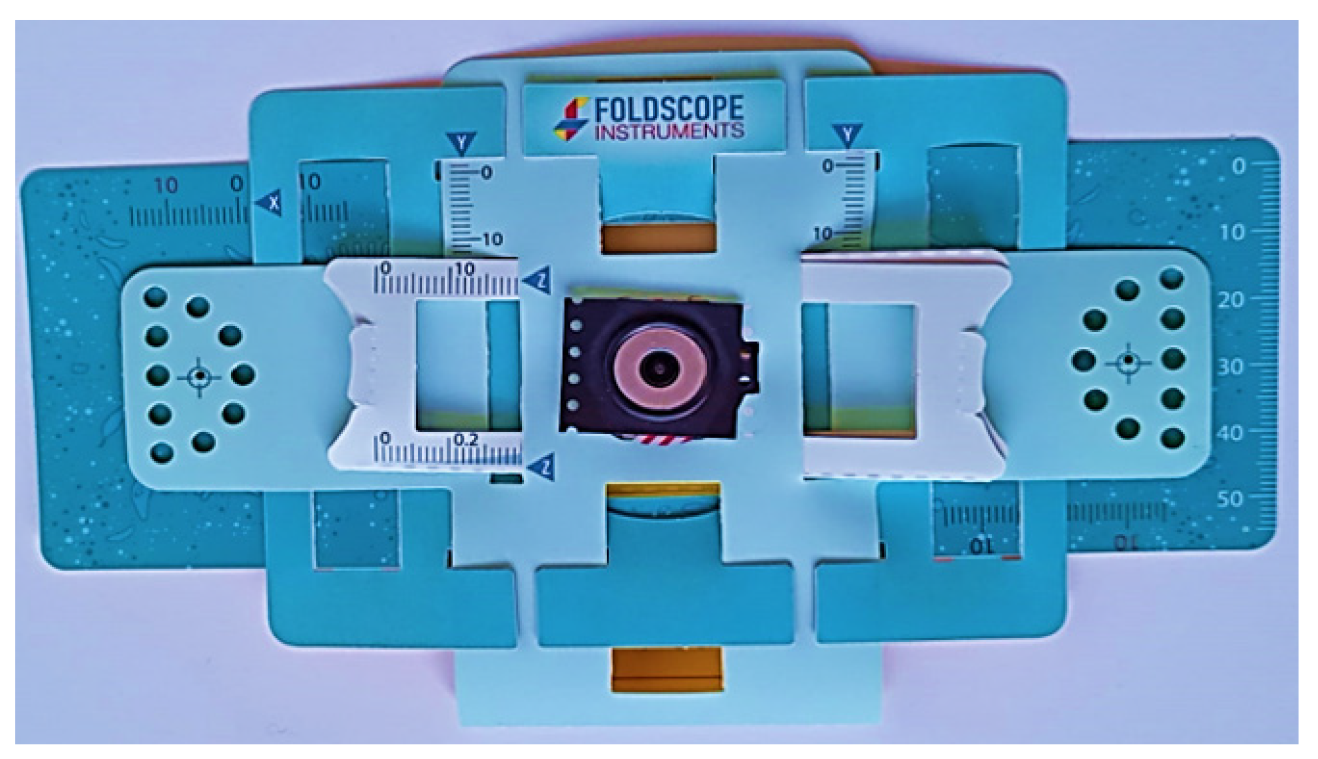
The pilot trial was offered to 92 students (18–19 years old) of the Plant Histology subject, which is part of the Biology Degree in the University of Salamanca, Spain. This Project was granted by the University of Salamanca as a program to innovate teaching, and was devised to address the exceptional circumstances of the COVID-19 lockdown. In this case, even though the microscopes were made available to all the students, two students did not want to participate. At the end of the academic year, the students had to return the Foldscopes and they were asked to complete a satisfaction survey that was available at the virtual campus (University online platform where students download or upload information related to their studies).

2.3. Instrument

To collect the students’ opinions, they were given a satisfaction survey with 4 questions (Table 1); 2 of them were dichotomic and the other 2 were Likert-type questions with a range of 4 options: very much, somewhat, not really, and not at all.

2.4. Use of the Foldscope

Each student received a Foldscope kit that contained a Foldscope, a 140-magnification lens, paper slides, instructions, and identification stickers, all in a nylon bag.
The first step the students had to take was the assembly of the Foldscope. After the final assembly, the students had to insert the lens. Once the microscope was completed, the students had to prepare their own plant samples, so they searched for a place to gather several plant tissues. The most common samples that were brought were petals, pollen, roots, stems, or leaves. One of the most common problems of microscopy is that light must pass through the tissues, and if the sample is too big or thick, it becomes opaque. Consequently, the students had to prepare the tissues; on the one hand, they could choose a small or thin sample (onion skin, pollen, small seeds), or on the other hand, they could make them thinner. To do this, they could cut the tissues with various tools (scissors, knifes) or tear them by hand. The prepared tissues were placed in the paper slide that was included in the kit with some tape, then, they were observed aiming the microscope towards a light source and focusing with their thumbs. The students had the opportunity to take some photographs with their own mobile phones, as the Foldscope integrates an accessory that allows for the attachment of a smartphone. Once the course was finished, the students returned the Foldscopes and they were asked to complete the aforementioned survey.
Moreover, to demonstrate the resolution of the Foldscope, several photographs of processed tissues were taken. The pictures were taken by placing the camera phone to touch the lens; the light source was the LED incorporated in the kit. In addition, the projection capacity of the Foldscope was tested; if an intense light crosses the sample, an image can be generated over a flat surface. Consequently, by using a smartphone flashlight, a projection of a sample can be observed on a wall.

3. Results

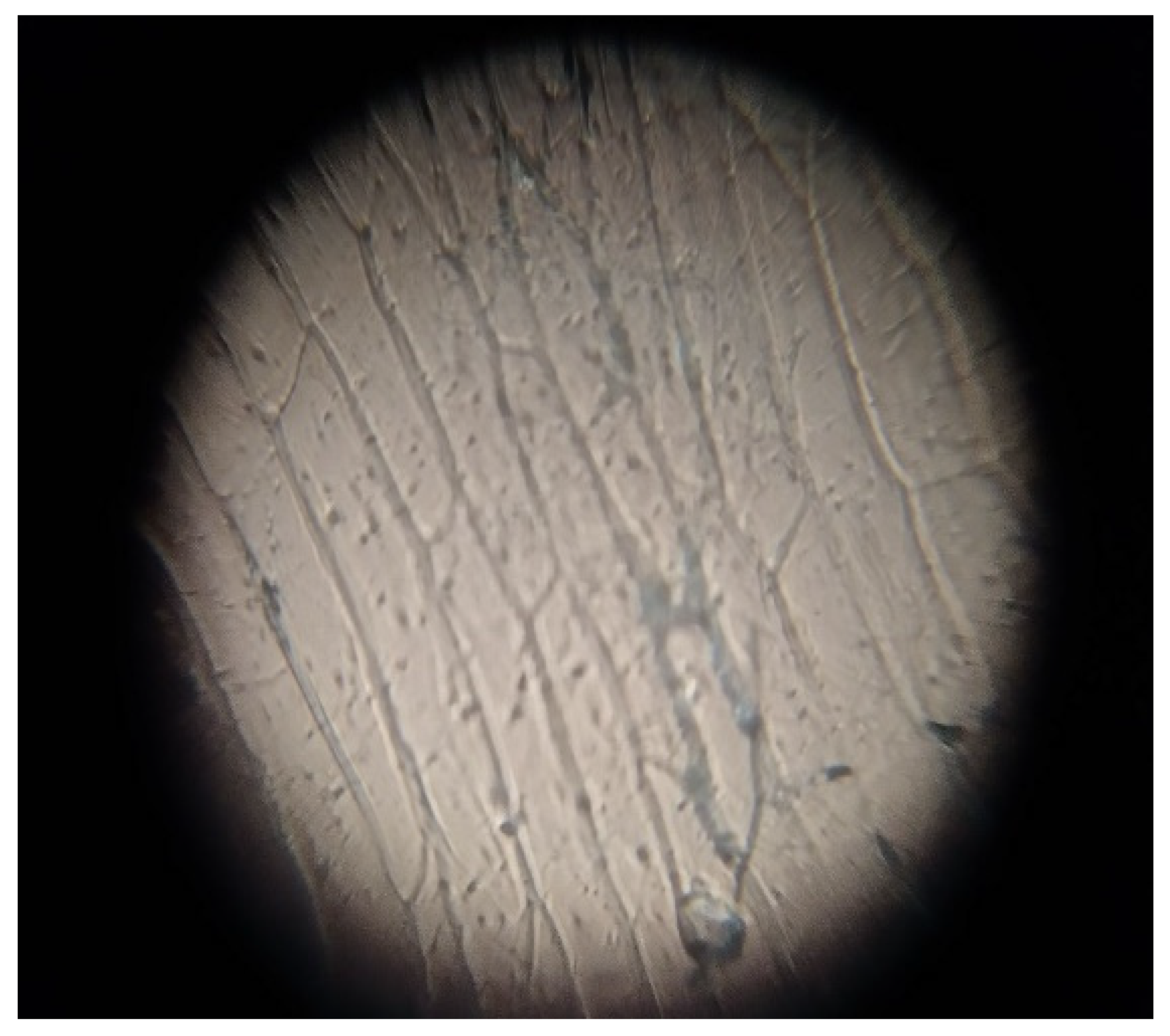
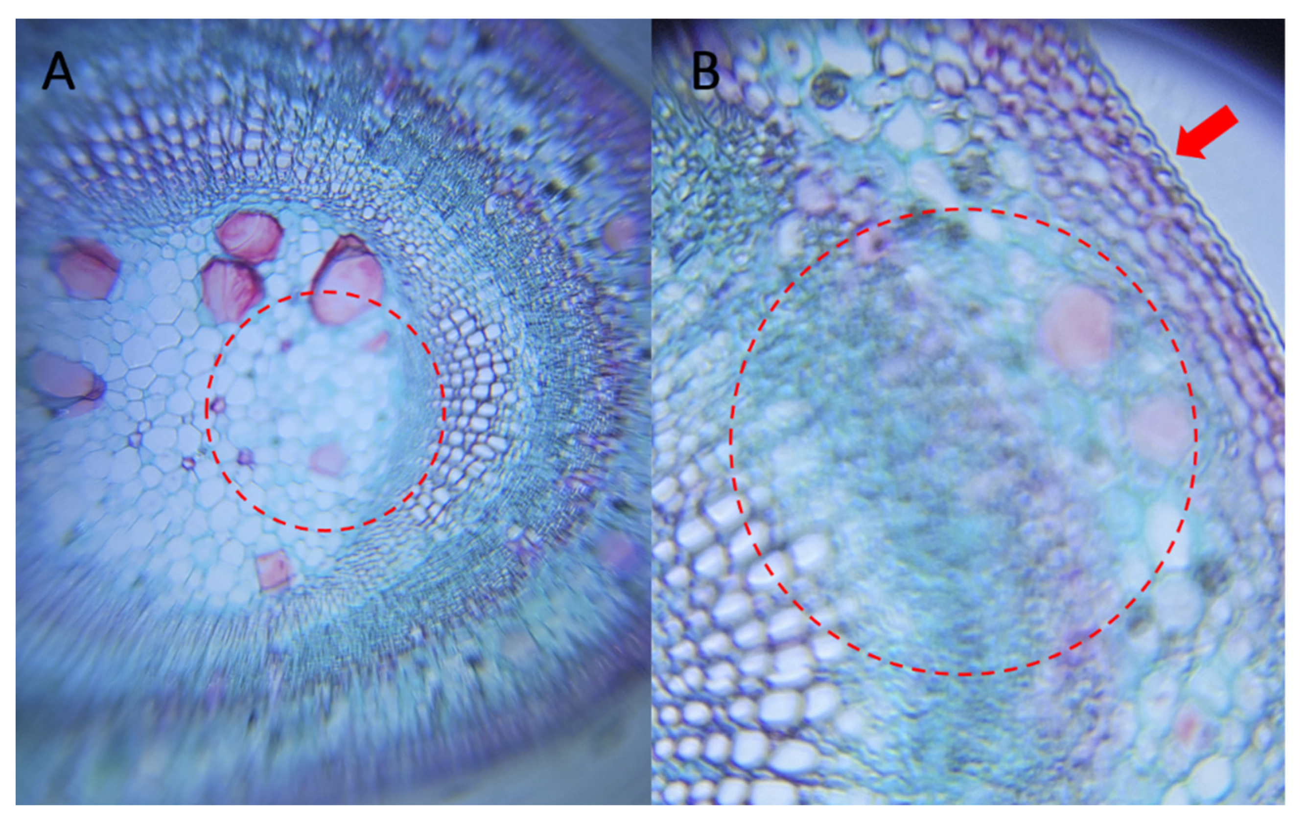
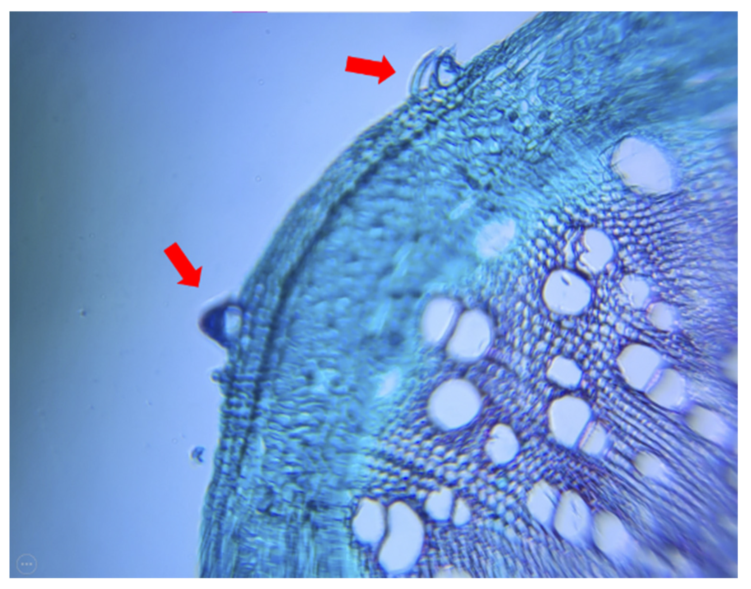
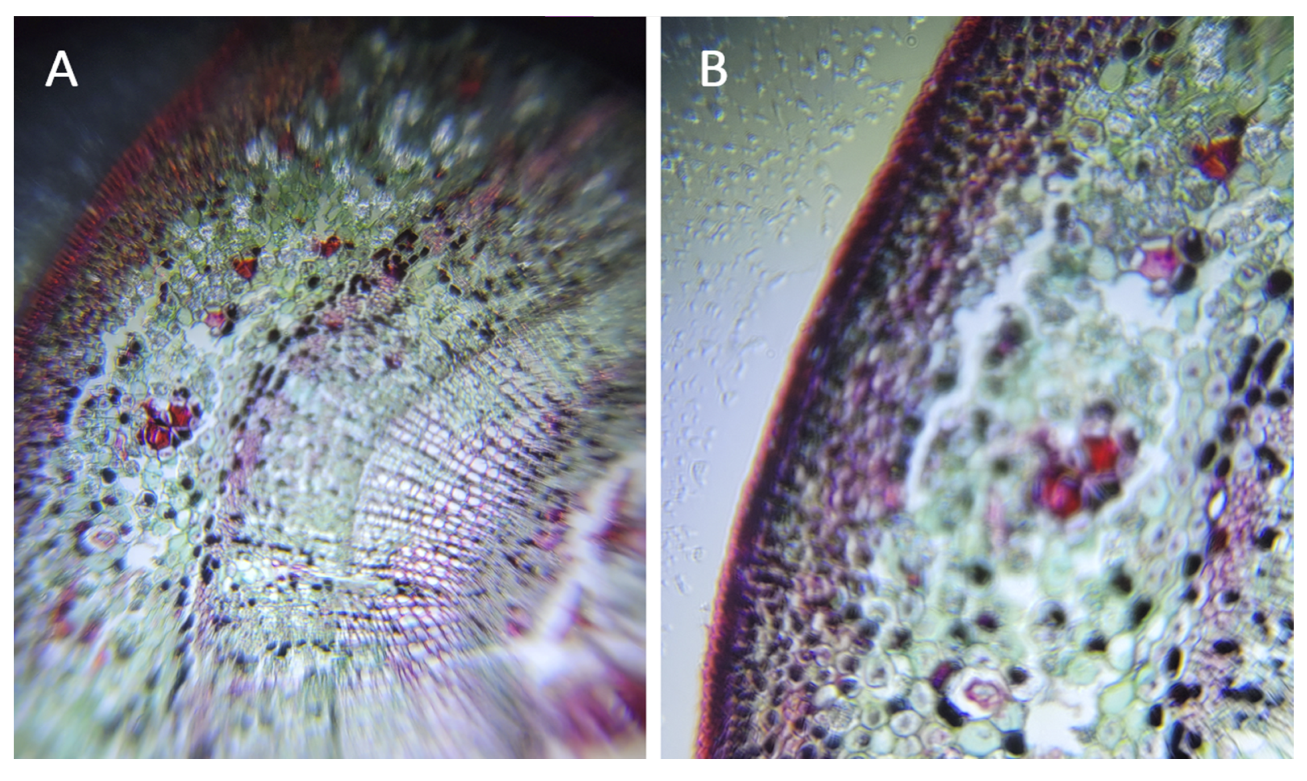
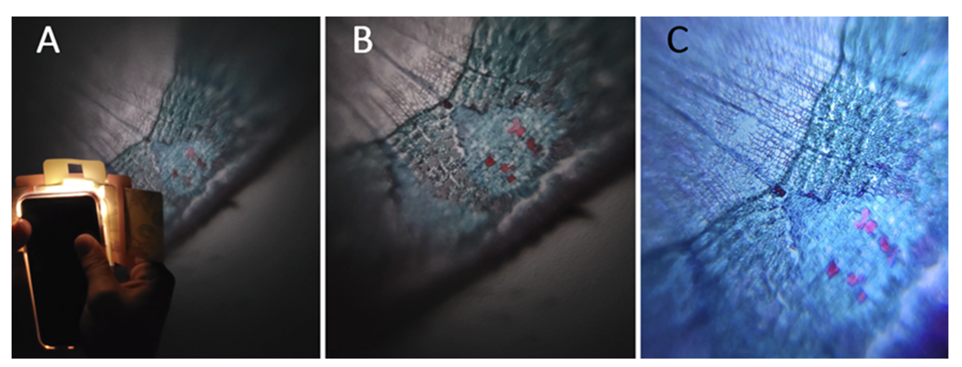
3.1. The Resolution of the Foldscope Is Enough to Distinguish Cell Structures

Onion skin is the classic vegetal tissue used to introduce microscopy and Plant Histology; therefore, the students selected this vegetable to learn the basics of the Foldscope. As shown in Figure 3, the unique cell part that can be distinguished from the plant cells without staining is the cell wall. Thanks to its structure, the polygonal form of the cells can be seen.
Unfortunately, there were few students who shared their own photographs. Besides, they did not have access to tissues processed by an expert; therefore, most of their photographs could not validate the actual resolution of the microscope. To do that, some micrographs of expert-processed tissue were taken. The plant structures selected were Tilia sp. in three different developmental stages: (1) meristematic stem (a plant tissue consisting of undifferentiated cells; Figure 4), (2) 1 year stem (Figure 5) and (3) 2 years stem.
Finally, apart from Tilia samples, various photographs of Cannabis sativa. stem were taken as well (Figure 5). Again, the Foldscope allowed the visualization of specific structures, such as the characteristic trichomes of Cannabis sativa.
As described, the projection capacity of the Foldscope was tested. To do so, the flashlight of a smartphone was used. The Foldscope only works in one light direction. In this case, the light must cross from the yellow part to the blue part, which allows the image to be generated against a surface. As can be seen in Figure 6 and Figure 7, the image quality is good enough to distinguish between the different structures. This can be helpful if it is necessary to project an image in a class to show the same structure to all the students.

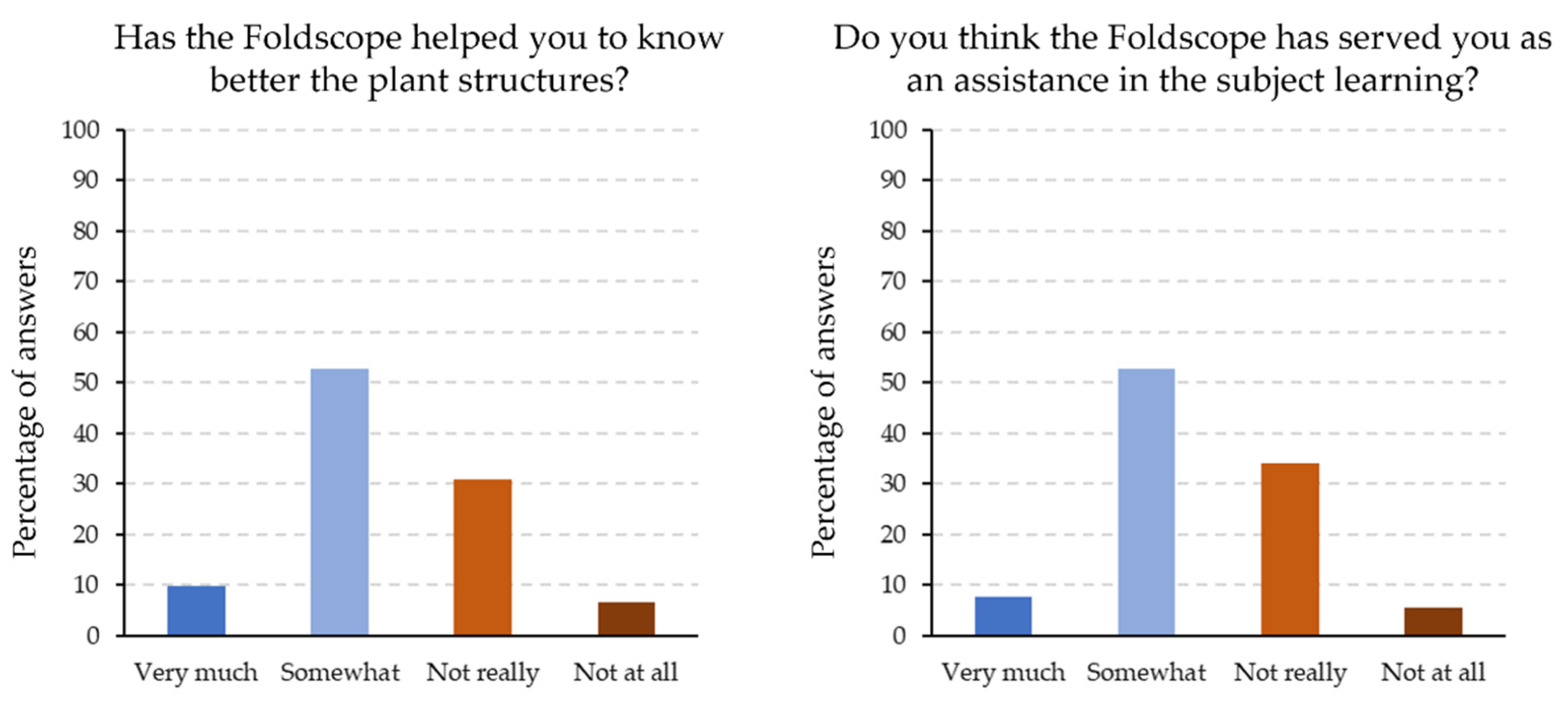
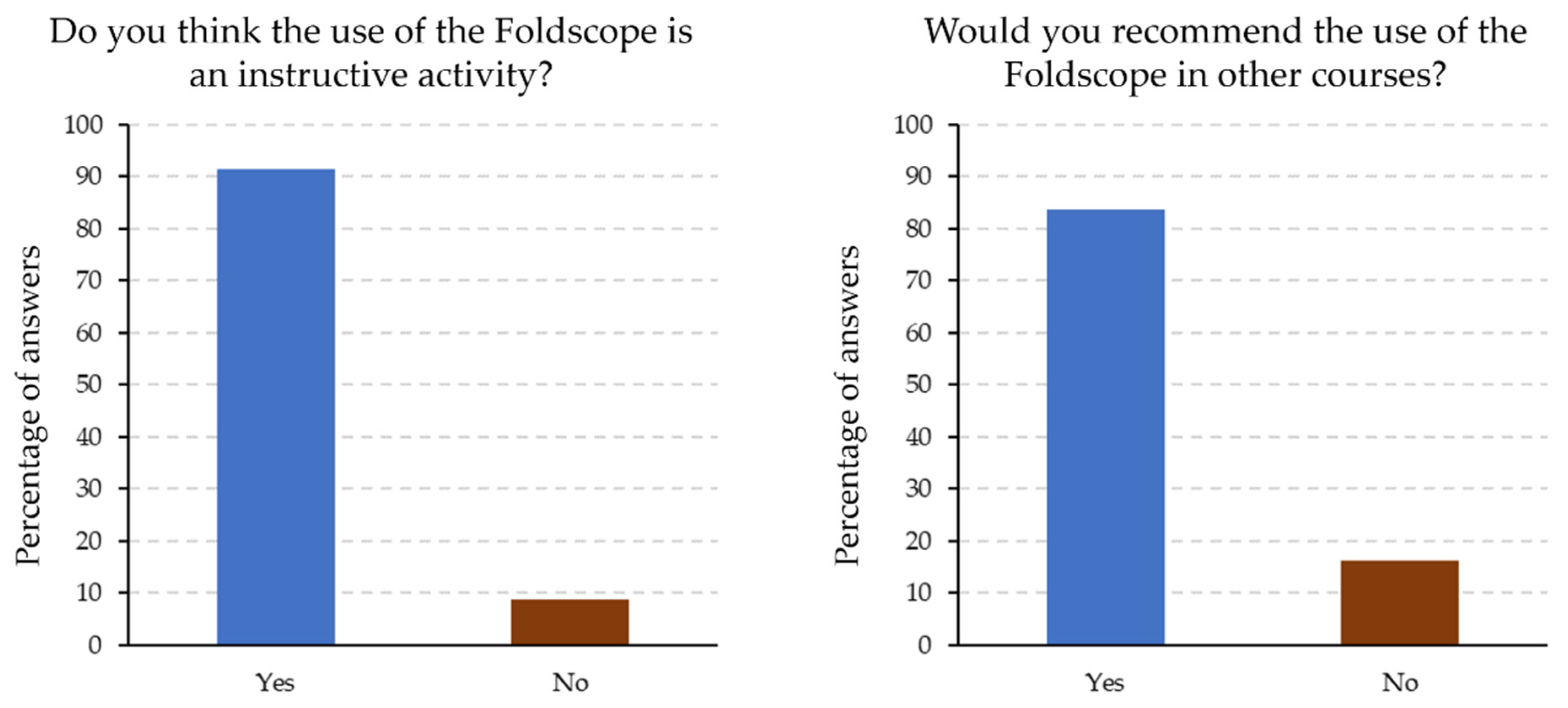
3.2. Students Have a Positive Opinion about the Foldscope

The students did not have a strong opinion about the use of the Foldscope: the percentage of students who thought that the Foldscope was very useful for learning the plant structures was about 10%. Secondly, the highest percentage corresponded to the students who thought it was somewhat useful; more than 50%. On the contrary, the percentage of the students who thought that the Foldscope was not really useful was 30%. Finally, the students who voted that the Foldscope was not useful at all was less than 10% (Figure 8). Some plant structures are distinguishable after proper treatment of the tissue, and as described before, the tissues were selected and treated by the students. Even though this method helps the students learn the basics of microscopy on their own, this could be the reason why they could not study all the structures. Previously, some pictures of tissues treated by a professional histologist were shown, and it was evident that the Foldscope provided enough resolution to study them. That is why it is logical to think that use of this tool with prepared tissues could give better results.
Regarding the question of “Do you think the Foldscope has served you as an assistance in the subject learning?”, the students had a similar opinion to the previous question. Less than 10% though that it was very useful as an assistance and more than 50% thought it was somewhat useful as an assistance. Additionally, to the contrary, more than 30% voted that it was not really useful as an assistance, and 5% voted that it was not an assistance at all (Figure 8). This scenario could be improved if the previous measure was applied. While it is true that processed samples could be provided to the students, they were not provided because the objective of the project was to promote autonomous learning. Providing samples would be against one of the main objectives, and would significantly increase the cost per student, which is also against the objectives of the project.
The students’ opinions were far better according to the dichotomic questions, as 90% of the students thought that the Foldscope project was an instructive activity. In addition, more than 80% of the students would recommend the use of the Foldscope in other courses (Figure 9). With all these data, it is obvious that whilst the use of the Foldscope in this educational scenario has room for improvement, the students are in favour of using the Foldscope in the project subject and others. It is obvious that using innovative tools can promote the teaching and learning process [22,23].

4. Discussion

Throughout the present research, an instrument was designed to evaluate students’ opinions about the use of low-cost technologies, such as the Foldscope, in laboratory practices.
The evaluation instrument was developed during the COVID-19 pandemic period. During the evaluation, two fundamental dimensions were studied: (i) didactic use of the Foldscope, (ii) opinion about Foldscope (Table 1).
When the results were analysed, it was found that University students considered the use of the Foldscope positively during their laboratory practices (Figure 8 and Figure 9). Thus, the specific objective of this work was achieved.
Previous studies agree with the results of the present investigation: a confirmation of (i) the pedagogical value of the Foldscope, although it is a low-cost microscope [11,12], and (ii) the motivation that this tool awakens [11,13,15,18,19] for the students, because they can have the Foldscope in their own homes for experimentation, and for the teachers, because it is economical and meets the minimum requirements. This microscope has positive features, such as durability, autonomy (no need to use electricity), and small size. The assessment of the use of the Foldscope as an alternative instrument to conventional microscopes agrees with previous studies’ results [11] that evidences its capacities in diverse disciplines.
There are numerous studies analysing teacher opinions about the innovative resources or methodologies; their general results demonstrate the need for innovative means to motivate the students, especially since the pandemic, where participation has been mostly virtual [22,23,24,25]. Knowing the opinion of students is necessary in order to promote use of the Foldscope as a portable tool for fixed use in university practices or in other epidemiological and clinical settings.
Regarding the use of the Foldscope during the laboratory teachings of superior studies, the obtained results confirm that students have a positive opinion about it (Figure 8 and Figure 9). In this sense, the results agree with others obtained previously, which show that the ease of transport of the microscope for the analysis of samples in any environment or the autonomous nature of its use guarantee a successful teaching–learning process [11,12,13,24]. This study also reveals the versatility of this microscope, making it a good candidate as an innovative tool for biomedical sciences degrees, as well as vaidating its use as a “collective microscope” due to its ability to project the information analysed on the screen or wall (Figure 7).

5. Conclusions

The use of the Foldscope is getting more and more attention in the sciences. This can be observed in the increment of investigations about the possibilities this tool offers from 2019 onwards.
Even though this study analysed the opinion of 92 Biology degree students, the obtained results could confirm that the Foldscope is a good low-cost alternative to more expensive microscopes.
The articles regarding the use of the Foldscope as a teaching tool are limited, because it was initially used as a clinical tool in less favourable regions. The number of participants and the quality of the instrument of this study are limited to the available means of the pandemic lockdown.
In summary, it would be appropriate to further study the benefits and possibilities that the Foldscope can offer to university, secondary and primary studies, as well as teachers’ opinions.

Author Contributions

Conceptualization, C.H.-P. and M.N.-S.; methodology, C.H.-P.; validation, C.H.-P.; formal analysis, C.H.-P.; data curation, C.H.-P.; writing—original draft preparation, C.H.-P. and M.N.-S.; writing—review and editing, C.H.-P. and M.N.-S.; supervision, C.H.-P. and M.N.-S. All authors have read and agreed to the published version of the manuscript.

Funding

University of Salamanca funded 100 Foldscopes for the project: “Sacar las clases practices del laboratorio a la calle: Foldscope. Uso de microscopios plegables como herramienta docente”.

Institutional Review Board Statement

The data collection process was approved by the direction council of the teaching innovation project of University of Salamanca titled “Sacar las clases prácticas del laboratorio a la calle: Foldscope. Uso de microscopios plegables como herramienta docente” (ID2019/1112), in September 2019. All data collected in this project were conducted in accordance with the Helsinki Declaration and were obtained with the informed consent of the participants under the data protection normative.

Informed Consent Statement

All participants were informed about the anonymous nature of their participation, why the research is being conducted, how their data will be used and that under no circumstances would their data be used to identify them.

Data Availability Statement

The data are not publicly available because they are part of a larger project involving more researchers. If you have any questions, please ask the contact author.

Conflicts of Interest

The authors declare no conflict of interest.

References

  1. Duroisin, N.; Beauset, R.; Tanghe, C. Education and digital inequalities during COVID-19 confinement: From the perspective of teachers in the French speaking Community of Belgium. Eur. J. Educ. 2021, 56, 515–535. [Google Scholar] [CrossRef] [PubMed]
  2. Ramos-Pla, A.; del Arco, I.; Flores Alarcia, Ò. University Professor Training in Times of COVID-19: Analysis of Training Programs and Perception of Impact on Teaching Practices. Educ. Sci. 2021, 11, 684. [Google Scholar] [CrossRef]
  3. Almonacid_Fierro, A.; Vargas-Vitoria, R.; Souza De Carvalho, R.; Almonacid Fierro, M. Impact on teaching in times of COVID-19 pandemic: A qualitative study. IJERE 2021, 10, 432–440. [Google Scholar] [CrossRef]
  4. Antón-Sancho, Á.; Vergara, D.; Fernández-Arias, P. Influence of country digitization level on digital pandemic stress. Behav. Sci. 2022, 12, 203. [Google Scholar] [CrossRef] [PubMed]
  5. Rosen, D.J.; Kelly, A.M. Working together or alone, near, or far: Social connections and communities of practice in in-person and remote physics laboratories. Phys. Rev. Phys. Educ. Res. 2022, 18, 010105. [Google Scholar] [CrossRef]
  6. Gamage, K.A.A.; Wijesuriya, D.I.; Ekanayake, S.Y.; Rennie, A.E.W.; Lambert, C.G.; Gunawardhana, N. Online Delivery of Teaching and Laboratory Practices: Continuity of University Programmes during COVID-19 Pandemic. Educ. Sci. 2020, 10, 291. [Google Scholar] [CrossRef]
  7. Mukhopadhyay, S.; Booth, A.L.; Calkins, S.M.; Doxtader, E.E.; Fine, S.W.; Gardner, J.M.; Gonzalez, R.S.; Mirza, K.M.; Jiang, X.S. Leveraging Technology for Remote Learning in the Era of COVID-19 and Social DistancingTips and Resources for Pathology Educators and Trainees. Arch. Pathol. Lab. Med. 2020, 144, 1027–1036. [Google Scholar] [CrossRef]
  8. Lidueña-Peréz, K.I.; Martelo-Solórzano, A.M.; Payares-Díaz, I.R.; Santos-Amaya, O.F.; Corredor-Prado, J.P. Ananas ananassoides (Baker) LB Sm. a bromeliad from the savanna: Seed morpho-anatomy and histochemistry. Braz. J. Biol. 2022, 84, 1–8. [Google Scholar] [CrossRef]
  9. Quang, N.K. A Simple Way to Demonstrate Fluorescent Image by Employing a Conventional Microscope and Transparent Colored Acrylic Pieces. Phys. Teach. 2021, 59, 57–59. [Google Scholar] [CrossRef]
  10. Jackson, C.; de Beer, J.; White, L. The affective affordances of frugal science using foldscopes during a Life Sciences water quality practical. Perspect. Educ. 2020, 38, 224–241. [Google Scholar] [CrossRef]
  11. Gupta, S.; Mathews, B.J.; Ghantaa, S.N.; Amerneni, K.C.; Karuna, T.; Pakhare, A.; Khadanga, S. Foldscope: Diagnostic accuracy and feasibility of its use in national malaria control program. J. Microsc. Ultrastruct. 2022, 10, 114. [Google Scholar] [CrossRef] [PubMed]
  12. Rameshbabu, R.; Priya, A.H.; Muthukumar, R.S.; Sivaraman, K.; Uthra, D. Evaluation of efficacy of foldscope–a paper microscope to be used as a chairside diagnostic tool in oral dysplastic lesions: A comparative study. Contemp. Clin. Dent. 2021, 12, 352. [Google Scholar] [CrossRef] [PubMed]
  13. PaaBen, B.; Bertsch, A.; Langer-Fischer, K.; Rüdian, S.; Wang, X.; Sinha, R.; Pinkwart, N. Analyzing Student Success and Mistakes in Virtual Microscope Structure Search Tasks. In Proceedings of the International Educational Data Mining Society, Paper presented at the International Conference on Educational Data Mining (EDM), International Educational Data Mining Society, Online, 29 June–2 July 2021; Available online: https://educationaldatamining.org/conferences/ (accessed on 8 September 2020).
  14. Hughes, S.; Evason, C.; Baldwin, S.; Nadarajah, H.; Leisemann, S.; Wright, S. STEM takes flight. Phys. Educ. 2019, 55, 025005. [Google Scholar] [CrossRef]
  15. Das, K.; Dutta, P.; Gogoi, J. ‘Foldscope’-A simple and economical microscope. J. Biol. Educ. 2021, 55, 217–222. [Google Scholar] [CrossRef]
  16. Rane, J.; George, P.; Pawar, K.R.; Kumar, M.; Singh, N.P.; Sebastian, J.S.V. Calibration and validation of the application of low-cost, lightweight, and portable paper microscopes for monitoring pollen traits as a screening tool for drought tolerance. S. Afr. J. Bot. 2022, 148, 694–703. [Google Scholar] [CrossRef]
  17. Kulshreshtha, P.; Gupta, S.; Shaikh, R.; Aggarwal, D.; Sharma, D.; Rahi, P. Foldscope Embedded Pedagogy in Stem Education: A Case Study of SDG4 Promotion in India. Sustainability 2022, 14, 13427. [Google Scholar] [CrossRef]
  18. Biswas, S.; Barma, S. A Large-Scale Fully Annotated Low-Cost Microscopy Image Dataset for Deep Learning Framework. IEEE Trans. Nanobiosci. 2021, 20, 507–515. [Google Scholar] [CrossRef]
  19. Dua, J.; Dhawan, S. Foldscope as a Teaching and Learning Tool: An Indian Perspective. Innovations 2021, 47, 64–71. [Google Scholar]
  20. Cybulski, J.S.; Clements, J.; Prakash, M. Foldscope: Origami-based paper microscope. PLoS ONE 2014, 9, e98781. [Google Scholar] [CrossRef] [Green Version]
  21. Kaur, T.; Dahiya, S.; Satija, S.H.; Nawal, S.J.; Kshetrimayum, N.; Ningthoujam, J.; Rao, A. Foldscope as a primary diagnostic tool for oral and urinary tract infections and its effectiveness in oral health education. J. Microsc. 2020, 279, 39–51. [Google Scholar] [CrossRef]
  22. Vergara, D.; Fernández-Arias, P.; Extremera, J.; Dávila, L.P.; Rubio, M.P. Educational trends post COVID-19 in engineering: Virtual laboratories. Mater. Today Proc. 2022, 49, 155–160. [Google Scholar] [CrossRef] [PubMed]
  23. Antón-Sancho, Á.; Nieto-Sobrino, M.; Fernández-Arias, P.; Vergara-Rodríguez, D. Usability of Memes and Humorous Resources in Virtual Learning Environments. Educ. Sci. 2022, 12, 208. [Google Scholar] [CrossRef]
  24. Antón-Sancho, Á.; Sánchez-Calvo, M. Influence of Knowledge Area on the Use of Digital Tools during the COVID-19 Pandemic among Latin American Professors. Educ. Sci. 2022, 12, 635. [Google Scholar] [CrossRef]
  25. Jadhav, V.D.; Bokefode, J.D.; Ghodake, A.K.; Borade, O.N.; Gidvir, A.S. Fold Scope: A Low-Cost Magnification Device and Its Applications in Various Fields. In Techno-Societal 2018; Springer: Berlin/Heidelberg, Germany, 2020; pp. 145–154. [Google Scholar] [CrossRef]
Figure 1. Scheme of a microscope and its section. Adapted from “Phase Contrast Microscopy”, by BioRender.com (2022).
Education 12 00927 g001
Figure 2. Photography of a Foldscope.
Education 12 00927 g002
Figure 3. Microphotography of onion skin. Image taken by a student with their own mobile phone.
Education 12 00927 g003
Figure 4. Tilia sp. meristem. The Foldscope resolution was good enough to allow to distinguish the different plant cells. As the Foldscope lens is a sphere, the central region of the generated images can be blurred ((A,B) red circle). On the other hand, the periphery of this blurred region presents good optic qualities as it is shown with a red arrow (B) that marks the external layer of the epidermis, a plant structure difficult to see without a good microscope.
Education 12 00927 g004
Figure 5. Cross section of Cannabis sativa. Red arrows mark trichomes, an outgrowth of the epidermis where the cannabinoids are stored.
Education 12 00927 g005
Figure 6. Micrographs of 2 years old Tilia stem. In (A) a cross section of a Tilia sp. stem can be distinguish. To take image (B) the 3× amplification lens of the mobile phone was used. The Foldscope includes a 140× magnification lens, this implies that using the 3× lens incorporated in some smartphones make possible the acquisition of photographs with 420 magnifications.
Education 12 00927 g006
Figure 7. 1 year old Tilia sp. stem. In (A) the disposition of the Foldscope with a mobile phone can be seen, in (B) the image is projected at 1 m and in (C) a photograph of the same plant structure is shown.
Education 12 00927 g007
Figure 8. Students’ opinion about the usefulness of the Foldscope. About 62% of the students thought that the Foldscope had helped (Very much or Somewhat) them to better understand the plant structures, while approximately 37% (Not really and Not at all) had a negative answer to this question. In addition, almost 60% (Very much and Somewhat) had a positive answer when asked if they thought that the Foldscope was an assistance for the subject learning, while almost 40% of the students had a negative answer.
Education 12 00927 g008
Figure 9. Students’ opinions about the Foldscope. 91% of the students gave a positive answer when asked if they thought that using the Foldscope was an instructive activity. In addition, 83% of the students would recommend the use of the Foldscope in other courses.
Education 12 00927 g009
Table 1. Dependent variables.
Do you think the use of the Foldscope is an instructive activity?YesNo
Would you recommend the use of the Foldscope in other courses?YesNo
Has the Foldscope helped you to know better the plant structures?Very muchSomewhatNot reallyNot at all
Do you think the Foldscope has served you as an assistance in the subject learning?Very muchSomewhatNot reallyNot at all
Publisher’s Note: MDPI stays neutral with regard to jurisdictional claims in published maps and institutional affiliations.

