

Immersive VR (Virtual Reality) Simulator for Vein Blood Sampling


Abstract
:1. Introduction
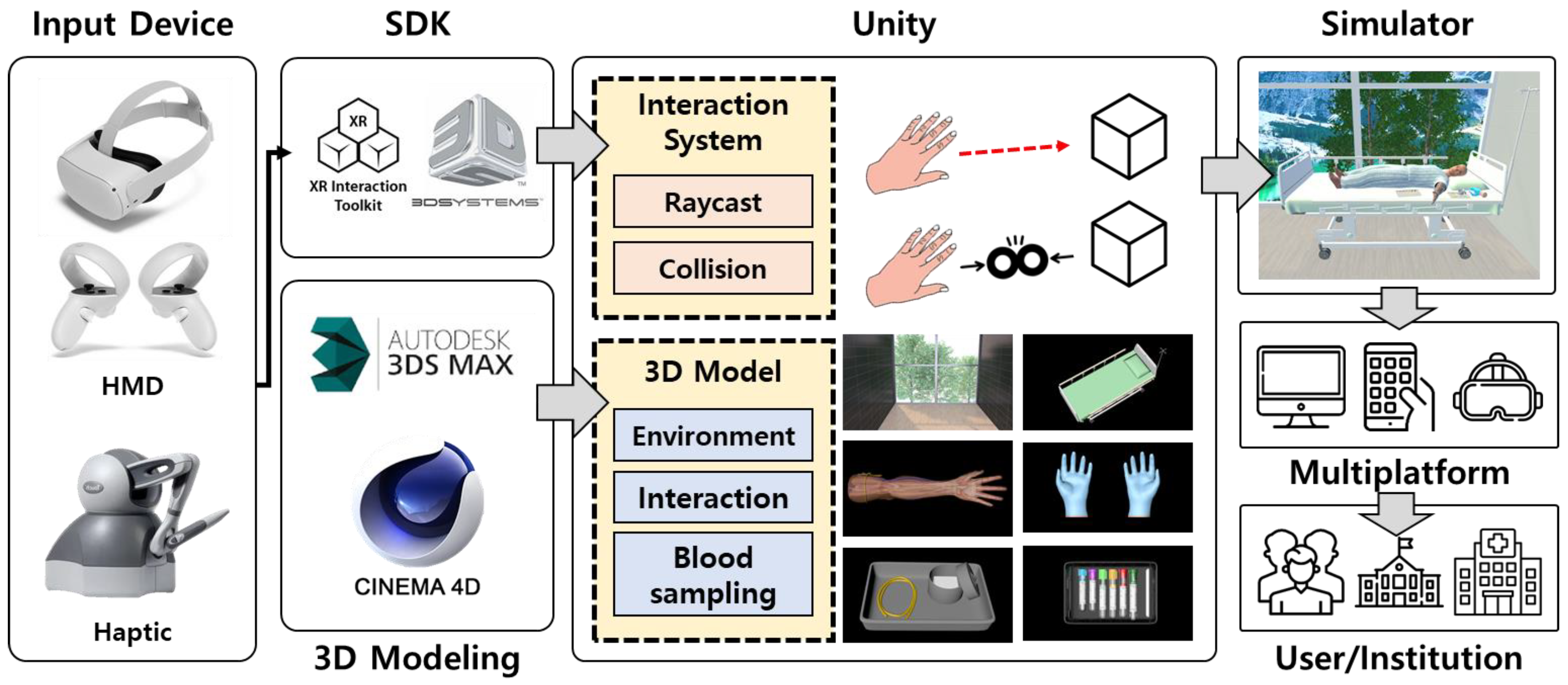
2. Materials and Methods
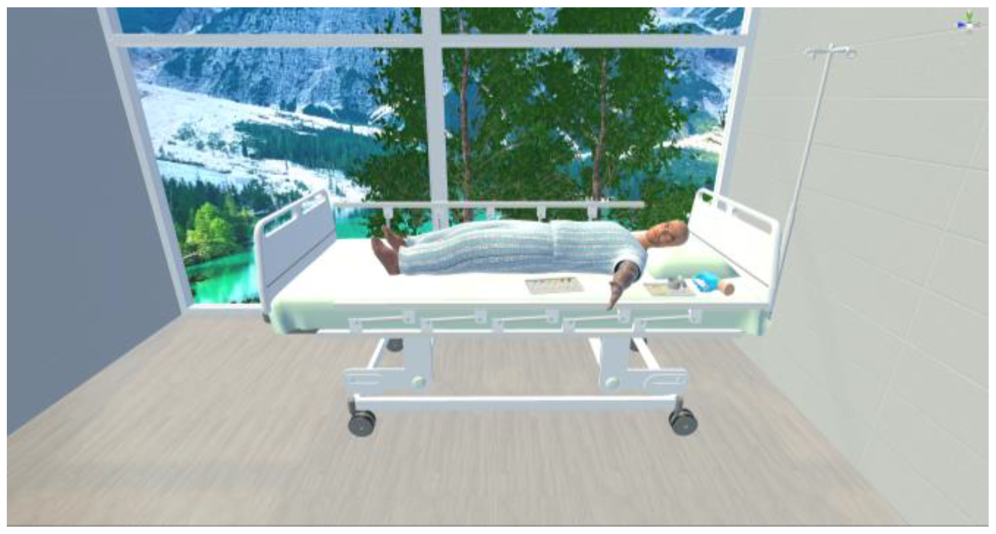
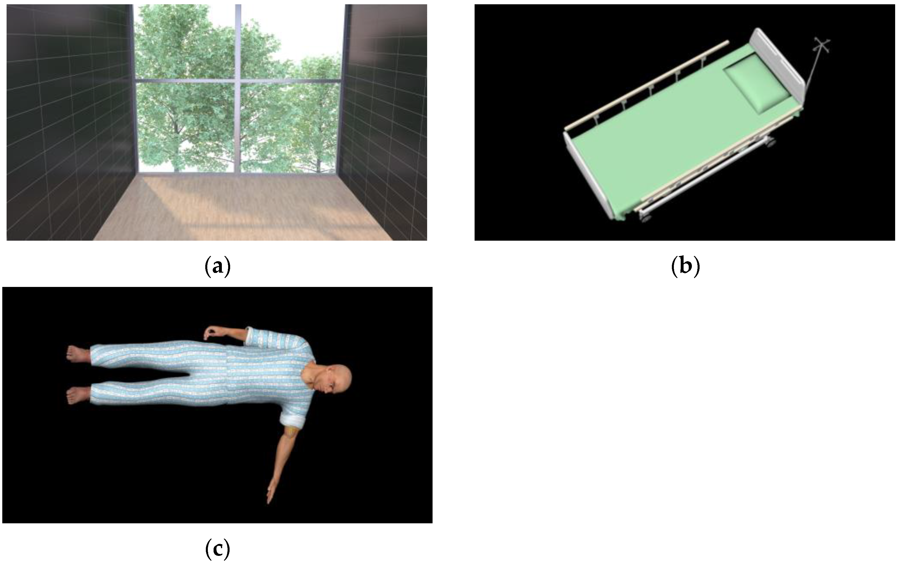
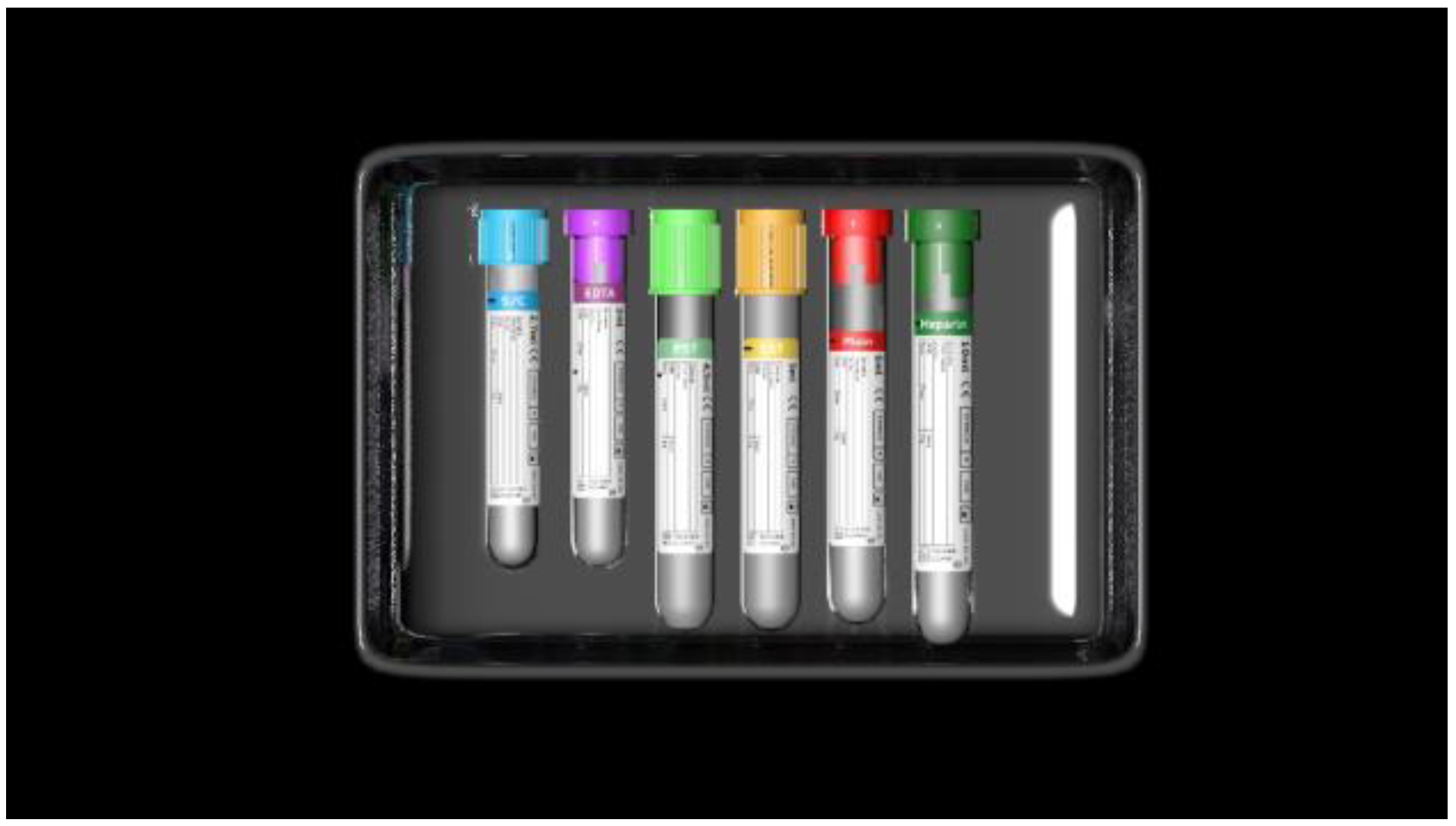
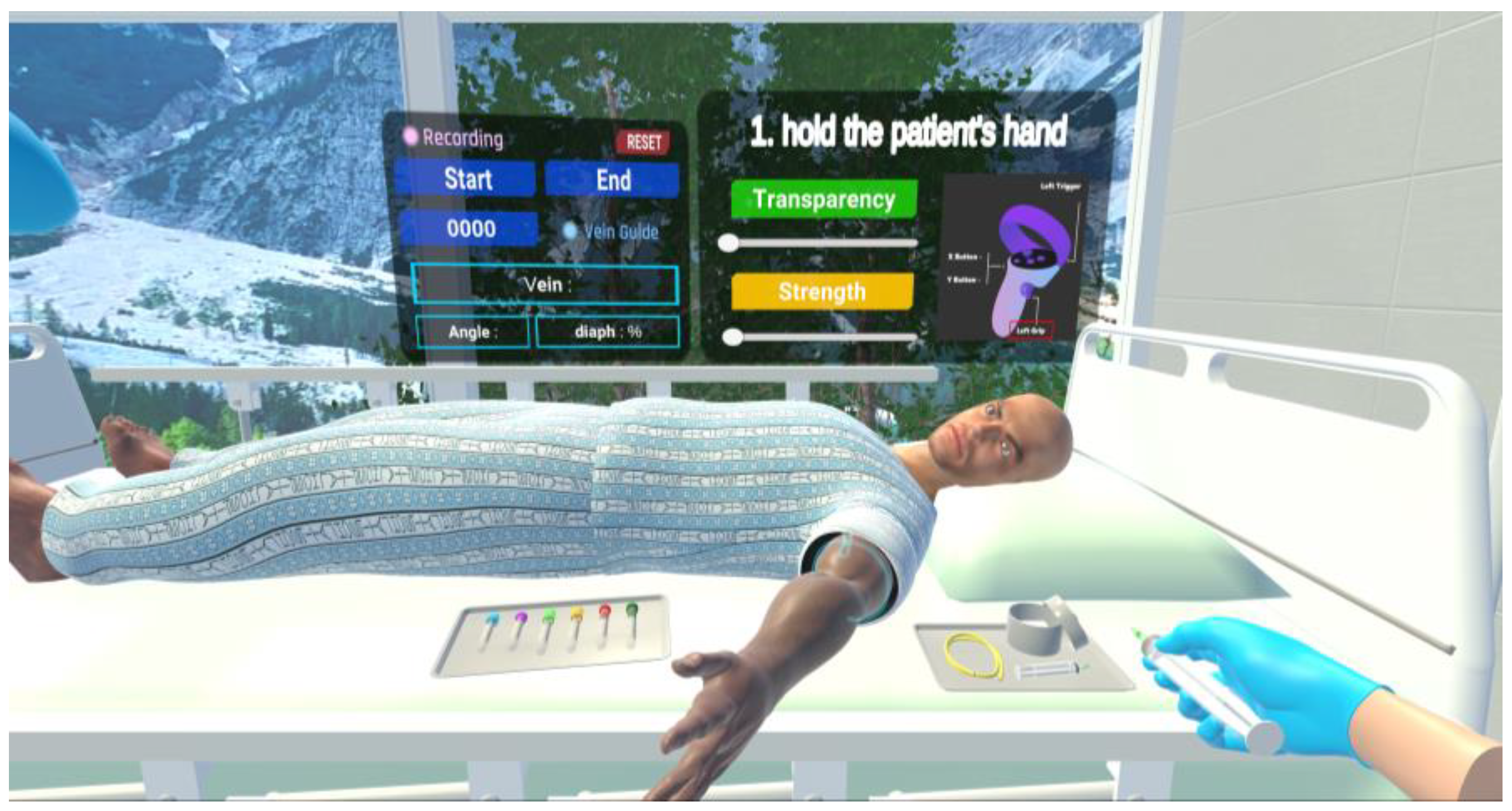
2.1. Production of Virtual Space for Vein Blood Sampling
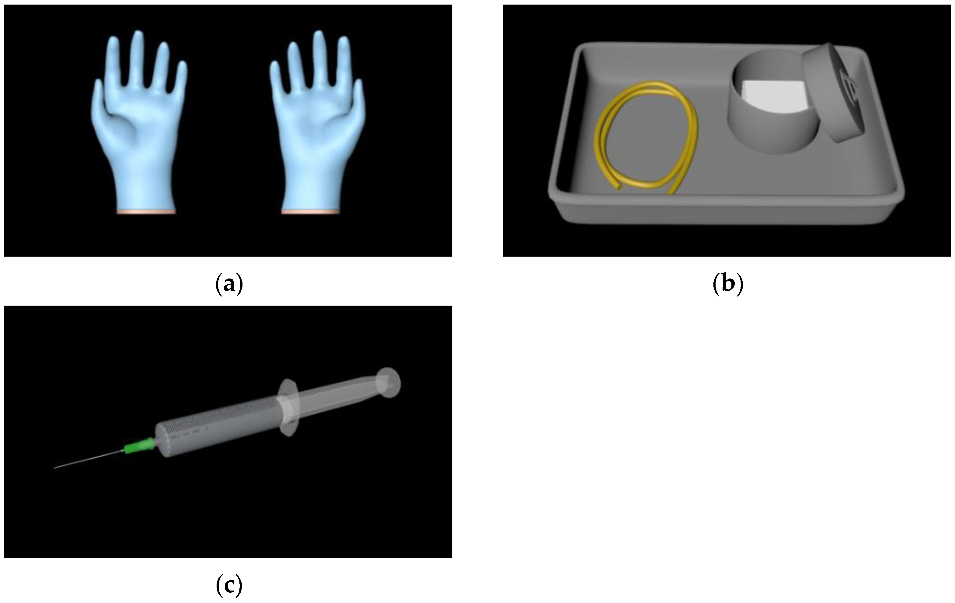
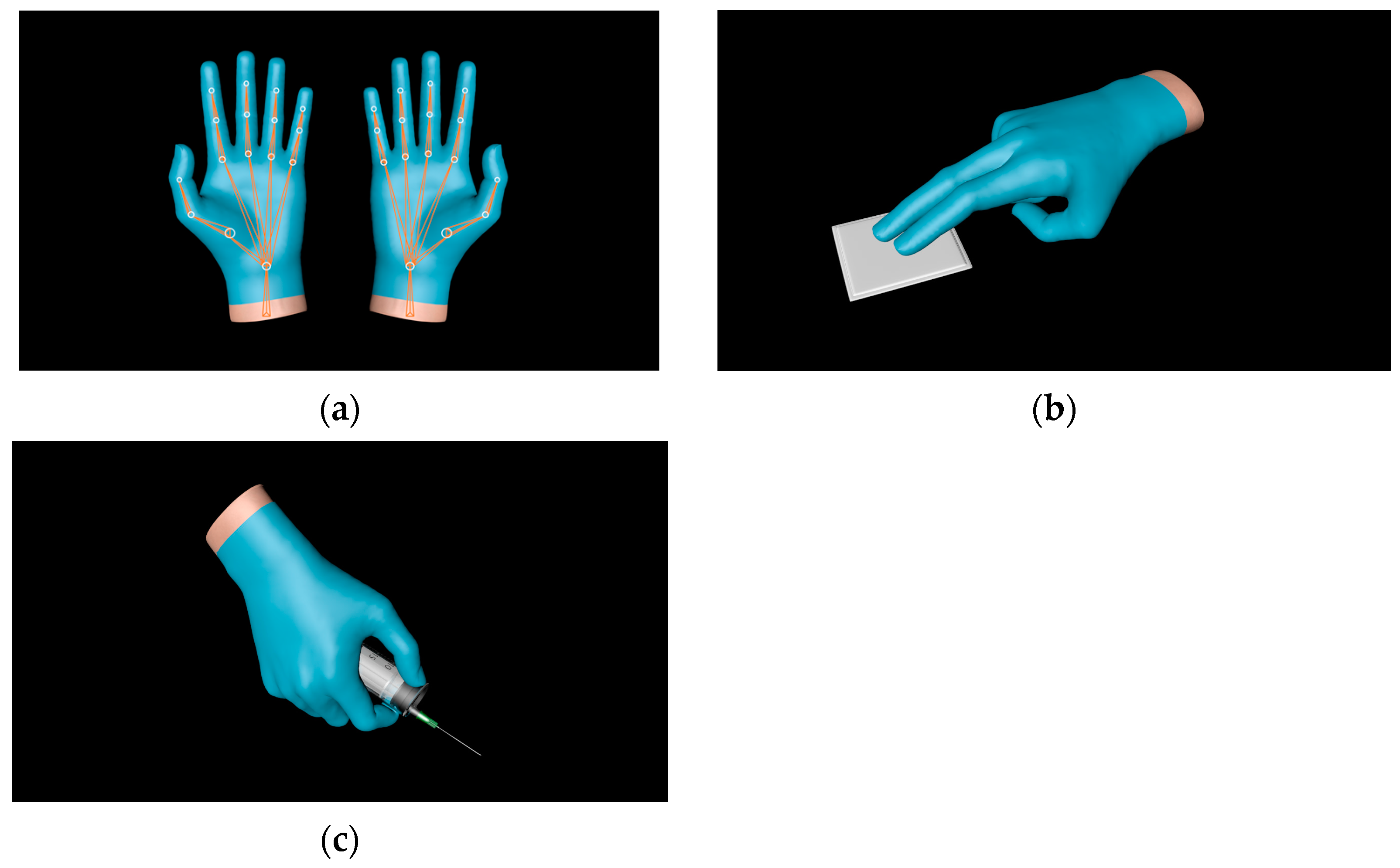
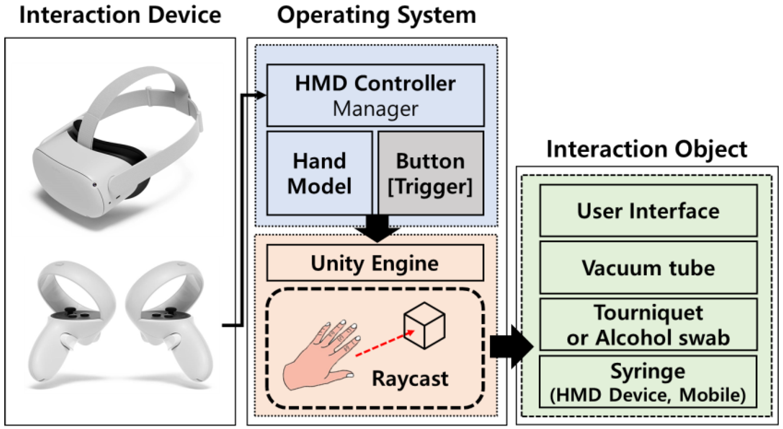
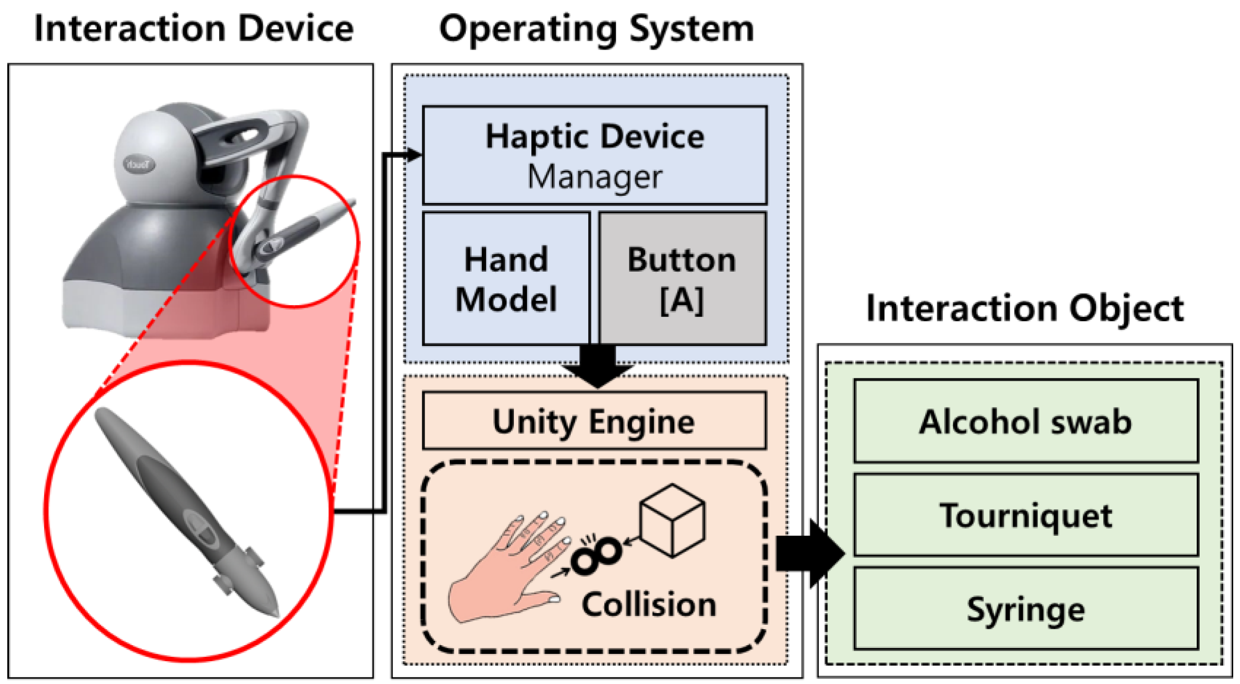
2.2. Implementation of Vein Blood Sampling Interaction
2.3. Evaluation of Vein Blood Sampling Simulator
3. Results
3.1. Implementation of Vein Blood Sampling Simulator
3.2. Application and Efficacy Evaluation of Vein Blood Sampling Simulator
4. Discussion
Author Contributions
Funding
Institutional Review Board Statement
Informed Consent Statement
Data Availability Statement
Conflicts of Interest
References
- Sun, J.I. Strategies for Effective Teaching in Clinical Clerkship. Hanyang Med. Rev. 2012, 32, 51–57. [Google Scholar] [CrossRef]
- Oh, Y.K. Importance of clinical medicine in medical education: Review of the articles in this issue. Korean J. Med. Educ. 2015, 27, 243–245. [Google Scholar] [CrossRef]
- Hem-Stokroos, H.V.D.; Scherpbier, A.J.J.A.; Vleuten, C.V.D.; Vries, H.D.; Haarman, H.T.M. How effective is a clerkship as a learning environment? Med. Teach. 2001, 23, 599–604. [Google Scholar] [CrossRef] [PubMed]
- Park, G.H.; Lee, Y.D.; Oh, J.H.; Choi, I.S.; Lim, Y.M.; Kim, Y.L. Program development of student internship (subinternship) in Gachon medical school. Korean J. Med. Educ. 2003, 15, 113–130. [Google Scholar] [CrossRef]
- Li, L.; Yu, F.; Shi, D.; Shi, J.; Tian, Z.; Yang, J.; Wang, X.; Jiang, Q. Application of virtual reality technology in clinical medicine. Am. J. Transl. Res. 2017, 9, 3867–3880. [Google Scholar]
- Kuehn, B.M. Virtual and augmented reality put a twist on medical education. JAMA 2019, 319, 756–758. [Google Scholar] [CrossRef] [PubMed]
- Pottle, J. Virtual reality and the transformation of medical education. Future Healthc. J. 2019, 6, 181–185. [Google Scholar] [CrossRef] [PubMed]
- Oxford Medical Simulation. Available online: https://oxfordmedicalsimulation.com (accessed on 1 August 2023).
- Falah, J.; Khan, S.; Alfalah, T.; Alfalah, S.F.M.; Chan, W.; Harrison, D.K.; Charissis, V. Virtual Reality medical training system for anatomy education. In Proceedings of the 2014 Science and Information Conference, London, UK, 27–29 August 2014; pp. 752–758. [Google Scholar] [CrossRef]
- Schild, J.; Misztal, S.; Roth, B.; Flock, L.; Luiz, T.; Lerner, D.; Herkersdorf, M.; Weaner, K.; Neuberaer, M.; Franke, A.; et al. Applying multi-user virtual reality to collaborative medical training. In Proceedings of the 2018 IEEE Conference on Virtual Reality and 3D User Interfaces (VR), Tuebingen/Reutlingen, Germany, 18–22 March 2018; pp. 775–776. [Google Scholar] [CrossRef]
- Ayoub, A.; Pulijala, Y. The application of virtual reality and augmented reality in Oral & Maxillofacial Surgery. BMC Oral Health 2019, 19, 238. [Google Scholar] [CrossRef] [PubMed]
- Ultraleap. Available online: https://www.ultraleap.com (accessed on 5 August 2023).
- Ropelato, S.; Menozzi, M.; Michel, D.; Siegrist, M. Augmented reality microsurgery: A tool for training micromanipulations in ophthalmic surgery using augmented reality. J. Soc. Simul. Healthc. 2020, 15, 122–127. [Google Scholar] [CrossRef] [PubMed]
- Rhienmora, P.; Gajananan, K.; Haddawy, P.; Dailey, M.N.; Suebnukarn, S. Augmented reality haptics system for dental surgical skills training. In Proceedings of the 17th ACM Symposium on Virtual Reality Software and Technology, Hong Kong, China, 22–24 November 2010; pp. 97–98. [Google Scholar] [CrossRef]
- Kata, H.; Billinghurst, M. Marker tracking and hmd calibration for a video-based augmented reality conferencing system. In Proceedings of the 2nd IEEE and ACM International Workshop on Augmented Reality (IWAR’99), San Francisco, CA, USA, 20–21 October 1999; pp. 85–94. [Google Scholar] [CrossRef]
- Si, W.X.; Liao, X.Y.; Qian, Y.L.; Sun, H.T.; Chen, X.D.; Wang, Q.; Heng, P.A. Assessing performance of augmented reality-based neurosurgical training. Vis. Comput. Ind. Biomed. Art 2019, 2, 1–10. [Google Scholar] [CrossRef] [PubMed]
- Microsoft. Available online: https://www.microsoft.com/en-us/hololens (accessed on 12 August 2023).
- AUTODESK. Available online: https://www.autodesk.com (accessed on 13 August 2023).
- Maxon. Available online: https://www.maxon.net (accessed on 13 August 2023).


| Environment Model | Interaction Model | Blood Sampling Model |
|---|---|---|
| Ward | data3D hand model | Vacuum tube |
| Medical bed | Syringe | Tray |
| 3D patient model | Tourniquet | |
| Alcohol swab |
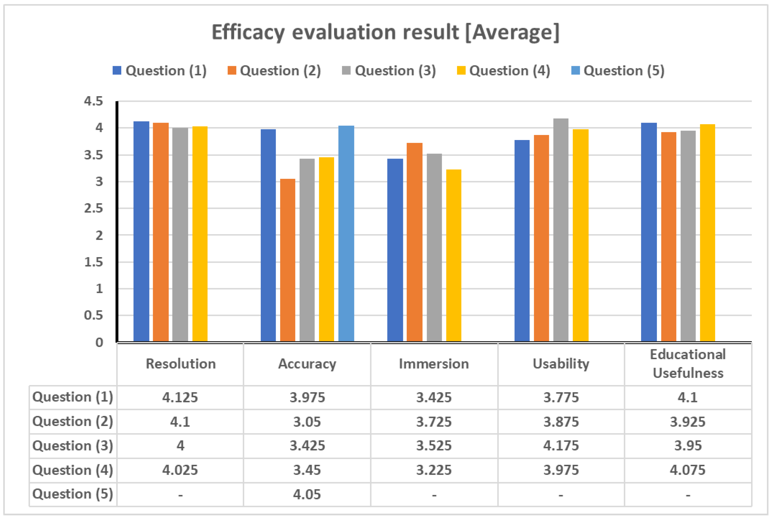
| Item | Question | Avg. | SD. |
|---|---|---|---|
| Resolution | Is the representation of the human anatomy 3D model clear? | 4.125 | 0.81202 |
| Is the human anatomy 3D model similar to the picture in the book? | 4.1 | 0.86023 | |
| Is the human anatomy 3D model representation clear when interacting with it? | 4 | 0.86603 | |
| Does the human anatomy 3D model hold up well during the vein blood sampling? | 4.025 | 0.8511 | |
| Accuracy | Is the vein blood sampling virtual environment implemented well? | 3.975 | 0.90795 |
| Are the viewpoints and hand positions consistent? | 3.05 | 1.13908 | |
| Does the movement of the virtual object match the movement of the educator? | 3.425 | 1.11552 | |
| Does the virtual object reflect minute movements? | 3.45 | 1.11692 | |
| Is the vein blood sampling step going well? | 4.05 | 0.80467 | |
| Immersion | Is there a sense of tactile reality? | 3.425 | 1.13771 |
| Is it visually realistic? | 3.725 | 1.09516 | |
| Are the controls user-friendly? | 3.525 | 1.04851 | |
| Are the manipulations as intended by the educator | 3.225 | 1.03652 | |
| Usability | Are the simulators professional? | 3.775 | 1.6037 |
| Can it be used for practical training? | 3.875 | 0.95361 | |
| Can it be used for patient explanation? | 4.175 | 0.77096 | |
| Are you thinking of using simulators for other medical procedures? | 3.975 | 1.01211 | |
| Educational utility | Is it helpful in understanding actual skills compared to books? | 4.1 | 0.91652 |
| Can I adapt to the actual vein blood sampling process? | 3.925 | 1.05801 | |
| Can you explain vein blood sampling to other medical personnel after learning with the simulator? | 3.95 | 0.9734 | |
| Can you explain the vein blood sampling process to the patient after learning with the simulator? | 4.075 | 0.81815 |
Disclaimer/Publisher’s Note: The statements, opinions and data contained in all publications are solely those of the individual author(s) and contributor(s) and not of MDPI and/or the editor(s). MDPI and/or the editor(s) disclaim responsibility for any injury to people or property resulting from any ideas, methods, instructions or products referred to in the content. |
© 2023 by the authors. Licensee MDPI, Basel, Switzerland. This article is an open access article distributed under the terms and conditions of the Creative Commons Attribution (CC BY) license (https://creativecommons.org/licenses/by/4.0/).
Share and Cite
Kim, J.-S.; Kim, K.-W.; Yang, S.-W.; Chung, J.-W.; Moon, S.-Y. Immersive VR (Virtual Reality) Simulator for Vein Blood Sampling. Technologies 2023, 11, 158. https://doi.org/10.3390/technologies11060158
Kim J-S, Kim K-W, Yang S-W, Chung J-W, Moon S-Y. Immersive VR (Virtual Reality) Simulator for Vein Blood Sampling. Technologies. 2023; 11(6):158. https://doi.org/10.3390/technologies11060158
Chicago/Turabian StyleKim, Jun-Seong, Kun-Woo Kim, Seong-Won Yang, Joong-Wha Chung, and Seong-Yong Moon. 2023. "Immersive VR (Virtual Reality) Simulator for Vein Blood Sampling" Technologies 11, no. 6: 158. https://doi.org/10.3390/technologies11060158