Review

Evolution and Role of Proteases in Campylobacter jejuni Lifestyle and Pathogenesis

Department of Biology, Division of Microbiology, Friedrich-Alexander-Universität Erlangen-Nürnberg, 91058 Erlangen, Germany
*
Author to whom correspondence should be addressed.
These authors contributed equally to this work.
Biomolecules 2023, 13(2), 323; https://doi.org/10.3390/biom13020323
Submission received: 14 December 2022 / Revised: 26 January 2023 / Accepted: 4 February 2023 / Published: 8 February 2023
(This article belongs to the Special Issue Molecular Targets in Campylobacter Infections)

Abstract

:
Infection with the main human food-borne pathogen Campylobacter jejuni causes campylobacteriosis that accounts for a substantial percentage of gastrointestinal infections. The disease usually manifests as diarrhea that lasts for up to two weeks. C. jejuni possesses an array of peptidases and proteases that are critical for its lifestyle and pathogenesis. These include serine proteases Cj1365c, Cj0511 and HtrA; AAA+ group proteases ClpP, Lon and FtsH; and zinc-dependent protease PqqE, proline aminopeptidase PepP, oligopeptidase PepF and peptidase C26. Here, we review the numerous critical roles of these peptide bond-dissolving enzymes in cellular processes of C. jejuni that include protein quality control; protein transport across the inner and outer membranes into the periplasm, cell surface or extracellular space; acquisition of amino acids and biofilm formation and dispersal. In addition, we highlight their role as virulence factors that inflict intestinal tissue damage by promoting cell invasion and mediating cleavage of crucial host cell factors such as epithelial cell junction proteins. Furthermore, we reconstruct the evolution of these proteases in 34 species of the Campylobacter genus. Finally, we discuss to what extent C. jejuni proteases have initiated the search for inhibitor compounds as prospective novel anti-bacterial therapies.

1. Introduction

Campylobacteriosis remains one of the most common zoonotic diseases of bacterial etiology worldwide. With 120,946 confirmed cases, campylobacteriosis was the most reported bacterial zoonosis in the European Union (EU) in 2020, followed by salmonellosis (52,702 cases) and yersiniosis (5668 cases) [1]. The EU numbers may likely be underestimated since the Centers for Disease Control and Prevention (CDC) reported over 60,000 cases in the USA annually, but estimated the actual number of Campylobacter infections to be as high as 1.3 million [2]. The main manifestations of campylobacteriosis include acute diarrheal illness often associated with abdominal cramping and fever [3]. In most patients, Campylobacter infections last for 1 to 2 weeks with the 24–48 h peak of the illness followed by self-limitation [4,5]. However, if not appropriately treated, the illness may relapse in approximately 20% of infected individuals [3]. In rare cases, post-infectious complications might result in developing serious gastrointestinal disorders including inflammatory bowel disease (IBD) or extra-gastrointestinal diseases such as Guillain-Barré syndrome (GBS), Miller Fisher syndrome (MFS) and reactive arthritis (RA) [6,7].
C. jejuni and C. coli are the two major Campylobacter species that cause campylobacteriosis in the EU with respectively 88.1% and 10.6% of the cases reported in 2020 [1]. Other non-jejuni Campylobacter species associated with enteritis account for a minor fraction of disease, including C. fetus, C. upsaliensis, C. lari, C. rectus, C. sputorum and C. hyointestinalis, among others [8]. Campylobacters asymptomatically inhabit diverse animal hosts with the capacity of being transmitted to humans [9]. Major reservoirs of Campylobacter spp. that may be transmitted to humans include consumption of contaminated poultry products, water and unpasteurized milk, contact with both domesticated and wild animals, and some other less common sources such as insects and protozoans [10]. The most common causative agent of campylobacteriosis, C. jejuni, primarily transmits to humans through inappropriately processed poultry meats, e.g., chicken, ducks and turkeys. Upon infection, C. jejuni is preferentially located in the intestinal tract, but has also been isolated from other organs. For instance, Hofreuter and co-authors isolated C. jejuni from the liver of mice in a mouse infection model [11]. A follow-up study confirmed that Campylobacter spp. could be isolated from the spleen, liver, gallbladder, follicle, upper and lower reproductive tracts and caecum of commercial Leghorn chicken [12].
The capability to colonize various sites of multiple hosts, and thus greatly changing environments, is enabled by a multitude of bacterial virulence and survival factors [13,14,15]. The flagellum is the major bacterial factor that provides effective C. jejuni movement, adherence and invasion functions [16]. It consists of the two important subunits, FlaA and FlaB, and can also serve as a type III secretion system (T3SS)-like syringe by secreting or injecting effector molecules such as FlaC, Campylobacter invasion antigens (Cia) and flagellar co-expressed determinants (Fed). Several adhesins including Campylobacter adhesion to fibronectin (CadF), fibronectin-like protein A (FlpA), C. jejuni lipoprotein A (JlpA) and others provide efficient bacterial attachment to host cells [14]. Various C. jejuni strains can release outer membrane vesicles (OMVs) containing cytolethal distending toxin (CDT), which leads to cell cycle arrest and cell death in host epithelia [17]. Finally, a range of proteases facilitates bacterial interaction with host epithelial cells, e.g., by disrupting cellular junctions like the high temperature requirement A (HtrA) protease [18,19,20]. Altogether, they facilitate bacterial survival in changing environments and adhesion to host tissues followed by invasion, replication and spread within the host.
Proteases are of special interest in pathogenesis due to their multiple critical roles in maintaining bacterial sustainability and virulence [21,22,23]. As a basic function, proteases provide cells with nutrients by degrading peptides of various natures [24]. Compared to other enterobacteria, the catabolic versatility of C. jejuni is very limited. Its specialized metabolism lacks the common bacterial pathways for processing and utilizing carbohydrates such as glucose and galactose [25,26]. Instead, C. jejuni largely depends on the consumption of amino acids, most notably aspartate, glutamate, asparagine, proline and serine, and catabolizes alternative carbohydrates such as lactate and intermediates of the citric acid cycle. Sensing of suitable nutrient sources is mediated by a vast array of chemotaxis proteins, which is followed by nutrient uptake into the bacterial cell. Acquisition of amino acids not only depends on an efficient uptake machinery, but likely also requires digestion of oligopeptides in the immediate surroundings of the bacteria, which is presumably orchestrated by various secreted proteases [25,26]. Further, proteases regulate the bacterial proteome in order to adapt to changing conditions, e.g., under stress pressure [27]. Energy-dependent proteases such as Lon and Clp control the expression of T3SSs in Gram-negative bacteria, mediating injection of pathogenic effector molecules [28]. Secreted HtrA proteases of various Gram-negative bacteria, e.g., C. jejuni or Helicobacter pylori, can cleave proteins of host cell junctions, which facilitates paracellular migration of the bacteria through the epithelial layer into deeper tissues [29]. Some Clostridia exhibit intracellular toxicity to their hosts via metalloproteases in the form of tetanus and botulinum neurotoxins [30]. In this review, we attempt to summarize recent knowledge of the role of Campylobacter proteases in pathogen-host interactions.
Several proteases have already been shown to be involved in C. jejuni virulence, and most of them are present in nearly all Campylobacter species (Table 1). We start with serine protease HtrA, which is to date the best-studied protease in C. jejuni virulence. We then discuss two other serine proteases, Cj1365c and Cj0511, and summarize how the ATPase-dependent (AAA+) proteases FtsH, ClpP, and Lon contribute to C. jejuni viability and virulence. Next, we highlight the role of protease PqqE and peptidases PepP, PepF and C26 in C. jejuni virulence and discuss the evolution of the proteases within the Campylobacter genus. Finally, we lay out possible future research directions.

2. Proteases

2.1. Serine Proteases

2.1.1. Serine Protease HtrA (Cj1228c)

Members of the family of high temperature requirement A (HtrA) serine proteases are expressed both by prokaryotic and eukaryotic organisms [20,31,32]. The C. jejuni-encoded HtrA operates as a bifunctional protein, comprising both protease and chaperone activities, and consists of a signal peptide at the N-terminus, a trypsin-like protease domain and two consecutive PDZ domains at the C-terminus [29]. The HtrA enzyme forms proteolytically active trimers, hexamers and dodecamers in the periplasm [33]. Like many other bacterial HtrAs, a major function of C. jejuni HtrA is the protection against a variety of external stress conditions [34,35]. Initially, the importance of HtrA during C. jejuni infection was studied in two mouse models, infant wild-type (wt) mice and gnotobiotic interleukin-10 (IL-10)-/- knockout mice [36]. In both animal models, the intestinal colonization loads of C. jejuni wt and HtrA mutant strains were similarly high. When infected with wt C. jejuni, IL-10-/- deficient mice revealed disturbed crypt structures and strong upregulation of colonic apoptotic cells [37]. This scenario resulted in the infiltration of immune cells such as monocytes, macrophages and neutrophils in the colon, which was associated with elevated release of nitric oxide (NO) and pro-inflammatory cytokines TNF, IFN-γ, IL-6 and MCP-1 [37,38]. Infected IL-10-/- knockout mice also displayed inflammatory reactions in the lung, liver and kidney, demonstrating that immune-relevant reactions were not restricted to the colon. Both phenotypes were dependent on HtrA and its protease activity. C. jejuni infection of infant mice revealed comparable results [39]. Taken together, HtrA emerged as a novel C. jejuni virulence factor, whose protease activity intensifies campylobacteriosis by causing apoptosis and pro-inflammatory immune pathology in mice [38].
Infection studies using cultured intestinal epithelial cells in vitro have shown that expression of HtrA is required to attach to and enter cells, probably mediated by its chaperone activity on one or more C. jejuni adhesins [18,35,40,41,42,43]. In addition, HtrA was shown to be secreted in the extracellular environment, either as a soluble enzyme or as a part of shed OMVs [18,44,45,46]. Remarkably, by using transwell filter systems it was demonstrated that wt C. jejuni can effectively and quickly transmigrate across polarized epithelial cells, a process that is associated with cell damage [18,47]. This phenotype is largely abolished during infection with a ∆htrA deletion mutant and with a mutant in which the protease function, but not the chaperone function, was inactivated. Those data suggested that HtrA protease activity is required for C. jejuni transmigration through the epithelium and led to the proposal that secreted HtrA may disrupt cell junctional proteins between neighboring intestinal epithelial cells. Indeed, C. jejuni HtrA was shown to cleave two tight junction proteins, occludin [47] and claudin-8 [19], and the exact cleavage sites were identified. In addition, HtrA cleaves the major protein of the adherens junctions, tumour suppressor E-cadherin, in vitro and during infection in vivo [18,48]. Cleavage of occludin, claudin-8 and E-cadherin results in the disruption of intestinal epithelial cell-to-cell junctions, which compromises epithelial barrier functions and paves the way for C. jejuni to travel across the intestinal epithelium using a paracellular pathway [49]. The capability for transepithelial migration can explain why C. jejuni expresses two adhesins with high affinity to fibronectin, CadF and FlpA [50]. Fibronectin is the natural ligand for the basal integrin-β1 complex that is important for cell entry of the bacteria [18,50,51,52,53,54]. This way, C. jejuni may also be capable of entering the lamina propria, the bloodstream and other organs. By using transwell filter assays, further work showed that C. jejuni also facilitated effective translocation of microbiota such as Lactococcus lactis or Escherichia coli through the epithelial layer. This HtrA-mediated transmigration of microbiota to the basolateral compartment, and thus deeper tissue layers, might be involved in the development of inflammatory bowel disease (IBD) [55].
Because of these important functions and the ubiquitous presence of the htrA gene in C. jejuni, this protease represents a supreme target for anti-C. jejuni therapy. Recent studies in mice and in cell culture systems showed that application of the polyphenolic compound curcumin reduced C. jejuni-triggered disruption of the epithelium [56]. Similar results were obtained using vitamin D that diminished C. jejuni transepithelial migration [57]. Therefore, vitamin D and curcumin might represent alternative options for the management of C. jejuni infections. Finally, HtrA could be a promising target for the development of a vaccine and small inhibitor compounds [58,59,60,61]. However, additional studies are required to improve the specificity of currently available HtrA inhibitors.

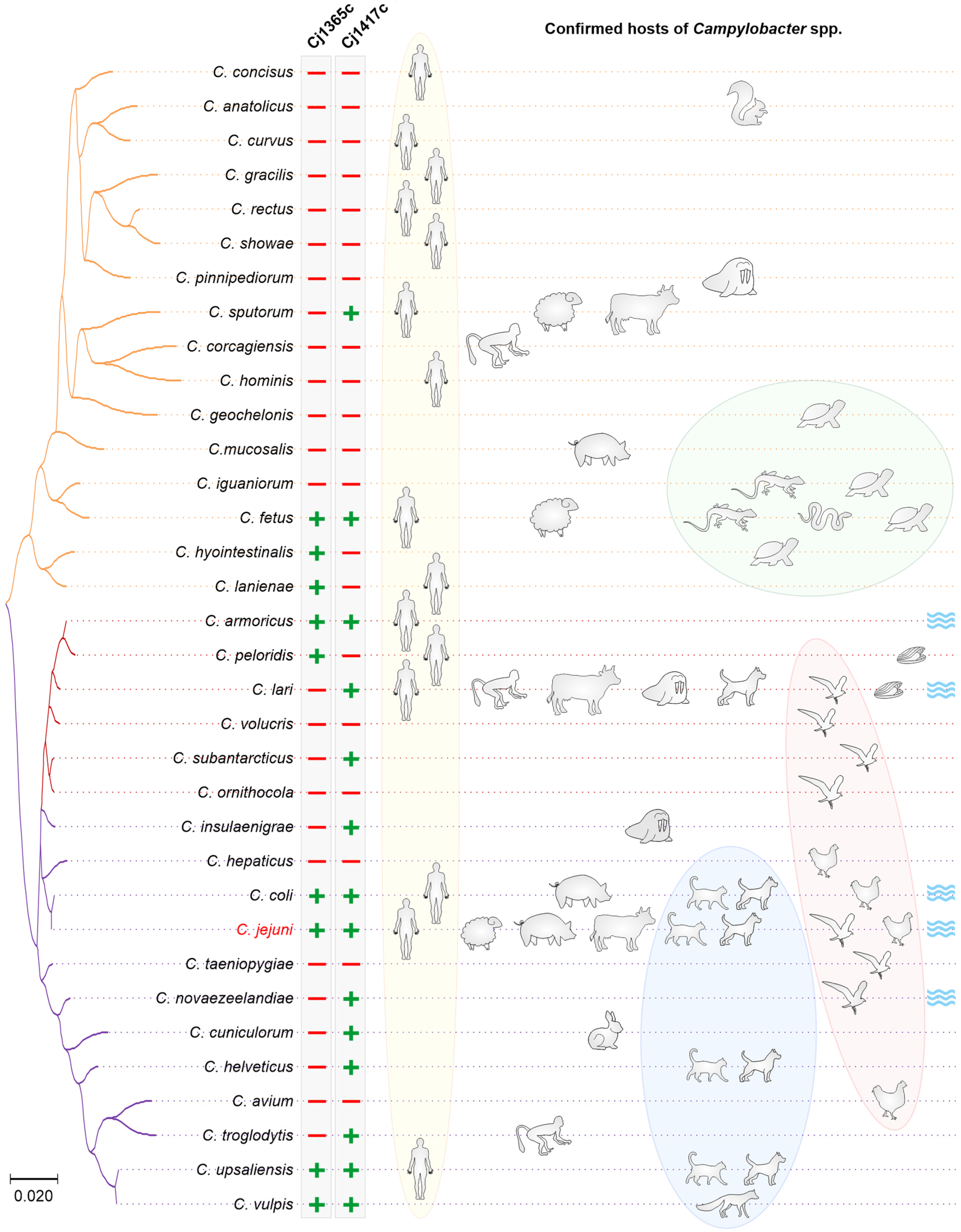
2.1.2. Serine Protease Cj1365c

C. jejuni protease Cj1365c belongs to the subtilisin-like family of serine proteases. Similar to HtrA, Cj1365c possesses a catalytic triad composed of a histidine, an aspartate and a serine, but the order of the residues in the protease (Asp-Ser-His) differs from that in HtrA (His-Asp-Ser). The domain architecture of Cj1365c consists of an N-terminal S8 peptidase domain and a C-terminal autotransporter domain that provides protease translocation across the membrane, consistent with a predicted location at the outer membrane (Table 1). Intriguingly, proteins containing autotransporter domains are often associated with bacterial virulence [62]. The assumption that Cj1365c may play a role in C. jejuni virulence was recently confirmed by a study in an in vitro model of intestinal epithelia. Cj1365c was found in secreted C. jejuni OMVs, along with serine proteases HtrA and Cj0511, as well as with approximately 150 other proteins, including important C. jejuni virulence factors such as CDT, the adhesins CadF and FlpA and the major antigenic protein Peb3 [44,45]. C. jejuni OMVs were shown to possess proteolytic activity that was associated with HtrA, Cj0511 and Cj1365c. Accordingly, proteolytic activity was reduced by deletion of either of the htrA, cj0511, or cj1365c genes [45]. Incubation of cultured T84 cells with OMVs efficiently hydrolyzed the host junctional proteins occludin and E-cadherin, an activity that was associated with HtrA and Cj1365c [45]. As expected from these observations, a knockout mutation of the cj1365c gene or pretreatment with serine protease inhibitors resulted in reduced adhesion and invasion of bacteria. In addition to junctional proteins, Cj1365c was proposed to cleave the major endoplasmic reticulum chaperone protein BiP/GRP78 [63], but the biological relevance of BiP/GRP78 protein cleavage in C. jejuni-host interactions remains to be elucidated. Interestingly, exposure of C. jejuni to physiologically relevant concentrations of sodium taurocholate, a bile salt secreted by the gallbladder into the digestive tract, resulted in increased concentrations of virulence-associated factors within the OMVs and in increased proteolytic activity [63].
However, not all C. jejuni isolates possess the Cj1365c protease. Our genome-wide screen revealed 2,359 genomes in the RefSeq Genome Database, of which only 1896 contained the cj1365c gene. The gene was predominantly found in clinical isolates and isolates from livestock, but not in isolates from environmental sources such as sand of a bathing beach [64]. This gene was prevalent in multiple C. jejuni clonal complexes (CCs), which are groups of related strains based on Multi Locus Sequence Typing (MLST). These CCs contained human clinical isolates as well as livestock isolates from cattle, chicken and turkey, indicating no specific host association. In contrast, CC45 and CC283 (mostly human and chicken isolates) and CC1332 (mostly from turkeys, chicken and humans) did not possess cj1365c [65].
Moreover, our analysis showed that only 60 out of the 1182 C. coli genomes in the NCBI RefSeq Genome Database possess the Cj1365c protease. It is missing in the majority of the isolates, including the C. coli type strain FDAARGOS_735. In those isolates that carry the gene, the protease shows 97.1% to 99.9% protein similarity to C. jejuni Cj1365c, in contrast to between 60% and 80% similarity in other proteins. These observations suggest a recent import of gene cj1365c from C. jejuni into C. coli by lateral DNA transfer. In addition, we searched for Cj1365c among a variety of Campylobacter species and found that this protease is absent from the majority of the analyzed species (Table 1). Further studies are required to confirm Cj1365c-mediated cleavage of occludin, E-cadherin and BiP/GRP78 during infection in vivo, and to unravel the biological relevance of BiP/GRP78 cleavage in C. jejuni-host interactions.

2.1.3. Serine Protease Cj0511

Another serine protease is Cj0511 [66], which is N-glycosylated in C. jejuni [67]. Cj0511 belongs to the family of S41 family peptidases, which are serine endopeptidases similar to the C-terminal-processing protease CtpA from Bartonella bacilliformis. Similar to HtrA, C-terminal-processing proteases (CTPs) are implicated in protein folding and processing in the periplasm and contain a PDZ domain that is assumed to mediate substrate recognition [68]. However, while the catalytic triads of both HtrA and Cj1365c are composed of a histidine, an aspartate and a serine residue (HDS-motif), CTPs possess a serine-lysine catalytic dyad (SK-motif). CTPs are critically important for post-translational protein processing, specifically for processing the C-terminus of their protein substrates, hence the name. In addition, CTPs have been associated with regulation of gene expression, stress response, maintenance of cell envelope integrity and bacterial virulence [68]. For example, the Legionella pneumophila protein Tsp is implicated in heat stress response and is essential for efficient infection of and intracellular growth in amoeba [69]. CtpA of Bordetella bronchiseptica is involved in post-translational processing of filamentous hemagglutinin FHA, a major virulence factor required for adhesion and persistence in the airways of an infected host. Accordingly, a CtpA-deficient mutant failed to persist in the lower respiratory tract [70]. Likewise, deletion mutants of CTPs from Acinetobacter baumannii [71], Brucella suis [72], Burkholderia mallei [73], Pseudomonas aeruginosa [74] and Staphylococcus aureus [75] exhibited reduced virulence compared to the respective wt bacteria.
Serine protease Cj0511 appears to play a pivotal role in C. jejuni biology, as it is present in all published C. jejuni genomes. The protease contains a signal peptide spanning 34 amino acid residues. In addition to a predicted cellular location at the cytoplasmic membrane and in the periplasm (Table 1), Cj0511 was suspected to be secreted [76], which is in agreement with the observation that Cj0511 was found in purified C. jejuni OMVs [44,63]. Cj0511 appears to play an important role during C. jejuni chicken colonization. Cj0511 was highly expressed in an efficient chicken-colonizer strain, but barely expressed in a strain that colonized chickens poorly [76]. Expectedly, a cj0511 gene deletion mutant was severely impaired in its ability to colonize chickens compared to the parental wt strain. Not only were more chickens of the cohort infected, but the wt strain also infected the chicken ileum and cecum in significantly higher numbers than did the cj0511 deletion mutant [66,77]. Likewise, deletion of cj0511 impaired the ability of C. jejuni to colonize mice, reducing bacterial numbers by over two logs [78]. The observed differences in colonization efficiency may largely be associated with the reduced ability of the Δcj0511 mutant to adhere to and invade IECs.
Exposure of C. jejuni to pancreas-secreted amylase triggered the production and secretion of a bacterial α-dextran, a response that depended on a functional Cj0511 [77]. The α-dextran production increased C. jejuni stress resistance and facilitated prolonged survival at extra-host temperatures (20 °C and 4 °C), suggesting a crucial role during transmission between hosts, including survival on meat and dairy products prepared for human consumption. In addition, the α-dextran greatly increased C. jejuni-induced mortality in the Galleria mellonella larvae infection model, an effect that was absent in the cj0511 deletion mutant, and promoted biofilm formation of C. jejuni bacteria in a Cj0511-dependent manner [77]. Yet, the exact mechanisms of how Cj0511 mediates interaction with IECs, how Cj0511 increases virulence during G. mellonella infection, how Cj0511 promotes colonization of the chicken ileum and how Cj0511 stimulates biofilm formation remain to be elucidated.

2.2. AAA+ Group Proteases

(AAA+) group proteases are widely distributed in all living organisms and are associated with diverse cellular activities. In bacteria, those proteases mostly facilitate survival under various stress conditions [27,79]. The AAA+ (or ATP-dependent) proteases share some distinct structural similarities and specifically contain ATPase (AAA+ module) and peptidase domains. The two major segments are responsible for substrate recognition, unfolding and degradation. Thus, AAA+ proteases regulate proteolysis and control the bacterial proteome in response to cellular needs. During infection, some pathogens strictly depend on the function of such proteases, such as the causative agent of leptospirosis in animals and humans, Leptospira interrogans [80]. Inactivation of the clpB gene that encodes the AAA+ protease in L. interrogans decreased bacterial tolerance to oxidative stress. In addition, gene deletion also hampered bacterial virulence in the gerbil model of acute leptospirosis [81]. Several lines of evidence indicate crucial roles of Clp protease in the virulence, resistance and persistence of Staphylococcus aureus, including methicillin-resistant (MRSA) strains [28,82]. As of today, FtsH, Lon and ClpP may be distinguished as AAA+ proteases playing significant roles in C. jejuni survival and virulence as discussed below.

2.2.1. ClpP Protease (Cj0192c)

As with most AAA+ proteases, the ClpP protease of C. jejuni is involved in heat stress response and protein quality control. Structurally, C. jejuni ClpP does not possess the ATPase module and instead forms hetero-oligomeric complexes with the ClpA and ClpX ATPases. In C. jejuni, ClpP plays various functions, contributing to both stress tolerance and virulence. Moreover, ClpP was shown to play a major role in the natural competence of C. jejuni as the deletion of the clpA or clpX genes reduced DNA uptake by 5 to 10-fold, respectively, and a ΔclpP mutant was virtually untransformable [83]. Knockout mutations of clpX and clpP, but not clpA, showed increased heat sensitivity at 42 °C, suggesting a crucial role of the ClpXP complex in the C. jejuni heat shock response [84]. Notably, ClpP was also proposed to promote C. jejuni survival at refrigerator temperatures [85]. Thus, similar to the Cj0511 serine protease discussed above, ClpP may increase survival in extra-host environments, and thus play an important role during transmission. While being involved in temperature adaption, ClpP does not seem to be involved in oxidative stress response; aerotolerant and non-aerotolerant C. jejuni strains displayed no significant differences in clpP gene expression under aerobic conditions [86].
Similar to HtrA, Cj0511, and Cj1365c, ClpP was found to be present in purified OMVs [63], in addition to a predicted cytoplasmic location in the bacterial cell (Table 1). Interestingly, loss of either clpX, clpA or clpP genes impaired bacterial invasion of IECs without affecting C. jejuni adherence to the cell surface [84]. Furthermore, the invasion-associated process of C. jejuni autoagglutination was dependent on ClpX and ClpP, but not on ClpA. The motility of bacteria was reduced upon deletion of clpP, but not clpA or clpX, which indicates that both ClpAP- and ClpXP-mediated proteolyses may be important in this process. Furthermore, ClpP protein expression was found upregulated in C. jejuni biofilm-associated cells.
Overall, the ClpP protease seems to play a vital role in both maintaining C. jejuni viability under heat stress conditions and during invasion of host organisms. We found this protease present in all C. jejuni isolates in the NCBI RefSeq Genome Database and that the protease is very conserved within the Campylobacter genus (Table 1). Interestingly, Ghunaim and co-authors found the clpP gene in only 83.3% of C. jejuni isolates [87]. The interstrain variability in virulence gene transcription could probably explain that ClpP was missing in a set of isolates. Thus, the two C. jejuni strains NCTC11168 and DFVF1099 displayed different levels of clpP transcription after 24 h incubation at 4 °C [88]. In contrast, Wurfel and co-authors found the clpP gene present in almost all C. jejuni strains isolated from poultry meat products in Brazil [89]. Further studies, including large-scale genome comparisons, are required to clarify whether the Clp protease complex genes are present in all C. jejuni strains.

2.2.2. Lon (Cj1073c)

Lon proteases are well characterized in numerous bacteria, including E. coli and S. aureus. In C. jejuni, the Lon protease, which is predicted to be a cytoplasmic enzyme (Table 1), was first identified in cells exposed to heat shock at 48 °C [90]. Dot blot assays revealed a rapid increase in the lon mRNA level already after 5 min incubation of C. jejuni at 48 °C, reaching the maximum level (about 6 to 8-fold) after 20–30 min. Along with ClpP and HtrA proteases, Lon maintains protein quality control in C. jejuni, preventing excessive accumulation of misfolded proteins. In particular, a C. jejuni ΔclpPlon double mutant was impaired in its growth after puromycin-triggered protein misfolding [84]. In contrast, the corresponding single ΔclpP and Δlon deletion mutants formed colonies similar to the wt C. jejuni, suggesting that both proteases eliminate misfolded proteins interchangeably. In line with the above observations, a ΔclpPlon double mutant, but not the single C. jejuni mutants, showed increased accumulation of misfolded proteins [84]. Finally, all ΔclpPlon double and single C. jejuni mutants exhibited reduced abilities to invade epithelial cells, indicating a major role of these quality control proteins in C. jejuni virulence, perhaps by processing precursors of several pathogenicity factors [84]. Overall, this chaperone and protease function appears to be crucial for the bacteria as our genome screens revealed presence of the lon gene in all C. jejuni genomes in the NCBI RefSeq Genome Database and in all 34 analyzed Campylobacter species (Table 1).

2.2.3. FtsH (Cj1116c)

Similar to Lon, FtsH proteases possess an AAA+ module linked to the protease domain. The N-terminal transmembrane domain is followed by the large and small AAA+ domains that form a single AAA+ module. Finally, the C-terminus of the AAA+ module is flanked by the protease domain. The major roles of FtsH in bacteria include quality control and degradation of membrane proteins, heat shock response and regulation of lipopolysaccharide biosynthesis [91]. FtsH was also shown to be essential in maintaining bacterial virulence of Edwardsiella piscicida during infection of Zebrafish; FtsH deficiency resulted in reduced bacterial adhesion, internalization and intracellular survival [92]. While it is likely that C. jejuni FtsH is involved in maintenance of membrane proteins and LPS, similar to FtsH in many other bacteria and consistent with the predicted location at the cytoplasmic membrane (Table 1), the specific role of FtsH in C. jejuni lifestyle and pathogenesis still needs to be investigated. Unfortunately, attempts to create a C. jejuni ΔftsH knockout mutant were so far unsuccessful [93]. This observation and the universal presence of FtsH homologs among C. jejuni isolates and in all analyzed Campylobacter species suggest an essential role of the protease for bacterial survival.

2.3. PqqE Protease (Cj0805)

C. jejuni protease PqqE belongs to the M16B family of zinc-dependent proteases. In eukaryotes, this protease is known as mitochondrial processing peptidase (MPP). Given the bacterial origin of the mitochondria, the cellular location of this peptidase points to an evolutionary bacterial origin of the enzyme. In most bacteria, including E. coli, this enzyme is located in the cytoplasm. The Vibrio vulnificus secreted insulin-degrading protease SidC, however, possesses an N-terminal signal peptide that is absent from PqqE homologs in other bacteria and was found secreted into the environment [94]. Mouse infection studies indicated an important role of SidC in V. vulnificus virulence. Compared to wt bacteria, a ΔsidC deletion mutant was severely impaired in its pathogenicity and barely colonized mice. In addition, SidC showed insulin-degrading activity, and the infection of diabetic mice resulted in higher bacterial numbers of both wt and ΔsidC bacteria compared to the infection of wt mice. Apparently, hyperglycemia favored bacterial survival, growth and proliferation in the infected host [94]. Similar to SidC, PqqE from H. pylori was identified as part of the secretome [95] and was found inside H. pylori OMVs [96]. PqqE from H. pylori was reported to compromise the integrity of the gastric epithelial layer by cleavage of the junctional adhesion molecule JAM-A during H. pylori interaction with the gastric epithelial layer [97]. However, these data were obtained by indirect experiments, warranting confirmation by additional data using a pqqE knockout mutant. Interestingly, unlike HtrA that cut the extracellular domain of E-cadherin, PqqE was proposed to cleave JAM-A at the cytoplasmic domain, and thus inside the host cells. While PqqE from H. pylori was secreted and released in OMVs, an analysis in PSORTb v3.0 [98] predicted a cytoplasmic location for PqqE from C. jejuni, suggesting that this protein is not located at the bacterial cell surface and is likely not being secreted. Thus, as expected from this analysis, PqqE was not present in C. jejuni OMVs [63]. In addition, infection experiments with C. jejuni and C. coli did not result in the disruption of JAM-A [97], suggesting different functions of H. pylori and C. jejuni PqqE proteases during pathogen-host interaction. In conclusion, the function of PqqE during C. jejuni pathogenesis remains to be unraveled. Unfortunately, the creation of a pqqE gene deletion mutant in C. jejuni may be challenging, because H. pylori pqqE (HP1012) is present in all isolates [99], and as mentioned above, attempts to create H. pylori ΔpqqE mutants have so far failed [97]. Furthermore, our genome screening revealed universal presence of pqqE among C. jejuni isolates, and PqqE homologs were found in all analyzed Campylobacter species (Table 1). Together, the data suggest that this bacterial protease might be essential, and attempts to generate a deletion mutant have been unsuccessful so far [97].

3. Peptidases

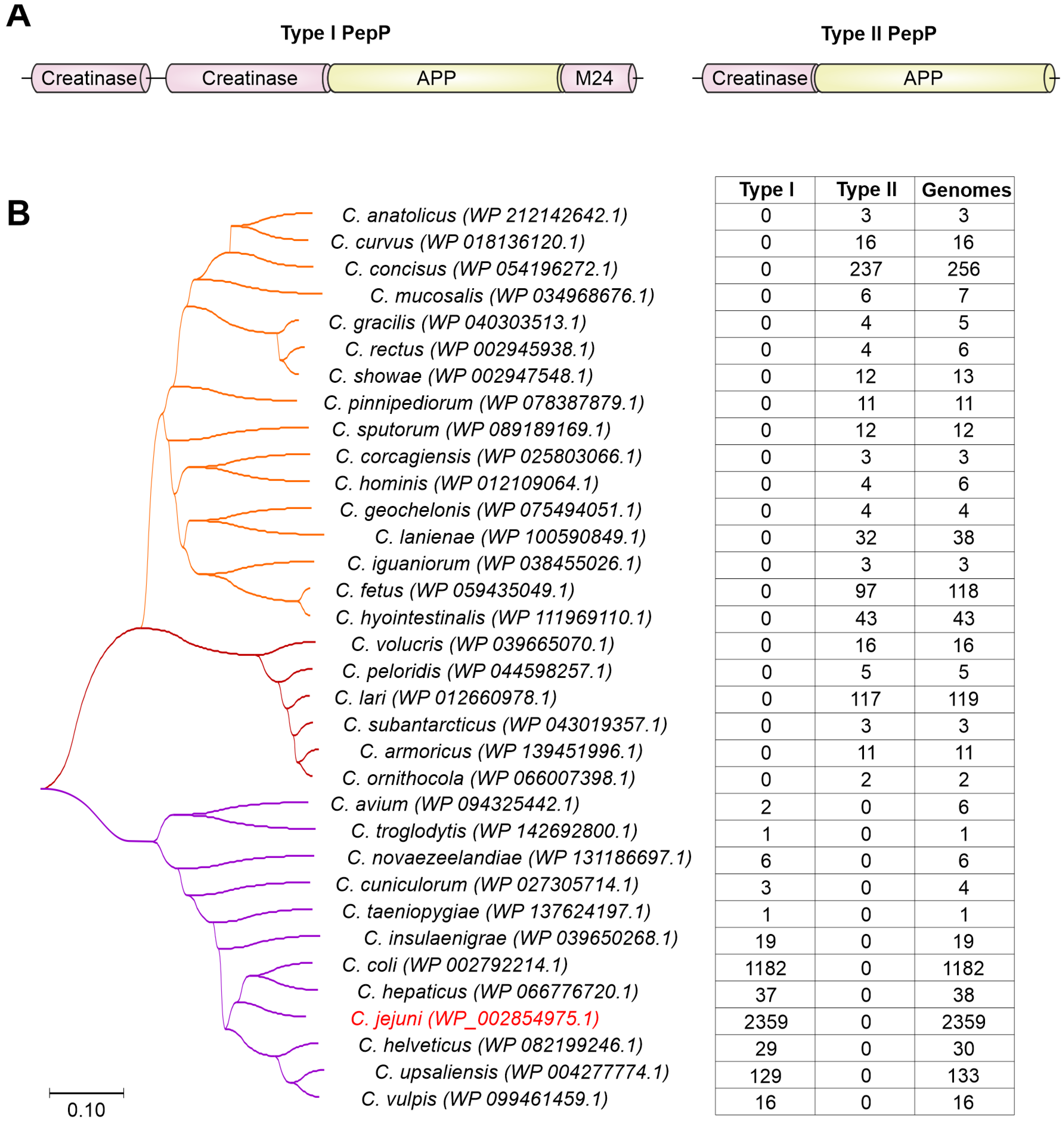
3.1. Proline Aminopeptidase PepP (Cj0653c)

Proline aminopeptidases are widely distributed from prokaryotes to eukaryotes. They play important roles in maintaining cell viability, virulence and other functions [100,101,102,103]. Aminopeptidase P from C. jejuni was first identified by two-dimensional gel electrophoresis that showed specific expression of the protein in a strain that robustly colonized the intestinal tract of chicken [76]. Recently, C. jejuni proline peptidase P (PepP) was identified as a potential virulence factor [103]. PepP was separated by SDS-PAGE as a ~70 kDa protein band, which was further defined as an M24 family metallopeptidase of 596 amino acids (aa) in size (locus WP_002854975.1). As described in the Pfam database, the corresponding sequence encoded two N-terminal creatinase/prolidase domains (creatinase N and creatinase N2) followed by a X-prolyl amino-peptidase (APP) domain, and a C-terminal M24 peptidase domain. Of those, the APP domain was shown to hydrolyze the N-terminal residue of a substrate at the Xaa-Pro site [104].
The PepP peptidases are highly conserved among C. jejuni strains and other Campylobacter species, including C. coli, C. hepaticus, C. upsaliensis and others (Table 1). Interestingly, we have identified a shorter PepP variant in other Campylobacter species with a size of approximately 341 aa. Therefore, we propose to name the 596 aa long and 341 aa long PepP peptidases as type I and type II, respectively. The shorter PepP peptidase (type II) harbors an APP domain at the C-terminus, while the N-terminus likely encodes a creatinase domain (Figure 1A), as predicted by Conserved Domain Database [105]. Intriguingly, all Campylobacter species have only one type of PepP protease (either type I or type II), separating them into two distinct groups (Figure 1B), the type I group with the important human pathogens C. jejuni and C. coli, and the type II group with animal pathogens and commensals such as C. fetus and C. lari.
While the function of the type II PepP in Campylobacter spp. is currently still unclear, the type I PepP from C. jejuni was shown to contribute in murine campylobacteriosis [103]. In particular, a C. jejuni ΔpepP mutant caused less severe symptoms in microbiota-depleted IL10−/− knockout mice and was associated with less pronounced apoptotic and innate immune cell (F4/80+) responses. Furthermore, intact PepP was required for an efficient C. jejuni-induced pro-inflammatory response in the intestine, including the release of interferon-γ (IFNγ), tumor necrosis factor (TNF), monocyte chemoattractant protein (MCP)-1 and IL-6. Although the pepP gene deletion did not compromise the colonization ability of C. jejuni in mice, it reduced the pro-inflammatory capacity of the bacterium. Interestingly, in other pathogens such as Eikenella corrodens, the prolyl aminopeptidase was reported to function as a hemolytic factor, significantly contributing to the pathogenicity of these bacteria [106]. Further research will shed more light on other potential activities of PepP in C. jejuni virulence.

3.2. Oligopeptidase PepF (Cj1099)

C. jejuni PepF is an oligopeptidase of the peptidase family M3B. PepF oligopeptidases are peptidases that commonly cleave short oligopeptides, but cannot degrade full-length proteins, hence the name. They have a broad substrate specificity and hydrolyse peptides between 5 and 21 amino acids in length [107,108]. Oligopeptide hydrolysis along with broad substrate specificity suggests that PepF peptidases are implemented in the degradation of cleavage products of various other proteases, and thus in protein recycling [109,110]. PepF oligopeptidases, including PepF from C. jejuni, contain a His-Glu-X-X-His motif that is followed by another glutamate residue, located 23 amino acids downstream. The two histidines and the downstream glutamate bind a single catalytic zinc ion and form the catalytic centre, while the glutamate residue embedded between the two histidines is believed to assist in the catalytic reaction by orienting the zinc ion and a water molecule [107].
In many bacteria, PepF peptidases are located in the cytoplasm. An analysis of the subcellular location using PSORT [98] confirmed a cytoplasmic localization for PepF from C. jejuni (Table 1). However, in Bacillus amyloliquefaciens PepF is secreted into the environment. Outside the bacterial cell, this peptidase apparently processes oligopeptides to generate pentapeptides, which represent small signal molecules that are subsequently imported into the cell and are involved in the initiation of sporulation [108]. While it is currently unknown whether C. jejuni PepF is involved in pathogen-host interactions and virulence, the presence of PepF peptidases in all C. jejuni isolates and in all examined Campylobacter species (Table 1) suggests conserved house-keeping functions such as peptide recycling or perhaps cell signaling.

3.3. C26 Peptidase (Cj1417c)

Another interesting peptidase that might play a major role in C. jejuni virulence is the C26 peptidase of the γ-glutamyl-gamma-aminobutyrate hydrolase family. Transposon mutagenesis coupled with sequencing approaches revealed significantly reduced C. jejuni colonization of mice upon inactivation of the C26 peptidase (CJJ81176_1416 in strain 81-176, Cj1417c in strain NCTC11168) [78]. The authors suggested that the peptidase may be involved in energy acquisition and/or protein biosynthesis that overall impacted the colonization capability by C. jejuni. These findings were consistent with a previous study that showed CJJ81176_1416 to be involved in chicken colonization by C. jejuni [111]. Furthermore, an earlier study showed that the acquisition of a gene encoding a γ-glutamyltranspeptidase enabled the bacteria to consume glutamine and glutathione, which enhanced their ability to colonize the intestine [11]. Interestingly, the observed growth or colonization effect may be strain-specific, because not all C. jejuni genomes possess the cj1417c gene. Our genome screen showed cj1417c presence in 1995 out of the 2359 genomes in the NCBI RefSeq Genome Database. In addition, 20 of the 34 analyzed Campylobacter species lack a Cj1417 protease homolog (Table 1). Thus, further studies focusing on the metabolic plasticity provided by peptidases may help to better understand the C. jejuni-driven pathogenesis.

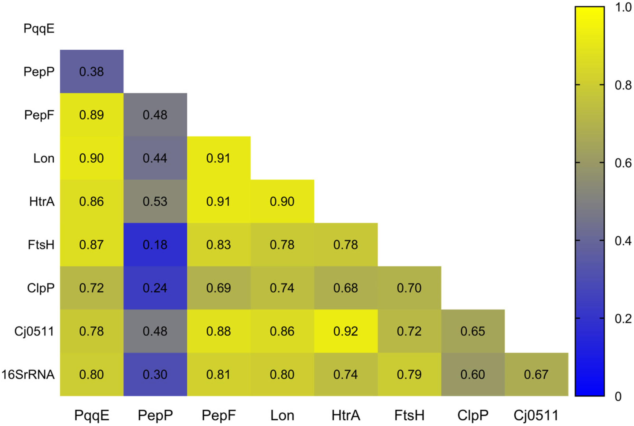
4. Evolution of Proteases and Peptidases in the Campylobacter Genus

Seven of the 10 discussed proteases, i.e., HtrA, Cj0511, ClpP, Lon, FtsH, PepF, and PqqE, were found to be present in all 34 analyzed Campylobacter species (Table 1), suggesting that these proteases were likely present in the ancestor of the genus. In fact, these seven proteases were also found present in other ε-Proteobacteria, including H. pylori, Helicobacter hepaticus, Wolinella suchinogenes and even in the environmental ε-Proteobacteria Sulfurospirillum arsenophilum and Nautilia profundicola, indicating a common evolutionary origin and implying that these proteases likely have important house-keeping functions in the bacterial cell. Some of those enzymes (HtrA, Cj0511, PqqE) acquired additional functions in the interaction with animal/human hosts. As a result of the common origin and subsequent evolution and diversification along with the other genes in the genomes, the pairwise similarity matrices of these genes were quite similar to each other and displayed high pairwise Pearson correlation coefficients ranging from r = 0.65 to r = 0.91 (Figure 2). In addition, the evolution was congruent with that of the 16S rRNA gene with scores of r = 0.60 to r = 0.81. In contrast, PepP showed low Pearson coefficients ranging from r = 0.18 to r = 0.53, indicating a different evolutionary trajectory.
Indeed, as mentioned above, protease PepP was present as two very distinct protein versions. Type I contained four protein domains (creatinase N, creatinase N2, APP, M24). The much shorter type II contained only a creatinase N domain (with weak homology) and an APP domain. In addition, the shorter type II split into two groups in the phylogenetic tree (Figure 1). Interestingly, superimposing the PepP protein variants onto the 16S rRNA-based phylogenetic tree revealed specific clustering of the PepP variants (Figure 3). Species carrying the short type II peptidase (orange tree branch, as in Figure 1) clustered together in a distinct branch of the phylogenetic tree. However, the other major branch of the 16S rRNA gene-based tree contained two groups of species possessing either type I or type II PepP peptidase variants (purple and brown tree branches, respectively). An analysis of the chromosomal location of pepP revealed that all type I pepP gene homologs are located at gene position cj0653c. In contrast, all type II pepP homologs from both orange and brown clades are located between cj0065c and cj0066c gene homologs. Given that the orange and brown species clades are located at different branches of the 16S rRNA-based phylogenetic tree, this suggests type II PepP to be ancestral in Campylobacter.
In addition, type II PepP homologs are also present in multiple other species of the ε-Proteobacteria, including the closely related genera Helicobacter, Wolinella and Sulfurospirillum, all of which belong to the order Campylobacterales, and the termophilic Nautilia of the order Nautiliales, supporting the proposed type II pepP gene ancestry in Campylobacter. In contrast, type I PepP peptidase homologs were only found in more distantly related genera, including Neisseria, Lautropia (both β-Proteobacteria), Acinetobacter and Pseudomonas (both γ-Proteobacteria), suggesting a secondary gene import from a currently unknown source into the ancestor of the purple species clade.
The C26 peptidase (Cj1417c) was only present in a subset of 14 Campylobacter species (Table 1). Notably, we found C26 peptidase homologs in W. succinogenes and H. hepaticus, but not in H. pylori and also not in the environmental Nautilia and Sulfurospirillum bacteria (Table 1). All 14 Campylobacter species harbored gene cj1417c at the same chromosomal location, suggesting a joint evolutionary origin of this peptidase in the genus. We found Cj1417c present in multiple species of the brown and purple clades, indicating loss of the cj1417c gene in individual species and lineages (Figure 3). In contrast, only two species (C. fetus, C. sputorum) in the orange clade possessed Cj1417c. Therefore, multiple species of the orange clade must have lost the gene. Alternatively, cj1417c may have been lost in the ancestor of the entire clade, followed by independent acquisition in the two species. However, the latter scenario is less likely, because the pairwise genetic distances between the 14 different Cj1417c homologs strongly support a common evolutionary origin and subsequent gene loss.
The evolutionary history of Cj1365c in Campylobacter appears to be different. First, this protease is only present in nine of the 34 analyzed species (Table 1, Figure 3). However, whether protease Cj1365c is present in all isolates of these nine species remains to be analyzed. Second, other ε-Proteobacteria such as the already mentioned W. succinogenes, H. hepaticus, H. pylori, N. profundicola and S. arsenophilum lack this protease (Table 1), which argues against a common evolutionary origin. Third, as outlined above, we found that not all C. jenuni and C. coli isolates possess this protease; we identified the cj1365c gene in 1845 of 2359 C. jejuni genomes in the RefSeq Genome Database. In addition, only a small subset of the C. coli genomes possess this gene, likely because of the recent gene import from C. jejuni into the same chromosomal location. While in C. jejuni and C. coli the gene is located at position cj1365c, it is located between cj0067 and cj0068 gene homologs in C. armoricus and C. peloridis, between cj1368 and cj1369 gene homologs in C. upsaliensis and C. vulpis, between the gene homologs of cj0027 and cj1615 in C. hyointestinalis and C. lanienae and inserted between two copies of the cj1153 gene homolog in C. fetus. These multiple chromosomal locations indicate that these species likely acquired the gene independently from (a) currently unknown source(s).

5. Conclusions and Perspectives

C. jejuni is the major food-borne pathogen of the human intestine. Infection is mainly caused by the consumption of contaminated poultry meat products or raw milk. The pathogen induces campylobacteriosis and—in rare, but relevant cases—severe post-infectious diseases as discussed above. These C. jejuni-associated sequelae result in enormous socioeconomic costs of billions of dollars yearly worldwide. However, the molecular mechanisms and bacterial factors implicated in disease development are not entirely understood. Here we identified and discussed the key functions of proteases in C. jejuni and related species. Proteases represent widespread enzymes with major physiological functions, and proteases also serve as efficient weapons during infection, as utilized by various other bacterial pathogens [21,22,23]. In case of C. jejuni, it appears that we are just beginning to unravel the multitude of proteases and their specific virulence properties. The only host cell substrates identified so far are the cell-to-cell junctional proteins occludin, claudin-8 and E-cadherin that are cleaved by serine proteases HtrA with verified cleavage sites [14]. Other data point to cleavage of junction proteins and of the endoplasmic reticulum chaperone protein BiP/GRP78 by Cj1365c [45], but these observations need to be confirmed by in vivo infection experiments. The potential host cell substrates and preferred cleavage sites of any other of the above discussed C. jejuni proteases are still unknown. Thus, more work needs to done to unravel the function of all these proteases in pathogen-host interactions. In addition, the catabolism of C. jejuni in the colon is highly specific, including the acquisition of amino acids such as aspartate and asparagine as mentioned above [25]. However, whether secreted C. jejuni proteases play a role in the acquisition of these amino acids, such as through degradation of extracellular proteins, is currently unknown and constitutes a topic for future studies. It may be speculative whether the presence of multiple proteases might help Campylobacter species to colonize/infect a variety of different hosts. However, there appears to be a tendency that Campylobacter species possessing both Cj1365c and Cj1417c (e.g., C. jejuni, C. coli, C. upsaliensis, C. fetus) are capable of colonizing multiple hosts, including hosts as different as mammals and birds or hosts as different as mammals and reptiles (Figure 3). Yet, there are exceptions; some species that possess both proteases are currently known to colonize only a single particular host, such as C. vulpis or C. armoricus. In addition, we need to keep in mind that not all Campylobacter species colonize the lower digestive tract, some were isolated from the oral cavity. In general, our knowledge of bacterial proteases and their importance in pathogenic processes is still limited, including whether proteases play a role in the development of specific diseases such as RA, GBS, MFS or IBD. In addition, the gut microbiota constitutes a complex microenvironment that affects human health and disease, and we are only beginning to unravel the complex interactions between host, microbiota and pathogens. Here, secreted bacterial proteases are of particular interest and constitute a special class of enzymes used by many bacteria to shape their surrounding ecosystem. Regarding the variety of activities and known targets of these proteases, only HtrA has so far been investigated for its potential as therapeutics against C. jejuni. Further research on HtrA and other C. jejuni proteases is an important task for the near future. Here, the array of C. jejuni proteases and their target molecules represent an increasingly important research topic area in the human health sector.

Author Contributions

Conceptualization, B.L., I.S. and S.B.; formal literature review, B.L., I.S. and S.B.; investigation, I.S. and B.L.; writing—original draft preparation, B.L., I.S., N.T. and S.B.; writing—review and editing, B.L. and S.B.; visualization, B.L. and I.S.; funding acquisition, S.B. All authors have read and agreed to the published version of the manuscript.

Funding

This work was supported by the German Federal Ministry of Education and Research (BMBF) through project IP9/01KI2007E to S.B. in the PAC-Campylobacter Research Consortium.

Data Availability Statement

Not applicable.

Conflicts of Interest

The authors declare no conflict of interest. The funders had no role in the design of the review; in the collection, analyses, or interpretation of data; and in the writing of the manuscript.

References

  1. European Food Safety, Authority; European Centre for Disease Prevention and Control. The European Union One Health 2020 Zoonoses Report. Efsa J. 2021, 19, e06971. [Google Scholar] [CrossRef]
  2. Laughlin, M.E.; Chatham-Stephens, K.; Geissler, A.L. Campylobacteriosis. In CDC Yellow Book 2020: Health Information for International Travel; Brunette, G.W., Nemhauser, J.B., Eds.; Oxford University Press: New York, NY, USA, 2019. [Google Scholar]
  3. Blaser, M.J. Epidemiologic and clinical features of Campylobacter jejuni infections. J. Infect. Dis. 1997, 176, S103–S105. [Google Scholar] [CrossRef] [PubMed]
  4. Heimesaat, M.M.; Backert, S.; Alter, T.; Bereswill, S. Human Campylobacteriosis-A Serious Infectious Threat in a One Health Perspective. In Fighting Campylobacter Infections; Current Topics in Microbiology and Immunology; Springer: Cham, Switzerland, 2021; Volume 431, pp. 1–23. [Google Scholar] [CrossRef]
  5. Burnham, P.M.; Hendrixson, D.R. Campylobacter jejuni: Collective components promoting a successful enteric lifestyle. Nat. Rev. Microbiol. 2018, 16, 551–565. [Google Scholar] [CrossRef] [PubMed]
  6. Mousavi, S.; Bereswill, S.; Heimesaat, M.M. Murine Models for the Investigation of Colonization Resistance and Innate Immune Responses in Campylobacter jejuni Infections. Curr. Top. Microbiol. Immunol. 2021, 431, 233–263. [Google Scholar] [CrossRef] [PubMed]
  7. Wakerley, B.R.; Uncini, A.; Yuki, N.; Group, G.B.S.C.; Group, G.B.S.C. Guillain-Barre and Miller Fisher syndromes--new diagnostic classification. Nat. Rev. Neurol. 2014, 10, 537–544. [Google Scholar] [CrossRef]
  8. de Sa, F.D.L.; Schulzke, J.D.; Bucker, R. Diarrheal Mechanisms and the Role of Intestinal Barrier Dysfunction in Campylobacter Infections. Curr. Top. Microbiol. Immunol. 2021, 431, 203–231. [Google Scholar] [CrossRef]
  9. Mozina, S.S.; Kurincic, M.; Klancnik, A.; Mavri, A. Campylobacter and its multi-resistance in the food chain. Trends Food Sci. Technol. 2011, 22, 91–98. [Google Scholar] [CrossRef]
  10. Kaakoush, N.O.; Castano-Rodriguez, N.; Mitchell, H.M.; Man, S.I.M. Global Epidemiology of Campylobacter Infection. Clin. Microbiol. Rev. 2015, 28, 687–720. [Google Scholar] [CrossRef] [PubMed]
  11. Hofreuter, D.; Novik, V.; Galan, J.E. Metabolic diversity in Campylobacter jejuni enhances specific tissue colonization. Cell Host Microbe 2008, 4, 425–433. [Google Scholar] [CrossRef] [Green Version]
  12. Cox, N.A.; Richardson, L.J.; Buhr, R.J.; Fedorka-Cray, P.J. Campylobacter species occurrence within internal organs and tissues of commercial caged Leghorn laying hens. Poult. Sci. 2009, 88, 2449–2456. [Google Scholar] [CrossRef] [PubMed]
  13. Croinin, T.O.; Backert, S. Host epithelial cell invasion by Campylobacter jejuni: Trigger or zipper mechanism? Front. Cell. Infect. Microbiol. 2012, 2, 25. [Google Scholar] [CrossRef]
  14. Tegtmeyer, N.; Sharafutdinov, I.; Harrer, A.; Soltan Esmaeili, D.; Linz, B.; Backert, S. Campylobacter Virulence Factors and Molecular Host-Pathogen Interactions. Curr. Top. Microbiol. Immunol. 2021, 431, 169–202. [Google Scholar] [CrossRef]
  15. Mourkas, E.; Taylor, A.J.; Meric, G.; Bayliss, S.C.; Pascoe, B.; Mageiros, L.; Calland, J.K.; Hitchings, M.D.; Ridley, A.; Vidal, A.; et al. Agricultural intensification and the evolution of host specialism in the enteric pathogen Campylobacter jejuni. Proc. Natl. Acad. Sci. USA 2020, 117, 11018–11028. [Google Scholar] [CrossRef]
  16. Young, G.M.; Schmiel, D.H.; Miller, V.L. A new pathway for the secretion of virulence factors by bacteria: The flagellar export apparatus functions as a protein-secretion system. Proc. Natl. Acad. Sci. USA 1999, 96, 6456–6461. [Google Scholar] [CrossRef]
  17. Lindmark, B.; Rompikuntal, P.K.; Vaitkevicius, K.; Song, T.; Mizunoe, Y.; Uhlin, B.E.; Guerry, P.; Wai, S.N. Outer membrane vesicle-mediated release of cytolethal distending toxin (CDT) from Campylobacter jejuni. BMC Microbiol. 2009, 9, 220. [Google Scholar] [CrossRef]
  18. Boehm, M.; Hoy, B.; Rohde, M.; Tegtmeyer, N.; Baek, K.T.; Oyarzabal, O.A.; Brondsted, L.; Wessler, S.; Backert, S. Rapid paracellular transmigration of Campylobacter jejuni across polarized epithelial cells without affecting TER: Role of proteolytic-active HtrA cleaving E-cadherin but not fibronectin. Gut Pathog. 2012, 4, 3. [Google Scholar] [CrossRef] [PubMed]
  19. Sharafutdinov, I.; Esmaeili, D.S.; Harrer, A.; Tegtmeyer, N.; Sticht, H.; Backert, S. Campylobacter jejuni Serine Protease HtrA Cleaves the Tight Junction Component Claudin-8. Front. Cell. Infect. Microbiol. 2020, 10, 590186. [Google Scholar] [CrossRef] [PubMed]
  20. Boehm, M.; Simson, D.; Escher, U.; Schmidt, A.-M.; Bereswill, S.; Tegtmeyer, N.; Backert, S.; Heimesaat, M.M. Function of Serine Protease HtrA in the Lifecycle of the Foodborne Pathogen Campylobacter jejuni. Eur. J. Microbiol. Immunol. 2018, 8, 70–77. [Google Scholar] [CrossRef]
  21. Lebrun, I.; Marques-Porto, R.; Pereira, A.S.; Pereira, A.; Perpetuo, E.A. Bacterial toxins: An overview on bacterial proteases and their action as virulence factors. Mini Rev. Med. Chem. 2009, 9, 820–828. [Google Scholar] [CrossRef]
  22. Solanki, P.; Putatunda, C.; Kumar, A.; Bhatia, R.; Walia, A. Microbial proteases: Ubiquitous enzymes with innumerable uses. 3 Biotech 2021, 11, 428. [Google Scholar] [CrossRef]
  23. Viana, F.; Peringathara, S.S.; Rizvi, A.; Schroeder, G.N. Host manipulation by bacterial type III and type IV secretion system effector proteases. Cell. Microbiol. 2021, 23, e13384. [Google Scholar] [CrossRef]
  24. Mohiuddin, S.G.; Ghosh, S.; Ngo, H.G.; Sensenbach, S.; Karki, P.; Dewangan, N.K.; Angardi, V.; Orman, M.A. Cellular Self-Digestion and Persistence in Bacteria. Microorganisms 2021, 9, 2269. [Google Scholar] [CrossRef]
  25. Hofreuter, D. Defining the metabolic requirements for the growth and colonization capacity of Campylobacter jejuni. Front. Cell. Infect. Microbiol. 2014, 4, 137. [Google Scholar] [CrossRef] [PubMed]
  26. Stahl, M.; Butcher, J.; Stintzi, A. Nutrient acquisition and metabolism by Campylobacter jejuni. Front. Cell. Infect. Microbiol. 2012, 2, 5. [Google Scholar] [CrossRef] [PubMed]
  27. Bittner, L.M.; Arends, J.; Narberhaus, F. ATP-dependent proteases in bacteria. Biopolymers 2016, 105, 505–517. [Google Scholar] [CrossRef]
  28. Frees, D.; Brondsted, L.; Ingmer, H. Bacterial proteases and virulence. Subcell Biochem. 2013, 66, 161–192. [Google Scholar] [CrossRef]
  29. Backert, S.; Bernegger, S.; Skorko-Glonek, J.; Wessler, S. Extracellular HtrA serine proteases: An emerging new strategy in bacterial pathogenesis. Cell. Microbiol. 2018, 20, e12845. [Google Scholar] [CrossRef] [PubMed]
  30. Humeau, Y.; Doussau, F.; Grant, N.J.; Poulain, B. How botulinum and tetanus neurotoxins block neurotransmitter release. Biochimie 2000, 82, 427–446. [Google Scholar] [CrossRef]
  31. Kim, D.Y.; Kim, K.K. Structure and function of HtrA family proteins, the key players in protein quality control. J. Biochem. Mol. Biol. 2005, 38, 266–274. [Google Scholar] [CrossRef] [Green Version]
  32. Krojer, T.; Sawa, J.; Huber, R.; Clausen, T. HtrA proteases have a conserved activation mechanism that can be triggered by distinct molecular cues. Nat. Struct. Mol. Biol. 2010, 17, 844–852. [Google Scholar] [CrossRef] [PubMed]
  33. Zarzecka, U.; Grinzato, A.; Kandiah, E.; Cysewski, D.; Berto, P.; Skorko-Glonek, J.; Zanotti, G.; Backert, S. Functional analysis and cryo-electron microscopy of Campylobacter jejuni serine protease HtrA. Gut Microbes 2020, 12, e1810532. [Google Scholar] [CrossRef] [PubMed]
  34. Brondsted, L.; Andersen, M.T.; Parker, M.; Jorgensen, K.; Ingmer, H. The HtrA protease of Campylobacter jejuni is required for heat and oxygen tolerance and for optimal interaction with human epithelial cells. Appl. Environ. Microbiol. 2005, 71, 3205–3212. [Google Scholar] [CrossRef]
  35. Baek, K.T.; Vegge, C.S.; Skorko-Glonek, J.; Brondsted, L. Different contributions of HtrA protease and chaperone activities to Campylobacter jejuni stress tolerance and physiology. Appl. Environ. Microbiol. 2011, 77, 57–66. [Google Scholar] [CrossRef]
  36. Heimesaat, M.M.; Bereswill, S. Murine infection models for the investigation of Campylobacter jejuni-host interactions and pathogenicity. Berl. Munch Tierarztl. Wochenschr. 2015, 128, 98–103. [Google Scholar] [PubMed]
  37. Heimesaat, M.M.; Alutis, M.; Grundmann, U.; Fischer, A.; Tegtmeyer, N.; Bohm, M.; Kuhl, A.A.; Gobel, U.B.; Backert, S.; Bereswill, S. The role of serine protease HtrA in acute ulcerative enterocolitis and extra-intestinal immune responses during Campylobacter jejuni infection of gnotobiotic IL-10 deficient mice. Front. Cell. Infect. Microbiol. 2014, 4, 77. [Google Scholar] [CrossRef]
  38. Schmidt, A.M.; Escher, U.; Mousavi, S.; Boehm, M.; Backert, S.; Bereswill, S.; Heimesaat, M.M. Protease Activity of Campylo-bacter jejuni HtrA Modulates Distinct Intestinal and Systemic Immune Responses in Infected Secondary Abiotic IL-10 Deficient Mice. Front. Cell. Infect. Microbiol. 2019, 9, 79. [Google Scholar] [CrossRef]
  39. Heimesaat, M.M.; Fischer, A.; Alutis, M.; Grundmann, U.; Boehm, M.; Tegtmeyer, N.; Gobel, U.B.; Kuhl, A.A.; Bereswill, S.; Backert, S. The impact of serine protease HtrA in apoptosis, intestinal immune responses and extra-intestinal histopathology during Campylobacter jejuni infection of infant mice. Gut Pathog. 2014, 6, 16. [Google Scholar] [CrossRef]
  40. Baek, K.T.; Vegge, C.S.; Brondsted, L. HtrA chaperone activity contributes to host cell binding in Campylobacter jejuni. Gut Pathog. 2011, 3, 13. [Google Scholar] [CrossRef]
  41. Boehm, M.; Haenel, I.; Hoy, B.; Brondsted, L.; Smith, T.G.; Hoover, T.; Wessler, S.; Tegtmeyer, N. Extracellular secretion of protease HtrA from Campylobacter jejuni is highly efficient and independent of its protease activity and flagellum. Eur. J. Microbiol. Immunol. 2013, 3, 163–173. [Google Scholar] [CrossRef] [Green Version]
  42. Boehm, M.; Lind, J.; Backert, S.; Tegtmeyer, N. Campylobacter jejuni serine protease HtrA plays an important role in heat tolerance, oxygen resistance, host cell adhesion, invasion, and transmigration. Eur. J. Microbiol. Immunol. 2015, 5, 68–80. [Google Scholar] [CrossRef] [PubMed]
  43. Simson, D.; Boehm, M.; Backert, S. HtrA-dependent adherence and invasion of Campylobacter jejuni in human vs avian cells. Lett. Appl. Microbiol. 2020, 70, 326–330. [Google Scholar] [CrossRef] [PubMed]
  44. Elmi, A.; Watson, E.; Sandu, P.; Gundogdu, O.; Mills, D.C.; Inglis, N.F.; Manson, E.; Imrie, L.; Bajaj-Elliott, M.; Wren, B.W.; et al. Campylobacter jejuni outer membrane vesicles play an important role in bacterial interactions with human intestinal epithelial cells. Infect. Immun. 2012, 80, 4089–4098. [Google Scholar] [CrossRef]
  45. Elmi, A.; Nasher, F.; Jagatia, H.; Gundogdu, O.; Bajaj-Elliott, M.; Wren, B.; Dorrell, N. Campylobacter jejuni outer membrane vesicle-associated proteolytic activity promotes bacterial invasion by mediating cleavage of intestinal epithelial cell E-cadherin and occludin. Cell. Microbiol. 2016, 18, 561–572. [Google Scholar] [CrossRef] [PubMed]
  46. Neddermann, M.; Backert, S. How many protein molecules are secreted by single Helicobacter pylori cells: Quantification of serine protease HtrA. Cell. Microbiol. 2019, 21, e13022. [Google Scholar] [CrossRef]
  47. Harrer, A.; Bucker, R.; Boehm, M.; Zarzecka, U.; Tegtmeyer, N.; Sticht, H.; Schulzke, J.D.; Backert, S. Campylobacter jejuni enters gut epithelial cells and impairs intestinal barrier function through cleavage of occludin by serine protease HtrA. Gut Pathog. 2019, 11, 4. [Google Scholar] [CrossRef]
  48. Hoy, B.; Geppert, T.; Boehm, M.; Reisen, F.; Plattner, P.; Gadermaier, G.; Sewald, N.; Ferreira, F.; Briza, P.; Schneider, G.; et al. Distinct roles of secreted HtrA proteases from gram-negative pathogens in cleaving the junctional protein and tumor suppressor E-cadherin. J. Biol. Chem. 2012, 287, 10115–10120. [Google Scholar] [CrossRef]
  49. Backert, S.; Boehm, M.; Wessler, S.; Tegtmeyer, N. Transmigration route of Campylobacter jejuni across polarized intestinal epithelial cells: Paracellular, transcellular or both? Cell Commun. Signal. 2013, 11, 72. [Google Scholar] [CrossRef]
  50. Eucker, T.P.; Konkel, M.E. The cooperative action of bacterial fibronectin-binding proteins and secreted proteins promote maximal Campylobacter jejuni invasion of host cells by stimulating membrane ruffling. Cell. Microbiol. 2012, 14, 226–238. [Google Scholar] [CrossRef]
  51. Monteville, M.R.; Konkel, M.E. Fibronectin-facilitated invasion of T84 eukaryotic cells by Campylobacter jejuni occurs preferentially at the basolateral cell surface. Infect. Immun. 2002, 70, 6665–6671. [Google Scholar] [CrossRef] [Green Version]
  52. van Alphen, L.B.; Bleumink-Pluym, N.M.; Rochat, K.D.; van Balkom, B.W.; Wosten, M.M.; van Putten, J.P. Active migration into the subcellular space precedes Campylobacter jejuni invasion of epithelial cells. Cell. Microbiol. 2008, 10, 53–66. [Google Scholar] [CrossRef] [PubMed]
  53. Bouwman, L.I.; Niewold, P.; van Putten, J.P. Basolateral invasion and trafficking of Campylobacter jejuni in polarized epithelial cells. PLoS ONE 2013, 8, e54759. [Google Scholar] [CrossRef] [PubMed]
  54. Krause-Gruszczynska, M.; Boehm, M.; Rohde, M.; Tegtmeyer, N.; Takahashi, S.; Buday, L.; Oyarzabal, O.A.; Backert, S. The signaling pathway of Campylobacter jejuni-induced Cdc42 activation: Role of fibronectin, integrin beta1, tyrosine kinases and guanine exchange factor Vav2. Cell Commun. Signal. 2011, 9, 32. [Google Scholar] [CrossRef]
  55. Sharafutdinov, I.; Tegtmeyer, N.; Musken, M.; Backert, S. Campylobacter jejuni Serine Protease HtrA Induces Paracellular Transmigration of Microbiota across Polarized Intestinal Epithelial Cells. Biomolecules 2022, 12, 521. [Google Scholar] [CrossRef] [PubMed]
  56. De Sa, F.D.L.; Butkevych, E.; Nattramilarasu, P.K.; Fromm, A.; Mousavi, S.; Moos, V.; Golz, J.C.; Stingl, K.; Kittler, S.; Seinige, D.; et al. Curcumin Mitigates Immune-Induced Epithelial Barrier Dysfunction by Campylobacter jejuni. Int. J. Mol. Sci. 2019, 20, 4830. [Google Scholar] [CrossRef]
  57. De Sa, F.D.L.; Heimesaat, M.M.; Bereswill, S.; Nattramilarasu, P.K.; Schulzke, J.D.; Bucker, R. Resveratrol Prevents Campylobacter jejuni-Induced Leaky gut by Restoring Occludin and Claudin-5 in the Paracellular Leak Pathway. Front. Pharmacol. 2021, 12, 640572. [Google Scholar] [CrossRef]
  58. Perna, A.M.; Rodrigues, T.; Schmidt, T.P.; Bohm, M.; Stutz, K.; Reker, D.; Pfeiffer, B.; Altmann, K.H.; Backert, S.; Wessler, S.; et al. Fragment-Based de novo Design Reveals a Small-Molecule Inhibitor of Helicobacter pylori HtrA. Angew. Chem. 2015, 54, 10244–10248. [Google Scholar] [CrossRef]
  59. Schmidt, T.P.; Perna, A.M.; Fugmann, T.; Bohm, M.; Hiss, J.; Haller, S.; Gotz, C.; Tegtmeyer, N.; Hoy, B.; Rau, T.T.; et al. Identification of E-cadherin signature motifs functioning as cleavage sites for Helicobacter pylori HtrA. Sci. Rep. 2016, 6, 23264. [Google Scholar] [CrossRef]
  60. Tegtmeyer, N.; Backert, S. Genetic and functional analyses reveal high conservation and crucial role of Helicobacter pylori serine protease HtrA. Helicobacter 2016, 21, 121. [Google Scholar]
  61. Wessler, S.; Schneider, G.; Backert, S. Bacterial serine protease HtrA as a promising new target for antimicrobial therapy? Cell Commun. Signal. 2017, 15, 4. [Google Scholar] [CrossRef]
  62. Benz, I.; Schmidt, M.A. Structures and functions of autotransporter proteins in microbial pathogens. Int. J. Med. Microbiol. 2011, 301, 461–468. [Google Scholar] [CrossRef] [PubMed] [Green Version]
  63. Elmi, A.; Dorey, A.; Watson, E.; Jagatia, H.; Inglis, N.F.; Gundogdu, O.; Bajaj-Elliott, M.; Wren, B.W.; Smith, D.G.E.; Dorrell, N. The bile salt sodium taurocholate induces Campylobacter jejuni outer membrane vesicle production and increases OMV-associated proteolytic activity. Cell. Microbiol. 2018, 20, e12814. [Google Scholar] [CrossRef]
  64. Champion, O.L.; Gaunt, M.W.; Gundogdu, O.; Elmi, A.; Witney, A.A.; Hinds, J.; Dorrell, N.; Wren, B.W. Comparative phylogenomics of the food-borne pathogen Campylobacter jejuni reveals genetic markers predictive of infection source. Proc. Natl. Acad. Sci. USA 2005, 102, 16043–16048. [Google Scholar] [CrossRef]
  65. Zautner, A.E.; Herrmann, S.; Corso, J.; Tareen, A.M.; Alter, T.; Gross, U. Epidemiological association of different Campylobacter jejuni groups with metabolism-associated genetic markers. Appl. Environ. Microbiol. 2011, 77, 2359–2365. [Google Scholar] [CrossRef]
  66. Karlyshev, A.V.; Thacker, G.; Jones, M.A.; Clements, M.O.; Wren, B.W. Campylobacter jejuni gene cj0511 encodes a serine peptidase essential for colonisation. FEBS Open Bio 2014, 4, 468–472. [Google Scholar] [CrossRef] [PubMed]
  67. Young, N.M.; Brisson, J.R.; Kelly, J.; Watson, D.C.; Tessier, L.; Lanthier, P.H.; Jarrell, H.C.; Cadotte, N.; St Michael, F.; Aberg, E.; et al. Structure of the N-linked glycan present on multiple glycoproteins in the Gram-negative bacterium, Campylobacter jejuni. J. Biol. Chem. 2002, 277, 42530–42539. [Google Scholar] [CrossRef] [PubMed]
  68. Sommerfield, A.G.; Darwin, A.J. Bacterial Carboxyl-Terminal Processing Proteases Play Critical Roles in the Cell Envelope and Beyond. J. Bacteriol. 2022, 204, e0062821. [Google Scholar] [CrossRef]
  69. Saoud, J.; Mani, T.; Faucher, S.P. The Tail-Specific Protease Is Important for Legionella pneumophila To Survive Thermal Stress in Water and inside Amoebae. Appl. Environ. Microbiol. 2021, 87. [Google Scholar] [CrossRef]
  70. Nash, Z.M.; Cotter, P.A. Regulated, sequential processing by multiple proteases is required for proper maturation and release of Bordetella filamentous hemagglutinin. Mol. Microbiol. 2019, 112, 820–836. [Google Scholar] [CrossRef]
  71. Roy, R.; You, R.I.; Chang, C.H.; Yang, C.Y.; Lin, N.T. Carboxy-Terminal Processing Protease Controls Production of Outer Membrane Vesicles and Biofilm in Acinetobacter baumannii. Microorganisms 2021, 9, 1336. [Google Scholar] [CrossRef]
  72. Bandara, A.B.; Sriranganathan, N.; Schurig, G.G.; Boyle, S.M. Carboxyl-terminal protease regulates Brucella suis morphology in culture and persistence in macrophages and mice. J. Bacteriol. 2005, 187, 5767–5775. [Google Scholar] [CrossRef] [PubMed] [Green Version]
  73. Bandara, A.B.; DeShazer, D.; Inzana, T.J.; Sriranganathan, N.; Schurig, G.G.; Boyle, S.M. A disruption of ctpA encoding carboxy-terminal protease attenuates Burkholderia mallei and induces partial protection in CD1 mice. Microb. Pathog. 2008, 45, 207–216. [Google Scholar] [CrossRef] [PubMed]
  74. Seo, J.; Darwin, A.J. The Pseudomonas aeruginosa periplasmic protease CtpA can affect systems that impact its ability to mount both acute and chronic infections. Infect. Immun. 2013, 81, 4561–4570. [Google Scholar] [CrossRef] [PubMed]
  75. Carroll, R.K.; Rivera, F.E.; Cavaco, C.K.; Johnson, G.M.; Martin, D.; Shaw, L.N. The lone S41 family C-terminal processing protease in Staphylococcus aureus is localized to the cell wall and contributes to virulence. Microbiology 2014, 160, 1737–1748. [Google Scholar] [CrossRef]
  76. Seal, B.S.; Hiett, K.L.; Kuntz, R.L.; Woolsey, R.; Schegg, K.M.; Ard, M.; Stintzi, A. Proteomic analyses of a robust versus a poor chicken gastrointestinal colonizing isolate of Campylobacter jejuni. J. Proteome Res. 2007, 6, 4582–4591. [Google Scholar] [CrossRef]
  77. Jowiya, W.; Brunner, K.; Abouelhadid, S.; Hussain, H.A.; Nair, S.P.; Sadiq, S.; Williams, L.K.; Trantham, E.K.; Stephenson, H.; Wren, B.W.; et al. Pancreatic amylase is an environmental signal for regulation of biofilm formation and host interaction in Campylobacter jejuni. Infect. Immun. 2015, 83, 4884–4895. [Google Scholar] [CrossRef]
  78. Gao, B.; Vorwerk, H.; Huber, C.; Lara-Tejero, M.; Mohr, J.; Goodman, A.L.; Eisenreich, W.; Galan, J.E.; Hofreuter, D. Metabolic and fitness determinants for in vitro growth and intestinal colonization of the bacterial pathogen Campylobacter jejuni. PLoS Biol. 2017, 15, e2001390. [Google Scholar] [CrossRef]
  79. Sauer, R.T.; Baker, T.A. AAA+ proteases: ATP-fueled machines of protein destruction. Annu. Rev. Biochem. 2011, 80, 587–612. [Google Scholar] [CrossRef]
  80. Kedzierska-Mieszkowska, S.; Arent, Z. AAA+ Molecular Chaperone ClpB in Leptospira interrogans: Its Role and Significance in Leptospiral Virulence and Pathogenesis of Leptospirosis. Int. J. Mol. Sci. 2020, 21, 6645. [Google Scholar] [CrossRef]
  81. Lourdault, K.; Cerqueira, G.M.; Wunder, E.A., Jr.; Picardeau, M. Inactivation of clpB in the pathogen Leptospira interrogans reduces virulence and resistance to stress conditions. Infect. Immun. 2011, 79, 3711–3717. [Google Scholar] [CrossRef]
  82. Ju, Y.; An, Q.; Zhang, Y.; Sun, K.; Bai, L.; Luo, Y. Recent advances in Clp protease modulation to address virulence, resistance and persistence of MRSA infection. Drug Discov. Today 2021, 26, 2190–2197. [Google Scholar] [CrossRef]
  83. Vegge, C.S.; Brondsted, L.; Ligowska-Marzeta, M.; Ingmer, H. Natural transformation of Campylobacter jejuni occurs beyond limits of growth. PLoS ONE 2012, 7, e45467. [Google Scholar] [CrossRef]
  84. Cohn, M.T.; Ingmer, H.; Mulholland, F.; Jorgensen, K.; Wells, J.M.; Brondsted, L. Contribution of conserved ATP-dependent proteases of Campylobacter jejuni to stress tolerance and virulence. Appl. Environ. Microbiol. 2007, 73, 7803–7813. [Google Scholar] [CrossRef]
  85. Kim, S.; Jeong, J.; Lee, H.; Lee, J.; Lee, S.; Ha, J.; Choi, Y.; Yoon, Y.; Choi, K.H. Kinetic Behavior of Campylobacter jejuni in Beef Tartare at Cold Temperatures and Transcriptomes Related to Its Survival. J. Food Prot. 2017, 80, 2127–2131. [Google Scholar] [CrossRef]
  86. Lee, H.; Lee, S.; Kim, S.; Ha, J.; Lee, J.; Choi, Y.; Oh, H.; Kim, Y.; Lee, Y.; Yoon, Y. The risk of aerotolerant Campylobacter jejuni strains in poultry meat distribution and storage. Microb. Pathog. 2019, 134, 103537. [Google Scholar] [CrossRef]
  87. Ghunaim, H.; Behnke, J.M.; Aigha, I.; Sharma, A.; Doiphode, S.H.; Deshmukh, A.; Abu-Madi, M.M. Analysis of resistance to antimicrobials and presence of virulence/stress response genes in Campylobacter isolates from patients with severe diarrhoea. PLoS ONE 2015, 10, e0119268. [Google Scholar] [CrossRef] [PubMed]
  88. Poli, V.F.; Thorsen, L.; Olesen, I.; Wik, M.T.; Jespersen, L. Differentiation of the virulence potential of Campylobacter jejuni strains by use of gene transcription analysis and a Caco-2 assay. Int. J. Food Microbiol. 2012, 155, 60–68. [Google Scholar] [CrossRef] [PubMed]
  89. Wurfel, S.D.R.; Prates, D.D.; Kleinubing, N.R.; Dalla Vecchia, J.; Vaniel, C.; Haubert, L.; Dellagostin, O.A.; da Silva, W.P. Comprehensive characterization reveals antimicrobial-resistant and potentially virulent Campylobacter isolates from poultry meat products in Southern Brazil. LWT-Food Sci. Technol. 2021, 149, 111831. [Google Scholar] [CrossRef]
  90. Thies, F.L.; Hartung, H.P.; Giegerich, G. Cloning and expression of the Campylobacter jejuni lon gene detected by RNA arbitrarily primed PCR. FEMS Microbiol. Lett. 1998, 165, 329–334. [Google Scholar] [CrossRef] [PubMed]
  91. Langklotz, S.; Baumann, U.; Narberhaus, F. Structure and function of the bacterial AAA protease FtsH. Biochim. Biophys. Acta 2012, 1823, 40–48. [Google Scholar] [CrossRef]
  92. Wang, W.; Jiang, J.; Chen, H.; Zhang, Y.; Liu, Q. FtsH is required for protein secretion homeostasis and full bacterial virulence in Edwardsiella piscicida. Microb. Pathog. 2021, 161, 105194. [Google Scholar] [CrossRef]
  93. Van Hoang, K.; Stern, N.J.; Saxton, A.M.; Xu, F.; Zeng, X.; Lin, J. Prevalence, development, and molecular mechanisms of bacteriocin resistance in Campylobacter. Appl. Environ. Microbiol. 2011, 77, 2309–2316. [Google Scholar] [CrossRef] [PubMed]
  94. Kim, I.H.; Kim, I.J.; Wen, Y.; Park, N.Y.; Park, J.; Lee, K.W.; Koh, A.; Lee, J.H.; Koo, S.H.; Kim, K.S. Vibrio vulnificus Secretes an Insulin-degrading Enzyme That Promotes Bacterial Proliferation in vivo. J. Biol. Chem. 2015, 290, 18708–18720. [Google Scholar] [CrossRef] [PubMed]
  95. Smith, T.G.; Lim, J.M.; Weinberg, M.V.; Wells, L.; Hoover, T.R. Direct analysis of the extracellular proteome from two strains of Helicobacter pylori. Proteomics 2007, 7, 2240–2245. [Google Scholar] [CrossRef] [PubMed]
  96. Mullaney, E.; Brown, P.A.; Smith, S.M.; Botting, C.H.; Yamaoka, Y.Y.; Terres, A.M.; Kelleher, D.P.; Windle, H.J. Proteomic and functional characterization of the outer membrane vesicles from the gastric pathogen Helicobacter pylori. Proteom. Clin. Appl. 2009, 3, 785–796. [Google Scholar] [CrossRef]
  97. Marques, M.S.; Costa, A.C.; Osorio, H.; Pinto, M.L.; Relvas, S.; Dinis-Ribeiro, M.; Carneiro, F.; Leite, M.; Figueiredo, C. Helicobacter pylori PqqE is a new virulence factor that cleaves junctional adhesion molecule A and disrupts gastric epithelial integrity. Gut Microbes 2021, 13, e1921928. [Google Scholar] [CrossRef]
  98. Yu, N.Y.; Wagner, J.R.; Laird, M.R.; Melli, G.; Rey, S.; Lo, R.; Dao, P.; Sahinalp, S.C.; Ester, M.; Foster, L.J.; et al. PSORTb 3.0: Improved protein subcellular localization prediction with refined localization subcategories and predictive capabilities for all prokaryotes. Bioinformatics 2010, 26, 1608–1615. [Google Scholar] [CrossRef]
  99. Gressmann, H.; Linz, B.; Ghai, R.; Pleissner, K.P.; Schlapbach, R.; Yamaoka, Y.; Kraft, C.; Suerbaum, S.; Meyer, T.F.; Achtman, M. Gain and loss of multiple genes during the evolution of Helicobacter pylori. PLoS Genet. 2005, 1, e43. [Google Scholar] [CrossRef]
  100. Peng, C.T.; Liu, L.; Li, C.C.; He, L.H.; Li, T.; Shen, Y.L.; Gao, C.; Wang, N.Y.; Xia, Y.; Zhu, Y.B.; et al. Structure-Function Relationship of Aminopeptidase P from Pseudomonas aeruginosa. Front. Microbiol. 2017, 8, 2385. [Google Scholar] [CrossRef] [PubMed]
  101. Feinbaum, R.L.; Urbach, J.M.; Liberati, N.T.; Djonovic, S.; Adonizio, A.; Carvunis, A.R.; Ausubel, F.M. Genome-wide identification of Pseudomonas aeruginosa virulence-related genes using a Caenorhabditis elegans infection model. PLoS Pathog. 2012, 8, e1002813. [Google Scholar] [CrossRef]
  102. Omar, M.N.; Abd Rahman, R.N.Z.R.; Noor, N.D.M.; Latip, W.; Knight, V.F.; Ali, M.S.M. Structure-Function and Industrial Relevance of Bacterial Aminopeptidase P. Catalysts 2021, 11, 1157. [Google Scholar] [CrossRef]
  103. Heimesaat, M.M.; Schmidt, A.M.; Mousavi, S.; Escher, U.; Tegtmeyer, N.; Wessler, S.; Gadermaier, G.; Briza, P.; Hofreuter, D.; Bereswill, S.; et al. Peptidase PepP is a novel virulence factor of Campylobacter jejuni contributing to murine campylobacteriosis. Gut Microbes 2020, 12, e1770017. [Google Scholar] [CrossRef] [PubMed]
  104. Lowther, W.T.; Matthews, B.W. Metalloaminopeptidases: Common functional themes in disparate structural surroundings. Chem. Rev. 2002, 102, 4581–4608. [Google Scholar] [CrossRef]
  105. Marchler-Bauer, A.; Derbyshire, M.K.; Gonzales, N.R.; Lu, S.N.; Chitsaz, F.; Geer, L.Y.; Geer, R.C.; He, J.; Gwadz, M.; Hurwitz, D.I.; et al. CDD: NCBI’s conserved domain database. Nucleic Acids Res. 2015, 43, D222–D226. [Google Scholar] [CrossRef]
  106. Mansur, F.J.; Takahara, S.; Yamamoto, M.; Shimatani, M.; Minnatul Karim, M.; Noiri, Y.; Ebisu, S.; Azakami, H. Purification and characterization of hemolysin from periodontopathogenic bacterium Eikenella corrodens strain 1073. Biosci. Biotechnol. Biochem. 2017, 81, 1246–1253. [Google Scholar] [CrossRef]
  107. Rawlings, N.D.; Barrett, A.J. Evolutionary families of metallopeptidases. Methods Enzymol. 1995, 248, 183–228. [Google Scholar] [CrossRef] [PubMed]
  108. Chao, S.H.; Cheng, T.H.; Shaw, C.Y.; Lee, M.H.; Hsu, Y.H.; Tsai, Y.C. Characterization of a novel PepF-like oligopeptidase secreted by Bacillus amyloliquefaciens 23-7A. Appl. Environ. Microbiol. 2006, 72, 968–971. [Google Scholar] [CrossRef]
  109. Mierau, I.; Kunji, E.R.; Leenhouts, K.J.; Hellendoorn, M.A.; Haandrikman, A.J.; Poolman, B.; Konings, W.N.; Venema, G.; Kok, J. Multiple-peptidase mutants of Lactococcus lactis are severely impaired in their ability to grow in milk. J. Bacteriol. 1996, 178, 2794–2803. [Google Scholar] [CrossRef] [PubMed]
  110. Kunji, E.R.; Mierau, I.; Poolman, B.; Konings, W.N.; Venema, G.; Kok, J. Fate of peptides in peptidase mutants of Lactococcus lactis. Mol. Microbiol. 1996, 21, 123–131. [Google Scholar] [CrossRef] [PubMed]
  111. Johnson, J.G.; Livny, J.; Dirita, V.J. High-throughput sequencing of Campylobacter jejuni insertion mutant libraries reveals mapA as a fitness factor for chicken colonization. J. Bacteriol. 2014, 196, 1958–1967. [Google Scholar] [CrossRef] [PubMed]
  112. Kumar, S.; Stecher, G.; Li, M.; Knyaz, C.; Tamura, K. MEGA X: Molecular Evolutionary Genetics Analysis across computing platforms. Mol. Biol. Evol. 2018, 35, 1547–1549. [Google Scholar] [CrossRef]
  113. Szczepanska, B.; Andrzejewska, M.; Spica, D.; Klawe, J.J. Prevalence and antimicrobial resistance of Campylobacter jejuni and Campylobacter coli isolated from children and environmental sources in urban and suburban areas. BMC Microbiol. 2017, 17, 80. [Google Scholar] [CrossRef]
  114. von Graevenitz, A. Revised nomenclature of Campylobacter laridis, Enterobacter intermedium, and “Flavobacterium branchiophila”. Int. J. Syst. Bacteriol. 1990, 40, 211. [Google Scholar] [CrossRef]
  115. Benjamin, J.; Leaper, S.; Owen, R.J.; Skirrow, M.B. Description of Campylobacter laridis, a New Species Comprising the Nalidixic-Acid Resistant Thermophilic Campylobacter (NARTC) Group. Curr. Microbiol. 1983, 8, 231–238. [Google Scholar] [CrossRef]
  116. Debruyne, L.; On, S.L.; De Brandt, E.; Vandamme, P. Novel Campylobacter lari-like bacteria from humans and molluscs: Description of Campylobacter peloridis sp. nov., Campylobacter lari subsp. concheus subsp. nov. and Campylobacter lari subsp. lari subsp. nov. Int. J. Syst. Evol. Microbiol. 2009, 59, 1126–1132. [Google Scholar] [CrossRef]
  117. Veron, M.; Chatelain, R. Taxonomic Study of Genus Campylobacter Sebald and Veron and Designation of Neotype Strain for Type Species, Campylobacter fetus (Smith and Taylor) Sebald and Veron. Int. J. Syst. Bacteriol. 1973, 23, 122–134. [Google Scholar] [CrossRef]
  118. Sandstedt, K.; Ursing, J. Description of Campylobacter upsaliensis sp-nov Previously Known as the CNW Group. Syst. Appl. Microbiol. 1991, 14, 39–45. [Google Scholar] [CrossRef]
  119. Boukerb, A.M.; Penny, C.; Serghine, J.; Walczak, C.; Cauchie, H.M.; Miller, W.G.; Losch, S.; Ragimbeau, C.; Mossong, J.; Megraud, F.; et al. Campylobacter armoricus sp. nov., a novel member of the Campylobacter lari group isolated from surface water and stools from humans with enteric infection. Int. J. Syst. Evol. Microbiol. 2019, 69, 3969–3979. [Google Scholar] [CrossRef]
  120. Roop, R.M.; Smibert, R.M.; Johnson, J.L.; Krieg, N.R. Designation of the Neotype Strain for Campylobacter sputorum (Prevot) Veron and Chatelain 1973. Int. J. Syst. Bacteriol. 1986, 36, 348. [Google Scholar] [CrossRef]
  121. Tanner, A.C.R.; Badger, S.; Lai, C.H.; Listgarten, M.A.; Visconti, R.A.; Socransky, S.S. Wolinella gen. nov., Wolinella succinogenes (Vibrio succinogenes Wolin et al.) comb. nov., and Description of Bacteroides gracilis sp. nov., Wolinella recta sp. nov., Campylobacter concisus sp. nov., and Eikenella corrodens from Humans with Periodontal Disease. Int. J. Syst. Bacteriol. 1981, 31, 432–445. [Google Scholar] [CrossRef]
  122. Cornelius, A.J.; Miller, W.G.; Lastovica, A.J.; On, S.L.W.; French, N.P.; Vandenberg, O.; Biggs, P.J. Complete Genome Sequence of Campylobacter concisus ATCC 33237(T) and Draft Genome Sequences for an Additional eight Well-Characterized C. concisus Strains. Genome Announc. 2017, 5, e00711-17. [Google Scholar] [CrossRef] [Green Version]
  123. Tanner, A.C.R.; Listgarten, M.A.; Ebersole, J.L. Wolinella curva sp nov-Vibrio succinogenes of Human Origin. Int. J. Syst. Bacteriol. 1984, 34, 275–282. [Google Scholar] [CrossRef]
  124. Vandamme, P.; Falsen, E.; Rossau, R.; Hoste, B.; Segers, P.; Tytgat, R.; De Ley, J. Revision of Campylobacter, Helicobacter, and Wolinella taxonomy: Emendation of generic descriptions and proposal of Arcobacter gen. nov. Int. J. Syst. Bacteriol. 1991, 41, 88–103. [Google Scholar] [CrossRef]
  125. Miller, W.G.; Yee, E. Complete Genome Sequence of Campylobacter gracilis ATCC 33236T. Genome Announc. 2015, 3, e01087-15. [Google Scholar] [CrossRef]
  126. Logan, J.M.; Burnens, A.; Linton, D.; Lawson, A.J.; Stanley, J. Campylobacter lanienae sp. nov., a new species isolated from workers in an abattoir. Int. J. Syst. Evol. Microbiol. 2000, 50, 865–872. [Google Scholar] [CrossRef]
  127. Etoh, Y.; Dewhirst, F.E.; Paster, B.J.; Yamamoto, A.; Goto, N. Campylobacter showae sp. nov., isolated from the human oral cavity. Int. J. Syst. Bacteriol. 1993, 43, 631–639. [Google Scholar] [CrossRef]
  128. Lawson, A.J.; Linton, D.; Stanley, J. 16S rRNA gene sequences of ‘Candidatus Campylobacter hominis’, a novel uncultivated species, are found in the gastrointestinal tract of healthy humans. Microbiology 1998, 144, 2063–2071. [Google Scholar] [CrossRef] [PubMed]
  129. Kaur, T.; Singh, J.; Huffman, M.A.; Petrzelkova, K.J.; Taylor, N.S.; Xu, S.; Dewhirst, F.E.; Paster, B.J.; Debruyne, L.; Vandamme, P.; et al. Campylobacter troglodytis sp. nov., isolated from feces of human-habituated wild chimpanzees (Pan troglodytes schweinfurthii) in Tanzania. Appl. Environ. Microbiol. 2011, 77, 2366–2373. [Google Scholar] [CrossRef] [PubMed]
  130. Koziel, M.; Lucid, A.; Bullman, S.; Corcoran, G.D.; Lucey, B.; Sleator, R.D. Draft Genome Sequence of Campylobacter corcagiensis Strain CIT045T, a Representative of a Novel Campylobacter Species Isolated from Lion-Tailed Macaques (Macaca silenus). Genome Announc. 2014, 2, e00248-14. [Google Scholar] [CrossRef]
  131. Jensen, A.N.; Dalsgaard, A.; Baggesen, D.L.; Nielsen, E.M. The occurrence and characterization of Campylobacter jejuni and C. coli in organic pigs and their outdoor environment. Vet. Microbiol. 2006, 116, 96–105. [Google Scholar] [CrossRef] [PubMed]
  132. Lawson, G.H.K.; Leaver, J.L.; Pettigrew, G.W.; Rowland, A.C. Some features of Campylobacter sputorum subsp mucosalis subsp nov, nom-rev and their taxonomic significance. Int. J. Syst. Bacteriol. 1981, 31, 385–391. [Google Scholar] [CrossRef]
  133. Roop, R.M.; Smibert, R.M.; Johnson, J.L.; Krieg, N.R. Campylobacter mucosalis (Lawson, Leaver, Pettigrew, and Rowland 1981) comb-nov-emended description. Int. J. Syst. Bacteriol. 1985, 35, 189–192. [Google Scholar] [CrossRef]
  134. Gebhart, C.J.; Edmonds, P.; Ward, G.E.; Kurtz, H.J.; Brenner, D.J. “Campylobacter hyointestinalis” sp. nov.: A new species of Campylobacter found in the intestines of pigs and other animals. J. Clin. Microbiol. 1985, 21, 715–720. [Google Scholar] [CrossRef] [PubMed]
  135. Jones, F.S.; Orcutt, M.; Little, R.B. Vibrios (Vibrio jejuni, N. Sp.) Associated with Intestinal Disorders of Cows and Calves. J. Exp. Med. 1931, 53, 853–863. [Google Scholar] [CrossRef] [PubMed]
  136. Foster, G.; Holmes, B.; Steigerwalt, A.G.; Lawson, P.A.; Thorne, P.; Byrer, D.E.; Ross, H.M.; Xerry, J.; Thompson, P.M.; Collins, M.D. Campylobacter insulaenigrae sp. nov., isolated from marine mammals. Int. J. Syst. Evol. Microbiol. 2004, 54, 2369–2373. [Google Scholar] [CrossRef] [PubMed]
  137. Gilbert, M.J.; Miller, W.G.; Leger, J.S.; Chapman, M.H.; Timmerman, A.J.; Duim, B.; Foster, G.; Wagenaar, J.A. Campylobacter pinnipediorum sp. nov., isolated from pinnipeds, comprising Campylobacter pinnipediorum subsp. pinnipediorum subsp. nov. and Campylobacter pinnipediorum subsp. caledonicus subsp. nov. Int. J. Syst. Evol. Microbiol. 2017, 67, 1961–1968. [Google Scholar] [CrossRef]
  138. Rossi, M.; Debruyne, L.; Zanoni, R.G.; Manfreda, G.; Revez, J.; Vandamme, P. Campylobacter avium sp. nov., a hippurate-positive species isolated from poultry. Int. J. Syst. Evol. Microbiol. 2009, 59, 2364–2369. [Google Scholar] [CrossRef]
  139. Van, T.T.H.; Elshagmani, E.; Gor, M.C.; Scott, P.C.; Moore, R.J. Campylobacter hepaticus sp. nov., isolated from chickens with spotty liver disease. Int. J. Syst. Evol. Microbiol. 2016, 66, 4518–4524. [Google Scholar] [CrossRef]
  140. Stanley, J.; Burnens, A.P.; Linton, D.; On, S.L.; Costas, M.; Owen, R.J. Campylobacter helveticus sp. nov., a new thermophilic species from domestic animals: Characterization, and cloning of a species-specific DNA probe. J. Gen. Microbiol. 1992, 138, 2293–2303. [Google Scholar] [CrossRef]
  141. Miller, W.G.; Yee, E.; Bono, J.L. Complete Genome Sequence of the Campylobacter helveticus Type Strain ATCC 51209. Genome Announc. 2017, 5, e00398-17. [Google Scholar] [CrossRef]
  142. Parisi, A.; Chiara, M.; Caffara, M.; Mion, D.; Miller, W.G.; Caruso, M.; Manzari, C.; Florio, D.; Capozzi, L.; D’Erchia, A.M.; et al. Campylobacter vulpis sp. nov. isolated from wild red foxes. Syst. Appl. Microbiol. 2021, 44, 126204. [Google Scholar] [CrossRef]
  143. Zanoni, R.G.; Debruyne, L.; Rossi, M.; Revez, J.; Vandamme, P. Campylobacter cuniculorum sp. nov., from rabbits. Int. J. Syst. Evol. Microbiol. 2009, 59, 1666–1671. [Google Scholar] [CrossRef]
  144. Aydin, F.; Abay, S.; Kayman, T.; Karakaya, E.; Mustak, H.K.; Mustak, I.B.; Bilgen, N.; Goncuoglu, M.; Duzler, A.; Guran, O.; et al. Campylobacter anatolicus sp. nov., a novel member of the genus Campylobacter isolated from feces of Anatolian Ground Squirrel (Spermophilus xanthoprymnus) in Turkey. Syst. Appl. Microbiol. 2021, 44, 126265. [Google Scholar] [CrossRef]
  145. Du, J.; Luo, J.; Huang, J.; Wang, C.; Li, M.; Wang, B.; Wang, B.; Chang, H.; Ji, J.; Sen, K.; et al. Emergence of Genetic Diversity and Multi-Drug Resistant Campylobacter jejuni From Wild Birds in Beijing, China. Front. Microbiol. 2019, 10, 2433. [Google Scholar] [CrossRef]
  146. Debruyne, L.; Broman, T.; Bergstrom, S.; Olsen, B.; On, S.L.W.; Vandamme, P. Campylobacter volucris sp. nov., isolated from black-headed gulls (Larus ridibundus). Int. J. Syst. Evol. Microbiol. 2010, 60, 1870–1875. [Google Scholar] [CrossRef]
  147. Debruyne, L.; Broman, T.; Bergstrom, S.; Olsen, B.; On, S.L.W.; Vandamme, P. Campylobacter subantarcticus sp nov., isolated from birds in the sub-Antarctic region. Int. J. Syst. Evol. Microbiol. 2010, 60, 815–819. [Google Scholar] [CrossRef]
  148. Bloomfield, S.; Wilkinson, D.; Rogers, L.; Biggs, P.; French, N.; Mohan, V.; Savoian, M.; Venter, P.; Midwinter, A. Campylobacter novaezeelandiae sp. nov., isolated from birds and water in New Zealand. Int. J. Syst. Evol. Microbiol. 2020, 70, 3775–3784. [Google Scholar] [CrossRef]
  149. Bryant, E.; Shen, Z.; Mannion, A.; Patterson, M.; Buczek, J.; Fox, J.G. Campylobacter taeniopygiae sp. nov., Campylobacter aviculae sp. nov., and Campylobacter estrildidarum sp. nov., Novel Species Isolated from Laboratory-Maintained Zebra Finches. Avian Dis. 2020, 64, 457–466. [Google Scholar] [CrossRef]
  150. Caceres, A.; Munoz, I.; Iraola, G.; Diaz-Viraque, F.; Collado, L. Campylobacter ornithocola sp. nov., a novel member of the Campylobacter lari group isolated from wild bird faecal samples. Int. J. Syst. Evol. Microbiol. 2017, 67, 1643–1649. [Google Scholar] [CrossRef]
  151. Harvey, S.; Greenwood, J.R. Isolation of Campylobacter fetus from a pet turtle. J. Clin. Microbiol. 1985, 21, 260–261. [Google Scholar] [CrossRef]
  152. Gilbert, M.J.; Kik, M.; Timmerman, A.J.; Severs, T.T.; Kusters, J.G.; Duim, B.; Wagenaar, J.A. Occurrence, diversity, and host association of intestinal Campylobacter, Arcobacter, and Helicobacter in reptiles. PLoS ONE 2014, 9, e101599. [Google Scholar] [CrossRef] [Green Version]
  153. Piccirillo, A.; Niero, G.; Calleros, L.; Perez, R.; Naya, H.; Iraola, G. Campylobacter geochelonis sp. nov. isolated from the western Hermann’s tortoise (Testudo hermanni hermanni). Int. J. Syst. Evol. Microbiol. 2016, 66, 3468–3476. [Google Scholar] [CrossRef]
  154. Gilbert, M.J.; Kik, M.; Miller, W.G.; Duim, B.; Wagenaar, J.A. Campylobacter iguaniorum sp. nov., isolated from reptiles. Int. J. Syst. Evol. Microbiol. 2015, 65, 975–982. [Google Scholar] [CrossRef] [Green Version]
Figure 1. Diversity of proline aminopeptidases found in Campylobacter spp. (A) Proposed schematic domain organization of two PepP proteases indicated as Type I and Type II. One or two N-terminal Creatinase domains are followed by C-terminal X-Prolyl Aminopeptidase (APP) domain. Type I PepP additionally has a short M24 peptidase domain at the C-terminus. (B) Phylogenetic tree of PepP proteases found in Campylobacter spp. Type I and Type II PepP proteases are indicated by purple and brown/orange branches, respectively. Values in the table correspond to the number of genomes possessing either type I or type II PepP peptidases as identified by tblastn searches against the NCBI RefSeq Genome Database, and the total number of genomes per species in the database. All species have either type I PepP or type II PepP, but not both. In most species, all genomes possess the pepP gene.
Biomolecules 13 00323 g001
Figure 2. Pairwise Pearson correlation coefficients r of similarity matrices from seven proteases/peptidases and the 16S rRNA gene from 34 Campylobacter species. High pairwise r values (yellow boxes) indicate congruent evolutionary history, low r values (blue boxes) indicate independent and distinct evolutionary trajectories. For each protease/peptidase and the 16S rRNA gene, pairwise similarity matrices were calculated for all 34 Campylobacter species in MEGA X [112]. Protein analyses were conducted using the Poisson correction model, with ambiguous pairwise positions removed (pairwise deletion option). The 16S rRNA gene matrix was calculated using the Maximum Composite Likelihood model with the pairwise deletion option to remove ambiguous positions. The pairwise Pearson correlation coefficients r of the eight resulting similarity matrices were calculated using the PEARSON function implemented in Microsoft Excel. The heatmap was generated in GraphPad Prism 9.
Biomolecules 13 00323 g002
Figure 3. 16S rRNA-based Neighbor-joining tree of 34 Campylobacter species, and corresponding hosts. The color-coded tree branches indicate type I (purple) and type II (orange and brown) PepP variants as in Figure 1. Presence or absence of Cj1365c or Cj1417c (C26) proteases in the Campylobacter species is indicated by “plus” and “minus” symbols, respectively. Confirmed human [113,114,115,116,117,118,119,120,121,122,123,124,125,126,127,128], monkey [115,129,130], sheep [117,120], pig [117,131,132,133,134], ruminant [115,117,120,135], marine mammal [115,136,137], poultry [113,117,138,139], cat/dog [113,115,118,140,141], fox [142], rabbit [143], squirrel [144], wild bird [115,145,146,147,148,149,150], reptile [151,152,153,154], and sea shell [116] hosts of Campylobacter spp., as well as water source [113,115,119,148], are shown on the right side by the corresponding symbols. Yellow, blue, red and green ellipses show the “human”, “cat/dog/fox”, “bird” and “reptile” host groups, respectively.
Biomolecules 13 00323 g003
Table 1. Distribution of various proteases within the Campylobacter genus and in other ε-Proteobacteria.
Protease
Locus Cj
HtrA
1228c

0511

1365c
ClpP
0192c
Lon
1073c
FtsH
1116c
PepP
0653c
PepF
1099
PqqE
0805
C26
1417c
Predicted cellular locationPCM, POM, ECCCCMCCC, CM, PC
C. jejuniYYYYYYYYYY
C. armoricusYYYYYYYYYY
C. vulpisYYYYYYYYYY
C. fetusYYYYYYYYYY
C. upsaliensisYYYYYYYYYY
C. coliYYYYYYYYYY
C. troglodytisYY-YYYYYYY
C. lariYY-YYYYYYY
C. cuniculorumYY-YYYYYYY
C. helveticusYY-YYYYYYY
C. insulaenigraeYY-YYYYYYY
C. subantarcticusYY-YYYYYYY
C. sputorumYY-YYYYYYY
C. novaezeelandiaeYY-YYYYYYY
C. peloridisYYYYYYYYY-
C. lanienaeYYYYYYYYY-
C. hyointestinalisYYYYYYYYY-
C. volucrisYY-YYYYYY-
C. concisusYY-YYYYYY-
C. corcagiensisYY-YYYYYY-
C. curvusYY-YYYYYY-
C. gracilisYY-YYYYYY-
C. pinnipediorumYY-YYYYYY-
C. rectusYY-YYYYYY-
C. showaeYY-YYYYYY-
C. mucosalisYY-YYYYYY-
C. aviumYY-YYYYYY-
C. geochelonisYY-YYYYYY-
C. hepaticusYY-YYYYYY-
C. hominisYY-YYYYYY-
C. iguaniorumYY-YYYYYY-
C. taeniopygiaeYY-YYYYYY-
C. anatolicusYY-YYYYYY-
C. ornithocolaYY-YYYYYY-
H. pyloriYY-YYYYYY-
H. hepaticusYY-YYYYYYY
W. succinogenesYY-YYYYYYY
S. arsenophilumYY-YYYYYY-
N. profundicolaYY-YYYYYY-
Y present; - absent; Cellular location as predicted by Motif Search (https://www.genome.jp/tools/motif/ accessed on 23 January 2023) and/or PSORTb v3.0: P periplasmic; CM cytoplasmic membrane; OM Outer membrane; EC extracellular; C cytoplasmic. Other ε-Proteobacteria: Helicobacter pylori, Helicobacter hepaticus, Wolinella succinogenes, Sulfurospirillum arsenophilum, Nautilia profundicola.
Disclaimer/Publisher’s Note: The statements, opinions and data contained in all publications are solely those of the individual author(s) and contributor(s) and not of MDPI and/or the editor(s). MDPI and/or the editor(s) disclaim responsibility for any injury to people or property resulting from any ideas, methods, instructions or products referred to in the content.

