Article

Upregulated Proteasome Subunits in COVID-19 Patients: A Link with Hypoxemia, Lymphopenia and Inflammation

by
Enrique Alfaro
1,†,
Elena Díaz-García
1,2,†,
Sara García-Tovar
1,
Ester Zamarrón
1,2,
Alberto Mangas
1,
Raúl Galera
1,2,
Eduardo López-Collazo
3,
Francisco García-Rio
1,2,4,* and
Carolina Cubillos-Zapata
1,2,*
1
Respiratory Diseases Group, Respiratory Service, La Paz University Hospital, IdiPAZ, 28029 Madrid, Spain
2
Biomedical Research Networking Center on Respiratory Diseases (CIBERES), 28029 Madrid, Spain
3
Innate Immune Response Group, IdiPAZ, 28029 Madrid, Spain
4
Faculty of Medicine, Autonomous University of Madrid, 28029 Madrid, Spain
*
Authors to whom correspondence should be addressed.
These authors contributed equally to this work.
Biomolecules 2022, 12(3), 442; https://doi.org/10.3390/biom12030442
Submission received: 28 February 2022 / Revised: 7 March 2022 / Accepted: 11 March 2022 / Published: 13 March 2022

Abstract

:
Severe COVID-19 disease leads to hypoxemia, inflammation and lymphopenia. Viral infection induces cellular stress and causes the activation of the innate immune response. The ubiquitin-proteasome system (UPS) is highly implicated in viral immune response regulation. The main function of the proteasome is protein degradation in its active form, which recognises and binds to ubiquitylated proteins. Some proteasome subunits have been reported to be upregulated under hypoxic and hyperinflammatory conditions. Here, we conducted a prospective cohort study of COVID-19 patients (n = 44) and age-and sex-matched controls (n = 20). In this study, we suggested that hypoxia could induce the overexpression of certain genes encoding for subunits from the α and β core of the 20S proteasome and from regulatory particles (19S and 11S) in COVID-19 patients. Furthermore, the gene expression of proteasome subunits was associated with lymphocyte count reduction and positively correlated with inflammatory molecular and clinical markers. Given the importance of the proteasome in maintaining cellular homeostasis, including the regulation of the apoptotic and pyroptotic pathways, these results provide a potential link between COVID-19 complications and proteasome gene expression.

1. Introduction

Coronavirus disease 2019 (COVID-19) is a new respiratory disorder caused by severe acute respiratory syndrome Coronavirus 2 (SARS-CoV-2). The clinical manifestations of COVID-19 range from asymptomatic to severe pneumonia. Growing evidence highlights the relevance of the immune response in severe COVID-19, including inflammatory cytokine storm, lymphopenia and non-T-cell response [1,2,3,4].
Lymphopenia has been frequently associated in COVID-19 patients with severe and fatal disease, indeed, it has been observed in up to 96.1% of patients with severe COVID-19 [2], and T cells are usually reduced in acute SARS-CoV-2 infection [5]. A meta-analysis found that lymphopenia is prominent in severe COVID-19, suggesting that lymphocyte counts lower than 1.5 × 10⁹/L might be useful for predicting clinical outcome [4]. Furthermore, SARS-CoV-2 infection has been proposed to cause lymphopenia, mainly through lymphocyte apoptosis and pyroptosis, and T-cell exhaustion [6,7,8].
It is well described that continuous exposure to high levels of cytokines result in a phenomenon called T-cell exhaustion. Functions of the exhausted T-cells are impaired so that inflammatory states extend without effective limitation of infection [9]. Furthermore, lymphopenia and T-cell exhaustion have been observed in recent viral pandemics such as H1N1 Influenza A [10,11], SARS-CoV [12,13] and Middle East respiratory syndrome (MERS)-CoV [14]. In fact, a higher expression of inhibitory receptors such as PD-1 and Tim-3 has been observed in PBMCs from patients with COVID-19 [3].
However, T-cell exhaustion might not be the only cause of lymphopenia in COVID-19 patients. In fact, patients with COVID-19 have elevated levels of serum lactate dehydrogenase, compared with healthy controls [15,16], which is accepted as a pyroptosis marker [17]. This type of programmed cell death, leads to an inflammatory exacerbation state in which cytoplasmic contents and pro-inflammatory cytokines, including IL-1β, are released [18]. In this way, pyroptosis links the two signature events in SARS-CoV-2 infection, i.e., lymphocyte depletion and hyperinflammation. In addition, recent reports show that RNA viruses promote the activation of the NLRP3 inflammasome, which leads to pyroptotic cell death as a consequence of gasdermin D (GSDMD) cleavage by caspase-1 [19].
Incidence of lymphopenia is related to sepsis pathophysiology [20,21] and is involved in clinical complications after heart transplant [22]. In septic patients, ARDS, cytokine storm and lymphopenia are concomitant, which is similar to the process occurring in severe COVID-19 patients [23,24]. Interestingly, in a recent report, Xue and colleagues identified a set of genes that were upregulated in septic patients with hypoxaemic phenotype. Among those, the authors found immune-related genes as NLRP3 and STAT3, which have been previously described in studies of sepsis and hypoxia pathogenesis. Strikingly, Xue et al. also reported the upregulation of some proteasome subunits, for which the relevance in hypoxemia and hyperinflammation remains to be elucidated [25]. Indeed, 20S proteasome has been described not only at the basis of several diseases but also has been highlighted as an early prognostic biomarker for sepsis development mainly related to lymphocyte apoptosis [26].
The proteasome is made up of two subcomplexes: a catalytic core particle (also known as the 20S proteasome) and one or two terminal regulatory particles. The 20S proteasome is composed of four heptameric rings, which consist of seven structurally related α and β subunits, displaying an α1-7, β1-7 setup [27,28]. The 19S regulatory particle recognize ubiquitylated proteins, deubiquitylate them for recycling of ubiquitin, and then unfolds and translocate them into the interior of the catalytic particle for degradation [29]. Ubiquitylation is, along with phosphorylation, one of the main regulatory modifications proteins can undergo [30]. In this context, the importance of the ubiquitin-proteasome system (UPS) in the regulation and cell signaling of immune cells has been described [31]. In response to viral infection, the UPS plays a key role in regulating the activity and stability of various key proteins as MAVS, TRAF3 or IKKα/β [32,33,34], which are involved in the activation of interferon response pathway and NF-κB signaling [35]. Additionally, UPS is implicated in the regulation of inflammation, with ubiquitylation acting as an important post-translational modification for NLRP3 regulation [36]. Furthermore, in most cells, oxidative stress, hypoxia or inflammatory cytokines are stimuli that lead to an elevated production of immunoproteasomes, which increase the production of peptides for the presentation on MHC class I molecules [37]. In addition to antigen processing, immunoproteasomes influence cytokine production and T cell differentiation, survival and function [38]. In fact, several viruses target the UPS to facilitate their entry by endocytosis [39,40]. Other viruses mimic ubiquitin ligases in order to shut down cellular defense proteins [41]. Additionally, the importance of UPS for efficient infection of Coronavirus has been reported [42]. In this line, MERS-CoV, SARS-CoV-1 and SARS-CoV-2 contain their own deubiquitynase-like enzyme, Nsp3-PLpro, which can target and dysregulate type-I interferon signaling [43,44,45].
Therefore, evidence suggests that the proteasome could play a key role during SARS-CoV-2 infection and disease complications. Here, we studied the expression of certain subunits of the proteasome core and regulatory particles in peripheral blood mononuclear cells (PBMCs) and their association with lymphopenia, hypoxemia and inflammation in COVID-19 patients.

2. Materials and Methods

2.1. Study Participants

A total of 20 healthy control (HC) subjects and 44 patients hospitalized for COVID-19 were consecutively recruited from La Paz University Hospital, Madrid, Spain. Inclusion criteria for the COVID-19 group were a positive result on a RT-PCR assay of an upper respiratory tract specimen for SARS-CoV-2; associated abnormalities or infiltrates on chest X-ray or chest CT; active fever or documented fever within 48 h or antipyretic use; and hypoxemia, defined as a room air oxygen saturation of less than 92% or requirement of supplemental oxygen. Exclusion criteria included aged younger than 18 years, onset of COVID-19 symptoms more than 14 days before hospital admission, concomitant systemic bacterial of fungal infection, history of immunodeficiency or neutropenia (absolute neutrophil count <1500/mm3), active neoplasia, history of severe pulmonary disease requiring home oxygen therapy or mechanical ventilation, history of current systemic autoimmune or auto-inflammatory disease, or previous therapy with long-term oral corticosteroids, anti-IL-1, anti-IL-6 or anti-TNF.
The COVID-19 pneumonia patients were treated according to institutional recommendations without drug restrictions. Blood samples for study measurements and hospital-based blood testing were obtained at the admission day and 7 days after. The ratio of arterial oxygen partial pressure (PaO2) to fractional inspired oxygen (FiO2) was also determined at day 1. Exploratory endpoints were mortality at day 60, admission to the ICU for intubation and mechanical ventilation and duration of hospitalization.
The study was approved by the local Ethics Committee (PI-4087) and informed consent was obtained from all the participants.

2.2. Peripheral Blood Mononuclear Cell Isolation

Peripheral blood mononuclear cells (PBMCs) from COVID-19 patients or HC were isolated using Ficoll-Paque Plus (Amersham Bioscience, Uppsala, Sweden) gradient by centrifugation (1500 rpm for 20 min at 24 °C). PBMCs were removed from the interphase and washed two times in PBS.

2.3. Hypoxia and PX-478 In Vitro Models

PBMCs were isolated from blood samples of healthy volunteers. After isolation, 2 × 10⁶ PBMCs/well were cultured in M6 plates in RPMI medium supplemented with 100 U/mL Penicillin and 100 μg/mL Streptomycin. Cells were cultured for 16 h in standard conditions (37 °C, 21% O2, 5% CO2) or using a specific hypoxic chamber (Oxycycler C42 from Biospherix, Parish, NY, USA) which can maintain 9% O2. For the PX478 inhibition assay, cells were treated with 30 μM of PX478 (an HIF1α inhibitor; MedKoo Biosciences Inc., Morrisville, NC, USA) [46,47] for 16 h.

2.4. Lipopolysaccharide (LPS) Stimulation Model

PBMCs were isolated from blood samples of healthy volunteers and COIVD-19 patients. After isolation, 2 × 10⁶ PBMCs/well were cultured in M6 plates in RPMI medium supplemented with 100 U/mL Penicillin and 100 μg/mL Streptomycin. Cells were cultured for 16 h in standard conditions (37 °C, 21% O2, 5% CO2) or with or without LPS (10 ng/mL) for 16 h.

2.5. mRNA Isolation and Quantification by Quantitative PCR

Then, mRNA was isolated from PBMCs using the High Pure RNA Isolation Kit (Roche Diagnostics, Basel, Switzerland), following the manufacturer’s protocol. After quantification, complementary DNA (cDNA) was obtained by reverse transcription of 1 ug RNA using the High-Capacity cDNA Reverse Transcription kit (Applied Biosystems, (Waltham, Massachusetts, USA). Then, human PSMA4,5, PSMB1-10, PSMD11,14, PSME1-3, HIF-1α, NF-κB, NLRP3, ASC, CAS-1, GSDMD, STAT3, c-FOS, c-JUN and 18S expression levels were measured by real-time quantitative polymerase chain reaction (qPCR) using the CFX96 Touch Real-Time PCR Detection System (Bio-Rad Laboratories, (Hercules, CA, USA) and QuantiMix Easy SYG kit from Biotools (Madrid, Spain) with specific primers, which are listed in Table S1, and then synthesized, desalted, and purified by Eurofins Scientific SE (Luxembourg). The cDNA copy number of each gene of interest was determined using a six-point standard curve and results were normalized to the expression of the house keeping gene 18S.

2.6. Flow Cytometry

Peripheral blood mononuclear cells (PBMCs) were treated following a standard protocol using the Transcription Factor Buffer Set (BD Biosciences, Franklin Lakes, NJ, USA) and stained with antibodies against CD14 (14A-100T, Inmunostep S.L., Salamanca, Spain) and NLRP3 (030-712, MiltenyiBiotec, Madrid, Spain). Appropriate isotype controls were used for each experiment. After staining for 30 min at 4 °C in the dark, cells were acquired using a BD FACSCalibur flow cytometry from BD Biosciences (Franklin Lakes, NJ, USA), and the collected data were analyzed using FlowJo vX.0.7 software (FlowJo, LLC, Franklin Lakes, NJ, USA). The gating strategy is depicted in Figure S1.

2.7. Statistical Analysis

Data are presented as mean and standard error of the mean. The means were compared using an unpaired t-test with Welch’s correction. For more than two groups, differences in mean were analysed by ANOVA test and multiple comparisons were assessed by Tukey’s test. Correlations were calculated with Spearman’s correlation test. Differences were considered significant at p < 0.05. The analyses were conducted using Prism 8.0 software (Graph Pad, Hercules, California, USA).

3. Results

3.1. Characteristics of the Study Subjects

COVID-19 pneumonia patients and healthy control subjects were homogeneous in sex (50% vs. 68% males, p = 0.243), age (52 ± 9 vs. 57 ± 13 years, p = 0.125) and body mass index (27.3 ± 5.6 vs. 29.7 ± 6.8 kg/m2, p = 0.405). Detailed clinical characteristics of patients with COVID-19 pneumonia are shown in Table 1. Initially, hypoxemia was treated with oxygen therapy, which was administered via the nasal canula, Venturi mask or high-flow nasal oxygen, or other non-invasive ventilation according to its severity and patient tolerance. In two patients (4.5%), respiratory failure progressed, requiring intubation and mechanical ventilation, and two patients (4.5%) died in the 60-day follow-up period.

3.2. COVID-19 Patients Overexpress Certain Proteasome Subunits

We examined the mRNA expression of some proteasome subunits in PBMCs from COVID-19 patients and healthy subjects (HC). Regarding the expression of 20S subunits in the α- and β-ring the mRNA expression of PSMA4, PSMA5, PSMB2, PSMB9, PSMB10 significantly increased in COVID-19 patients (COV) when compared to healthy control (HC). Moreover, PSMB1, PSMB4, PSMB8 were not overexpressed in COVID-19 patients PBMCs (Figure 1A–C). Interestingly, PSMB5, PSMB6, and PSMB7 also did not reveal a significant difference between COVID-19 patients and HC (Figure S1A), suggesting an overexpression of the immunoproteasome rather than constitutive proteasome. In addition, we assessed some subunits from the 19S and 11S regulatory particles and found that PSME1 and PSMD14 were significantly elevated in COVID-19 patients in comparison to HC, while the other subunits assessed did not reach significance (Figure 1D and Figure S1B).

3.3. Proteasome Subunits Gene Expresion Are Related to Hypoxia

Severe COVID-19 patients might develop hypoxemia, which often requires oxygen therapy. In line with this, we determined the Hypoxia-inducible factor 1-alpha (HIF-1α) mRNA expression in PBMCs from COVID-19 patients to corroborate the hypoxemia condition in these patients (Figure S1D). Strikingly, proteasome-subunit mRNA expression was strongly associated with HIF-1α mRNA expression in patients (Figure 2A–D and Figure S1D,E). These results suggest an association between proteasome mRNA expression and hypoxemia induced by SARS-CoV-2 infection.
In order to verify the contribution of HIF1-α to the upregulation of proteasome subunits in COVID-19 patients, we exposed PBMCs from healthy volunteers to hypoxic conditions in combination with PX-478 (S-2-amino-3-[4V-N, N-bis (2-chloroethyl) amino]-phenyl propionic acid N-oxide dihydrochloride), which suppresses constitutive and hypoxia-induced levels of HIF1α (Figure S2A). The mRNA levels from all of the proteasome subunits studied (unless PSMB5, PSMB6) increased under hypoxic condition, and this increment was impaired when the cells were treated with PX-478 (Figure 3A–D and Figure S2B,C).

3.4. Some Proteasome Subunits Are Related to Poor Clinical Outcome

Severe COVID-19 patients are characterized by lymphopenia development during the first days of infection. Therefore, we explored the association between proteasome subunits expression and lymphocyte count in patients. Patients with high PSMA4 and PSMA5 mRNA expression suggested more acute lymphopenia, more severe hypoxemia and higher concentrations of C-reactive protein and ferritin in plasma (Table 2).

3.5. Some Proteasome Subunits Are Associated with Inflammatory Markers

Given the acute inflammation and lymphopenia in COVID-19 patients, we tested whether the gene expression of proteasome subunit might be involved in the inflammatory pathways leading to cell death. First, we observed that PBMCs from patients tended to overexpress nuclear factor kappa B (NF-κB) and NLRP3 when compared to HC although not reaching statistical significance (Figure S3A,B). Interestingly, all the studied proteasome subunits were positively correlated with NF-κB. Besides, the majority of proteasome subunits studied (unless PSMB9 and PSME1) positively correlated with NLRP3 mRNA expression in COVID-19 patients (Figure 4A and Figure S3F).
In this line, we evaluated NLRP3 protein expression in CD14+ monocytes (gated strategy in Figure S3C). Our data suggested that patients characterized by elevated expression of the proteasome subunits PSMA5; PSMB1,2,4; PSMB5, PBMB8, had higher levels of NLRP3 protein expression than patients characterized by low proteasome subunits mRNA expression (Figure 4B and Figure S3D). Furthermore, NLRP3 inflammasome components were upregulated in COVID-19 patients compared to HC (Figure S3E). In fact, we observed a direct correlation between the mRNA expression of the majority of proteasome subunits studied and the mRNA expression of members of the NLRP3 inflammasome complex such as apoptotic speck-like protein containing a caspase recruitment domain (ASC) and caspase-1 (CAS-1) (Figure 4A and Figure S3F). Finally, the mRNA expression of the proteasome subunits studied (unless PSMB8 and PSME2) were also positively correlated with gasdermin D (GSDMD) (Figure 4A), the main effector molecule for the lytic and highly inflammatory form of programmed cell death known as pyroptosis.

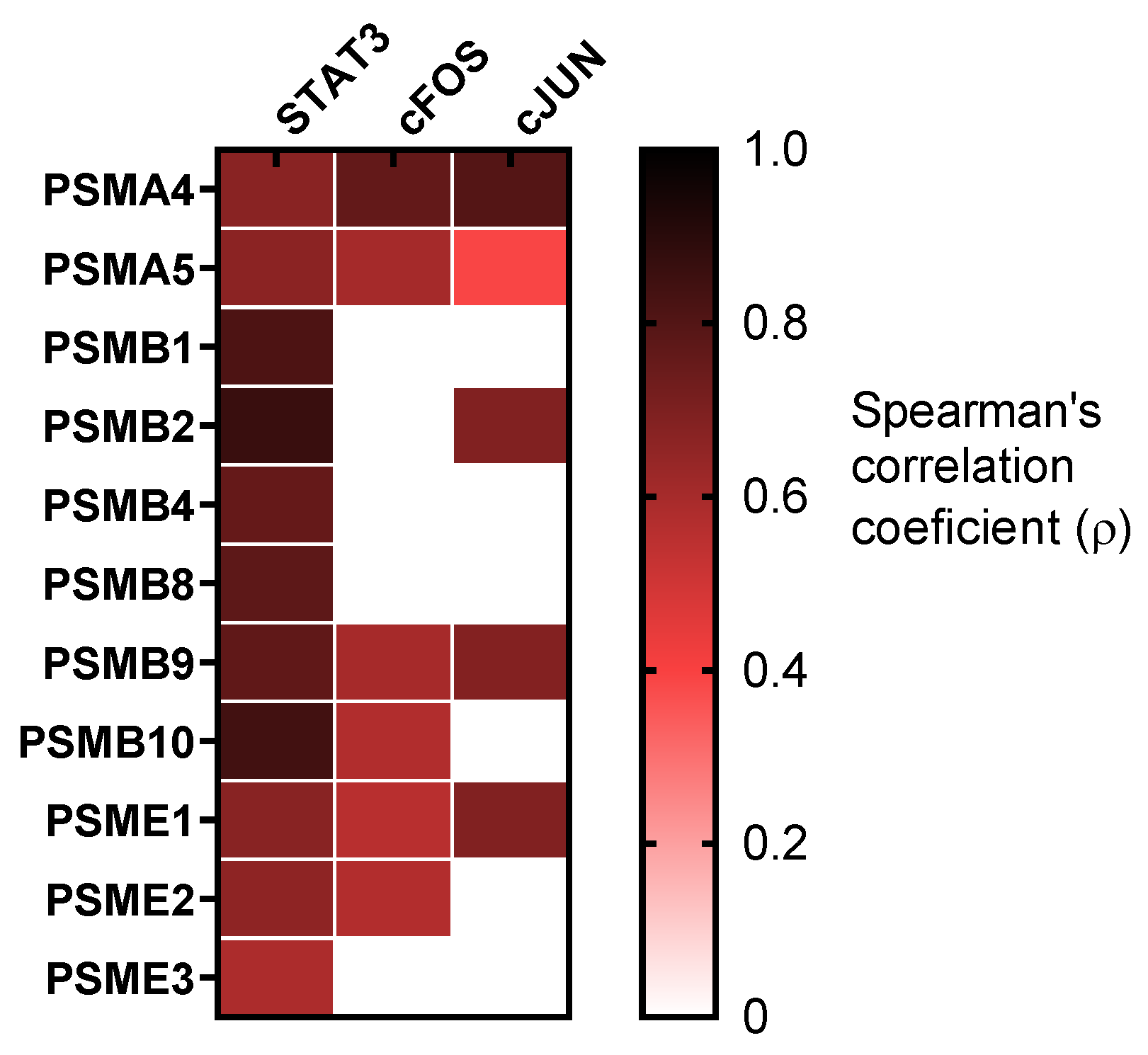
3.6. Proteasome Subunitswere Related with STAT3 Cascade

Furthermore, STAT3 was identified to be upregulated in the context of intensive inflammation and hypoxemia, and its potential role in COVID-19 pathogenesis has been recently reviewed [48,49]. Here, we reported the mRNA upregulation of STAT3 together with its well-known downstream effectors c-FOS and c-JUN in COVID-19 patients compared with healthy controls (Figure S3G). A positive correlation was observed between all the proteasome subunits studied and STAT3. In addition, some proteasome subunits correlated with c-FOS and c-JUN (Figure 5 and Figure S3F).

3.7. Proteasome Subunits Upregulation May Be a Cause and Not a Consequence of Inflammation

Finally, in order to clarify whether inflammation is a cause or consequence of proteasome upregulation in COVID-19, patients we treated HC and COVID-19 patients PBMCs with LPS (10 ng/mL) during 16 h. Our in vitro data suggested that inflammation may not have an impact on proteasome subunit expression (Figure S4). Therefore, considering the proteasome subunits overexpression data in COVID-19 patients, we speculate a possible role of proteasome subunits mRNA expression in the inflammatory condition of these patients.

4. Discussion

Severe COVID-19 patients are well characterized by hypoxemia, a reduced number of T cells and inflammatory storm during the first days after SARS-CoV-2 infection [2,50]. Additionally, there is a wide range of evidence supporting the idea that proteasome activity play a role in the inflammatory response [51]. Indeed, ubiquitylation maintains a balance between the activation and inhibition of the immune system response due to infection [52]. In our study, we assessed the expression of 16 genes from the catalytic core and regulatory particles of the 20S proteasome. Our data showed an increased expression in seven of the subunits of proteasome (PSMA4,5; PSMB2,9,10; PSMD14 and PSME1) in PBMCs from COVID-19 patients, suggesting a potential increase in proteasome activity, since other studies have already related an overexpression of proteasome subunits with a higher proteasome activity [53,54]. Besides, many studies have attempted to elucidate the mechanisms underlying the regulation of proteasome subunit expression, leading to the identification of multiple transcription factors which in turn are regulated by several interrelated pathways [55,56,57,58,59]. This suggests that the regulation of proteasome subunit expression is intricate and complex, and supports the fact that only seven of the proteasome subunits studied are upregulated in COVID-19 patients, while the others are not. Consequently, further studies are required in order to elucidate the mechanism underlying proteasome subunits expression in a physiological and COVID-19 pathological context.
Both PSMA4 and PSMA5 are part of the α-rings of the catalytic core proteasome [60], which comprise the outer rings of the 20S proteasome [61]. We observed PSMA4 and PSMA5 overexpression in COVID-19 patients. In agreement with these findings, PSMA5 mRNA expression was elevated in patients with sepsis [25]. Furthermore, PSMB1-10 are part of the β-rings of the catalytic core proteasome (20S) [28], which comprise the inner rings of the 20S proteasome. The β-rings form a proteolytic chamber where only the subunits encoded by PSMB5, PSMB6 and PSMB7 genes demonstrate hydrolytic activity [62,63]. Furthermore, β-type proteasome subunits PSMB8, PSMB9 and PSMB10, which are homologous to PSMB5, PSMB6 and PSMB7, respectively, form the immunoproteasome, a type of proteasome with a specialized role in processing intracellular antigens for presentation to the immune system [64]. Interestingly, we only observed an increase in PSMB9 and PSMB10, suggesting mediation of the immunoproteasome in COVID-19 pathology. Curiously, mutations in genes encoding for immunoproteasome subunits have been related to the development of proteasome-associated auto-inflammatory syndromes in mouse models [65] and humans [66]. In addition, PSMD11 and PSMD14 are part of the lid of the 19S regulatory particle [28]. Our data showed an increase in PSMD14 expression. As is known, PSMD14 encodes the metalloisopeptidase Rpn11, which plays an essential role in the de-ubiquitylation of captured substrates to facilitate their degradation, which constitutes the main biochemical activity of 19S lid [67]. Besides, PSMD14 is implicated in viral replication and pathogenesis in hepatitis B [68]. Moreover, PSME1-3 are subunits of the 11S (also called PA28 or REG) regulatory particles. PSME1 and PSME2 assemble to form a complex (also called proteasome activator PA28alpha/beta) that binds the ends of the 20S proteasome and dramatically accelerates the generation of a subset of MHC class I-presented antigenic peptides. Our data revealed the upregulation of PSME1 in COVID-19 patients. Interestingly, the role of 11S proteasome during viral infection has already been reported [69,70].
Our data suggested proteasome subunits overexpression to be related to the hypoxic condition in COVID-19 patients. In agreement, some proteasome subunits have been previously reported to increase under hypoxaemic conditions [25,71,72,73]. Thus, we hypothesised that hypoxia might play a role in the upregulation of proteasome subunits in COVID-19 patients, according to the patient and in vitro model data. Other authors have highlighted the role of 20S proteasome in septic patients suggesting its involvement in immunological activity [74]. Moreover, in a recent study, 20S proteasome serum content was related to requirement of oxygen therapy in COVID-19 patients [75]. Along with hypoxia development, COVID-19 severity is tightly bounded to hyperinflammatory state.
Our data revealed a relation of the proteasome subunits with inflammation markers. Similarly, ubiquitin-proteasome axis has been described to be dually essential for inflammation via the NF-κB pathway and NLRP3 inflammasome [76]. On the one hand, all proteasome subunits studied correlated with NF-κB and an LPS in vitro model showed no effect on proteasome subunits expression, suggesting that proteasome upregulation might be implicated in COVID-19 inflammatory condition development. On the other hand, we identified a direct relation between the majority of proteasome subunits studied and NLRP3 inflammasome components (NLPR3, ASC, CAS-1). The ubiquitin-proteasome system (UPS) is involved in the regulation of NLRP3 and ASC stability by selective ubiquitilation [36,77]. Rodrigues and colleagues demonstrated NLRP3 inflammasome is activated in response to SARS-CoV-2 infection [78]. Furthermore, NLRP3 inflammasome activation drives cells into pyroptosis, a process leading to cell-death by mean of gasdermin D [79], which may be the prominent cause of lymphopenia, and high levels of inflammation. Overall, we hypothesized that proteasome subunits upregulation is related to inflammation and pyroptosis, highlighting a potential connection between UPS regulatory activities and COVID-19 clinical features. Upregulation of proteasome has also been described in critically ill septic patients and trauma patients, both sharing extensive inflammatory conditions [74].
Concomitantly to pyroptosis, lymphopenia may also be related with hyperinflammation, which is able to induce lymphocyte exhaustion and apoptosis. Although apoptotic cell death was not assessed in this study, we related proteasome subunits with STAT3. Recent reports show that the IL-6/STAT3 pathway can drive the upregulation of the transcription of TGF-β [80], which is related to T-cell exhaustion and mediates apoptosis of virus-specific CD8+ T cells [81], although it has been typically described as an anti-apoptotic pathway [82]. Furthermore, activated STAT3 can induce P53 transcription, enhancing COVID-19 patients’ lymphopenia [83]. Interestingly, the IL-6/STAT3 axis could contribute to an exacerbated immune response and COVID-19 complications such as thrombosis and lung fibrosis [49]. Proteasome and its subunits also play a direct role in apoptosis. Indeed, the overexpression of proteasome subunits has been identified to protect different lung and colon cancer cells from apoptosis [54,84,85,86].
In this line, we suggested that hypoxia might upregulate proteasome subunits mRNA expression, which could be implicated in lymphocyte cell-death. Many studies discuss that proteasome inhibitors are capable to induce apoptosis; indeed, they are frequently used in the treatment of cancer [87,88]. Furthermore, an inhibition of proteasome has been described to induce apoptosis in human monocyte-derived dendritic cells, suggesting a protective role of proteasome in the immune system [89]. This protective role is supported by a proteasome-dependent degradation of proteins such as NLRP3 and P53. However, other studies have demonstrated a requirement of proteasomes during apoptosis in non-cycling and differentiated cells [90]. In addition, evidence has suggested that viruses and their infected hosts exploit the proteasomal degradation machinery to promote efficient viral replication and restrict invading viruses in the infected host [42,91]. In regard, proteasome inhibitors have been demonstrated to be efficient in limiting viral life cycle, including in SARS-CoV [92,93]. Conversely other authors report that the proteasome inhibitor can also enhance viral disease [94]. In the context of COVID-19, Longhitano et al. hypothesized the therapeutic value of proteasome inhibitors [95]. The issue of whether proteasome subunits overexpression exerts a protective or pathogenic role remains to be elucidated.
The study has several limitations which we acknowledge. Firstly, the study is limited to mRNA expression quantification although other studies have related mRNA expression of proteasome subunits with a higher proteasome activity [53,54]. Secondly, the procedures with infected cells were restricted due to local biologically secure conditions. Thirdly, we have not considered the possible impact on proteasome subunits expression of the differences in PBMCs subpopulation described in COVID-19 patients. Therefore, the results obtained allow us to hypothesize involvement between the pathogenic pathways and limit the mechanistic and causative description of the processes involved.
In this study, we explored the proteasome subunits in COVID-19 patients to determine the potential association between hypoxemia, lymphopenia and hyperinflammation. Collectively, these findings suggested a link between proteasome subunits expression and the clinical manifestations in COVID-19 patients. In this line, a recent study by Wendt et al. also suggests a relation between proteasome expression and the hyperactivation of the immune system and tissue damage in COVID-19 patients [75]. Future studies should focus on the proteasome activity in SARS-CoV-2 infection severity, which might corroborate our findings on the relationship between proteasome activity and lymphopenia, hypoxemia and inflammation. (Figure 6). Additional work is needed to corroborate the causative relations of the upregulation of proteasome subunits and COVID-19 severity.

Supplementary Materials

The following supporting information can be downloaded at: https://www.mdpi.com/article/10.3390/biom12030442/s1, Figure S1: Proteasome and HIF-1α upregulation in COVID-19 patients; Figure S2: In vitro model of the effect of hypoxia on proteasome subunits expression; Figure S3: Elevated mRNA expression of genes related to inflammation or STAT3 pathway in COVID-19 patients; Figure S4: In vitro model of the effect of inflammatory response is proteasome subunits expression; Table S1: Primers used for qPCR; Table S2: Clinical parameters of COVID-19 patients grouped by proteasome subunits expression.

Author Contributions

Conceptualization, F.G.-R. and C.C.-Z.; methodology, E.A., E.D.-G., S.G.-T., E.Z., A.M., R.G., E.L.-C., F.G.-R. and C.C.-Z.; formal analysis, E.A., E.D.-G., F.G.-R. and C.C.-Z.; patient recruitment and sample collection, E.Z., A.M. and R.G.; writing—original draft preparation, F.G.-R. and C.C.-Z.; supervision, F.G.-R. and C.C.-Z.; project administration, F.G.-R. and C.C.-Z.; funding acquisition, F.G.-R. and C.C.-Z. All authors have read and agreed to the published version of the manuscript.

Funding

This research was funded by Health Research Fund (Fondo de Investigación Sanitario [FIS])-European Regional Development Fund (FEDER), Spain through PI19/01612 (F.G.-R.) and COV20/00207 and PI19-01363 (C.C.-Z.) and ISCIII (CP18/00028), co-funded by ESF, “Investing in your future”.

Institutional Review Board Statement

The study was conducted in accordance with the Declaration of Helsinki and approved by the Institutional Review Board (or Ethics Committee) of Hospital Universitario de la Paz (Madrid) (PI-4087).

Informed Consent Statement

Informed consent was obtained from all subjects involved in the study.

Data Availability Statement

The data that support the findings of this study are available from the corresponding authors upon reasonable request. Individual level participant data will not be made available to others due to privacy concerns.

Acknowledgments

We thank the blood donor service of La Paz University Hospital for helping in the recruitment of healthy controls.

Conflicts of Interest

The authors declare no conflict of interest.

References

  1. Vabret, N.; Britton, G.J.; Gruber, C.; Hegde, S.; Kim, J.; Kuksin, M.; Levantovsky, R.; Malle, L.; Moreira, A.; Park, M.D.; et al. Immunology of COVID-19: Current State of the Science. Immunity 2020, 52, 910–941. [Google Scholar] [CrossRef] [PubMed]
  2. Jafarzadeh, A.; Jafarzadeh, S.; Nozari, P.; Mokhtari, P.; Nemati, M. Lymphopenia an important immunological abnormality in patients with COVID-19: Possible mechanisms. Scand. J. Immunol. 2021, 93, e12967. [Google Scholar] [CrossRef] [PubMed]
  3. Diao, B.; Wang, C.; Tan, Y.; Chen, X.; Liu, Y.; Ning, L.; Chen, L.; Li, M.; Wang, G.; Yuan, Z.; et al. Reduction and Functional Exhaustion of T Cells in Patients With Coronavirus Disease 2019 (COVID-19). Front. Immunol. 2020, 11, 827. [Google Scholar] [CrossRef] [PubMed]
  4. Zhao, Q.; Meng, M.; Kumar, R.; Wu, Y.; Huang, J.; Deng, Y.; Weng, Z.; Yang, L. Lymphopenia is associated with severe coronavirus disease 2019 (COVID-19) infections: A systemic review and meta-analysis. Int. J. Infect. Dis. 2020, 96, 131–135. [Google Scholar] [CrossRef] [PubMed]
  5. Li, T.; Qiu, Z.; Zhang, L.; Han, Y.; He, W.; Liu, Z.; Ma, X.; Fan, H.; Lu, W.; Xie, J.; et al. Significant changes of peripheral T lymphocyte subsets in patients with severe acute respiratory syndrome. J. Infect. Dis. 2004, 189, 648–651. [Google Scholar] [CrossRef] [PubMed] [Green Version]
  6. Kroemer, A.; Khan, K.; Plassmeyer, M.; Alpan, O.; Haseeb, M.A.; Gupta, R.; Fishbein, T.M. Inflammasome activation and pyroptosis in lymphopenic liver patients with COVID-19. J. Hepatol. 2020, 73, 1258–1262. [Google Scholar] [CrossRef] [PubMed]
  7. Fathi, N.; Rezaei, N. Lymphopenia in COVID-19: Therapeutic opportunities. Cell Biol. Int. 2020, 44, 1792–1797. [Google Scholar] [CrossRef]
  8. Cizmecioglu, A.; Akay Cizmecioglu, H.; Goktepe, M.H.; Emsen, A.; Korkmaz, C.; Esenkaya Tasbent, F.; Colkesen, F.; Artac, H. Apoptosis-induced T-cell lymphopenia is related to COVID-19 severity. J. Med. Virol. 2021, 93, 2867–2874. [Google Scholar] [CrossRef]
  9. Bucks, C.M.; Norton, J.A.; Boesteanu, A.C.; Mueller, Y.M.; Katsikis, P.D. Chronic antigen stimulation alone is sufficient to drive CD8+ T cell exhaustion. J. Immunol. 2009, 182, 6697–6708. [Google Scholar] [CrossRef] [Green Version]
  10. Cao, B.; Li, X.W.; Mao, Y.; Wang, J.; Lu, H.Z.; Chen, Y.S.; Liang, Z.A.; Liang, L.; Zhang, S.J.; Zhang, B.; et al. Clinical features of the initial cases of 2009 pandemic influenza A (H1N1) virus infection in China. N. Engl. J. Med. 2009, 361, 2507–2517. [Google Scholar] [CrossRef] [Green Version]
  11. Perez-Padilla, R.; Fernandez, R.; Garcia-Sancho, C.; Franco-Marina, F.; Aburto, O.; Lopez-Gatell, H.; Bojorquez, I. Pandemic (H1N1) 2009 virus and Down syndrome patients. Emerg. Infect. Dis. 2010, 16, 1312–1314. [Google Scholar] [CrossRef] [PubMed]
  12. Li, L.; Wo, J.; Shao, J.; Zhu, H.; Wu, N.; Li, M.; Yao, H.; Hu, M.; Dennin, R.H. SARS-coronavirus replicates in mononuclear cells of peripheral blood (PBMCs) from SARS patients. J. Clin. Virol. 2003, 28, 239–244. [Google Scholar] [CrossRef]
  13. Poutanen, S.M.; Low, D.E.; Henry, B.; Finkelstein, S.; Rose, D.; Green, K.; Tellier, R.; Draker, R.; Adachi, D.; Ayers, M.; et al. Identification of severe acute respiratory syndrome in Canada. N. Engl. J. Med. 2003, 348, 1995–2005. [Google Scholar] [CrossRef] [PubMed]
  14. Assiri, A.; Al-Tawfiq, J.A.; Al-Rabeeah, A.A.; Al-Rabiah, F.A.; Al-Hajjar, S.; Al-Barrak, A.; Flemban, H.; Al-Nassir, W.N.; Balkhy, H.H.; Al-Hakeem, R.F.; et al. Epidemiological, demographic, and clinical characteristics of 47 cases of Middle East respiratory syndrome coronavirus disease from Saudi Arabia: A descriptive study. Lancet Infect. Dis. 2013, 13, 752–761. [Google Scholar] [CrossRef] [Green Version]
  15. Guan, W.J.; Ni, Z.Y.; Hu, Y.; Liang, W.H.; Ou, C.Q.; He, J.X.; Liu, L.; Shan, H.; Lei, C.L.; Hui, D.S.C.; et al. Clinical Characteristics of Coronavirus Disease 2019 in China. N. Engl. J. Med. 2020, 382, 1708–1720. [Google Scholar] [CrossRef] [PubMed]
  16. Garvin, M.R.; Alvarez, C.; Miller, J.I.; Prates, E.T.; Walker, A.M.; Amos, B.K.; Mast, A.E.; Justice, A.; Aronow, B.; Jacobson, D. A mechanistic model and therapeutic interventions for COVID-19 involving a RAS-mediated bradykinin storm. Elife 2020, 9, e59177. [Google Scholar] [CrossRef] [PubMed]
  17. Shamaa, O.R.; Mitra, S.; Gavrilin, M.A.; Wewers, M.D. Monocyte Caspase-1 Is Released in a Stable, Active High Molecular Weight Complex Distinct from the Unstable Cell Lysate-Activated Caspase-1. PLoS ONE 2015, 10, e0142203. [Google Scholar] [CrossRef] [PubMed] [Green Version]
  18. Bergsbaken, T.; Fink, S.L.; Cookson, B.T. Pyroptosis: Host cell death and inflammation. Nat. Rev. Microbiol. 2009, 7, 99–109. [Google Scholar] [CrossRef] [Green Version]
  19. da Costa, L.S.; Outlioua, A.; Anginot, A.; Akarid, K.; Arnoult, D. RNA viruses promote activation of the NLRP3 inflammasome through cytopathogenic effect-induced potassium efflux. Cell Death Dis. 2019, 10, 346. [Google Scholar] [CrossRef] [PubMed]
  20. Roth, G.; Moser, B.; Krenn, C.; Brunner, M.; Haisjackl, M.; Almer, G.; Gerlitz, S.; Wolner, E.; Boltz-Nitulescu, G.; Ankersmit, H.J. Susceptibility to programmed cell death in T-lymphocytes from septic patients: A mechanism for lymphopenia and Th2 predominance. Biochem. Biophys. Res. Commun. 2003, 308, 840–846. [Google Scholar] [CrossRef]
  21. Hotchkiss, R.S.; Osmon, S.B.; Chang, K.C.; Wagner, T.H.; Coopersmith, C.M.; Karl, I.E. Accelerated lymphocyte death in sepsis occurs by both the death receptor and mitochondrial pathways. J. Immunol. 2005, 174, 5110–5118. [Google Scholar] [CrossRef] [PubMed] [Green Version]
  22. Ankersmit, H.J.; Moser, B.; Roedler, S.; Teufel, I.; Zuckermann, A.; Roth, G.; Lietz, K.; Back, C.; Gerlitz, S.; Wolner, E.; et al. Death-inducing receptors and apoptotic changes in lymphocytes of patients with heart transplant vasculopathy. Clin. Exp. Immunol. 2002, 127, 183–189. [Google Scholar] [CrossRef] [PubMed]
  23. Pedersen, P.B.; Hrobjartsson, A.; Nielsen, D.L.; Henriksen, D.P.; Brabrand, M.; Lassen, A.T. Prevalence and prognosis of acutely ill patients with organ failure at arrival to hospital: Protocol for a systematic review. Syst. Rev. 2017, 6, 227. [Google Scholar] [CrossRef] [PubMed] [Green Version]
  24. Mehta, P.; McAuley, D.F.; Brown, M.; Sanchez, E.; Tattersall, R.S.; Manson, J.J.; Hlh Across Speciality Collaboration, U.K. COVID-19: Consider cytokine storm syndromes and immunosuppression. Lancet 2020, 395, 1033–1034. [Google Scholar] [CrossRef]
  25. Xue, M.; Zhang, S.; Xie, J.; Zhang, X.; Liu, F.; Huang, Y.; Liu, L.; Liu, S.; Guo, F.; Yang, Y.; et al. Differential expression of genes associated with T lymphocytes function in septic patients with hypoxemia challenge. Ann. Transl. Med. 2019, 7, 810. [Google Scholar] [CrossRef] [PubMed]
  26. Yousef, A.A.; Suliman, G.A.; Mabrouk, M.M. The value of correlation of serum 20S proteasome concentration and percentage of lymphocytic apoptosis in critically ill patients: A prospective observational study. Crit. Care 2010, 14, R215. [Google Scholar] [CrossRef] [Green Version]
  27. Kravtsova-Ivantsiv, Y.; Ciechanover, A. The ubiquitin-proteasome system and activation of NF-kappaB: Involvement of the ubiquitin ligase KPC1 in p105 processing and tumor suppression. Mol. Cell Oncol. 2015, 2, e1054552. [Google Scholar] [CrossRef] [Green Version]
  28. Murata, S.; Yashiroda, H.; Tanaka, K. Molecular mechanisms of proteasome assembly. Nat. Rev. Mol. Cell Biol. 2009, 10, 104–115. [Google Scholar] [CrossRef] [PubMed]
  29. Eldridge, A.G.; O’Brien, T. Therapeutic strategies within the ubiquitin proteasome system. Cell Death Differ. 2010, 17, 4–13. [Google Scholar] [CrossRef] [Green Version]
  30. Rieser, E.; Cordier, S.M.; Walczak, H. Linear ubiquitination: A newly discovered regulator of cell signalling. Trends Biochem. Sci. 2013, 38, 94–102. [Google Scholar] [CrossRef]
  31. Filardy, A.A.; He, J.; Bennink, J.; Yewdell, J.; Kelsall, B.L. Posttranscriptional control of NLRP3 inflammasome activation in colonic macrophages. Mucosal. Immunol. 2016, 9, 850–858. [Google Scholar] [CrossRef] [PubMed] [Green Version]
  32. Israel, A. The IKK complex, a central regulator of NF-kappaB activation. Cold Spring Harb. Perspect Biol. 2010, 2, a000158. [Google Scholar] [CrossRef] [PubMed] [Green Version]
  33. Kayagaki, N.; Phung, Q.; Chan, S.; Chaudhari, R.; Quan, C.; O’Rourke, K.M.; Eby, M.; Pietras, E.; Cheng, G.; Bazan, J.F.; et al. DUBA: A deubiquitinase that regulates type I interferon production. Science 2007, 318, 1628–1632. [Google Scholar] [CrossRef] [PubMed]
  34. Liu, B.; Zhang, M.; Chu, H.; Zhang, H.; Wu, H.; Song, G.; Wang, P.; Zhao, K.; Hou, J.; Wang, X.; et al. The ubiquitin E3 ligase TRIM31 promotes aggregation and activation of the signaling adaptor MAVS through Lys63-linked polyubiquitination. Nat. Immunol. 2017, 18, 214–224. [Google Scholar] [CrossRef] [PubMed]
  35. Ren, Z.; Ding, T.; Zuo, Z.; Xu, Z.; Deng, J.; Wei, Z. Regulation of MAVS Expression and Signaling Function in the Antiviral Innate Immune Response. Front. Immunol. 2020, 11, 1030. [Google Scholar] [CrossRef] [PubMed]
  36. Song, H.; Liu, B.; Huai, W.; Yu, Z.; Wang, W.; Zhao, J.; Han, L.; Jiang, G.; Zhang, L.; Gao, C.; et al. The E3 ubiquitin ligase TRIM31 attenuates NLRP3 inflammasome activation by promoting proteasomal degradation of NLRP3. Nat. Commun. 2016, 7, 13727. [Google Scholar] [CrossRef] [PubMed] [Green Version]
  37. Ferrington, D.A.; Gregerson, D.S. Immunoproteasomes: Structure, function, and antigen presentation. Prog. Mol. Biol. Transl. Sci. 2012, 109, 75–112. [Google Scholar] [CrossRef] [PubMed] [Green Version]
  38. Groettrup, M.; Kirk, C.J.; Basler, M. Proteasomes in immune cells: More than peptide producers? Nat. Rev. Immunol. 2010, 10, 73–78. [Google Scholar] [CrossRef] [PubMed] [Green Version]
  39. Barrow, E.; Nicola, A.V.; Liu, J. Multiscale perspectives of virus entry via endocytosis. Virol. J. 2013, 10, 177. [Google Scholar] [CrossRef] [PubMed] [Green Version]
  40. Nicola, A.V.; Aguilar, H.C.; Mercer, J.; Ryckman, B.; Wiethoff, C.M. Virus entry by endocytosis. Adv. Virol. 2013, 2013, 469538. [Google Scholar] [CrossRef] [PubMed]
  41. Randow, F.; Lehner, P.J. Viral avoidance and exploitation of the ubiquitin system. Nat. Cell Biol. 2009, 11, 527–534. [Google Scholar] [CrossRef] [PubMed]
  42. Raaben, M.; Posthuma, C.C.; Verheije, M.H.; te Lintelo, E.G.; Kikkert, M.; Drijfhout, J.W.; Snijder, E.J.; Rottier, P.J.; de Haan, C.A. The ubiquitin-proteasome system plays an important role during various stages of the coronavirus infection cycle. J. Virol. 2010, 84, 7869–7879. [Google Scholar] [CrossRef] [PubMed] [Green Version]
  43. Shin, D.; Mukherjee, R.; Grewe, D.; Bojkova, D.; Baek, K.; Bhattacharya, A.; Schulz, L.; Widera, M.; Mehdipour, A.R.; Tascher, G.; et al. Papain-like protease regulates SARS-CoV-2 viral spread and innate immunity. Nature 2020, 587, 657–662. [Google Scholar] [CrossRef] [PubMed]
  44. Bailey-Elkin, B.A.; Knaap, R.C.; Johnson, G.G.; Dalebout, T.J.; Ninaber, D.K.; van Kasteren, P.B.; Bredenbeek, P.J.; Snijder, E.J.; Kikkert, M.; Mark, B.L. Crystal structure of the Middle East respiratory syndrome coronavirus (MERS-CoV) papain-like protease bound to ubiquitin facilitates targeted disruption of deubiquitinating activity to demonstrate its role in innate immune suppression. J. Biol. Chem. 2014, 289, 34667–34682. [Google Scholar] [CrossRef] [PubMed] [Green Version]
  45. Chen, X.; Yang, X.; Zheng, Y.; Yang, Y.; Xing, Y.; Chen, Z. SARS coronavirus papain-like protease inhibits the type I interferon signaling pathway through interaction with the STING-TRAF3-TBK1 complex. Protein. Cell 2014, 5, 369–381. [Google Scholar] [CrossRef] [PubMed] [Green Version]
  46. Spivak-Kroizman, T.R.; Hostetter, G.; Posner, R.; Aziz, M.; Hu, C.; Demeure, M.J.; Von Hoff, D.; Hingorani, S.R.; Palculict, T.B.; Izzo, J.; et al. Hypoxia triggers hedgehog-mediated tumor-stromal interactions in pancreatic cancer. Cancer Res. 2013, 73, 3235–3247. [Google Scholar] [CrossRef] [PubMed] [Green Version]
  47. Schwartz, D.L.; Powis, G.; Thitai-Kumar, A.; He, Y.; Bankson, J.; Williams, R.; Lemos, R.; Oh, J.; Volgin, A.; Soghomonyan, S.; et al. The selective hypoxia inducible factor-1 inhibitor PX-478 provides in vivo radiosensitization through tumor stromal effects. Mol. Cancer Ther. 2009, 8, 947–958. [Google Scholar] [CrossRef] [PubMed] [Green Version]
  48. Matsuyama, T.; Kubli, S.P.; Yoshinaga, S.K.; Pfeffer, K.; Mak, T.W. An aberrant STAT pathway is central to COVID-19. Cell Death Differ. 2020, 27, 3209–3225. [Google Scholar] [CrossRef] [PubMed]
  49. Jafarzadeh, A.; Nemati, M.; Jafarzadeh, S. Contribution of STAT3 to the pathogenesis of COVID-19. Microb. Pathog. 2021, 154, 104836. [Google Scholar] [CrossRef]
  50. Cui, R.; Wang, C.; Li, T.; Hua, J.; Zhao, T.; Ren, L.; Wang, Y.; Li, Y. Carboxypeptidase N1 is anticipated to be a synergy metrics for chemotherapy effectiveness and prognostic significance in invasive breast cancer. Cancer Cell Int. 2021, 21, 571. [Google Scholar] [CrossRef]
  51. Qureshi, N.; Morrison, D.C.; Reis, J. Proteasome protease mediated regulation of cytokine induction and inflammation. Biochim. Biophys. Acta 2012, 1823, 2087–2093. [Google Scholar] [CrossRef] [PubMed] [Green Version]
  52. Luo, H. Interplay between the virus and the ubiquitin-proteasome system: Molecular mechanism of viral pathogenesis. Curr. Opin. Virol. 2016, 17, 1–10. [Google Scholar] [CrossRef] [PubMed]
  53. Chen, L.; Madura, K. Increased proteasome activity, ubiquitin-conjugating enzymes, and eEF1A translation factor detected in breast cancer tissue. Cancer Res. 2005, 65, 5599–5606. [Google Scholar] [CrossRef] [PubMed] [Green Version]
  54. Arlt, A.; Bauer, I.; Schafmayer, C.; Tepel, J.; Muerkoster, S.S.; Brosch, M.; Roder, C.; Kalthoff, H.; Hampe, J.; Moyer, M.P.; et al. Increased proteasome subunit protein expression and proteasome activity in colon cancer relate to an enhanced activation of nuclear factor E2-related factor 2 (Nrf2). Oncogene 2009, 28, 3983–3996. [Google Scholar] [CrossRef] [PubMed] [Green Version]
  55. Motosugi, R.; Murata, S. Dynamic Regulation of Proteasome Expression. Front. Mol. Biosci. 2019, 6, 30. [Google Scholar] [CrossRef] [PubMed] [Green Version]
  56. Xu, H.; Fu, J.; Ha, S.W.; Ju, D.; Zheng, J.; Li, L.; Xie, Y. The CCAAT box-binding transcription factor NF-Y regulates basal expression of human proteasome genes. Biochim. Biophys. Acta 2012, 1823, 818–825. [Google Scholar] [CrossRef] [PubMed] [Green Version]
  57. Vilchez, D.; Boyer, L.; Morantte, I.; Lutz, M.; Merkwirth, C.; Joyce, D.; Spencer, B.; Page, L.; Masliah, E.; Berggren, W.T.; et al. Increased proteasome activity in human embryonic stem cells is regulated by PSMD11. Nature 2012, 489, 304–308. [Google Scholar] [CrossRef] [PubMed]
  58. Vangala, J.R.; Dudem, S.; Jain, N.; Kalivendi, S.V. Regulation of PSMB5 protein and beta subunits of mammalian proteasome by constitutively activated signal transducer and activator of transcription 3 (STAT3): Potential role in bortezomib-mediated anticancer therapy. J. Biol. Chem. 2014, 289, 12612–12622. [Google Scholar] [CrossRef] [PubMed] [Green Version]
  59. Radhakrishnan, S.K.; Lee, C.S.; Young, P.; Beskow, A.; Chan, J.Y.; Deshaies, R.J. Transcription factor Nrf1 mediates the proteasome recovery pathway after proteasome inhibition in mammalian cells. Mol. Cell 2010, 38, 17–28. [Google Scholar] [CrossRef] [Green Version]
  60. Chen, I.Y.; Moriyama, M.; Chang, M.F.; Ichinohe, T. Severe Acute Respiratory Syndrome Coronavirus Viroporin 3a Activates the NLRP3 Inflammasome. Front. Microbiol. 2019, 10, 50. [Google Scholar] [CrossRef] [Green Version]
  61. Panfair, D.; Ramamurthy, A.; Kusmierczyk, A.R. Alpha-ring Independent Assembly of the 20S Proteasome. Sci. Rep. 2015, 5, 13130. [Google Scholar] [CrossRef] [PubMed] [Green Version]
  62. Arendt, C.S.; Hochstrasser, M. Identification of the yeast 20S proteasome catalytic centers and subunit interactions required for active-site formation. Proc. Natl. Acad. Sci. USA 1997, 94, 7156–7161. [Google Scholar] [CrossRef] [PubMed] [Green Version]
  63. Heinemeyer, W.; Ramos, P.C.; Dohmen, R.J. The ultimate nanoscale mincer: Assembly, structure and active sites of the 20S proteasome core. Cell Mol. Life Sci. 2004, 61, 1562–1578. [Google Scholar] [CrossRef] [PubMed]
  64. Murata, S.; Takahama, Y.; Kasahara, M.; Tanaka, K. The immunoproteasome and thymoproteasome: Functions, evolution and human disease. Nat. Immunol. 2018, 19, 923–931. [Google Scholar] [CrossRef] [PubMed]
  65. Kanazawa, N.; Hemmi, H.; Kinjo, N.; Ohnishi, H.; Hamazaki, J.; Mishima, H.; Kinoshita, A.; Mizushima, T.; Hamada, S.; Hamada, K.; et al. Heterozygous missense variant of the proteasome subunit beta-type 9 causes neonatal-onset autoinflammation and immunodeficiency. Nat. Commun. 2021, 12, 6819. [Google Scholar] [CrossRef] [PubMed]
  66. Sarrabay, G.; Mechin, D.; Salhi, A.; Boursier, G.; Rittore, C.; Crow, Y.; Rice, G.; Tran, T.A.; Cezar, R.; Duffy, D.; et al. PSMB10, the last immunoproteasome gene missing for PRAAS. J. Allergy Clin. Immunol. 2020, 145, 1015–1017e1016. [Google Scholar] [CrossRef] [PubMed] [Green Version]
  67. Verma, R.; Aravind, L.; Oania, R.; McDonald, W.H.; Yates, J.R., III; Koonin, E.V.; Deshaies, R.J. Role of Rpn11 metalloprotease in deubiquitination and degradation by the 26S proteasome. Science 2002, 298, 611–615. [Google Scholar] [CrossRef] [PubMed]
  68. Kong, F.; You, H.; Kong, D.; Zheng, K.; Tang, R. The interaction of hepatitis B virus with the ubiquitin proteasome system in viral replication and associated pathogenesis. Virol. J. 2019, 16, 73. [Google Scholar] [CrossRef] [PubMed] [Green Version]
  69. Sijts, A.; Sun, Y.; Janek, K.; Kral, S.; Paschen, A.; Schadendorf, D.; Kloetzel, P.M. The role of the proteasome activator PA28 in MHC class I antigen processing. Mol. Immunol. 2002, 39, 165–169. [Google Scholar] [CrossRef]
  70. McCarthy, M.K.; Weinberg, J.B. The immunoproteasome and viral infection: A complex regulator of inflammation. Front. Microbiol. 2015, 6, 21. [Google Scholar] [CrossRef] [PubMed] [Green Version]
  71. Li, C.; Su, F.; Zhang, L.; Liu, F.; Fan, W.; Li, Z.; Ma, J. Identifying Potential Diagnostic Genes for Diabetic Nephropathy Based on Hypoxia and Immune Status. J. Inflamm. Res. 2021, 14, 6871–6891. [Google Scholar] [CrossRef] [PubMed]
  72. Yang, C.; Yu, P.; Yang, F.; He, Q.; Jiang, B.; Zheng, L.; Wang, Q.; Wang, J.; Qiu, H.; Wang, H.; et al. PSMB4 inhibits cardiomyocyte apoptosis via activating NF-kappaB signaling pathway during myocardial ischemia/reperfusion injury. J. Mol. Histol. 2021, 52, 693–703. [Google Scholar] [CrossRef] [PubMed]
  73. Wang, J.; Xu, L.; Yun, X.; Yang, K.; Liao, D.; Tian, L.; Jiang, H.; Lu, W. Proteomic analysis reveals that proteasome subunit beta 6 is involved in hypoxia-induced pulmonary vascular remodeling in rats. PLoS ONE 2013, 8, e67942. [Google Scholar] [CrossRef] [PubMed] [Green Version]
  74. Roth, G.A.; Moser, B.; Krenn, C.; Roth-Walter, F.; Hetz, H.; Richter, S.; Brunner, M.; Jensen-Jarolim, E.; Wolner, E.; Hoetzenecker, K.; et al. Heightened levels of circulating 20S proteasome in critically ill patients. Eur. J. Clin. Invest. 2005, 35, 399–403. [Google Scholar] [CrossRef] [PubMed]
  75. Wendt, R.; Lingitz, M.T.; Laggner, M.; Mildner, M.; Traxler, D.; Graf, A.; Krotka, P.; Moser, B.; Hoetzenecker, K.; Kalbitz, S.; et al. Clinical Relevance of Elevated Soluble ST2, HSP27 and 20S Proteasome at Hospital Admission in Patients with COVID-19. Biology 2021, 10, 1186. [Google Scholar] [CrossRef] [PubMed]
  76. Palombella, V.J.; Rando, O.J.; Goldberg, A.L.; Maniatis, T. The ubiquitin-proteasome pathway is required for processing the NF-kappa B1 precursor protein and the activation of NF-kappa B. Cell 1994, 78, 773–785. [Google Scholar] [CrossRef]
  77. Guan, K.; Wei, C.; Zheng, Z.; Song, T.; Wu, F.; Zhang, Y.; Cao, Y.; Ma, S.; Chen, W.; Xu, Q.; et al. MAVS Promotes Inflammasome Activation by Targeting ASC for K63-Linked Ubiquitination via the E3 Ligase TRAF3. J. Immunol. 2015, 194, 4880–4890. [Google Scholar] [CrossRef] [PubMed]
  78. Rodrigues, T.S.; de Sa, K.S.G.; Ishimoto, A.Y.; Becerra, A.; Oliveira, S.; Almeida, L.; Goncalves, A.V.; Perucello, D.B.; Andrade, W.A.; Castro, R.; et al. Inflammasomes are activated in response to SARS-CoV-2 infection and are associated with COVID-19 severity in patients. J. Exp. Med. 2021, 218, e20201707. [Google Scholar] [CrossRef] [PubMed]
  79. Deng, C.; Wu, S.; Zhang, L.; Yang, M.; Lin, Q.; Xie, Q.; Ding, H.; Lian, N.; Gao, S.; Huang, Y.; et al. Role of monocyte tissue factor on patients with non-small cell lung cancer. Clin. Respir. J. 2018, 12, 1125–1133. [Google Scholar] [CrossRef] [PubMed]
  80. Li, C.; Iness, A.; Yoon, J.; Grider, J.R.; Murthy, K.S.; Kellum, J.M.; Kuemmerle, J.F. Noncanonical STAT3 activation regulates excess TGF-beta1 and collagen I expression in muscle of stricturing Crohn’s disease. J. Immunol. 2015, 194, 3422–3431. [Google Scholar] [CrossRef] [PubMed] [Green Version]
  81. Tinoco, R.; Alcalde, V.; Yang, Y.; Sauer, K.; Zuniga, E.I. Cell-intrinsic transforming growth factor-beta signaling mediates virus-specific CD8+ T cell deletion and viral persistence in vivo. Immunity 2009, 31, 145–157. [Google Scholar] [CrossRef] [PubMed] [Green Version]
  82. Takeda, K.; Kaisho, T.; Yoshida, N.; Takeda, J.; Kishimoto, T.; Akira, S. Stat3 activation is responsible for IL-6-dependent T cell proliferation through preventing apoptosis: Generation and characterization of T cell-specific Stat3-deficient mice. J. Immunol. 1998, 161, 4652–4660. [Google Scholar] [CrossRef] [PubMed] [Green Version]
  83. Xiong, Y.; Liu, Y.; Cao, L.; Wang, D.; Guo, M.; Jiang, A.; Guo, D.; Hu, W.; Yang, J.; Tang, Z.; et al. Transcriptomic characteristics of bronchoalveolar lavage fluid and peripheral blood mononuclear cells in COVID-19 patients. Emerg. Microbes. Infect. 2020, 9, 761–770. [Google Scholar] [CrossRef] [PubMed]
  84. Liu, Y.; Liu, P.; Wen, W.; James, M.A.; Wang, Y.; Bailey-Wilson, J.E.; Amos, C.I.; Pinney, S.M.; Yang, P.; de Andrade, M.; et al. Haplotype and cell proliferation analyses of candidate lung cancer susceptibility genes on chromosome 15q24-25.1. Cancer Res. 2009, 69, 7844–7850. [Google Scholar] [CrossRef] [PubMed] [Green Version]
  85. Lv, J.; Zhang, S.; Wu, H.; Lu, J.; Lu, Y.; Wang, F.; Zhao, W.; Zhan, P.; Lu, J.; Fang, Q.; et al. Deubiquitinase PSMD14 enhances hepatocellular carcinoma growth and metastasis by stabilizing GRB2. Cancer Lett. 2020, 469, 22–34. [Google Scholar] [CrossRef] [PubMed]
  86. Walter, R.F.H.; Sydow, S.R.; Berg, E.; Kollmeier, J.; Christoph, D.C.; Christoph, S.; Eberhardt, W.E.E.; Mairinger, T.; Wohlschlaeger, J.; Schmid, K.W.; et al. Bortezomib sensitivity is tissue dependent and high expression of the 20S proteasome precludes good response in malignant pleural mesothelioma. Cancer Manag. Res. 2019, 11, 8711–8720. [Google Scholar] [CrossRef] [PubMed] [Green Version]
  87. McConkey, D.J.; Zhu, K. Mechanisms of proteasome inhibitor action and resistance in cancer. Drug Resist. Updat. 2008, 11, 164–179. [Google Scholar] [CrossRef] [PubMed]
  88. Bazzaro, M.; Lee, M.K.; Zoso, A.; Stirling, W.L.; Santillan, A.; Shih Ie, M.; Roden, R.B. Ubiquitin-proteasome system stress sensitizes ovarian cancer to proteasome inhibitor-induced apoptosis. Cancer Res. 2006, 66, 3754–3763. [Google Scholar] [CrossRef] [PubMed] [Green Version]
  89. Nencioni, A.; Garuti, A.; Schwarzenberg, K.; Cirmena, G.; Dal Bello, G.; Rocco, I.; Barbieri, E.; Brossart, P.; Patrone, F.; Ballestrero, A. Proteasome inhibitor-induced apoptosis in human monocyte-derived dendritic cells. Eur. J. Immunol. 2006, 36, 681–689. [Google Scholar] [CrossRef] [PubMed] [Green Version]
  90. Neutzner, A.; Li, S.; Xu, S.; Karbowski, M. The ubiquitin/proteasome system-dependent control of mitochondrial steps in apoptosis. Semin. Cell Dev. Biol. 2012, 23, 499–508. [Google Scholar] [CrossRef] [PubMed]
  91. Proulx, J.; Borgmann, K.; Park, I.W. Role of Virally-Encoded Deubiquitinating Enzymes in Regulation of the Virus Life Cycle. Int. J. Mol. Sci. 2021, 22, 4438. [Google Scholar] [CrossRef] [PubMed]
  92. Schneider, M.; Ackermann, K.; Stuart, M.; Wex, C.; Protzer, U.; Schatzl, H.M.; Gilch, S. Severe acute respiratory syndrome coronavirus replication is severely impaired by MG132 due to proteasome-independent inhibition of M-calpain. J. Virol. 2012, 86, 10112–10122. [Google Scholar] [CrossRef] [PubMed] [Green Version]
  93. Ma, X.Z.; Bartczak, A.; Zhang, J.; Khattar, R.; Chen, L.; Liu, M.F.; Edwards, A.; Levy, G.; McGilvray, I.D. Proteasome inhibition in vivo promotes survival in a lethal murine model of severe acute respiratory syndrome. J. Virol. 2010, 84, 12419–12428. [Google Scholar] [CrossRef] [PubMed] [Green Version]
  94. Raaben, M.; Grinwis, G.C.; Rottier, P.J.; de Haan, C.A. The proteasome inhibitor Velcade enhances rather than reduces disease in mouse hepatitis coronavirus-infected mice. J. Virol. 2010, 84, 7880–7885. [Google Scholar] [CrossRef] [PubMed] [Green Version]
  95. Longhitano, L.; Tibullo, D.; Giallongo, C.; Lazzarino, G.; Tartaglia, N.; Galimberti, S.; Volti, G.L.; Palumbo, G.A.; Liso, A. Proteasome Inhibitors as a Possible Therapy for SARS-CoV-2. Int. J. Mol. Sci. 2020, 21, 3622. [Google Scholar] [CrossRef] [PubMed]
Figure 1. Upregulation of proteasome subunits in COVID-19 patients. mRNA expression in PBMCs from healthy controls (HC) compared to COVID-19 patients (COV) of (A) 20S proteasome alpha ring subunits: PSMA4 (HC n = 20; COV n = 32), PSMA5 (HC n = 15; COV n = 34); (B) 20S proteasome beta ring non-catalytic subunits: PSMB1 (HC n = 11; COV n = 15), PSMB2 (HC n = 12; COV n = 14), PSMB4 (HC n = 12; COV n = 13); (C) 20S immunoproteasome beta ring catalytic subunits: PSMB8 (HC n = 13; COV n = 18), PSMB9 (HC n = 12; COV n = 17), PSMB10 (HC n = 13; COV n = 14); and (D) 11S proteasome subunits: PSME1 (HC n = 11; COV n = 18), PSME2 (HC n = 14; COV n = 19), PSME3 (HC n = 12; COV n = 21). Mean differences were analysed using unpaired Student’s t-test analysis with Welch’s correction. Error bars: standard error of the mean. *: p < 0.05, **: p < 0.01.
Biomolecules 12 00442 g001
Figure 2. Hypoxia-inducible factor 1-alpha correlation with proteasome subunits. Correlation of hypoxia-inducible factor 1-alpha (HIF-1α) mRNA expression and mRNA expression of (A) 20S proteasome alpha ring subunits: PSMA4 (n = 31), PSMA5 (n = 32); (B) 20S proteasome beta ring non-catalytic subunits: PSMB1 (n = 14), PSMB2 (n = 14), PSMB4 (n = 13); (C) 20S immunoproteasome beta ring catalytic subunits: PSMB8 (n = 16), PSMB9 (n = 17), PSMB10 (n = 15); and (D) 11S proteasome subunits: PSME1 (n = 19), PSME2 (n = 19), PSME3 (n = 19). Correlations were analysed using Spearman’s analysis. p-value and Spearman’s correlation coefficient (ρ) are shown.
Biomolecules 12 00442 g002
Figure 3. In vitro model of the effect of hypoxia on proteasome subunits expression. mRNA expression of proteasome subunits from healthy volunteers PBMCs (n = 5) cultured in normoxia (N) or hypoxia (H) and treated with PX478 (PX) or untreated (C). (A) 20S proteasome alpha ring subunits: PSMA4, PSMA5; (B) 20S proteasome beta ring non-catalytic subunits: PSMB1, PSMB2, PSMB4; (C) 20S immunoproteasome beta ring catalytic subunits: PSMB8, PSMB9, PSMB10; and (D) 11S proteasome subunits: PSME1, PSME2, PSME3. Mean differences were analysed using mixed-effects analysis with Bonferroni’s multiple comparison test. Error bars: standard error of the mean. *: p < 0.05, **: p < 0.01.
Biomolecules 12 00442 g003
Figure 4. Proteasome subunits relationship with inflammation components. (A) Heatmap represents Spearman’s correlation coefficient (ρ) between proteasome subunits mRNA expression and genes related to inflammation mRNA expression. White gaps represent non-significant correlations, while the rest of correlations are significant (p < 0.05). (B) NLRP3 expression in monocytes analysed by flow cytometry in monocytes from healthy controls (HC), and COVID-19 patients (COV) presenting LOW or HIGH expression of proteasome subunits. Mean differences were analysed using one-way ANOVA analysis with Tukey’s multiple comparisons test. Error bars: standard error of the mean. *: p < 0.05, **: p < 0.01, ***: p < 0.001.
Biomolecules 12 00442 g004
Figure 5. Proteasome subunits relationship with STAT3 pathway genes. Graph represents Spearman’s correlation coefficient (ρ) between proteasome subunits mRNA expression and STAT3, cFOS and cJUN. White gaps represent non-significant correlations, while the rest of correlations are significant (p < 0.05).
Biomolecules 12 00442 g005
Figure 6. Proteasome might play an important role in COVID-19 pathogenesis. SARS-CoV-2 infection induces hypoxia which, we suggested, leads to overexpression of proteasome subunits in PBMCs from patients with COVID-19, potentially leading to an overactivation of the ubiquitin-proteasome system. The pleiotropic roles of proteasome in immune biology include regulation of master proteins such as, NF-κB, NLRP3, GSDMD and STAT3, which have been related to COVID-19 complications, mainly lymphopenia and hyperinflammation.
Biomolecules 12 00442 g006
Table 1. General characteristics of the COVID-19.
Age, year57 ± 13
Sex, Male/Female30/14
Days since onset of symptoms7.7 ± 4.0
Symptoms at admission
Cough28 (64)
Active fever32 (73)
Dyspnea31 (70)
Myalgia15 (34)
Sputum production8 (18)
Chest tightness3 (7)
Headache8 (18)
Fatigue12 (27)
Anorexia5 (11)
Nausea3 (7)
Diarrhea7 (16)
Chest pain5 (11)
Anosmia6 (14)
Comorbidities
Hypertension12 (27)
Coronary artery disease3 (7)
Diabetes mellitus13 (30)
Obesity10 (23)
Chronic lung disease4 (9)
Chronic kidney disease1 (2)
Hypothyroidism2 (3)
Smoking history
Current, n (%)31 (70)
Former, (%)3 (7)
Never, n (%)10 (23)
Pneumonia severity scores
CURB-650.84 ± 0.88
Fine risk class2.4 ± 1.3
Laboratory findings
PaO2, mmHg65.0 ± 25.9
PaO2/FiO2 ratio282.5 ± 113.5
PaCO2, mmHg36.8 ± 14.7
White cell count, 103 cells/µL6.74 ± 4.41
Neutrophils, 103 cells/µL4.90 ± 3.10
Lymphocytes, 103 cells/µL1.31 ± 2.04
Eosinophils, 103 cells/µL0.02 ± 0.03
Platelets, 103 cells/µL227.7 ± 101.8
Hemoglobin, g/dL13.9 ± 4.7
C-reactive protein, mg/L68.8 ± 57.7
Aspartate aminotransferase, U/L48.8 ± 38.5
Alanine aminotransferase, IU/L43.7 ± 29.6
Υ-Glutamyltransferase, IU/L84.8 ± 81.0
Bilirubin, µmol/L0.51 ± 0.28
Albumin, g/L4.34 ± 1.82
Ferritin, ng/mL839.1 ± 899.4
Lactate dehydrogenase, U/L304.2 ± 163.6
D-dimer, ng/mL697.4 ± 677.4
Fibrinogen, mg/dL672.1 ± 270.9
Evolution results
Duration of hospital admission, days9.9 ± 5.6
ICU admission, n (%)2 (4.5)
Exitus, n (%)2 (4.5)
Table 2. Clinical parameters of COVID-19 patients grouped by proteasome subunits expression.
GeneLymphocytes
(×103 Cells/mm3)
PaO2/FiO2 RatioC-Reactive Protein (mg/L)Ferritin (mg/mL)
LowHighpLowHighpLowHighpLowHighp
PSMA41.018 ± 0.2930.740 ± 0.1680.0084320.0 ± 92.90232.9 ± 105.00.02846.97 ± 32.6390.42 ± 67.200.044486.1 ± 532.21361 ± 11580.024
PSMA51.035 ± 0.4760.734 ± 0.1640.0498302.2 ± 75.82229.5 ± 88.980.042950.36 ± 34.3294.5 ± 66.770.0470473.8 ± 475.6927.6 ± 4710.0339
PSMB10.983 ± 0.3060.655 ± 0.1990.056288.3 ± 100.8247.5 ± 82.380.43841.93 ± 38.447.1 ± 29.80.791707.7 ± 684.4876.5 ± 696.40.659
PSMB20.983 ± 0.3060.626 ± 0.2070.048288.3 ± 100.8245.7 ± 88.810.44141.93 ± 38.447.20 ± 32.20.796707.7 ± 684.4796.3 ± 711.20.824
PSMB41.030 ± 0.3800.784 ± 0.3600.301295.7 ± 106.5247.7 ± 90.970.40748.25 ± 35.9537.51 ± 30.140.576772.3 ± 621.6775.3 ± 734.70.994
PSMB80.957 ± 0.2870.919 ± 0.5280.164284.3 ± 92.6262.6 ± 95.120.66342.57 ± 35.0847.81 ± 29.880.763812.0 ± 683.0747.5 ± 672.70.857
PSMB90.873 ± 0.3170.976 ± 0.4710.623302.0 ± 85.14244.4 ± 88.270.20952.13 ± 35.4638.77 ± 26.520.423804.9 ± 690.5707.1 ± 639.20.777
PSMB100.798 ± 0.2720.963 ± 0.4560.441296.2 ± 91.72241.6 ± 85.970.28358.45 ± 34.2634.71 ± 29.010.202936.0 ± 654.0705.3 ± 708.50.541
PSME10.920 ± 0.3110.885 ± 0.4890.867285.6 ± 89.74249.2 ± 86.460.40956.75 ± 37.7442.46 ± 33.010.422614.1 ± 618.9795.3 ± 687.20.576
PSME20.831 ± 0.3810.974 ± 0.4230.491289.4 ± 93.14245.9 ± 81.420.32558.65 ± 37.1740.77 ± 32.650.312641.8 ± 621.7770.8 ± 691.20.691
PSME30.939 ± 0.3700.866 ± 0.4430.728294.6 ± 91.56241.2 ± 79.860.22352.24 ± 32.6946.47 ± 38.630.743779.6 ± 626.1648.2 ± 687.50.686
Clinical parameters (lymphocyte count, PAFI ratio, C-reactive protein concentration and ferritin concentration) comparison in COVID-19 patients grouped by high or low expression of proteasome subunits (PSMA4/5, PSMB1/2/4/8/9/10 and PSME1-3). Data are mean ± standard deviation. p-values are shown. Differences were considered significant p < 0.05.
Publisher’s Note: MDPI stays neutral with regard to jurisdictional claims in published maps and institutional affiliations.

